Branchial Cleft and Pouch Anomalies
|
|
|
- Derek Atkins
- 5 years ago
- Views:
Transcription


1 Branchial Cleft and Pouch Anomalies Prof.Mohamed Hesham Alexandria Faculty of Medicine Alexandria, Egypt Emberyological Basis


2 Branchial Clefts 1st 2nd Pinna EAC 3rd 4th 4th 6th Cervical sinus Branchial Pouches 1st 2nd 3rd 4th 6th
3 Types of Anomalies Skin Sinus External Internal Fistula Cyst Gut Branchial cleft and pouches anomalies Incidence 2nd cleft&pouch 95% 1st 5% 3rd and 4th rarely reported


4 First Branchial Cleft Anomalies The first branchial cleft is the only cleft that not become obliterated by the 8 th week. First cleft anomalies are a result of incomplete closure of the cleft First Branchial Cleft Anomalies Classification Arnot,1971 Work,1972 Olsen etal,1980


5 First Branchial Cleft Anomalies Site Preauricular and cervical region above hyoid bone The incidence of malformation is higher at the top of the triangle Trigalia et al,1998 Otological Manifestations Parotid Manifestations Neck Manifestations Pochet s triangle First Branchial Cleft Anomalies Presentation Cyst Sinus Fistula Infection Prior Surgery


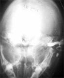
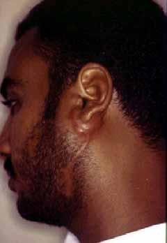
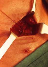
6 First Branchial Cleft Anomalies Presentation First Branchial Cleft Anomalies Presentation


7 First Branchial Cleft Anomalies Presentation First Branchial Cleft Anomalies Presentation


8 First Branchial Cleft Anomalies Presentation First Branchial Cleft Anomalies Presentation Branchio-Oto-renal syndrome Autosomal dominant Renal, 1st and 2nd arches Hearing impairment 73% Preauricular pits 70% Renal abnormalities 67% Branchial cleft cyst or sinus 60%
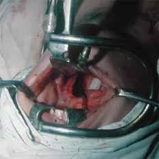
9 First Branchial Cleft Anomalies Facial Nerve The relationship of the branchial anomaly to the facial nerve is variable The fate of branchial apparatus is complete by 6 or 7 week Parotid gland appear at 6th week Facial nerve and muscles migrate upward between 6-8 week First Branchial Cleft Anomalies Facial Nerve In the Majority of cases, The anomaly is lateral to the nerve Miller et al, 1984; Sinuses lateral to the facial nerve Fistulae medial to facial nerve Solares etal, 2003; Complete fisulae are usually deep to the facial nerve


10 First Branchial Cleft Anomalies Facial Nerve First Branchial Cleft Anomalies Facial Nerve


11 First Branchial Cleft Anomalies Facial Nerve Cyst First Branchial Cleft Anomalies Facial Nerve

12 First Branchial Cleft Anomalies Facial Nerve First Branchial Cleft Anomalies Facial Nerve

13 First Branchial Cleft Anomalies Facial Nerve First Branchial Cleft Anomalies Facial Nerve The facial nerve is vulnerable to injury Variable relation of the facial nerve Prior surgeries Prior infection 15-30% Facial nerve insult

14 First Branchial Cleft Anomalies Second Branchial Cleft Anomalies Tons. Skin Ext. opening: Ant. Border of sternomastoid muscle in the mid or lower neck.

15 Second Branchial Cleft Anomalies Tons. Skin Course: Deep to the platysma, along the carotid sheath, then between 2 carotids, superficial to nerve XII, IX deep to stylohyoid ligament and post. Belly of the digastric muscle Internal opening: Anterior aspect of the upper half of the post. pillar of the tonsil Second Branchial Cleft Anomalies? Sinus or Fistula How Why


16 Second Branchial Cleft Anomalies Second Branchial Cleft Anomalies False negative Not canalized along the whole length Obstructed by infection

17 Second Branchial Cleft Anomalies How? Clinical examination Sinogram Intraoperative MB injection Intraoperative tract searching


18 Why? For complete excision To prevent recurrence Second Branchial Cleft Anomalies Pull Through Branchial Fistulectomy Prof.M.Talaat


19 Second Branchial Cleft Anomalies Pull Through Branchial Fistulectomy Prof.M.Talaat Second Branchial Cleft Anomalies Pull Through Branchial Fistulectomy Prof.M.Talaat


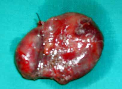
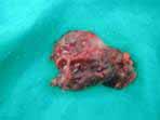
20 Second Branchial Cleft Anomalies Pull Through Branchial Fistulectomy Prof.M.Talaat Second Branchial Cleft Anomalies Cyst


21 Second Branchial Cleft Anomalies Cyst Second Branchial Cleft Anomalies Cyst


22 Second Branchial Cleft Anomalies Cyst Second Branchial Cleft Anomalies Cyst


23 Second Branchial Cleft Anomalies Cyst Second Branchial Cleft Anomalies Cyst


24 Second Branchial Cleft Anomalies Infected Cyst Second Branchial Cleft Anomalies Infected Cyst


25 Second Branchial Cleft Anomalies Infected Cyst Second Branchial Cleft Anomalies Infected Cyst


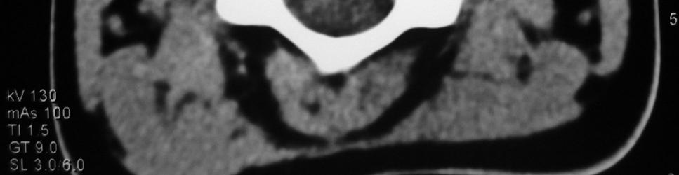
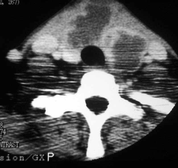
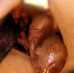
26 3 rd and 4 th Branchial Cleft Anomalies Rarely reported Failure of the pouches to obliterate 3 rd : Inferior parathyroid, Thymus 4 th : Superior Parathyroid, PFC Pharyngobranchial duct 3 rd and 4 th Branchial Cleft Anomalies Presentation Cyst in close relation to the thyroid gland Sinus tract ; pyriform sinus External openining

27 3 rd and 4 th Branchial Cleft Anomalies Presentation Cyst in close relation to the thyroid gland Sinus tract ; pyriform sinus External openining 3 rd and 4 th Branchial Cleft Anomalies Presentation Cyst in close relation to the thyroid gland Sinus tract ; pyriform sinus External openning

28 3 rd and 4 th Branchial Cleft Anomalies Presentation Cyst in close relation to the thyroid gland Sinus tract ; pyriform sinus External openning 3 rd and 4 th Branchial Cleft Anomalies Presentation Cyst in close relation to the thyroid gland Sinus tract ; pyriform sinus External openning


29 3 rd and 4 th Branchial Cleft Anomalies Presentation Cyst in close relation to the thyroid gland Sinus tract ; pyriform sinus External openning 3 rd and 4 th Branchial Cleft Anomalies Presentation Cyst in close relation to the thyroid gland Sinus tract ; pyriform sinus External openning

30 3 rd and 4 th Branchial Cleft Anomalies Presentation Cyst in close relation to the thyroid gland Sinus tract ; pyriform sinus External openning 3 rd and 4 th Branchial Cleft Anomalies Presentation Cyst in close relation to the thyroid gland Sinus tract ; pyriform sinus External openning


31 Midline Cervical Cleft It is not a "true" cleft, as it does not involve Medial a gap cleft between adjacent skin flaps Median and only fissure extends of the partially neck through Midline the cervical skin layers. cord Midline cervical webbing The Pterygium cleft is often colli weeping medianum at birth, then toughens and dries, healing with a scar A subcutaneous fibrous cord that originates from the deep layer of the skin tag and ends in the subcutaneous tissue of the chin Midline Cervical Cleft
32 Conclusion Branchial arch anomalies should be bared in mind in all cysts or sinuses located from the root of the helix to the clavicle Other congenital anomalies Should be excluded Complete fistula has to be diagnosed Pull through branchial fistulectomy for complete fistula of the 2 nd arch 1st branchial arch anomalies should be excised with wide approach with care of the facial nerve
THYROID & PARATHYROID. By Prof. Saeed Abuel Makarem & Dr. Sanaa Al-Sharawy
 THYROID & PARATHYROID By Prof. Saeed Abuel Makarem & Dr. Sanaa Al-Sharawy 1 OBJECTIVES By the end of the lecture, the student should be able to: Describe the shape, position, relations and structure of
 Drawings illustrating the human pharyngeal apparatus. Drawings illustrating the human pharyngeal apparatus. Drawings illustrating the human pharyngeal apparatus. Drawings illustrating the human pharyngeal
REVIEW OF CLINICAL EMBRYOLOGY OF HEAD AND NECK
 REVIEW OF CLINICAL EMBRYOLOGY OF HEAD AND NECK OUTLINE - EMBRYOLOGY UNDERLYING CLINICAL CONDITIONS I. EARLY DEVELOPMENT OF FACE: CLEFT LIP, CLEFT PALATE, OBSTRUCTED NASOLACRIMAL DUCT II. BRANCHIAL ARCHES
Congenital Neck Masses C. Stefan Kénel-Pierre, MD
 Congenital Neck Masses C. Stefan Kénel-Pierre, MD SUNY-LICH Medical Center Department of Surgery Case Presentation xx year old male presents with sudden onset left lower neck swelling x 1 week Denies pain,
04 Development of the Face and Neck. Development of the Face Development of the neck
 04 Development of the Face and Neck Development of the Face Development of the neck Development of the face Overview of facial development The fourth week ~ the twelfth week of prenatal development Between
Neck-2. Dr. Heba Kalbouneh Associate Professor of Anatomy and Histology
 Neck-2 ` Dr. Heba Kalbouneh Associate Professor of Anatomy and Histology Triangles of the neck Side of the neck Midline Lower border of mandible Line between angle of mandible and mastoid Superior nuchal
Pharyngeal Apparatus. Pouches Endoderm Grooves Ectoderm Arch Neural Crest Somitomeres Aortic Arch - Vessel
 Pharyngeal Apparatus Pouches Endoderm Grooves Ectoderm Arch Neural Crest Somitomeres Aortic Arch - Vessel Segmental Organization Humans: Arch 1-4 prominent Arch 5 absent Arch 6 - transient First Arch Face
Anterior triangle of neck
 Anterior triangle of neck Dept. of Anatomy Zhou Hong Ying Outline boundary and subdivisions of ant. triangle contents of the triangle Muscles: suprahyoid muscles, infrahyoid muscles Nerves: CNⅩ, CNⅪ, CNⅫ,
Thyroid gland. importance. relations and connections. external laryngeal nerves. malformations.
 Thyroid gland 1. Recognize and understand the coverings of the thyroid gland and their clinical importance. 2. Recognize and understand the main parts of the thyroid gland and their locations, relations
Anatomy: head and Neck (6 questions) 1. Prevertebral Flexor Musculature (lying in front of the vertebrae) include all, EXCEPT: Longus Colli.
 1. Prevertebral Flexor Musculature (lying in front of the vertebrae) include all, EXCEPT: Longus Colli.") Anatomy: head and Neck (6 questions) 1. Prevertebral Flexor Musculature (lying in front of the vertebrae) include all, EXCEPT: Longus Colli. Rectus Capitis Anterior. Rectus Capitis Lateralis. Rectus Capitis
Anatomy: head and Neck (6 questions) 1. Prevertebral Flexor Musculature (lying in front of the vertebrae) include all, EXCEPT: Longus Colli. Rectus Capitis Anterior. Rectus Capitis Lateralis. Rectus Capitis
ANTERIOR CERVICAL TRIANGLE (Fig. 2.1 )
") 2 Neck Anatomy ANTERIOR CERVICAL TRIANGLE (Fig. 2.1 ) The boundaries are: Lateral: sternocleidomastoid muscle Superior: inferior border of the mandible Medial: anterior midline of the neck This large triangle
2 Neck Anatomy ANTERIOR CERVICAL TRIANGLE (Fig. 2.1 ) The boundaries are: Lateral: sternocleidomastoid muscle Superior: inferior border of the mandible Medial: anterior midline of the neck This large triangle
PEDIATRICS WK 3 HEAD AND NECK ALISON WALLACE MD, PHD
 PEDIATRICS WK 3 HEAD AND NECK ALISON WALLACE MD, PHD Topics 1. Cervical lymphadenopathy 2. Lymphatic malformation 3. Thyroglossal duct cysts 4. Branchial cleft cysts 5. Thyroid masses CASE 1 Case 1 A 2
Preface... Contributors... 1 Embryology... 3
 Contents Preface... Contributors... vii xvii I. Pediatrics 1 Embryology... 3 Pearls... 3 Branchial Arch Derivatives... 3 Branchial Arch Anomalies: Cysts, Sinus, Fistulae... 4 Otologic Development... 4
OTOLARYNGOLOGY ONLINE. Preauricular sinus. Management. Dr. T. Balasubramanian 6/17/2010
 1 OTOLARYNGOLOGY ONLINE Preauricular sinus Management Dr. T. Balasubramanian 6/17/2010 This e book discusses the Etiopathogenesis of preauricular sinus. Even though it is commonly encountered surgical
CERVICAL LYMPH NODES
 CERVICAL LYMPH NODES (ANATOMY & EXAMINATION) Hemant (DTCD 1 st YEAR) 1. Lymphatic Tissues: A Type of connective tissue that contains large numbers of lymphocytes. 2. Lymphatic Vessels: Are Tubes that assist
A clinical study on branchial arch anomalies
 IOSR Journal of Dental and Medical Sciences (IOSR-JDMS) e-issn: 2279-0853, p-issn: 2279-0861.Volume 18, Issue 1 Ver. 5 (January. 2019), PP 05-10 www.iosrjournals.org Ashim Sarkar 1, Ritam Ray 2 1 (Clinical
REVIEW/PREVIEW OF HEAD AND NECK ANATOMY FOR ENT EXAM
 REVIEW/PREVIEW OF HEAD AND NECK ANATOMY FOR ENT EXAM - 2017 PALPATE CAROTID ARTERY: AT LEVEL OF CAROTID BIFURCATION VERTEBRAL LEVEL C4 Sternocleidomastoid Muscle INTERNAL CAROTID EXTERNAL CAROTID COMMON
ORIGINAL ARTICLE. and histological data. An additional confounding factor is infection, which oftendivertsthephysician sattention.
 ORIGINAL ARTICLE First Branchial Cleft Anomalies A Study of 39 Cases and a Review of the Literature Jean-Michel Triglia, MD; Richard Nicollas, MD; Vincent Ducroz, MD; Peter J. Koltai, MD; Erea-Noël Garabedian,
Chapter 13: Mass in the Neck. Raymond P. Wood II:
 Chapter 13: Mass in the Neck Raymond P. Wood II: In approaching the problem of a mass in the neck, one immediately encounters the fact that there are normally palpable masses in the neck (eg, almost all
PCM1 Physical Exam Skills Session: Head and Neck FACILITATOR & STUDENT COPY
 PATIENT CENTERED MEDICINE - 1 GOALS & OUTCOMES: PCM1 Physical Exam Skills Session: Head and Neck FACILITATOR & STUDENT COPY 1. To introduce the applied anatomy relevant for the examination of the head
Distribution of branchial anomalies in a paediatric Asian population
 Singapore Med J 2015; 56(4): 203-207 doi: 10.11622/smedj.2015060 Distribution of branchial anomalies in a paediatric Asian population Neville Wei Yang Teo 1, MBBS, MRCS, Shahrul Izham Ibrahim 2, MBBCh,
Case Report A Case of Pyriform Sinus Fistula Infection with Double Tracts
 Case Reports in Otolaryngology, Article ID 126840, 5 pages http://dx.doi.org/10.1155/2014/126840 Case Report A Case of Pyriform Sinus Fistula Infection with Double Tracts Masato Shino, Yoshihito Yasuoka,
Chapter 20: Branchial cleft anomalies, thyroglossal cysts and fistulae. P. D. M. Ellis. Branchial cleft anomalies. Embryology
 Chapter 20: Branchial cleft anomalies, thyroglossal cysts and fistulae P. D. M. Ellis Branchial cleft anomalies and thyroglossal cysts and fistulae are the end result of defects in development in the neck
Pharyngeal apparatus. - At the third week, it is a 3 layered structure: ectoderm, mesoderm and endoderm. This is called trilaminar disc
 Pharyngeal apparatus Remember from the first year embryology - The embryo was disc shaped in the second week of development (this is called embryonic disc) and it is a 2 layered disc (composed of two layers)---bilaminar
Thyroid and Parathyroid Glands
 Thyroid and Parathyroid Glands Please view our Editing File before studying this lecture to check for any changes. Color Code Important Doctors Notes Notes/ explanation Objectives: By the end of the lecture,
Neck lumps in children
 Neck lumps in children Midline Lateral Midline neck lumps Thyroglossal cyst - 80% Dermoid cyst Submental lymph node Ectopic thyroid Some rare lesions Thyroglossal cyst Diagnosis: midline, usually overlying
Alexander C Vlantis. Selective Neck Dissection 33
 05 Modified Radical Neck Dissection Type II Alexander C Vlantis Selective Neck Dissection 33 Modified Radical Neck Dissection Type II INCISION Various incisions can be used for a neck dissection. The incision
Lecture 01. The Thyroid & Parathyroid Glands. By: Dr Farooq Khan PMC Date: 12 th March. 2018
 Lecture 01 The Thyroid & Parathyroid Glands By: Dr Farooq Khan PMC Date: 12 th March. 2018 INTRODUCTION LAYERS OF THE NECK The neck has four major compartments or layer which are enclosed by an outer musculofascial
The Neck the lower margin of the mandible above the suprasternal notch and the upper border of the clavicle
 The Neck is the region of the body that lies between the lower margin of the mandible above and the suprasternal notch and the upper border of the clavicle below Nerves of the neck Cervical Plexus Is formed
SCHOOL OF ANATOMICAL SCIENCES Mock Run Questions. 4 May 2012
 SCHOOL OF ANATOMICAL SCIENCES Mock Run Questions 4 May 2012 1. With regard to the muscles of the neck: a. the platysma muscle is supplied by the accessory nerve. b. the stylohyoid muscle is supplied by
PANELISTS. Controversial Issues In Common Interventions In ORL 4/10/2014
 Controversial Issues In Common Interventions In ORL Mohamed Hesham,MD Alexandria Faculty of Medicine PANELISTS Prof. Ahmed Eldaly Prof. Hamdy EL-Hakim Prof. Hossam Thabet Prof. Maged El-Shenawy Prof. Prince
Surgical Anatomy of the Neck. M. J. Jurkiewicz, John Bostwick. Surgical Clinics of North America, Vol 54, No 6, December 1974.
 Surgical Anatomy of the Neck M. J. Jurkiewicz, John Bostwick Surgical Clinics of North America, Vol 54, No 6, December 1974. The radical neck dissection is a safe, effective therapeutic procedure for eradication
locomotice system Plastinated specimensⅠ: Silicone specimens Regional specimens and organs
 locomotice system Plastinated specimensⅠ: Silicone specimens Regional specimens and organs Art-No. Name Description The locomotor system SL001 Two hundred pieces of plastinated bones (without six The bones
Posterior Triangle of the Neck By Prof. Dr. Muhammad Imran Qureshi
 Posterior Triangle of the Neck By Prof. Dr. Muhammad Imran Qureshi For the purpose of anatomical description the neck is sub divided into two major triangles, the Anterior and the Posterior by muscle bellies
Case Report Duplication of the External Auditory Canal: Two Cases and a Review of the Literature
 Case Reports in Otolaryngology Volume 2012, Article ID 924571, 4 pages doi:10.1155/2012/924571 Case Report Duplication of the External Auditory Canal: Two Cases and a Review of the Literature John K. Goudakos,
Prevertebral Region, Pharynx and Soft Palate
 Unit 20: Prevertebral Region, Pharynx and Soft Palate Dissection Instructions: Step1 Step 2 Step 1: Insert your fingers posterior to the sternocleidomastoid muscle, vagus nerve, internal jugular vein,
Lec [8]: Mandibular nerve:
![Lec [8]: Mandibular nerve:](/thumbs/94/121295776.jpg "Lec [8]: Mandibular nerve:") Lec [8]: Mandibular nerve: The mandibular branch from the trigeminal ganglion lies in the middle cranial fossa lateral to the cavernous sinus. With the motor root of the trigeminal nerve [motor roots lies
Lec [8]: Mandibular nerve: The mandibular branch from the trigeminal ganglion lies in the middle cranial fossa lateral to the cavernous sinus. With the motor root of the trigeminal nerve [motor roots lies
THE ANGULAR TRACT: AN ANATOMICAL
 British Journal of Oral Surgery (1981) 19, 116-120 0 The British Association of Oral Surgeons 0007-117X/81/00170116$02.00 THE ANGULAR TRACT: AN ANATOMICAL OF SURGICAL SIGNIFICANCE STRUCTURE HAITHEM A.
Neckmasses in infancy and childhood: Clinical and radiological classification and imaging approaches M. Mearadji
 Neckmasses in infancy and childhood: Clinical and radiological classification and imaging approaches M. Mearadji International Foundation for Pediatric Imaging Aid Introduction Neck masses are a frequent
Auricular Sinus ORIGINAL ARTICLE. Introduction. Fig 1: The various positions of the sinus opening. 286 Med J Malaysia VoI 48 No 3 Se pt 1993
 ORIGINAL ARTICLE Auricular Sinus, r' :'- t" R. Esa, MS M. Solahuddin, MD Department of ENT,Fq(:u/ty.of 0eFlicrine,t!niv;er~iti KebangsacmMa/aysia, Ja/an Raia Muda Abdu/ Aziz, 50300 Kua/a Lumpur,t. Introduction
Ultrasound Interpretation of Non-Thyroid Neck Pathology
 Ultrasound Interpretation of Non-Thyroid Neck Pathology Kevin T. Brumund, M.D., F.A.C.S. Associate Professor of Surgery Head and Neck Surgery University of California, San Diego Health Sciences VA Medical
The Neck. BY: Lina Abdullah & Rahaf Jreisat
 The Neck BY: Lina Abdullah & Rahaf Jreisat Boundaries of the Neck: generally from base of the skull to root of the neck Superior margin :From superior nuchal line of occipital bone up to mastoid process
Evaluation and Management of Pediatric Neck masses
 Evaluation and Management of Pediatric Neck masses Steven T. Wright, M.D. Faculty Advisor: Ronald Deskin, M.D. The University of Texas Medical Branch Department of Otolaryngology Grand Rounds Presentation
Veins of the Face and the Neck
 Veins of the Face and the Neck Facial Vein The facial vein is formed at the medial angle of the eye by the union of the supraorbital and supratrochlear veins. connected through the ophthalmic veins with
Head & Neck Contouring
 Head & Neck Contouring Presented by James Wheeler, MD Center for Cancer Care Goshen, IN 46526 September 12, 2014 Special Thanks to: Spencer Boulter, Director of Operations (AAMD) Adam Moore, RT(T), CMD
Case Presentation. x year old African American male seen in Pediatric Surgery Clinic. History: NKDA
 Case Presentation ALIREZA SADEGHI MD Kings County Hospital Center University Hospital of Brooklyn Downstate Medical Center Division of Pediatric Surgery July 7 th 2006 Case Presentation x year old African
SYLLABUS BDS I PROFESSIONAL GENERAL HUMAN ANATOMY INCLUDING EMBRYOLOGY AND HISTOLOGY
 GENERAL HUMAN ANATOMY INCLUDING EMBRYOLOGY AND HISTOLOGY I. General Anatomy 1. Anatomical terms 2. Skin, superficial fascia & deep fascia 3. Cardiovascular system, portal system, collateral circulation
Asian Journal of Pharmacy and Life Science ISSN Vol.3 (2), April-June, 2013
, April-June, 2013") RECURRENT NECK INFECTION ASSOCIATED WITH FOURTH BRANCHIAL POUCH SINUS: A RARE CLINICAL ENTITY WITH DELAYED DIAGNOSIS Bhawana Pant* 1, Sanjay Gaur 2, Shahzad Ahmad 1 1. Department of E.N.T, Government Medical
RECURRENT NECK INFECTION ASSOCIATED WITH FOURTH BRANCHIAL POUCH SINUS: A RARE CLINICAL ENTITY WITH DELAYED DIAGNOSIS Bhawana Pant* 1, Sanjay Gaur 2, Shahzad Ahmad 1 1. Department of E.N.T, Government Medical
Development of the Pharyngeal Arches
 Development of the Pharyngeal Arches Thomas A. Marino, Ph.D. Temple University School of Medicine Competencies: Upon completion of this section of the course, the student must be able to: 1. Recall the
Surgical Privileges Form: ORL - HNS
 Surgical Privileges Form: ORL - HNS Clinical Privileges Request Applicant s Name:. No. (If Any):... Date:... Scope of Practice:. License Facility:.. Place of Work:. CATEGORY I: OTOLOGY PROCEDURES Privileges
Bilateral Second Branchial Arch Fistula in a 19 year old - A Case Report
 CASE REPORT Bilateral Second Branchial Arch Fistula in a 19 year old - A Case Report 1 1 1 2 Abubakar Adamu, Hamman Ibrahim Grandawa, Yusuf Bukar Ngamdu, Haruna Ngadda. SUMMARY Developmental anomalies
For the following questions, indicate the letter that corresponds to the SINGLE MOST APPROPRIATE ANSWER
 GROSS ANATOMY EXAMINATION May 15, 2000 For the following questions, indicate the letter that corresponds to the SINGLE MOST APPROPRIATE ANSWER 1. Pain associated with an infection limited to the middle
Oral cavity : consist of two parts: the oral vestibule and the oral cavity proper. Oral vestibule : is slit like space between.
 Oral cavity Oral cavity : consist of two parts: the oral vestibule and the oral cavity proper Oral vestibule : is slit like space between the teeth, buccal gingiva, lips, and cheeks 1 Oral cavity Oral
Lies in front and sides of the neck. Consists of two lobe connected anterior to the trachea by an isthmus.
 THYROID GLAND 1 Lies in front and sides of the neck. Consists of two lobe connected anterior to the trachea by an isthmus. A small pyramidal lobe projects upwards from the left lobe in 40% of cases. The
HEAD & NECK SWELLINGS
 HEAD & NECK SWELLINGS EXCLUDING GOITRE FAISAL GHANI SIDDIQUI MBBS; FCPS; MCPS-HPE; PGDIP-BIOETHICS PROFESSOR OF SURGERY J I N N A H S I N D H M E D I C A L U N I V E R S I T Y MIDLINE SWELLINGS NECK SWELLINGS
OBJECTIVE: To obtain a fundamental knowledge of the root of the neck with respect to structure and function
 The root of the neck Jeff Dupree, Ph.D. e mail: jldupree@vcu.edu OBJECTIVE: To obtain a fundamental knowledge of the root of the neck with respect to structure and function READING ASSIGNMENT: Moore and
APRIL
 APRIL - 2003 OCTOBER - 2003 February 2009 [KU 652] Sub. Code : 4131 FIRST B.D.S DEGREE EXAMINATION (Modified Regulations III) Paper I HUMAN ANATOMY, HISTOLOGY AND EMBRYOLOGY Time : Three hours
Surgical anatomy of thyroid and parathyroid glands
 Head & Neck Surgery Course Surgical anatomy of thyroid and parathyroid glands Dr Pierfrancesco PELLICCIA Pr Benjamin LALLEMANT Service ORL et CMF CHU de Nîmes CH de Arles Thyroid glands Dr Pierfrancesco
Case Report Cervical Thymic Cyst: A Rare Differential Diagnosis in Lateral Neck Swelling
 Case Reports in Otolaryngology Volume 2013, Article ID 350502, 4 pages http://dx.doi.org/10.1155/2013/350502 Case Report Cervical Thymic Cyst: A Rare Differential Diagnosis in Lateral Neck Swelling Vijendra
Anatomy of the Thyroid Gland
 Anatomy of the Thyroid Gland Introduction Nomenclature G, thyreos= shield, eidos= like Location Root of the neck ventrally (C5-T1) Function endocrine gland that secretes: Thyroxine (T4) T3 Calcitonin LWW,
Clinical Anatomy of the Thyroid and Adrenal Glands
 Clinical Anatomy of the Thyroid and Adrenal Glands Handout download: http://www.oucom.ohiou.edu/dbms-witmer/gs-rpac.htm 28 October 2003 Lawrence M. Witmer, PhD Department of Biomedical Sciences College
Tikrit University College of Dentistry Dr.Ban I.S. head & neck anatomy 2 nd y.
 Lec [3]/The scalp The scalp extends from the supraorbital margins anteriorly to the nuchal lines at the back of the skull and down to the temporal lines at the sides. The forehead, from eyebrows to hairline,
PAPILLARY THYROID CARCINOMA PRESENTING AS A LATERAL NECK MASS MASS. Dr. Pamela Hanson DO PGY3
 PAPILLARY THYROID CARCINOMA PRESENTING AS A LATERAL NECK MASS MASS Dr. Pamela Hanson DO PGY3 MK CASE PRESENTATION 28 yo Female presented to the ENT Clinic in October 2016, with the complaint of chronic
THIEME. Scalp and Superficial Temporal Region
 CHAPTER 2 Scalp and Superficial Temporal Region Scalp Learning Objectives At the end of the dissection of the scalp, you should be able to identify, understand and correlate the clinical aspects: Layers
Tikrit University collage of dentistry Dr.Ban I.S. head & neck anatomy 2 nd y. Lec [5] / Temporal fossa :
![Tikrit University collage of dentistry Dr.Ban I.S. head & neck anatomy 2 nd y. Lec [5] / Temporal fossa :](/thumbs/88/115294566.jpg "Tikrit University collage of dentistry Dr.Ban I.S. head & neck anatomy 2 nd y. Lec [5] / Temporal fossa :") Lec [5] / Temporal fossa : Borders of the Temporal Fossa: Superior: Superior temporal line. Inferior: gap between zygomatic arch and infratemporal crest of sphenoid bone. Anterior: Frontal process of the
Lec [5] / Temporal fossa : Borders of the Temporal Fossa: Superior: Superior temporal line. Inferior: gap between zygomatic arch and infratemporal crest of sphenoid bone. Anterior: Frontal process of the
Dr.Ban I.S. head & neck anatomy 2 nd y. جامعة تكريت كلية طب االسنان املرحلة الثانية أ.م.د. بان امساعيل صديق 6102/6102
 جامعة تكريت كلية طب االسنان التشريح مادة املرحلة الثانية أ.م.د. بان امساعيل صديق 6102/6102 Parotid region The part of the face in front of the ear and below the zygomatic arch is the parotid region. The
ANTERIOR CERVICAL TRIANGLE (FIG. 2.1 )
") 2 Neck Anatomy ANTERIOR CERVICAL TRIANGLE (FIG. 2.1 ) The boundaries are: Lateral: sternocleidomastoid muscle Superior: inferior border of the mandible Medial: anterior midline of the neck This large triangle
2 Neck Anatomy ANTERIOR CERVICAL TRIANGLE (FIG. 2.1 ) The boundaries are: Lateral: sternocleidomastoid muscle Superior: inferior border of the mandible Medial: anterior midline of the neck This large triangle
Superior View of the Skull (Norma Verticalis) Anteriorly the frontal bone articulates with the two parietal bones AT THE CORONAL SUTURE
 Anteriorly the frontal bone articulates with the two parietal bones AT THE CORONAL SUTURE") Superior View of the Skull (Norma Verticalis) Anteriorly the frontal bone articulates with the two parietal bones AT THE CORONAL SUTURE 1 The two parietal bones articulate in the midline AT THE SAGITTAL
Superior View of the Skull (Norma Verticalis) Anteriorly the frontal bone articulates with the two parietal bones AT THE CORONAL SUTURE 1 The two parietal bones articulate in the midline AT THE SAGITTAL
Lecture 01 Internal surface of anterolateral abdominal wall. BY Dr Farooq Khan Aurakzai
 Lecture 01 Internal surface of anterolateral abdominal wall BY Dr Farooq Khan Aurakzai Dated: 21.12.2017 Internal surface of the anterolateral abdominal wall The internal ( posterior ) surface of the anterolateral
Case Report Right-Sided Pyriform Sinus Fistula: A Case Report and Review of the Literature
 Case Reports in Otolaryngology Volume 2012, Article ID 934968, 5 pages doi:10.1155/2012/934968 Case Report Right-Sided Pyriform Sinus Fistula: A Case Report and Review of the Literature Rachel B. Cain,
Embryology of the Neck and Neck Masses June 2005
 TITLE: Embryology of the Neck and Neck Masses SOURCE: Grand Rounds Presentation, UTMB, Dept. of Otolaryngology DATE: June 8, 2005 RESIDENT PHYSICIAN: Steven T. Wright, MD FACULTY ADVISOR: Shawn D. Newlands,
Lecture 07. Lymphatic's of Head & Neck. By: Dr Farooq Amanullah Khan PMC
 Lecture 07 Lymphatic's of Head & Neck By: Dr Farooq Amanullah Khan PMC Dated: 28.11.2017 Lymphatic Vessels Of the 800 lymph nodes in the human body, 300 are in the Head & neck region. The lymphatic vessels
Parotid Gland, Temporomandibular Joint and Infratemporal Fossa
 M1 - Anatomy Parotid Gland, Temporomandibular Joint and Infratemporal Fossa Jeff Dupree Sanger 9-057 jldupree@vcu.edu Parotid gland: wraps around the mandible positioned between the mandible and the sphenoid
1TRUNK: BODY WALL AND SPINE
 TRUNK: BODY WALL AND SPINE SURFACE ANATOMY SKELETON JOINTS & LIGAMENTS MUSCLES VASCULATURE NERVES SPINAL CORD & VERTEBRAL CANAL ANTERIOR BODY WALL & MAMMARY GLAND LATERAL BODY WALL INGUINAL REGION SUPERFICIAL
Infratemporal fossa: Tikrit University college of Dentistry Dr.Ban I.S. head & neck Anatomy 2 nd y.
 Infratemporal fossa: This is a space lying beneath the base of the skull between the lateral wall of the pharynx and the ramus of the mandible. It is also referred to as the parapharyngeal or lateral pharyngeal
Thyroglossal cyst our experience
 Volume 3 Issue 1 2013 ISSN: 2250-0359 Thyroglossal cyst our experience Balasubramanian Thiagarajan 1 Ulaganathan Venkatesan 2 Geetha Ramamoorthy 1 1 Stanley Medical College 2 Meenakshi Medical College
Pyriform Sinus Fistula
 ISSN: 2250-0359 Case Report Volume 6 Issue 3: 124 2016 Pyriform Sinus Fistula Vijay Abraham*, Anjali Lepcha, Betty Simon, BS Chandran Department of Surgery Unit 3, Christian Medical College and Hospital,
By : Prof Saeed Abuel Makarem & Dr.Sanaa Alshaarawi
 By : Prof Saeed Abuel Makarem & Dr.Sanaa Alshaarawi OBJECTIVES By the end of the lecture, students shouldbe able to: List the nuclei of the deep origin of the trigeminal and facial nerves in the brain
Histology. Dr.shatarat
 Histology Dr.shatarat Dr.shatarat Dr.shatarat Cells of the adenohypophysis adenohypophysis Dr.shatarat Dr.shatarat Adenohypophysis high power acidopill basophill Chromophobes Dr.shatarat 1-Chromophils
University of Palestine. Midterm Exam 2013/2014 Total Grade:
 Course No: DNTS2208 Course Title: Head and Neck Anatomy Date: 09/11/2013 No. of Questions: (50) Time: 1hour Using Calculator (No) University of Palestine Midterm Exam 2013/2014 Total Grade: Instructor
Temporal region. temporal & infratemporal fossae. Zhou Hong Ying Dept. of Anatomy
 Temporal region temporal & infratemporal fossae Zhou Hong Ying Dept. of Anatomy Temporal region is divided by zygomatic arch into temporal & infratemporal fossae. Temporal Fossa Infratemporal fossa Temporal
Tympanic Bulla Temporal Bone. Digastric Muscle. Masseter Muscle
 Superior view Hyoid Bone The hyoid bone does not articulate with any other bones. It is held in place by ligaments to the styloid process of the temporal bone and the thyroid cartilage of the larynx. It
Larynx. Rudimentary. Behind the posterior surface : -stylopharyngeus - salpingopharyngeus -platopharyngeus
 Larynx The larynx is an organ that provides a protective sphincter at the inlet of the air passages and is responsible for voice production. It extends from C3-C6: *Posterior: the pharynx *Lateral: the
Chapter 5: Other mediastinal structures. The Large Arteries. The Aorta. Ascending aorta
 Chapter 5: Other mediastinal structures The Large Arteries The Aorta The aorta is the main arterial trunk of the systemic circulation and in the healthy state its wall contain a large amount of yellow
Clinical Study Branchial Anomalies: Diagnosis and Management
 International Journal of Otolaryngology, Article ID 237015, 9 pages http://dx.doi.org/10.1155/2014/237015 Clinical Study Branchial Anomalies: Diagnosis and Management Sampath Chandra Prasad, 1 Arun Azeez,
Clinical evaluation. Imaging Surgical treatment
 Parapharyngeal Space Khalid adhussain AL-Qahtani a MD,MSc,FRCS(c) Assistant Professor Consultant of Otolaryngology Advance Head & Neck Oncology, Thyroid & Parathyroid,Microvascular Reconstruction, ti and
The ear: some applied basic science
 Chapter 1 The ear: some applied basic science The pinna The external ear or pinna is composed of cartilage with closely adherent perichondrium and skin. It is developed from six tubercles of the first
Pediatric Temporal Bone
 Pediatric Temporal Bone Suresh K. Mukherji, MD, FACR Professor and Chief of Neuroradiology Professor of Radiology, Otolaryngology Head Neck Surgery, Radiation Oncology and Periodontics & Oral Medicine
A. Incorrect! Think of a therapy that reduces prostaglandin synthesis. B. Incorrect! Think of a therapy that reduces prostaglandin synthesis.
 USMLE Step 1 - Problem Drill 02: Embryology Question No. 1 of 10 1. A premature infant is born with a patent ductus arteriosis. Which of the following treatments may be used as part of the treatment regimen?
3-Deep fascia: is absent (except over the parotid gland & buccopharngeal fascia covering the buccinator muscle)
") The Face 1-Skin of the Face The skin of the face is: Elastic Vascular (bleed profusely however heal rapidly) Rich in sweat and sebaceous glands (can cause acne in adults) It is connected to the underlying
The Face 1-Skin of the Face The skin of the face is: Elastic Vascular (bleed profusely however heal rapidly) Rich in sweat and sebaceous glands (can cause acne in adults) It is connected to the underlying
DEVELOPMENTAL ANATOMY OF THE FACE, JAW AND NECK. O.M. Oluwatosin Department of Surgery
 DEVELOPMENTAL ANATOMY OF THE FACE, JAW AND NECK O.M. Oluwatosin Department of Surgery 1 2 By the end of this lecture, you should be able to: Discuss the embryology of the face Relate congenital anomalies
2015 Otolaryngology Survival Guide
 2015 Otolaryngology Survival Guide Chapter 12: Scope Procedures Know All Your New Sinus Endoscopy Procedures Choices You have three codes in the CPT manual for sinus endoscopy procedures that are meant
Neck of Condylar. Process. Anterior Border of Ramus. Mandibular. Foramen. Posterior Border of Ramus Incisive Fossa.
 Learning Outcomes The Mandible Surface Anatomy Muscle Attachments The (FOM) Muscles of the FOM The Tongue Muscles of the Tongue The Submandibular Region Submandibular Gland Sublingual Gland Lingual The
be very thin and variable. Facial nerve branches that exit the parotid gland are deep to the SMAS.
 The Superficial musculoaponeurotic system (SMAS) fascia is a fanlike fascia that envelops the face and provides a suspensory sheet which distributes forces of facial expression.. The SMAS is continuous
Head and Neck I. PHARYNGEAL APPARATUS (FIGURE 12.1; TABLE 12.1)
") chapter 12 Head and Neck I. PHARYNGEAL APPARATUS (FIGURE 12.1; TABLE 12.1) The pharyngeal apparatus consists of the pharyngeal arches, pharyngeal pouches, pharyngeal grooves, and pharyngeal membranes,
chapter 12 Head and Neck I. PHARYNGEAL APPARATUS (FIGURE 12.1; TABLE 12.1) The pharyngeal apparatus consists of the pharyngeal arches, pharyngeal pouches, pharyngeal grooves, and pharyngeal membranes,
Face. Definition: The area between the two ears and from the chin to the eye brows. The muscles of the face
 Face Definition: The area between the two ears and from the chin to the eye brows. The muscles of the face The muscle of facial expression (include the muscle of the face and the scalp). All are derived