MICROSTRUCTURES SMALL INTESTIN LARGE INTESTIN PANCREAS LIVER GALLBLADDER SALIVARY GLANDS ADRENALS THYROID AND PARATHYROID GLANDS
|
|
|
- Myrtle Nicholson
- 6 years ago
- Views:
Transcription
1 MICROSTRUCTURES SMALL INTESTIN LARGE INTESTIN PANCREAS LIVER GALLBLADDER SALIVARY GLANDS ADRENALS THYROID AND PARATHYROID GLANDS
 or that includes a cavity (as of the heart or urinary bladder) The parenchyma are the functional parts of an organ in the body The stroma refers to the connective, non-functional")

2 HUMAN ANATOMY: MICROSTRUCTURES CLASSIFICATION: LOCATION AND BOUNDARIES, FORM, FUNCTION, MICROSCOPIC STRUCTURE: A hollow organ is a visceral organ that is a hollow tube or pouch (as the stomach or intestine) or that includes a cavity (as of the heart or urinary bladder) The parenchyma are the functional parts of an organ in the body The stroma refers to the connective, non-functional supportive framework of an organ You always recognize a stroma (supporting tissue, formed by capsule and trabeculae) and parenchyma (functional tissue). It is usually covered with a capsule of connective tissue that penetrates inside the body. We recognize overlapping layers of different tissue origin. Usually by the outer layer are: Tunica serosa (tunica adventitia in esophagus and rectum) Tunica muscularis Tunica submucosa Tunica mucosa

3 The small intestine is divided into three structural parts: Duodenum 26 cm in length Jejunum 2.5 m Ileum 3.5 m duodenum

4 The small intestine is divided into three structural parts: Duodenum 26 cm in length Jejunum 2.5 m Ileum 3.5 m 2 3 duodenum

5 4 Jejunum

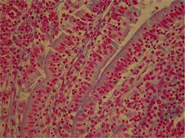
6 ILEUM WALL

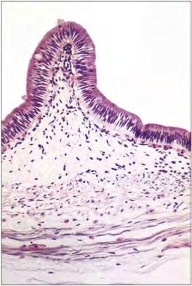
7 ILEUM 3 L V 1 2 aggregated lymphoid nodules in the digestive system (Peyer's patches)
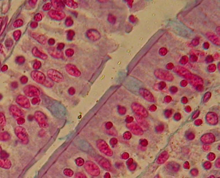
8 INTESTINAL VILLI e c e a a
9 Parts of the large intestine are: Cecum the first part of the large intestine Taeniae coli three bands of smooth muscle Haustra bulges caused by contraction of taeniae coli Epiploic appendages small fat accumulations on the viscera Appendix (or vermiform appendix; also cecal (or caecal) appendix; also vermix Locations along the colon are: The ascending colon The right colic flexure (hepatic) The transverse colon The transverse mesocolon The left colic flexure (splenic) The descending colon The sigmoid colon the v-shaped region of the large intestine

10 LARGE INTESTINE

11 CECAL APPENDIX

12 CECAL APPENDIX


13 CECAL APPENDIX A B 4

14 RECTUM
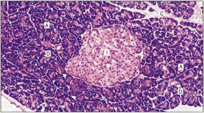
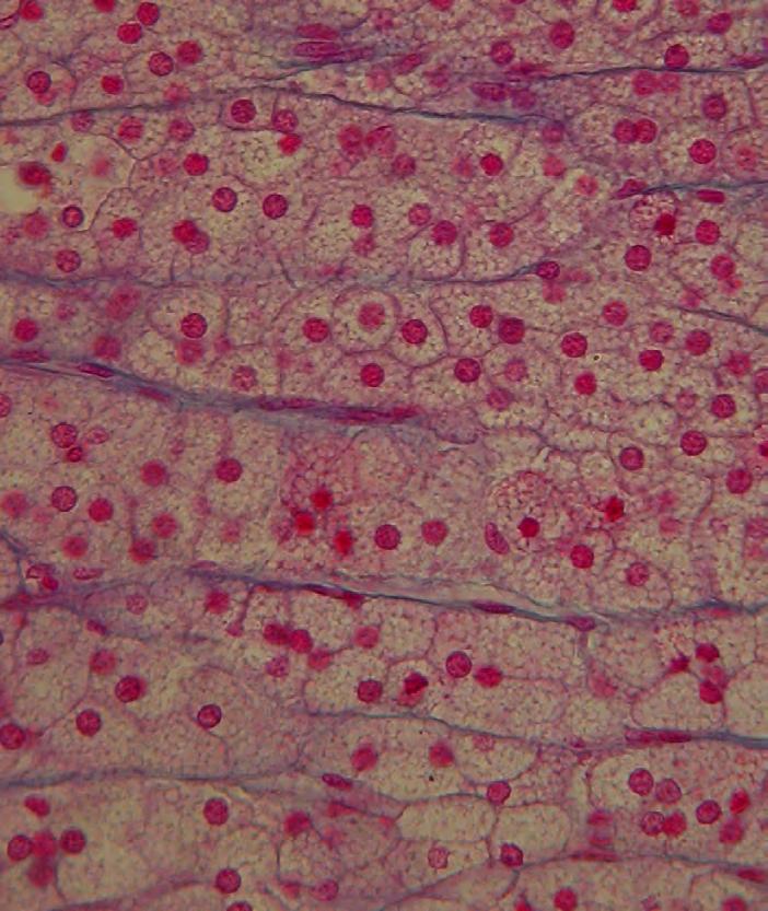
15 Pancreatic tissue

16 PANCREAS s L s A V s s s A V
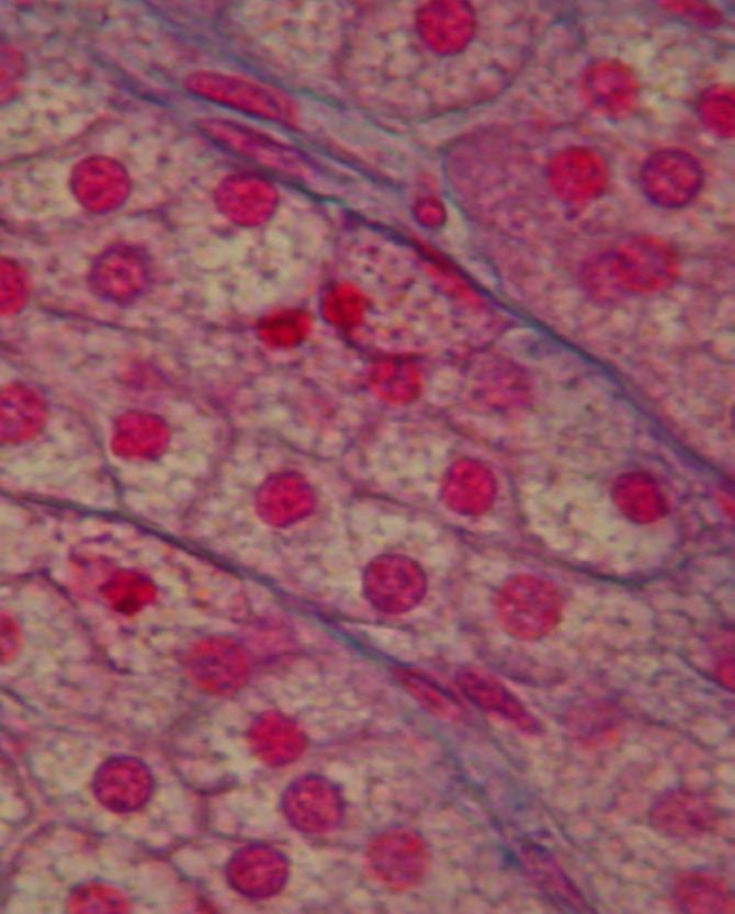
 C C Exo")
17 endocrine islet (of Langerhans) C C Exo Endo Exo

18 HIGHER MAGNIFICATION s s L s s

19 islet surrounding by serous acini

20 ISLET VESSELS V V V

21 ENDOCRINE CELLS

22 serous acini of parotid gland s D

23 The structural organization of human liver tissue

24 LIVER L vcl SP vcl s v D vcl vcl s

25 PORTAL TRIADS L1 PS A V L3 B L2

26 Central vein and sinusoids E V S

27 Sinusoids S E V

28 Cell cords of hepatocytes V C

29 LIVER Sinusoids small irregularly shaped blood vessels

30 Hepatic microstructure
31 Gallbladder wall

32 Gallbladder wall high magnification M pc bm

33 Gallbladder wall high magnification bm Mi M

34 Anastomosis Gallbladder MUCOSA - a cross-connection between adjacent channels, tubes, fibers, pits or other parts of a network. - a connection made surgically between adjacent blood vessels, parts of the intestine, or other channels of the body, or the operation in which this is constructed. M

35 Gallbladder mucosa epithelium E p pm

36 gallbladder mucosa E A bm V E

37 Common bile duct structure

38 parotid submaxillary or submandibular sublingual The salivary glands parotid gland sublingual gland submandibular gland
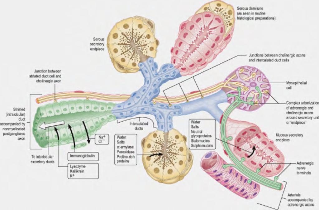
39
 MUCOUS ACINI SUBLINGUAL GLAND (mucous tubules and acini predominate)")
40 PAROTID GLAND (almost exclusively serous acini) SUBMANDIBULAR GLAND (predominantly serous acini) MUCOUS ACINI SUBLINGUAL GLAND (mucous tubules and acini predominate)
 is produced by the parotid gland. 2 4 v 1 L 1 4 2 L 3 3")
41 PAROTID GLAND (almost exclusively serous acini) Serous fluid (as opposed to mucous fluid) is produced by the parotid gland. 2 4 v 1 L L 3 3
 1")
42 PAROTID GLAND (almost exclusively serous acini) 1 A 3 1 2

43 SUBMANDIBULAR GLAND (predominantly serous acini) 2 S A M 1 4 L v n v serous demilune, a layer of lysozyme-secreting serous cells resembling a half moon (Giannuzzi s).
 A 3")
44 SUBMANDIBULAR GLAND (predominantly serous acini) A 3 S M 4 v A S A S
 1 2 4 3 v n L 1 3 1")
45 SUBLINGUAL GLAND (mucous tubules and acini predominate) v n L L

46 MAGNIFICATION OF SUBLINGUAL GLAND ACINI M A S M S M n S S A M 4 S M M
, zona reticularis (ZR), and the highly vascular")
47 adult suprarenal gland (adrenal gland) zona granulosa (ZG), zona fasciculata (ZF), zona reticularis (ZR), and the highly vascular medulla (M).

48 adrenal gland FC L G F R C M V

49 adrenal gland FC G 1 C F 2 R 3 M 4 V

50 zona granulosa (ZG) FC ZG F
 ZF c c ZR")
51 zona fasciculata (ZF) zona reticularis (ZR) ZF c c ZR
52 Zona Fascicolata (ZF) S A B

53 Zona Reticularis (ZR) sinusoid
")
54 Medulla (M)
 of the thyroid")
55 The parathyroid glands are four or more small glands, about the size of a grain of rice, located on the posterior surface (back side) of the thyroid gland

56 human thyroid follicles ( ) reticular connective tissue

57 Parathyroid gland the parathyroid oxyphil cell is larger and paler than the parathyroid chief cell. These cells can be found in clusters in the center of the section and at the periphery. Oxyphil cells appear at the onset of puberty, but have no known function. With nuclear medicine scans, they selectively take up the Technetiumsestamibi complex radiotracer dye to allow delineation of glandular anatomy oxyphil cells

58 human thyroid follicles CT colloidal thyroglobulin calcitonin-secreting C cells follicular epithelium

59 thyroid colloid ( )
")
60 Thyrocyte (T) and basal membrane (BM) T BM
61 calcitonin-secreting C cells C1 C2
ANATOMY AND BASIC FUNCTION OF THE ENDOCRINE GLANDS
 ANATOMY AND BASIC FUNCTION OF THE ENDOCRINE GLANDS Know these endocrine organs of the cat: thymus, thyroid, pancreas, adrenal glands, ovaries, and testes. Review and know microslides, hormones, and structures
The Digestive System Laboratory
 The Digestive System Laboratory 1 The Digestive Tract The alimentary canal is a continuous tube stretching from the mouth to the anus. Liver Gallbladder Small intestine Anus Parotid, sublingual, and submaxillary
The Digestive System
 The Digestive System Identify the Structure and Function. Mesentery of the Large Intestine The mesentery functions to connect the visceral organs to the abdominal wall. Identify the Structure. Nasal Cavity
ACTIVITY 11: RESPIRATORY AND DIGESTIVE SYSTEMS RESPIRATORY SYSTEM
 ACTIVITY 11: RESPIRATORY AND DIGESTIVE SYSTEMS OBJECTIVES: 1) How to get ready: Read Chapters 25 and 26, McKinley et al., Human Anatomy, 4e. All text references are for this textbook. 2) Identify structures
RESPIRATORY SYSTEM. described: pp. 744,746 fig. 25.1, described: p. 746 fig described: p. 776 fig. 26.3
 ACTIVITY 11: RESPIRATORY AND DIGESTIVE SYSTEMS OBJECTIVES: 1) How to get ready: Read Chapters 25 and 26, McKinley et al., Human Anatomy, 5e. All text references are for this textbook. 2) Identify structures
Bio 322 Human Anatomy Objectives for the laboratory exercise Digestive System
 Bio 322 Human Anatomy Objectives for the laboratory exercise Digestive System Required reading before beginning this lab: Saladin, KS: Human Anatomy 5 th ed (2017) Chapter 24 For this lab you will use
Gastrointestinal Tract
 CTO Lab #5 GI TRACT & GLANDS; ENDOCRINE SYSTEM Page 1 Gastrointestinal Tract Slide 126 This section through the esophagus shows the characteristic layers of the gastrointestinal tract. Examine the non-keratinized
HISTOLOGY VIRTUAL LABORATORY GASTROINTESTINAL SYSTEM
 HISTOLOGY VIRTUAL LABORATORY GASTROINTESTINAL SYSTEM LIP (Slides GI 1, 2) Identify the outer portion lined by stratified squamous (keratinized) epithelium. Note the hair follicles and sebaceous glands
BIOL& 253 Lab Manual for Practical #2 Page 1 Rausch. For all slides, know a function for structures marked with a single asterisk (*).
.") BIOL& 253 Lab Manual for Practical #2 Page 1 Rausch Lab equipment: slides, models SLIDES For all slides, know a function for structures marked with a single asterisk (*). DIGESTIVE SYSTEM Layers of the
BIOL& 253 Lab Manual for Practical #2 Page 1 Rausch Lab equipment: slides, models SLIDES For all slides, know a function for structures marked with a single asterisk (*). DIGESTIVE SYSTEM Layers of the
THE ORAL CAVITY
 THE ORAL CAVITY WALL OF ABDOMEN (ANTERIOR) The paraumbilical vein drains into the portal vein and then through the liver. This is an important clinical connection. THE ABDOMINAL VISCERA The small
Exercise. Digestive System. Digestive system function. 1. Define the following terms: a. Chemical digestionb. Mechanical digestionc.
 Exercise 7 The Digestive System NAME: DATE: INSTRUCTOR: SECTION: Digestive system function 1. Define the following terms: a. Chemical digestionb. Mechanical digestionc. Ingestiond. Digestione. Absorptionf.
The stomach is formed of three parts: -
 The stomach is formed of three parts: - (a) CARDIAC STOMACH: - It receives the oesophagus through Cardiac aperture guarded by a cardiac sphincter which prevents regurgitation of food. (b) FUNDIC PART:
Bio 104 Digestive System
 13 Lecture Outline: Digestive System Hole s HAP [Chapters 17 & 18] General Characteristics of the Alimentary Canal A. Functions 1. Ingestion 2. Mechanical digestion 3. Chemical digestion 4. Propulsion
Digestive System. In one end and out the other.
 Digestive System In one end and out the other. Overview Every cell in the body needs nourishment, yet most cells cannot leave their position in the body and travel to a food source, so the food must be
Midterm 2 is Tuesday 5/28/13
 Business Reminder: No class Monday (Memorial Day) Midterm 2 is Tuesday 5/28/13 Optional review session tomorrow @ 5pm Homework due in Lab 1. PreLab 8 (1pt) 2. Replace a Missing Assignment (4 pts) Homework
The Digestive System and Body Metabolism
 14 PART B The Digestive System and Body Metabolism PowerPoint Lecture Slide Presentation by Jerry L. Cook, Sam Houston University ESSENTIALS OF HUMAN ANATOMY & PHYSIOLOGY EIGHTH EDITION ELAINE N. MARIEB
Slide 154: Pancreas, H&E
 Slide 154: Pancreas, H&E the pancreas, located adjacent to the duodenum, is a mixed exocrine and endocrine gland; it is usually readily identifiable by the presence of the interspersed endocrine pancreatic
MICROSTRUCTURES LIPS TOOTH TONGUE OESOPHAGUS STOMACH, CARDIAC, PYLORIC FUNDIC GLANDS
 MICROSTRUCTURES LIPS TOOTH TONGUE OESOPHAGUS STOMACH, CARDIAC, PYLORIC FUNDIC GLANDS HUMAN ANATOMY: MICROSTRUCTURES CLASSIFICATION: LOCATION AND BOUNDARIES, FORM, FUNCTION, MICROSCOPIC STRUCTURE: A hollow
Lab activity manual - Histology of the digestive system. Lab activity 1: esophagus stomach - small intestines
 Lab activity manual - Histology of the digestive system Jeanne Adiwinata Pawitan Prerequisite: Histology of the 4 basic tissues In this module we learn about the histology of the digestive system, from
Epithelia will be discussed according to the following scheme: Type Number of layers Shape Line drawing. Squamous Cuboidal Columnar
 Epithelia Epithelia will be discussed according to the following scheme: Type Number of layers Shape Line drawing Simple Squamous Cuboidal Columnar Covering and Lining epithelium Pseudostratified Stratified
Gastrointestinal System!
 Gastrointestinal System! Assoc. Prof. Prasit Suwannalert, Ph.D. (Email: prasit.suw@mahidol.ac.th)! Objectives: After learning, student should be able to describe and discuss in topics of! 1. Anatomical
- Digestion occurs during periods of low activity - Produces more energy than it uses. - Mucosa
 Introduction Digestive System Chapter 29 Provides processes to break down molecules into a state easily used by cells - A disassembly line: Starts at the mouth and ends at the anus Digestive functions
Biology Human Anatomy Abdominal and Pelvic Cavities
 Biology 351 - Human Anatomy Abdominal and Pelvic Cavities Please place your name and I.D. number on the back of the last page of this exam. You must answer all questions on this exam. Because statistics
The Digestive System. Chapter 25
 The Digestive System Chapter 25 Introduction Structure of the digestive system A tube that extends from mouth to anus Accessory organs are attached Functions include Ingestion Movement Digestion Absorption
Chapter 14: The Digestive System
 Chapter 14: The Digestive System Digestive system consists of Muscular tube (digestive tract) alimentary canal Accessory organs teeth, tongue, glandular organs 6 essential activities 1. 2. 3. 4. 5. 6.
consists of: Muscular, hollow tube (= digestive tract ) + Various accessory organs
 + Various accessory organs") DIGESTIVE SYSTEM consists of: Muscular, hollow tube (= digestive tract ) + Various accessory organs FUNCTION Individual parts function in: ingestion mechanical digestion chemical and enzymatic digestion
DIGESTIVE SYSTEM consists of: Muscular, hollow tube (= digestive tract ) + Various accessory organs FUNCTION Individual parts function in: ingestion mechanical digestion chemical and enzymatic digestion
The endocrine system
 The endocrine system Overview I. Pituitary II. Adrenals III. Pancreas IV. Thyroid V. Parathyroids VI. Ovaries, testis fenestrated capillaries are general structures http://www.ama-assn.org/ama/pub/category/7157.html
Includes mouth, pharynx, esophagus, stomach, small intestine, large intestine, rectum, anus. Salivary glands, liver, gallbladder, pancreas
 Chapter 14 The Digestive System and Nutrition Digestive System Brings Nutrients Into the Body The digestive system includes Gastrointestinal (GI) tract (hollow tube) Lumen: space within this tube Includes
Lab 8: Digestive System
 BIOL 221 A&P II Lab 8: Digestive System Become familiar with the gross anatomy of the digestive system (Exercise 38) using the models, Fig. 38.1 (Activity 1), and the rat. Recognize and know the functions
Lab 5 Digestion and Hormones of Digestion. 7/16/2015 MDufilho 1
 Lab 5 Digestion and Hormones of Digestion 1 Figure 23.1 Alimentary canal and related accessory digestive organs. Mouth (oral cavity) Tongue* Parotid gland Sublingual gland Submandibular gland Salivary
- Digestion occurs during periods of low activity - Produces more energy than it uses. 3 Copyright 2016 by Elsevier Inc. All rights reserved.
 Introduction Digestive System Chapter 29 Provides processes to break down molecules into a state easily used by cells - A disassembly line: Starts at the mouth and ends at the anus Digestive functions
A. Incorrect! The esophagus connects the pharynx and the stomach.
 Human Physiology - Problem Drill 19: Digestive Physiology and Nutrition Question No. 1 of 10 Instructions: (1) Read the problem and answer choices carefully, (2) Work the problems on paper as 1. This organ
PRACTICAL ROADMAP. GLANDS AFFECTING LIFESTYLE WJ van der Spuy & T Tshabalala
 PRACTICAL ROADMAP GLANDS AFFECTING LIFESTYLE WJ van der Spuy & T Tshabalala GLANDS AFFECTING LIFESTYLE Submandibular gland (salivary gland) Liver Pancreas Hypophysis (pituitary gland) Thyroid Suprarenal
The Digestive System. Chapter
 The Digestive System Chapter 15.1 Functions: mechanical and chemical breakdown of food *absorption of nutrients Consists of alimentary canal and accessory organs Wall of the Alimentary Canal 15.2 Characteristics
Histology Lab. looking at microscopic pictures of tissues, for more information use Junqueira book and you can use BlueHistolgy website
 Done By: Aseel Twaijer & Laith Sorour Histology Lab *These notes help in differentiating tissues and you must read them while looking at microscopic pictures of tissues, for more information use Junqueira
DIGESTIVE SYSTEM. Chapter 25
 DIGESTIVE SYSTEM Chapter 25 DIGESTIVE SYSTEM Digestive Tract Mouth Pharynx Esophagus Stomach Small intestines Large intestines Anus Accessory Organs Teeth Tongue Salivary glands Pancreas Liver Gallbladder
General Structure of Digestive Tract
 Dr. Nabil Khouri General Structure of Digestive Tract Common Characteristics: Hollow tube composed of a lumen whose diameter varies. Surrounded by a wall made up of 4 principal layers: Mucosa Epithelial
Digestive System 7/15/2015. Outline Digestive System. Digestive System
 Digestive System Biology 105 Lecture 18 Chapter 15 Outline Digestive System I. Functions II. Layers of the GI tract III. Major parts: mouth, pharynx, esophagus, stomach, small intestine, large intestine,
Anatomy & Physiology Revealed Instructions. 1. From the Module dropdown menu, chose the 12. Digestive system.
 #10 - Objectives: Examine the histology of selected body organs using Anatomy & Physiology Revealed software and microscope slides. Be able to identify each organ and the specific structures indicated
Practical Histology o
 Practical Histology o 1.. Contents: Histology of the : Stomach Esophagus Small intestine Large intestine Liver Gallbladder Exocrine pancreas Spleen GNT Block Things you need to know before the exam : o
This lab activity is aligned with Visible Body s Human Anatomy Atlas app. Learn more at visiblebody.com/professors
 1 This lab activity is aligned with Visible Body s Human Anatomy Atlas app. Learn more at visiblebody.com/professors 2 A. Digestive System Overview To Start: Go to the Views menu and scroll down to the
Endocrine Histology Lab GUIDE TO MICROSCOPES IN LAB
 Endocrine Histology Lab GUIDE TO MICROSCOPES IN LAB The micrographs that appear on this review page are typical views of the tissues seen in the laboratory. The descriptions that accompany them are designed
Peritoneum: Def. : It is a thin serous membrane that lines the walls of the abdominal and pelvic cavities and clothes the viscera.
 Peritoneum: Def. : It is a thin serous membrane that lines the walls of the abdominal and pelvic cavities and clothes the viscera. Layers of the peritoneum: 1. Outer Layer ( Parietal Peritoneum) : lines
Chapter 9. The digestive system. Glossary. Louise McErlean
 Chapter 9 The digestive system Louise McErlean Glossary Absorption Process whereby the products of digestion move into the blood or lymph fluid. Acini glands Produce pancreatic juice. Amylase Carbohydrate
Laboratory exercises for abdominal organs
 Laboratory exercises for abdominal organs Slide #77 (C007- H- 107A). Pancreas, dog. pancreatic islets CENTROACINAR CELLS ARE THE BEGINNING CELLS OF THE INTERCALATED DUCTS THAT DRAIN THE SECRETORY ACINI
The Digestive System. Chapter 16. Introduction. Overview of Digestive System. Histological Organization. Movement and Mixing of Digestive Materials
 The Digestive System Chapter 16 Introduction Structure of the digestive system A tube that extends from mouth to anus Accessory organs are attached Functions include Ingestion Movement Digestion Absorption
Respiratory & Digestive Organs of the Head and Neck, Human;
 Name Date Lab Exercise 5: Lab Exercise 6: Lab Exercise 7: Lab Exercise 8: Respiratory & Digestive Organs of the Head and Neck, Human; Histology of the Respiratory System Digestive System Models, Human
DIGESTIVE SYSTEM II ACCESSORY DIGESTIVE ORGANS
 DIGESTIVE SYSTEM II ACCESSORY DIGESTIVE ORGANS Dr. Larry Johnson Texas A& M University Objectives Distinguish between the parotid and submandibular salivary glands. Understand and identify the structural
Soft palate elevates, closing off the nasopharynx. Hard palate Tongue Bolus Epiglottis. Glottis Larynx moves up and forward.
 The Cephalic Phase Chemical and mechanical digestion begins in the mouth Saliva is an exocrine secretion Salivary secretion is under autonomic control Softens and lubricates food Chemical digestion: salivary
Lab activity manual Histology of the digestive system
 Lab activity manual Histology of the digestive system Jeanne Adiwinata Pawitan Prerequisite: Histology of the 4 basic tissues In this module we learn about the histology of the digestive system, from the
1. To describe the gross structure of the pituitary gland and be able to identify the pars nervosa, pars intermedia and pars distalis.
 ENDOCRINE Objectives 1. To describe the gross structure of the pituitary gland and be able to identify the pars nervosa, pars intermedia and pars distalis. 2. Identify and describe the histological features
DIGESTIVE SYSTEM ALIMENTARY CANAL / GI TRACT & ACCESSORY ORGANS. Mar 16 10:34 PM
 DIGESTIVE SYSTEM ALIMENTARY CANAL / GI TRACT & ACCESSORY ORGANS Mar 16 10:34 PM 1 I. Digestive System Functions > Ingestion the taking in of food > Propulsion movement caused by force > Digestion breakdown
Histology 3. We will continue talking about a few things from last lecture, starting with M cells:
 Histology 3 This is the last Histology lecture in the GI system. Enjoy! There are some extra notes listed as footnotes. We will continue talking about a few things from last lecture, starting with M cells:
DIGESTIVE TRACT ESOPHAGUS
 DIGESTIVE TRACT From the lower esophagus to the lower rectum four fundamental layers comprise the wall of the digestive tube: mucosa, submucosa, muscularis propria (externa), and adventitia or serosa (see
BIO 116 Anatomy & Physiology II Practice Assignment 3 - The Lymphatic, Immune and Digestive Systems This is not a required assignment
 BIO 116 Anatomy & Physiology II Practice Assignment 3 - The Lymphatic, Immune and Digestive Systems This is not a required assignment 1. Which are components of the lymphatic system? a: Thyroid gland b:
Small Plicae Circularis. Short Closely packed together. Sparse, completely absent at distal part Lymphoid Nodule
 Intestines Differences Between Jejunum and Ileum Types Jejunum Ileum Color Deeper red Paler pink Calibre Bigger Smaller Thickness of wall Thick and Heavy Thin and Lighter Vascularity Highly vascularised
Small intestine. Small intestine
 General features Tubular organ longest part; 5-6 m most of chemical digestion absorption of nutrients reabsorption of H2O occurs. Two structural features; maximize the lumenal surface area villi microvilli
I. The Alimentary Canal (GI track)
") A. About 9 meters long B. Passes through the ventral cavity. C.Movements of the Tube 1. Mixing movements- smooth muscles contract rhythmically. 2. Propelling movements- a wavelike motion called peristalsis.
A. About 9 meters long B. Passes through the ventral cavity. C.Movements of the Tube 1. Mixing movements- smooth muscles contract rhythmically. 2. Propelling movements- a wavelike motion called peristalsis.
The Digestive System. What is the advantage of a one-way gut? If you swallow something, is it really inside you?
 The Digestive System What is the advantage of a one-way gut?! If you swallow something, is it really inside you? Functions and Processes of the Digestive System: Move nutrients, water, electrolytes from
Lab Monitor Images Dissection of the Abdominal Vasculature + Lower Digestive System
 Lab Monitor Images Dissection of the Abdominal Vasculature + Lower Digestive System Stomach & Duodenum Frontal (AP) View Nasogastric tube 2 1 3 4 Stomach Pylorus Duodenum 1 Duodenum 2 Duodenum 3 Duodenum
Overview of the Digestive System Organs are divided into two groups
 C H A P T E R 23 The Digestive System Overview of the Digestive System Organs are divided into two groups The alimentary canal Mouth, pharynx, and esophagus Stomach, small intestine, and large intestine
Chapter 26 The Digestive System
 Chapter 26 The Digestive System Digestive System Gastroenterology is the study of the stomach and intestine. Digestion Catabolism Absorption Anabolism The actions of the digestive system are controlled
Netter's Anatomy Flash Cards Section 4 List 4 th Edition
 Netter's Anatomy Flash Cards Section 4 List 4 th Edition https://www.memrise.com/course/1577335/ Section 4 Abdomen (31 cards) Plate 4-1 Bony Framework of Abdomen 1.1 Costal cartilages 1.2 Iliac crest 1.3
Endocrine System. Organs and Tissues: Pituitary Adrenals Pancreas Thyroid Parathyroids
 Endocrine System Organs and Tissues: Pituitary Adrenals Pancreas Thyroid Parathyroids Bruce A. Fenderson, Ph.D. Pathology, Anatomy & Cell Biology Sidney Kimmel Medical College Bruce.Fenderson@Jefferson.edu
Chapter 16. Lecture and Animation Outline
 Chapter 16 Lecture and Animation Outline To run the animations you must be in Slideshow View. Use the buttons on the animation to play, pause, and turn audio/text on or off. Please Note: Once you have
Small Intestine, Large Intestine and anal cannel
 Small Intestine, Large Intestine and anal cannel 32409 Small intestine Large intestine Small intestine General Structure of the Digestive Tract rat 32409 Epithelium with goblet cells and absorptive cells
Two main groups Alimentary canal continuous coiled hollow tube Accessory digestive organs
 Digestion Breakdown of ingested food Absorption of nutrients into the blood Metabolism Production of cellular energy (ATP) Constructive and degradative cellular activities Two main groups Alimentary canal
Accessory Glands of Digestive System
 Accessory Glands of Digestive System The liver The liver is soft and pliable and occupies the upper part of the abdominal cavity just beneath the diaphragm. The greater part of the liver is situated under
Alimentary Canal (I)
") Alimentary Canal (I) Esophagus and Stomach (Objectives) By the end of this lecture, the student should be able to discuss the microscopic structure in correlation with the function of the following organs:
Alimentary Canal (I) Esophagus and Stomach (Objectives) By the end of this lecture, the student should be able to discuss the microscopic structure in correlation with the function of the following organs:
Al s 202 study guide answers Answers Respiratory System 1 External nares (nostrils) 33 Carina 2 Vestibule 34 Left primary bronchus 3 Nasal cavity 35
 33 Carina 2 Vestibule 34 Left primary bronchus 3 Nasal cavity 35") Trachea & Respiratory Histology 1 Epiglottis 26 Capillary 2 Larynx 27 Alveolar sac 3 Thyroid cartilage 28 Alveoli/Alveolus 4 Cricoid cartilage 29 Basement membrane 5 Vocal folds (True vocal cords) 30 Cilia
Trachea & Respiratory Histology 1 Epiglottis 26 Capillary 2 Larynx 27 Alveolar sac 3 Thyroid cartilage 28 Alveoli/Alveolus 4 Cricoid cartilage 29 Basement membrane 5 Vocal folds (True vocal cords) 30 Cilia
BIO 132 Anatomy and Physiology II Spring, 2016 Exam 1 Name: BIO 132 ID Number. Section 1 Answer questions 1 40 on the scan sheet.
 BIO 132 Anatomy and Physiology II Spring, 2016 Exam 1 Name: BIO 132 ID Number Section 1 Answer questions 1 40 on the scan sheet. 1. The homeostatic value of a particular controlled variable in the body
ANATOMY & PHYSIOLOGY ONLINE COURSE - SESSION 13 THE DIGESTIVE SYSTEM
 ANATOMY & PHYSIOLOGY ONLINE COURSE - SESSION 13 THE DIGESTIVE SYSTEM The digestive system also known as the alimentary canal or gastrointestinal tract consists of a series of hollow organs joined in a
The Digestive System PowerPoint Lecture Presentations prepared by Steven Bassett Southeast Community College Lincoln, Nebraska
 25 The Digestive System PowerPoint Lecture Presentations prepared by Steven Bassett Southeast Community College Lincoln, Nebraska Introduction The digestive system consists of: The digestive tract Accessory
Duodenum retroperitoneal
 Duodenum retroperitoneal C shaped Initial region out of stomach into small intestine RETROperitoneal viscus Superior 1 st part duodenal cap ; moves upwards and backwards to lie on the R crura medial to
Human Body Systems. The human body consists of 11 major systems.
 Human Body Systems The human body consists of 11 major systems. Systems Skeletal Muscular Cardiovascular or Circulatory Digestive Respiratory Urinary Endocrine Nervous Integumentary Lymphatic or the Immune
21 Endocrine organs and cells
 21 Endocrine organs and cells The endocrine system consists of discrete organs, portions of organs and distributed cells that secrete hormones into surrounding tissues or structures. Objectives You should
HISTOLOGY. GIT Block 432 Histology Team. Lecture 1: Alimentary Canal (1) (Esophagus & Stomach) Done by: Ethar Alqarni Reviewed by: Ibrahim Alfuraih
 (Esophagus & Stomach) Done by: Ethar Alqarni Reviewed by: Ibrahim Alfuraih") HISTOLOGY Lecture 1: Alimentary Canal (1) (Esophagus & Stomach) Done by: Ethar Alqarni Reviewed by: Ibrahim Alfuraih Color Guide: Black: Slides. Red: Important. Green: Doctor s notes. Blue: Explanation.
HISTOLOGY Lecture 1: Alimentary Canal (1) (Esophagus & Stomach) Done by: Ethar Alqarni Reviewed by: Ibrahim Alfuraih Color Guide: Black: Slides. Red: Important. Green: Doctor s notes. Blue: Explanation.
ORGANS OF THE DIGESTIVE SYSTEM
 ORGANS OF THE DIGESTIVE SYSTEM OBJECTIVES: 1. List and describe the major activities of the digestive system. 2. Identify and give the functions of the organs in and along the digestive tract. MAJOR ACTIVITIES
Energy, Chemical Reactions and Enzymes
 Phosphorylation Hydrolysis Energy, Chemical Reactions and Enzymes Chapter 2 (selections) What is Energy? Energy is the capacity to do work Potential Energy Kinetic Energy Chemical Bond Energy Like a rechargeable
The Digestive System and Body Metabolism Premedical Biology
 The Digestive System and Body Metabolism Premedical Biology Copyright 2003 Pearson Education, Inc. publishing as Benjamin Cummings The Digestive System and Body Digestion Metabolism Breakdown of ingested
DIGESTIVE. CHAPTER 17 Lecture: Part 1 Part 2 BIO 212: ANATOMY & PHYSIOLOGY II
 BIO 212: ANATOMY & PHYSIOLOGY II CHAPTER 17 Lecture: DIGESTIVE Part 1 Part 2 Dr. Lawrence G. Altman www.lawrencegaltman.com Some illustrations are courtesy of McGraw-Hill. SMALL INTESTINE DUODENUM > JEJUNUM
Digestive system L 4. Lecturer Dr. Firdous M. Jaafar Department of Anatomy/Histology section
 Digestive system L 4 Lecturer Dr. Firdous M. Jaafar Department of Anatomy/Histology section objectives 1-Describe the structure of liver. 2-Define liver lobule, and identify its zones. 3-Define portal
Digestive Anatomy Lab
 Digestive Anatomy Lab In-Lab Exercises I have included the word list in this document. Any descrepencies between this document and the wordlist, you should default to this document. There is a lot of repetition
THE DIGESTIVE SYSTEM THE FOUR STEPS OF DIGESTION
 THE DIGESTIVE SYSTEM WHAT IS THE DIGESTIVE SYSTEM? the digestive system is a long, muscular tube The entire system is approximately 9m in length and is usually a one way tract WHY DO WE NEED A DIGESTIVE
DIGESTIVE SYSTEM CLASS NOTES. tube along with several
 DIGESTIVE SYSTEM CLASS NOTES Digestion Breakdown of food and the of nutrients in the bloodstream. Metabolism Production of for and cellular activities. The digestive system is composed of the canal which
Nasogastric tube. Stomach. Pylorus. Duodenum 1. Duodenum 2. Duodenum 3. Duodenum 4
 Esophagus Barium Swallow Stomach and Duodenum 4 year old Upper GI Nasogastric tube Stomach and Duodenum 4 year old Upper GI Nasogastric tube Stomach Pylorus Duodenum 1 Duodenum 2 Duodenum 3 Duodenum 4
Jhia Anjela D. Rivera 1 1. BS Biology, Department of Biology, College of Science, Polytechnic University of the Philippines
 DIGESTIVE SYSTEM Jhia Anjela D. Rivera 1 1 BS Biology, Department of Biology, College of Science, Polytechnic University of the Philippines DIGESTIVE SYSTEM Consists of the digestive tract (gastrointestinal
(A) Diarrhea. (B) Stomach cramps. (C) Dehydration due to excess fluid loss. (D) A, B, and C are correct. (E) Only answer B is correct.
 Diarrhea. (B) Stomach cramps. (C) Dehydration due to excess fluid loss. (D) A, B, and C are correct. (E) Only answer B is correct.") Human Anatomy - Problem Drill 21: The Digestive System Question No. 1 of 10 1. A 26-year-old male is treated in the emergency department for severe gastrointestinal disturbance. Which of the following
Human Anatomy - Problem Drill 21: The Digestive System Question No. 1 of 10 1. A 26-year-old male is treated in the emergency department for severe gastrointestinal disturbance. Which of the following
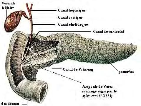
Anatomy of the liver and pancreas
 Anatomy of the liver and pancreas Prof. Abdulameer Al-Nuaimi E-mail: a.al-nuaimi@sheffield.ac.uk abdulameerh@yahoo.com Liver Aorta Pulm. Trunk Rt. At, Duct. Art. Lt. Ven. Rt. Ven. Internal Posterior
Lungs a. d. b. c. e.
 Lungs d. e. Lungs Right superior lobe Right middle lobe Right inferior lobe d. Left superior lobe e. Left inferior lobe Sinuses d. Nasal Cavity & Sinuses g. g. i. Nasal Cavity & Sinuses g. h. d. f. e.
BLOCK IV: OFFICIAL BODY PARTS LIST FOR ANTERIOR ABDOMINAL WALL AND ABDOMINAL CONTENTS
 BLOCK IV: OFFICIAL BODY PARTS LIST FOR ANTERIOR ABDOMINAL WALL AND ABDOMINAL CONTENTS External oblique muscle Muscular portion Aponeurotic portion Superficial inguinal ring Lateral (inferior) crus Medial
Digestive System II - Lower tract Revised
 ANAT D502 Basic Histology Digestive System II - Lower tract Revised 10.12.12 Outline: I. Small intestine II. Enterocyte digestion II. Hepatic portal system IV. Large intestine V. Enteric nervous system
MICROSCOPIC STRUCTURE OF LIVER, GALLBLADDER, GALL DUCTS, AND PANCREAS OVERVIEW OF DEVELOPMENT OF THE ALIMENTARY CANAL
 Lecture 2 ESS_3rd semester MICROSCOPIC STRUCTURE OF LIVER, GALLBLADDER, GALL DUCTS, AND PANCREAS OVERVIEW OF DEVELOPMENT OF THE ALIMENTARY CANAL MICROSCOPIC STRUCTURE OF LIVER - is the largest gland of
LYMPHATIC ANATOMY LAB. BIO 139 ANATOMY AND PHYSIOLOGY II MARY CATHERINE FLATH, Ph.D.
 LYMPHATIC ANATOMY LAB BIO 139 ANATOMY AND PHYSIOLOGY II MARY CATHERINE FLATH, Ph.D. THE LYMPHATIC SYSTEM ORGANS PRIMARY BONE MARROW THYMUS SECONDARY LYMPH NODES SPLEEN FUNCTIONS CONTROL DISEASE TRANSPORT
Lecture 1. Introduction to splanhnology. Digestive system
 Lecture 1 Introduction to splanhnology. Digestive system SPLANCHNOLOGY (THE SCIENCE OF THE VISCERA) SOMATOLOGY SPLANCHNOLOGY The general cover body (skin) The skeletal system The connection system The
Digestive System. Human Anatomy & Physiology University of Washington PMT
 Digestive System Human Anatomy & Physiology University of Washington PMT Digestive System Function Acquires nutrients from environment Anabolism Uses raw materials to synthesize essential compounds Catabolism
NOTES: The Digestive System (Ch 14, part 2)
") NOTES: The Digestive System (Ch 14, part 2) PANCREAS Structure of the pancreas: The pancreas produces PANCREATIC JUICE that is then secreted into a pancreatic duct. The PANCREATIC DUCT leads to the The
NOTES: The Digestive System (Ch 14, part 2) PANCREAS Structure of the pancreas: The pancreas produces PANCREATIC JUICE that is then secreted into a pancreatic duct. The PANCREATIC DUCT leads to the The
The Human Body: An Overview of Anatomy. Anatomy. Physiology. Anatomy - Study of internal and external body structures
 C H A P T E R 1 The Human Body: An Orientation An Overview of Anatomy Anatomy The study of the structure of the human body Physiology The study of body function Anatomy - Study of internal and external
GASTROINTESTINAL SYSTEM
 GASTROINTESTINAL SYSTEM Topographic Anatomy of the Abdomen Surface Landmarks Xiphoid process T9/T10 Inferior costal margin L2/L3 Iliac Crest L4 level ASIS L5/S1 level Pubic symphysis level of greater trochanter
In the name ofgod. Abdomen 3. Dr. Zahiri
 In the name ofgod Abdomen 3 Dr. Zahiri Peritoneum Peritoneum It is the serous membrane(a type of loose connective tissue and is covered by mesothelium) that lines the abdominal cavity. Extensions of the