Practical Histology o
|
|
|
- Bethany Barton
- 5 years ago
- Views:
Transcription


1 Practical Histology o 1.. Contents: Histology of the : Stomach Esophagus Small intestine Large intestine Liver Gallbladder Exocrine pancreas Spleen GNT Block

2 Things you need to know before the exam : o The pictures in the exam will be the same as the ones included in the slides. o Do t tr to take short uts duri g the e a so avoid usi g a reviatio s so you don t lose marks. o Please keep in mind that this work is done by students, so if there are any mistakes please inform us. o This work is not by any means a reference. o Please study hard and don t worry the exam will be easy!! Identify the *م جدا جدا جدا جدا تبربط ن الص ر ب لمع مة ب الختب ر بيجي سؤال : مالحظة هذا اكثر سؤال يجي structure مثال stomach انه يجي ص ره لل, * الدكت ر نبه ع ى شيء يخط ن فيه كثير من الطال Mucosa and submucosa,ect هن الز تكت الاليرز ك م ة Mention the 4 layers ي ل Epithelium, lamina propria,ect هن تكت subdivisions of the mucosa بس لمن ي ل


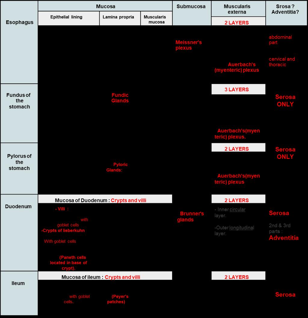
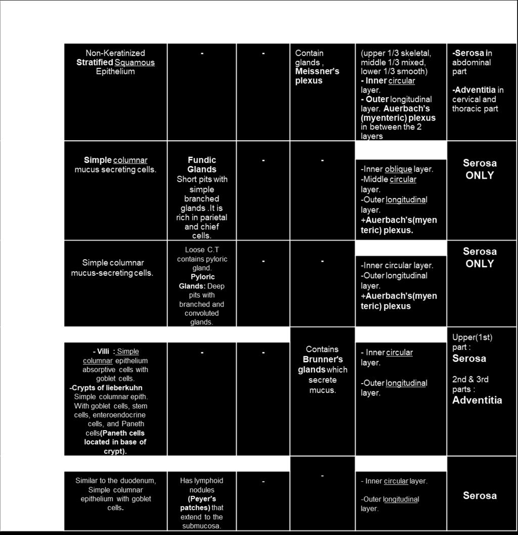
3 Esophagus 1- Mucosa: Epithelium Non-Keratinized Stratified Squamous Epithelium. Lamina propria Muscluaris Mucosae 2- Submucosa Submucosal Glands Meissner's plexuses 3- Muscularis Externa Inner circular M e teri ple uses Auer a h s Outer longitudinal layer. 4- Serosa ( in abdominal part ) or Adventitia ( in cervical and thoracic parts ) 4

 Outer longitudinal 4-Serosa Peptic cells (basophili Parietal cells ( acidophilic appears pink in")
4 1- Mucosa: Fundus of the stomach Fundic Glands Epithelium: simple columnar epithelium Lamina propria Muscluaris Mucosae Fundic Glands: Parietal cells Peptic cells Mucous neck cells Stem cells Enteroendocrine cells Short pits 2- Submucosa NO submucosal glands meissner's plexuses 3- Muscularis Externa Inner oblique Middle circular M e teri ple uses Auer a h s d) Outer longitudinal 4-Serosa Peptic cells (basophili Parietal cells ( acidophilic appears pink in colour )
 cells")

5 1- Mucosa: Pylorus of the stomach Epithelium: simple columnar epithelium Pyloric Glands : Mucous neck cells Few parietal cells Their pits are deep Stem cells No peptic ( chief ) cells Enteroendocrine cells Lamina propria d) Muscluaris Mucosae 2- Submucosa NO submucosal glands Meissner's plexuses 3- Muscularis Externa Inner circular M e teri ple uses Auer a h s Outer longitudinal 4-Serosa


 e)")
6 Villus Intestinal villi and crypts Crypt 1-Villi: Covering epithelium Central core of C.T contains : Lymphocytes Lacteal 2-Crypts : Columnar absorptive cells. Goblet cells Enteroendocrine (EE) cells d) Paneth cells ( at the base of the crypt ) e) Stem cells Paneth cells Paneth cells


 4.")

 meissner's plexuses 3- Muscularis")
7 Duodenum 1- Mucosa (Shows villi and crypts) : Epithelium lining : Villi : 1. simple columnar epithelium (brush border) with goblet cells crypts of lieberkuhn : 1. Paneth cells 2. Goblet cells 3. Simple columnar epithelium (brush border) 4. Stem cells 5. M cells over lymphoid nodules Lamina propria Muscluaris Mucosae 2- Submucosa Bru er s gla ds (seromucus glands) meissner's plexuses 3- Muscularis Externa Inner circular M e teri ple uses Auer a h s Outer longitudinal 4-Serosa Recommended by the doctor Brunner's glands the centre is whitish -
 : simple columnar epithelium with")


 Outer longitudinal 4-Serosa Peyer's")
8 1- Mucosa: Ileum Epithelium lining (Similar to the deudenum) : simple columnar epithelium with goblet cells M cells over lymphoid nodules Lamina propria Has lymphoid nodules (Peyer's patches) that extend to the submucosa. Muscluaris Mucosae 2- Submucosa 3- Muscularis Externa: Inner circular Myenteric plexuses ( Auer a h s ) Outer longitudinal 4-Serosa Peyer's patches Peyer's patches

9 Ileum Recommended by the doctor

 Muscluaris Mucosae NO VILLI")

.")

10 1- Mucosa (Shows only crypts) : Epithelium lining : simple columnar epithelium with goblet cells Lamina propria : Contains frequent lymphatic nodules (White circle) Muscluaris Mucosae NO VILLI 2- Submucosa 3- Muscularis Externa Inner circular M e teri ple uses Auer a h s Outer longitudinal (has Teniae coli). 4-Serosa (has appendices epiploicae). Cells lining the crypts of the colon are: Surface columnar absorptive cells. Goblet cells. Enteroendocrinecells. Stem cells. M-cells. Colon
 :")


")
11 Appendix 1- Mucosa: Epithelium lining (Similar to the Colon) : simple columnar epithelium with goblet cells Lamina propria Contains lymphoid nodules Muscluaris Mucosae 2- Submucosa 3- Muscularis Externa: Inner circular Myenteric plexuses ( Auer a h s ) Outer longitudinal 4-Serosa

 C.T.")
 Hepatocytes.")

12 Liver 1- Features: Classical hepatic lobule. Central hepatic vein. Portal area. d) C.T. Mention four cells in the square: d) Hepatocytes. Ito Cells ( stellate cells ) Kuppfer Cells Cells Of Endothelium Reticular fibers in liver

 Kuppfer Cells Cells")
13 Portal Area 1- Features: d) Branch of portal vein. Bile duct. Branch of hepatic artery C.T Mention four cells in the square: d) Hepatocytes. Ito Cells ( stellate cells ) Kuppfer Cells Cells Of Endothelium


14 Gallbladder 1- Mucosa: Epithelium lining (Similar to the Colon) : simple columnar epithelium Lamina propria Contains contains mucous glands in the neck of gall bladder. Muscluaris Mucosae 2- Muscularis Externa: Smooth muscle fibres are oriented in all directions M e teri ple uses Auer a h s 3- Adventitia except the fundus which is Serosa


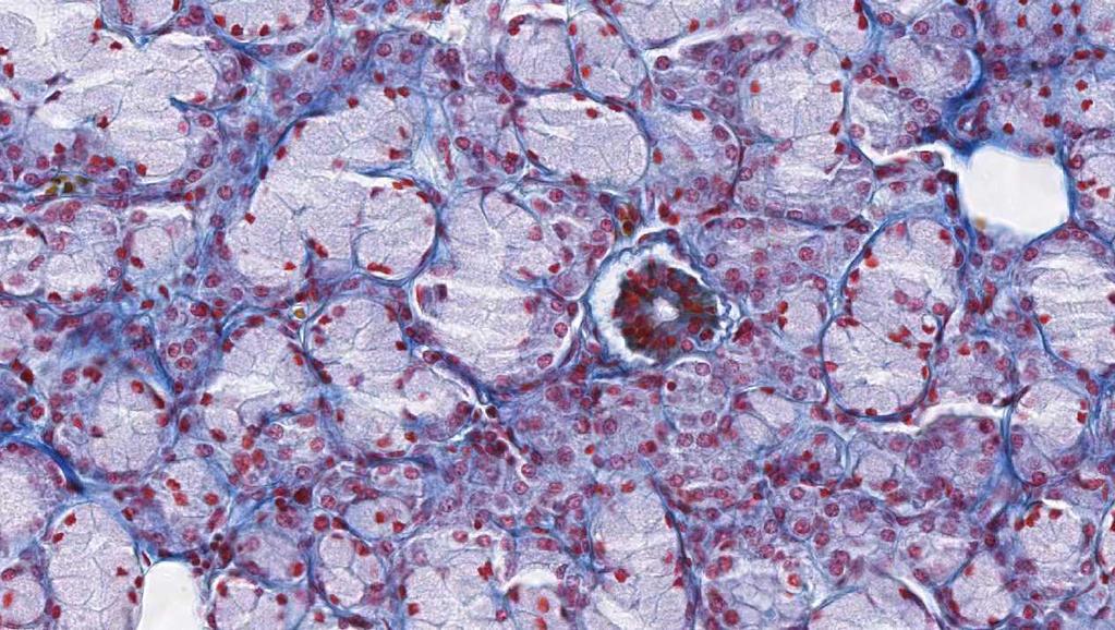
15 Pancreas 1- Features: 1-Islets of Langerhans 2-Pancreatic acini : 3-Centroacinar cells: pancreatic acinar cells 2-Features of the pancreatic acinar cells : Basal part basophilic due to abundant rer Apical part acidophilic due to granules
: housing B lymphocytes, plasma cells and")
 cords (Bilroth s cord): Extravasated blood cells,")
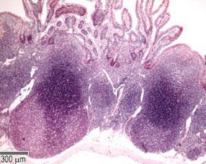
16 Spleen 1- Features: White pulp Periarterial lymphatic sheaths (PALS): housing T lymphocytes. Lymphoid follicles (with germinal centers): housing B lymphocytes, plasma cells and macrophages. Central artery Red pulp: Splenic (pulp) cords (Bilroth s cord): Extravasated blood cells, plasma cells, B and T lymphocytes, macrophages, reticular cells and fibers. Splenic blood sinusoids.
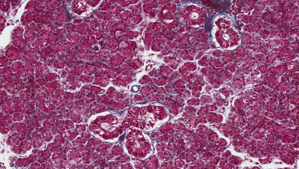
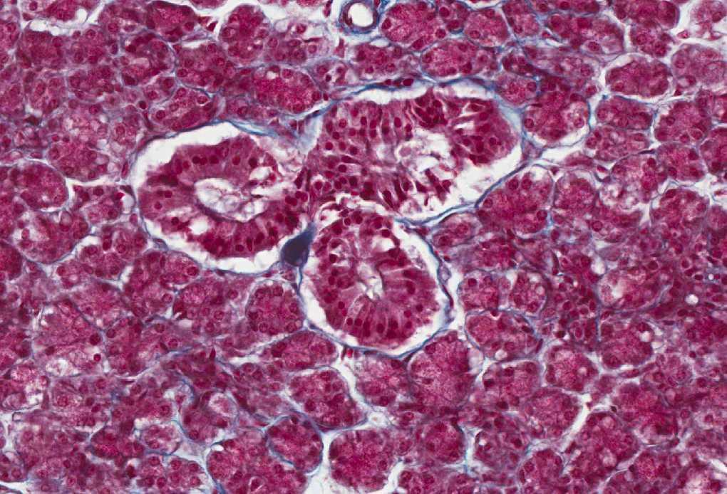
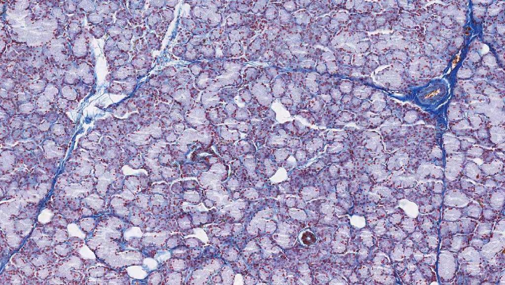
17 Sublingual Gland Parotid Gland
18 SUMMERY
19 SUMMERY Colon Mucosa of colon : Crypts ONLY 2 LAYERS Serosa Simple columnar epithelium with goblet cells. Contains frequent lymphatic nodules -Inner circular layer. - -Outer longitudinal layer. (has Teniae coli) (has appendices epiploicae) Cells lining the crypts are: 1. Surface columnar absorptive cells. 2. Goblet cells. 3. Enteroendocrine Cells. 4. Stem cells. 5. M-cells Appendix Mucosa of Appendix : shallow Crypts ONLY Similar to the colon, Simple columnar epithelium with goblet cells. Contains lymphatic nodules. 2 LAYERS - - Serosa -Inner circular layer. - Outer longitudinal layer Mucosa of GallBladder : highly folded GallBladder Simple columnar epithelium contains mucous glands in the neck of gallbladder - - Srosa or adventitia


20 Thank you & good luck - Histology team Done by: Rana Barasain Omar Turkistani Faisal Alrabaii
Small Intestine, Large Intestine and anal cannel
 Small Intestine, Large Intestine and anal cannel 32409 Small intestine Large intestine Small intestine General Structure of the Digestive Tract rat 32409 Epithelium with goblet cells and absorptive cells
Alimentary Canal (I)
") Alimentary Canal (I) Esophagus and Stomach (Objectives) By the end of this lecture, the student should be able to discuss the microscopic structure in correlation with the function of the following organs:
Alimentary Canal (I) Esophagus and Stomach (Objectives) By the end of this lecture, the student should be able to discuss the microscopic structure in correlation with the function of the following organs:
HISTOLOGY. GIT Block 432 Histology Team. Lecture 1: Alimentary Canal (1) (Esophagus & Stomach) Done by: Ethar Alqarni Reviewed by: Ibrahim Alfuraih
 (Esophagus & Stomach) Done by: Ethar Alqarni Reviewed by: Ibrahim Alfuraih") HISTOLOGY Lecture 1: Alimentary Canal (1) (Esophagus & Stomach) Done by: Ethar Alqarni Reviewed by: Ibrahim Alfuraih Color Guide: Black: Slides. Red: Important. Green: Doctor s notes. Blue: Explanation.
HISTOLOGY Lecture 1: Alimentary Canal (1) (Esophagus & Stomach) Done by: Ethar Alqarni Reviewed by: Ibrahim Alfuraih Color Guide: Black: Slides. Red: Important. Green: Doctor s notes. Blue: Explanation.
HISTOLOGY VIRTUAL LABORATORY GASTROINTESTINAL SYSTEM
 HISTOLOGY VIRTUAL LABORATORY GASTROINTESTINAL SYSTEM LIP (Slides GI 1, 2) Identify the outer portion lined by stratified squamous (keratinized) epithelium. Note the hair follicles and sebaceous glands
Lab activity manual - Histology of the digestive system. Lab activity 1: esophagus stomach - small intestines
 Lab activity manual - Histology of the digestive system Jeanne Adiwinata Pawitan Prerequisite: Histology of the 4 basic tissues In this module we learn about the histology of the digestive system, from
The Digestive System Laboratory
 The Digestive System Laboratory 1 The Digestive Tract The alimentary canal is a continuous tube stretching from the mouth to the anus. Liver Gallbladder Small intestine Anus Parotid, sublingual, and submaxillary
Dr Nadine Gravett School of Anatomical Sciences Room 2B10B
 Dr Nadine Gravett School of Anatomical Sciences Room 2B10B Nadine.Gravett@wits.ac.za Oral cavity Mechanical breakdown Formation of bolus Oesophagus Conduit from mouth to stomach Stomach Digestion Temporary
The doctor mentioned a few things about the esophagus from the previous lecture:
 السالم عليكم [HISOLOGY 2] April 27, 2014 The doctor mentioned a few things about the esophagus from the previous lecture: Esophagus - It is about 25 cm in length (from the incisor it is 45 cm) Histological
Slide 154: Pancreas, H&E
 Slide 154: Pancreas, H&E the pancreas, located adjacent to the duodenum, is a mixed exocrine and endocrine gland; it is usually readily identifiable by the presence of the interspersed endocrine pancreatic
DIGESTIVE TRACT ESOPHAGUS
 DIGESTIVE TRACT From the lower esophagus to the lower rectum four fundamental layers comprise the wall of the digestive tube: mucosa, submucosa, muscularis propria (externa), and adventitia or serosa (see
Histology 3. We will continue talking about a few things from last lecture, starting with M cells:
 Histology 3 This is the last Histology lecture in the GI system. Enjoy! There are some extra notes listed as footnotes. We will continue talking about a few things from last lecture, starting with M cells:
Lab 8: Digestive System
 BIOL 221 A&P II Lab 8: Digestive System Become familiar with the gross anatomy of the digestive system (Exercise 38) using the models, Fig. 38.1 (Activity 1), and the rat. Recognize and know the functions
Small intestine. Small intestine
 General features Tubular organ longest part; 5-6 m most of chemical digestion absorption of nutrients reabsorption of H2O occurs. Two structural features; maximize the lumenal surface area villi microvilli
Dana Alrafaiah. Dareen Abu Shalbak. Mohammad Almuhtaseb. 1 P a g e
 2 Dana Alrafaiah Dareen Abu Shalbak Mohammad Almuhtaseb 1 P a g e Esophagus: A muscular tube that is 25 cm long, but if measured from the incisors it would be 45cm long. Extends from C6 of cervical vertebra,
Alimentary Canal (I) Salivatory Glands. (Esophagus and Stomach) Color index: Slides.. Important..Notes..Extra..
 Salivatory Glands. (Esophagus and Stomach) Color index: Slides.. Important..Notes..Extra..") Alimentary Canal (I) (Esophagus and Stomach) Salivatory Glands Color index: Slides.. Important..Notes..Extra.. Objectives: 1. By the end of this lecture, the student should be able to discuss the microscopic
Alimentary Canal (I) (Esophagus and Stomach) Salivatory Glands Color index: Slides.. Important..Notes..Extra.. Objectives: 1. By the end of this lecture, the student should be able to discuss the microscopic
General Structure of Digestive Tract
 Dr. Nabil Khouri General Structure of Digestive Tract Common Characteristics: Hollow tube composed of a lumen whose diameter varies. Surrounded by a wall made up of 4 principal layers: Mucosa Epithelial
Digestive Anatomy Lab
 Digestive Anatomy Lab In-Lab Exercises I have included the word list in this document. Any descrepencies between this document and the wordlist, you should default to this document. There is a lot of repetition
Histology Lab. looking at microscopic pictures of tissues, for more information use Junqueira book and you can use BlueHistolgy website
 Done By: Aseel Twaijer & Laith Sorour Histology Lab *These notes help in differentiating tissues and you must read them while looking at microscopic pictures of tissues, for more information use Junqueira
Lab activity manual Histology of the digestive system
 Lab activity manual Histology of the digestive system Jeanne Adiwinata Pawitan Prerequisite: Histology of the 4 basic tissues In this module we learn about the histology of the digestive system, from the
الله الر ح م ن الر ح يم مسب
 بسم رلا هللارلا هللا This is the second histology lecture in the GI system. In this lecture, we will discuss the histology of the esophagus, stomach, and small intestine so prepare yourself.this sheet
(b) Stomach s function 1. Dilution of food materials 2. Acidification of food (absorption of dietary Fe in small intestine) 3. Partial chemical digest
 Stomach s function 1. Dilution of food materials 2. Acidification of food (absorption of dietary Fe in small intestine) 3. Partial chemical digest") (1) General features a) Stomach is widened portion of gut-tube: between tubular and spherical; Note arranged of smooth muscle tissue in muscularis externa. 1 (b) Stomach s function 1. Dilution of food
(1) General features a) Stomach is widened portion of gut-tube: between tubular and spherical; Note arranged of smooth muscle tissue in muscularis externa. 1 (b) Stomach s function 1. Dilution of food
Anatomy & Histology of The Small intestine
 Anatomy & Histology of The Small intestine Prof. Abdulameer Al-Nuaimi E-mail: a.al-nuaimi@sheffield.ac.uk E. mail: abdulameerh@yahoo.com Jejunum Ileum Histology: Duodenum, jejunum, and ileum
Connective tissue The Digestive System
 Connective tissue The Digestive System Part 1 Structure of digestive system Functions Basic Structure of the Alimentary Canal Wall Tube is made up of four layers: 1. Mucosa 2. Submucosa 3. Muscularis externa
Digestive system L 2. Lecturer Dr. Firdous M. Jaafar Department of Anatomy/Histology section
 Digestive system L 2 Lecturer Dr. Firdous M. Jaafar Department of Anatomy/Histology section objectives 1-Describe the general structure of digestive tract: a-mucosa. b-submucosa. c-muscularis externa d-adventitia
MICROSTRUCTURES SMALL INTESTIN LARGE INTESTIN PANCREAS LIVER GALLBLADDER SALIVARY GLANDS ADRENALS THYROID AND PARATHYROID GLANDS
 MICROSTRUCTURES SMALL INTESTIN LARGE INTESTIN PANCREAS LIVER GALLBLADDER SALIVARY GLANDS ADRENALS THYROID AND PARATHYROID GLANDS HUMAN ANATOMY: MICROSTRUCTURES CLASSIFICATION: LOCATION AND BOUNDARIES,
Epithelia will be discussed according to the following scheme: Type Number of layers Shape Line drawing. Squamous Cuboidal Columnar
 Epithelia Epithelia will be discussed according to the following scheme: Type Number of layers Shape Line drawing Simple Squamous Cuboidal Columnar Covering and Lining epithelium Pseudostratified Stratified
DIGESTIVE SYSTEM. Chapter 25
 DIGESTIVE SYSTEM Chapter 25 DIGESTIVE SYSTEM Digestive Tract Mouth Pharynx Esophagus Stomach Small intestines Large intestines Anus Accessory Organs Teeth Tongue Salivary glands Pancreas Liver Gallbladder
ACTIVITY 11: RESPIRATORY AND DIGESTIVE SYSTEMS RESPIRATORY SYSTEM
 ACTIVITY 11: RESPIRATORY AND DIGESTIVE SYSTEMS OBJECTIVES: 1) How to get ready: Read Chapters 25 and 26, McKinley et al., Human Anatomy, 4e. All text references are for this textbook. 2) Identify structures
DIGESTIVE. CHAPTER 17 Lecture: Part 1 Part 2 BIO 212: ANATOMY & PHYSIOLOGY II
 BIO 212: ANATOMY & PHYSIOLOGY II CHAPTER 17 Lecture: DIGESTIVE Part 1 Part 2 Dr. Lawrence G. Altman www.lawrencegaltman.com Some illustrations are courtesy of McGraw-Hill. SMALL INTESTINE DUODENUM > JEJUNUM
Laboratory exercises for abdominal organs
 Laboratory exercises for abdominal organs Slide #77 (C007- H- 107A). Pancreas, dog. pancreatic islets CENTROACINAR CELLS ARE THE BEGINNING CELLS OF THE INTERCALATED DUCTS THAT DRAIN THE SECRETORY ACINI
1. Approximately 21 ft. long: duodenum (one ft.), jejunum (eight ft.), and ileum (twelve ft.)
, jejunum (eight ft.), and ileum (twelve ft.)") IV. Small Intestines A. General features and functions 1. Approximately 21 ft. long: duodenum (one ft.), jejunum (eight ft.), and ileum (twelve ft.) 2. Functions: move forward chyme, continue digestion,
IV. Small Intestines A. General features and functions 1. Approximately 21 ft. long: duodenum (one ft.), jejunum (eight ft.), and ileum (twelve ft.) 2. Functions: move forward chyme, continue digestion,
RESPIRATORY SYSTEM. described: pp. 744,746 fig. 25.1, described: p. 746 fig described: p. 776 fig. 26.3
 ACTIVITY 11: RESPIRATORY AND DIGESTIVE SYSTEMS OBJECTIVES: 1) How to get ready: Read Chapters 25 and 26, McKinley et al., Human Anatomy, 5e. All text references are for this textbook. 2) Identify structures
ANATOMY AND BASIC FUNCTION OF THE ENDOCRINE GLANDS
 ANATOMY AND BASIC FUNCTION OF THE ENDOCRINE GLANDS Know these endocrine organs of the cat: thymus, thyroid, pancreas, adrenal glands, ovaries, and testes. Review and know microslides, hormones, and structures
Anatomy of the liver and pancreas
 Anatomy of the liver and pancreas Prof. Abdulameer Al-Nuaimi E-mail: a.al-nuaimi@sheffield.ac.uk abdulameerh@yahoo.com Liver Aorta Pulm. Trunk Rt. At, Duct. Art. Lt. Ven. Rt. Ven. Internal Posterior
The Digestive System and Body Metabolism
 14 PART B The Digestive System and Body Metabolism PowerPoint Lecture Slide Presentation by Jerry L. Cook, Sam Houston University ESSENTIALS OF HUMAN ANATOMY & PHYSIOLOGY EIGHTH EDITION ELAINE N. MARIEB
Gastrointestinal System!
 Gastrointestinal System! Assoc. Prof. Prasit Suwannalert, Ph.D. (Email: prasit.suw@mahidol.ac.th)! Objectives: After learning, student should be able to describe and discuss in topics of! 1. Anatomical
Digestive System II - Lower tract Revised
 ANAT D502 Basic Histology Digestive System II - Lower tract Revised 10.12.12 Outline: I. Small intestine II. Enterocyte digestion II. Hepatic portal system IV. Large intestine V. Enteric nervous system
BIOL& 253 Lab Manual for Practical #2 Page 1 Rausch. For all slides, know a function for structures marked with a single asterisk (*).
.") BIOL& 253 Lab Manual for Practical #2 Page 1 Rausch Lab equipment: slides, models SLIDES For all slides, know a function for structures marked with a single asterisk (*). DIGESTIVE SYSTEM Layers of the
BIOL& 253 Lab Manual for Practical #2 Page 1 Rausch Lab equipment: slides, models SLIDES For all slides, know a function for structures marked with a single asterisk (*). DIGESTIVE SYSTEM Layers of the
Gastrointestinal Tract
 CTO Lab #5 GI TRACT & GLANDS; ENDOCRINE SYSTEM Page 1 Gastrointestinal Tract Slide 126 This section through the esophagus shows the characteristic layers of the gastrointestinal tract. Examine the non-keratinized
DIGESTIVE SYSTEM II ACCESSORY DIGESTIVE ORGANS
 DIGESTIVE SYSTEM II ACCESSORY DIGESTIVE ORGANS Dr. Larry Johnson Texas A& M University Objectives Distinguish between the parotid and submandibular salivary glands. Understand and identify the structural
Respiratory & Digestive Organs of the Head and Neck, Human;
 Name Date Lab Exercise 5: Lab Exercise 6: Lab Exercise 7: Lab Exercise 8: Respiratory & Digestive Organs of the Head and Neck, Human; Histology of the Respiratory System Digestive System Models, Human
The Digestive System
 The Digestive System Identify the Structure and Function. Mesentery of the Large Intestine The mesentery functions to connect the visceral organs to the abdominal wall. Identify the Structure. Nasal Cavity
Esophagus. Transport is achieved by peristaltic contractions and relaxation of the esophageal sphincters (upper and lower)
") GI Histology 2 Esophagus is a muscular tube whose function is to transport foodstuffs from the mouth to the stomach and to prevent the retrograde flow of gastric contents Transport is achieved by peristaltic
GI Histology 2 Esophagus is a muscular tube whose function is to transport foodstuffs from the mouth to the stomach and to prevent the retrograde flow of gastric contents Transport is achieved by peristaltic
Connective tissue The Digestive System
 Connective tissue The Digestive System Part 1 Structure of digestive system Functions Basic Structure of the Alimentary Canal Wall Tube is made up of four layers: 1. Mucosa 2. Submucosa 3. Muscularis externa
Human Structure and Function GI Tract Exercises
 GI Tract Exercises Study Exercises. Review of the Elements of the Alimentary Tube. On the following two pages is a chart or matrix of blank spaces. Each space is the intersection of a horizontal row and
LIVER & SPLEEN. Color index: Slides.. Important..Notes..Extra..
 LIVER & SPLEEN Color index: Slides.. Important..Notes..Extra.. Objectives: By the end of this lecture, the student should be able to describe: 1. The histological structure of liver with special emphasis
Chapter 9. The digestive system. Glossary. Louise McErlean
 Chapter 9 The digestive system Louise McErlean Glossary Absorption Process whereby the products of digestion move into the blood or lymph fluid. Acini glands Produce pancreatic juice. Amylase Carbohydrate
The Digestive System and Body Metabolism Premedical Biology
 The Digestive System and Body Metabolism Premedical Biology Copyright 2003 Pearson Education, Inc. publishing as Benjamin Cummings The Digestive System and Body Digestion Metabolism Breakdown of ingested
Chapter 14: The Digestive System
 Chapter 14: The Digestive System Digestive system consists of Muscular tube (digestive tract) alimentary canal Accessory organs teeth, tongue, glandular organs 6 essential activities 1. 2. 3. 4. 5. 6.
Gastrointestinal Anatomy and Physiology. Bio 219 Napa Valley College Dr. Adam Ross
 Gastrointestinal Anatomy and Physiology Bio 219 Napa Valley College Dr. Adam Ross Functions of digestive system Digestion Breakdown of food (chemically) using enzymes, acid, and water Absorption Nutrients,
Bio 322 Human Anatomy Objectives for the laboratory exercise Digestive System
 Bio 322 Human Anatomy Objectives for the laboratory exercise Digestive System Required reading before beginning this lab: Saladin, KS: Human Anatomy 5 th ed (2017) Chapter 24 For this lab you will use
Paneth Cells. Road Map to the Finish. No Review this Friday. Today 11/29 Finish digestion/accessory organs. Wednesday 12/1 Immune System I
 Road Map to the Finish No Review this Friday Today 11/29 Finish digestion/accessory organs Wednesday 12/1 Immune System I Paneth Cells - base of intestinal glands -! large -! intense acidophilic granules
THE ORAL CAVITY
 THE ORAL CAVITY WALL OF ABDOMEN (ANTERIOR) The paraumbilical vein drains into the portal vein and then through the liver. This is an important clinical connection. THE ABDOMINAL VISCERA The small
Digestive System. Presented by: Dr M. Arianmanesh PhD in Reproductive and Developmental Biology Dept. of Anatomical Sciences
 Digestive System Presented by: Dr M. Arianmanesh PhD in Reproductive and Developmental Biology Dept. of Anatomical Sciences Today we will discuss: Histological layers of alimentary canal Oral cavity Lip
The Digestive System. Chapter 16. Introduction. Overview of Digestive System. Histological Organization. Movement and Mixing of Digestive Materials
 The Digestive System Chapter 16 Introduction Structure of the digestive system A tube that extends from mouth to anus Accessory organs are attached Functions include Ingestion Movement Digestion Absorption
Anatomy & Physiology Revealed Instructions. 1. From the Module dropdown menu, chose the 12. Digestive system.
 #10 - Objectives: Examine the histology of selected body organs using Anatomy & Physiology Revealed software and microscope slides. Be able to identify each organ and the specific structures indicated
consists of: Muscular, hollow tube (= digestive tract ) + Various accessory organs
 + Various accessory organs") DIGESTIVE SYSTEM consists of: Muscular, hollow tube (= digestive tract ) + Various accessory organs FUNCTION Individual parts function in: ingestion mechanical digestion chemical and enzymatic digestion
DIGESTIVE SYSTEM consists of: Muscular, hollow tube (= digestive tract ) + Various accessory organs FUNCTION Individual parts function in: ingestion mechanical digestion chemical and enzymatic digestion
LYMPHATIC ANATOMY LAB. BIO 139 ANATOMY AND PHYSIOLOGY II MARY CATHERINE FLATH, Ph.D.
 LYMPHATIC ANATOMY LAB BIO 139 ANATOMY AND PHYSIOLOGY II MARY CATHERINE FLATH, Ph.D. THE LYMPHATIC SYSTEM ORGANS PRIMARY BONE MARROW THYMUS SECONDARY LYMPH NODES SPLEEN FUNCTIONS CONTROL DISEASE TRANSPORT
Digestive System. In one end and out the other.
 Digestive System In one end and out the other. Overview Every cell in the body needs nourishment, yet most cells cannot leave their position in the body and travel to a food source, so the food must be
Exercise. Digestive System. Digestive system function. 1. Define the following terms: a. Chemical digestionb. Mechanical digestionc.
 Exercise 7 The Digestive System NAME: DATE: INSTRUCTOR: SECTION: Digestive system function 1. Define the following terms: a. Chemical digestionb. Mechanical digestionc. Ingestiond. Digestione. Absorptionf.
The Digestive System. Chapter 25
 The Digestive System Chapter 25 Introduction Structure of the digestive system A tube that extends from mouth to anus Accessory organs are attached Functions include Ingestion Movement Digestion Absorption
Large Intestine. The large intestine consists of a mucosal membrane with no folds except in its distal (rectal) portion
 portion") GI Histology 3 Large Intestine The large intestine consists of a mucosal membrane with no folds except in its distal (rectal) portion No villi are present in this portion of the intestine The intestinal
GI Histology 3 Large Intestine The large intestine consists of a mucosal membrane with no folds except in its distal (rectal) portion No villi are present in this portion of the intestine The intestinal
Midterm 2 is Tuesday 5/28/13
 Business Reminder: No class Monday (Memorial Day) Midterm 2 is Tuesday 5/28/13 Optional review session tomorrow @ 5pm Homework due in Lab 1. PreLab 8 (1pt) 2. Replace a Missing Assignment (4 pts) Homework
458 Essentials of Human Anatomy and Physiology
 458 Essentials of Human Anatomy and Physiology Visceral peritoneum Intrinsic nerve plexuses: Myenteric nerve plexus Submucosal nerve plexus Submucosal glands Mucosa: Surface epithelium Lamina propria Muscle
The stomach is formed of three parts: -
 The stomach is formed of three parts: - (a) CARDIAC STOMACH: - It receives the oesophagus through Cardiac aperture guarded by a cardiac sphincter which prevents regurgitation of food. (b) FUNDIC PART:
Overview of the Digestive
 Overview of the Digestive System Bởi: OpenStaxCollege The function of the digestive system is to break down the foods you eat, release their nutrients, and absorb those nutrients into the body. Although
Tissues and organs PART 1
 Tissues and organs PART 1 Animals and plants are multicellular (made of many cells). Cells become specialised according to their function Tissues: Many cells that perform one or several functions; they
DIGESTIVE SYSTEM ALIMENTARY CANAL / GI TRACT & ACCESSORY ORGANS. Mar 16 10:34 PM
 DIGESTIVE SYSTEM ALIMENTARY CANAL / GI TRACT & ACCESSORY ORGANS Mar 16 10:34 PM 1 I. Digestive System Functions > Ingestion the taking in of food > Propulsion movement caused by force > Digestion breakdown
Sinusoids and venous sinuses
 LYMPHOID SYSTEM General aspects Consists of organs that are made of lymphoid tissue; Immune defense Breakdown of red blood cells. 1 Sinusoids In place of capillaries Endothelium; often fenestrated More
The Digestive System PowerPoint Lecture Presentations prepared by Steven Bassett Southeast Community College Lincoln, Nebraska
 25 The Digestive System PowerPoint Lecture Presentations prepared by Steven Bassett Southeast Community College Lincoln, Nebraska Introduction The digestive system consists of: The digestive tract Accessory
Overview of the Digestive System Organs are divided into two groups
 C H A P T E R 23 The Digestive System Overview of the Digestive System Organs are divided into two groups The alimentary canal Mouth, pharynx, and esophagus Stomach, small intestine, and large intestine
Gastric Contrac,le Ac,vity. Regula,on of Gastric Emptying
 Gastric Contrac,le Ac,vity Figure 23.18 Regula,on of Gastric Emptying Gastric emptying is regulated by: Neural enterogastric reflex Hormonal (enterogastrone) mechanisms In the presence of gastric gastrin
Soft palate elevates, closing off the nasopharynx. Hard palate Tongue Bolus Epiglottis. Glottis Larynx moves up and forward.
 The Cephalic Phase Chemical and mechanical digestion begins in the mouth Saliva is an exocrine secretion Salivary secretion is under autonomic control Softens and lubricates food Chemical digestion: salivary
بسم هللا الرحمن الرحيم
 بسم هللا الرحمن الرحيم Today, we will leave all hormones and start with another topic which is GI system This lecture is talking about general histology of Gastrointestinal system.. The gastrointestinal
Al s 202 study guide answers Answers Respiratory System 1 External nares (nostrils) 33 Carina 2 Vestibule 34 Left primary bronchus 3 Nasal cavity 35
 33 Carina 2 Vestibule 34 Left primary bronchus 3 Nasal cavity 35") Trachea & Respiratory Histology 1 Epiglottis 26 Capillary 2 Larynx 27 Alveolar sac 3 Thyroid cartilage 28 Alveoli/Alveolus 4 Cricoid cartilage 29 Basement membrane 5 Vocal folds (True vocal cords) 30 Cilia
Trachea & Respiratory Histology 1 Epiglottis 26 Capillary 2 Larynx 27 Alveolar sac 3 Thyroid cartilage 28 Alveoli/Alveolus 4 Cricoid cartilage 29 Basement membrane 5 Vocal folds (True vocal cords) 30 Cilia
the serous membranes lining the peritoneal cavity continuously produce what?
 Basic A & P II Dr. L. Bacha Chapter Outline (Martini & Nath 2010) - two groups of organs form the digestive system (see Fig. 22-1): 1. digestive tract what is it also called? list the organs that make
The Digestive system
 The Digestive system The GI tract (gastrointestinal tract) Mouth Pharynx Esophagus Stomach Small intestine Large intestine Anus The accessory digestive organs Supply secretions contributing to the breakdown
For more information about how to cite these materials visit
 Author(s): Matthew Velkey, 2009 License: Unless otherwise noted, this material is made available under the terms of the Creative Commons Attribution Non-Commercial Share Alike 3.0 License: http://creativecommons.org/licenses/by-nc-sa/3.0/
The peripheral (secondary) lymphoid tissues
 lymphoid tissues") The peripheral (secondary) lymphoid tissues The peripheral (secondary) lymphoid tissues : are the lymph nodes, spleen, Mucosal associated lymphoid tissue (MALT). All secondary lymphoid organs have one
The peripheral (secondary) lymphoid tissues The peripheral (secondary) lymphoid tissues : are the lymph nodes, spleen, Mucosal associated lymphoid tissue (MALT). All secondary lymphoid organs have one
Digestive system L 4. Lecturer Dr. Firdous M. Jaafar Department of Anatomy/Histology section
 Digestive system L 4 Lecturer Dr. Firdous M. Jaafar Department of Anatomy/Histology section objectives 1-Describe the structure of liver. 2-Define liver lobule, and identify its zones. 3-Define portal
14 Cardiovascular System
 14 Cardiovascular System The goal of this topic is to examine and understand the structure of the heart, blood vessels and the lymphatic vessels. You should aim to understand how the structure of blood
Bio 104 Digestive System
 13 Lecture Outline: Digestive System Hole s HAP [Chapters 17 & 18] General Characteristics of the Alimentary Canal A. Functions 1. Ingestion 2. Mechanical digestion 3. Chemical digestion 4. Propulsion
Biology Human Anatomy Abdominal and Pelvic Cavities
 Biology 351 - Human Anatomy Abdominal and Pelvic Cavities Please place your name and I.D. number on the back of the last page of this exam. You must answer all questions on this exam. Because statistics
Nutrition. Autotrophs. plants, some protists & bacteria producers
 Nutrition Autotrophs plants, some protists & bacteria producers Nutrition Heterotrophs animals, fungi, some protists & bacteria consumers Animal Nutrition Most obtain food by ingestion take in their food
BIO 139 ANATOMY AND PHYSIOLOGY II
 BIO 139 ANATOMY AND PHYSIOLOGY II THE DIGESTIVE SYSTEM LAB ANALOGY PAGES 248-265 MARY CATHERINE FLATH, Ph.D. DIGESTIVE ORGANS ALIMENTARY CANAL MOUTH PHARYNX ESOPHAGUS STOMACH SMALL INTESTINE LARGE INTESTINE
BIO 139 ANATOMY AND PHYSIOLOGY II. THE DIGESTIVE SYSTEM LAB ANALOGY PAGES MARY CATHERINE FLATH, Ph.D.
 BIO 139 ANATOMY AND PHYSIOLOGY II THE DIGESTIVE SYSTEM LAB ANALOGY PAGES 248-265 MARY CATHERINE FLATH, Ph.D. DIGESTIVE ORGANS ALIMENTARY CANAL MOUTH PHARYNX ESOPHAGUS STOMACH SMALL INTESTINE LARGE INTESTINE
HISTOLOGY OF THE RESPIRATORY SYSTEM I. Introduction A. The respiratory system provides for gas exchange between the environment and the blood. B.
 HISTOLOGY OF THE RESPIRATORY SYSTEM I. Introduction A. The respiratory system provides for gas exchange between the environment and the blood. B. The human respiratory system may be subdivided into two
Organs Associated with the Digestive Tract. Dr. Emad I H Shaqoura M.D, M.Sc. Anatomy Faculty of Medicine, IUG March, 2016
 Organs Associated with the Digestive Tract Dr. Emad I H Shaqoura M.D, M.Sc. Anatomy Faculty of Medicine, IUG March, 2016 2 Salivary Glands Salivary Glands Major 90% of saliva Minor 10% of saliva Parotid
MICROSTRUCTURES LIPS TOOTH TONGUE OESOPHAGUS STOMACH, CARDIAC, PYLORIC FUNDIC GLANDS
 MICROSTRUCTURES LIPS TOOTH TONGUE OESOPHAGUS STOMACH, CARDIAC, PYLORIC FUNDIC GLANDS HUMAN ANATOMY: MICROSTRUCTURES CLASSIFICATION: LOCATION AND BOUNDARIES, FORM, FUNCTION, MICROSCOPIC STRUCTURE: A hollow
HUMAN NUTRITION: ABSORPTION & ASSIMILATION 14 MAY 2014
 HUMAN NUTRITION: ABSORPTION & ASSIMILATION 14 MAY 2014 In this lesson, we: Absorption Lesson Description Examine and understand absorption Define absorption and describe where it occurs Study the structure
GI Histology Lab 1. Prepared by: Zeina Kalaji
 GI Histology Lab 1 Prepared by: Zeina Kalaji Lip ORAL MUCOSA -Arrow shows labial salivary glands in the submucosa. VERMILLION transitional zone. SKIN Stratified Squamous epithelium, keratinized -Arrow
Lab 5 Digestion and Hormones of Digestion. 7/16/2015 MDufilho 1
 Lab 5 Digestion and Hormones of Digestion 1 Figure 23.1 Alimentary canal and related accessory digestive organs. Mouth (oral cavity) Tongue* Parotid gland Sublingual gland Submandibular gland Salivary
Digestive System 7/15/2015. Outline Digestive System. Digestive System
 Digestive System Biology 105 Lecture 18 Chapter 15 Outline Digestive System I. Functions II. Layers of the GI tract III. Major parts: mouth, pharynx, esophagus, stomach, small intestine, large intestine,
Chapter 22: Digestive System
 Chapter 22: Digestive System Converts food into raw materials necessary for cell maintenance and growth Digestive System: Main Divisions of Digestive System: 1) Alimentary Canal (gastrointestinal tract)
Organs Histology D. Sahar AL-Sharqi. Digestive System
 Digestive System The digestive system consists of the digestive tract oral cavity, esophagus, stomach, small and large intestines, and anus and its associated glands salivary glands, liver, and pancreas.
Chapter 12 The Digestive Glands
 Chapter 12 The Digestive Glands Lyu Zhengmei Department of Histology and Embryology, Anhui Medical University Components of digestive glands large salivary glands, pancreas, liver, gallbladder. These organs
Upper Respiratory Histology
 Upper Respiratory Histology - Today we ll discuss the histology of larynx, trachea, primary, secondary, and tertiary bronchus. *First: The Larynx: -The picture below represents a section in the larynx,
Chapter 24, Part 2! The Digestive System!
 Chapter 24, Part 2! The Digestive System! SECTION 24-6! The small intestine digests and absorbs nutrients, and associated glandular organs assist with the digestive process 2 1! Small Intestine Regions!
The Digestive System. What is the advantage of a one-way gut? If you swallow something, is it really inside you?
 The Digestive System What is the advantage of a one-way gut?! If you swallow something, is it really inside you? Functions and Processes of the Digestive System: Move nutrients, water, electrolytes from
Section 1.1: What is the function of digestion?
 Section 1.1: What is the function of digestion? When you have completed this section, you should be able to: Describe the overall function of the GI tract. Describe the processes involved in digestion.