Specialespecifikt kursus i Patologisk Anatomi 2009: Fordøjelseskanalens patologi APPENDIX
|
|
|
- Jeffry Bradley
- 5 years ago
- Views:
Transcription

1 Specialespecifikt kursus i Patologisk Anatomi 2009: Fordøjelseskanalens patologi APPENDIX
2

3
4 Appendix Occurrence of lesions (%) Acute appendicitis 72 Normal 16 Fibrosis 3 (Cyst-)Adenoma 3 Diverticulitis 2 Periappendicitis 1 Hyperplastic polyp 1 Chronic appendicitis 1 Metastatic carcinoma 1 Carcinoid tumour 0.4 Endometriosis 0.3 Granulomatous inflam. 0.2 Adenocarcinoma 0.1 Aalborg : ~ 4,400 elective appendectomies Primary diagnosis only
5 Appendix - examination Lenght Normal: 5 12 cm (2 20) Diameter Normal: ~ 0.7 cm Serosal and cut surface Discoloration and exsudate Diverticula Tumor Fecalith

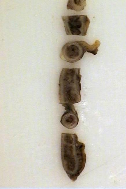
6 Appendix - examination Alternating longitudinal and transverse sections

7

8 Appendix - examination


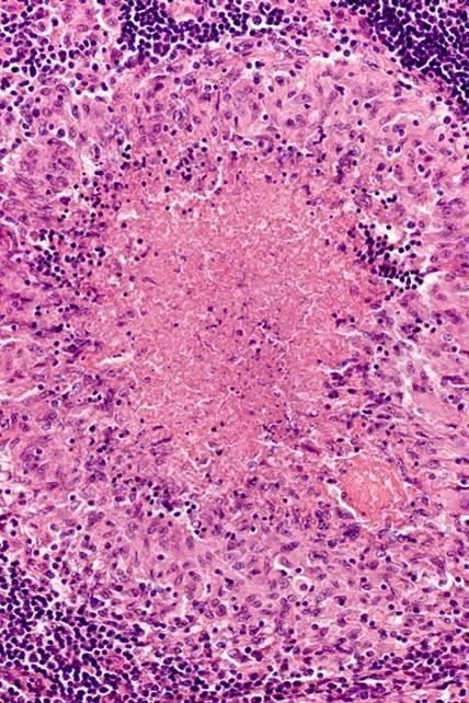
9 Idiopathic acute appendicitis Life time risk: 5% Peak age: y Range: From neonatal to ancient Pathogenesis: Luminal obstruction infection Fecalith, food fragments, lymphoid hyperplasia, tumour


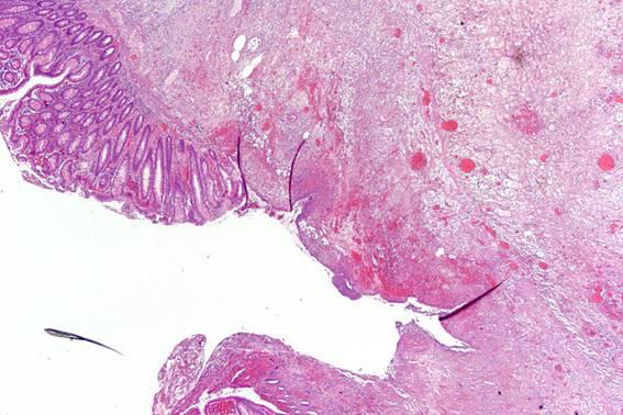
10 Gross: hyperaemia fibrinopurulent ex. black pus in lumen ectatic lumen ulceration perforation

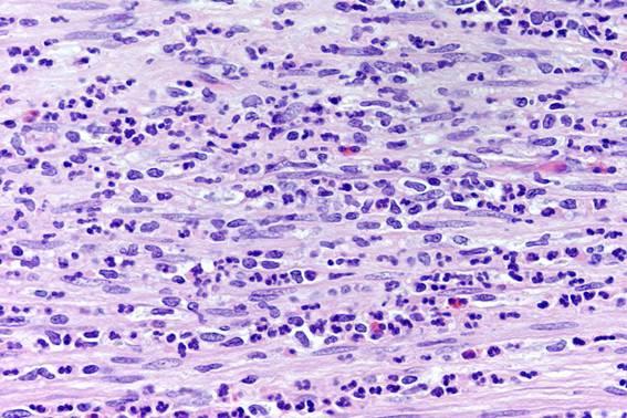
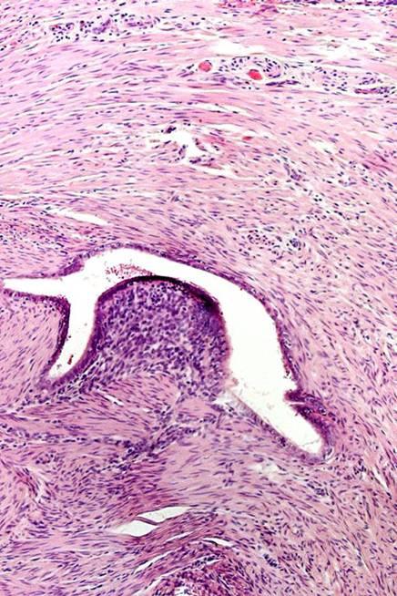
11 Idiopathic acute appendicitis Histology (classic) Erosion Crypt abscesses Ulceration Mucosal neutrophilic infiltration Transmural neutrophilic infiltration Myocytolysis
12
 Acute phlegmonous Acute necrotitizing")
13 Idiopathic acute appendicitis Acute superficial Acute (NOS) Acute phlegmonous Acute necrotitizing Gangrene


14 Complications of acute appendicitis Perforation 25% (higher in young and old) Periappendiceal absces 5% Pylephlebitis Hepatic absces

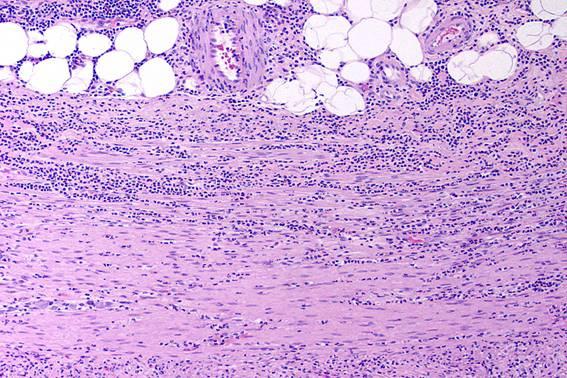
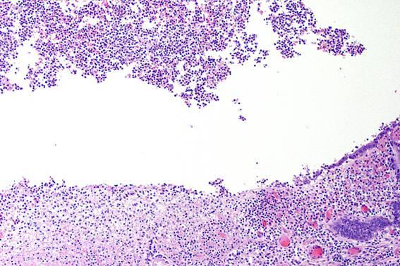
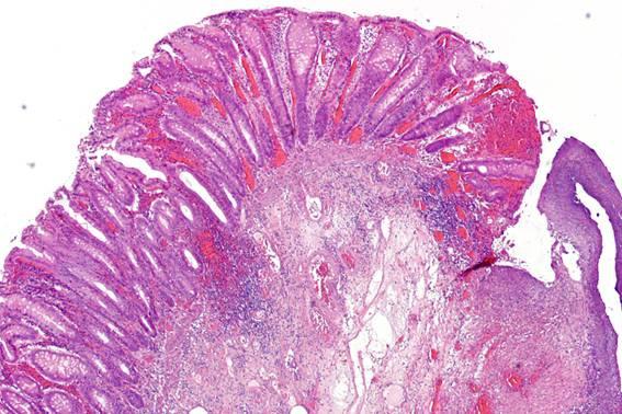
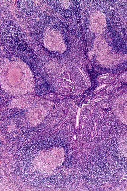
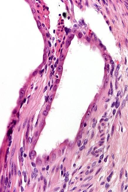
15 Protracted inflammation Resolution Crypt distortion Chronic inflammation Granulomatous inflammation Submucosal fibrosis Luminal obliteration Neuronal hyperplasia

16 Protracted inflammation Resolution Crypt distortion Chronic inflammation Granulomatous inflammation Submucosal fibrosis Luminal obliteration Neuronal hyperplasia

17 Protracted inflammation Resolution Crypt distortion Chronic inflammation Granulomatous inflammation Submucosal fibrosis Luminal obliteration Neuronal hyperplasia

18 Protracted inflammation Resolution Crypt distortion Chronic inflammation Granulomatous inflammation Submucosal fibrosis Luminal obliteration Neuronal hyperplasia


19 Protracted inflammation Resolution Crypt distortion Chronic inflammation Granulomatous inflammation Submucosal fibrosis Luminal obliteration Neuronal hyperplasia

20 Protracted inflammation Resolution Crypt distortion Chronic inflammation Granulomatous inflammation Submucosal fibrosis Luminal obliteration Neuronal hyperplasia


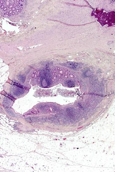
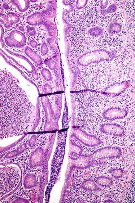
21 Appendix diverticulitis 90% of diverticuli acquired Often secondary to luminal obstruction Often multiple Localized inflammation Early perforation Older patients Protracted course
22


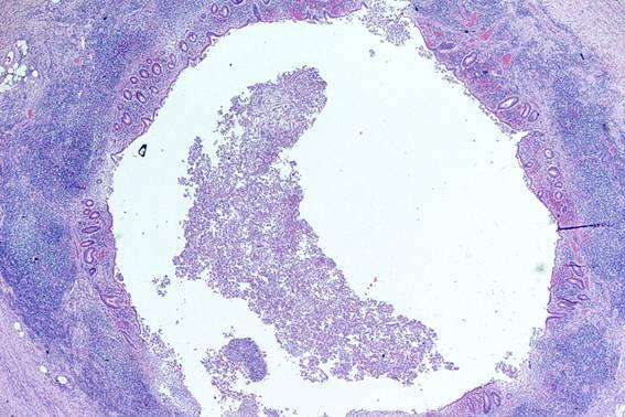
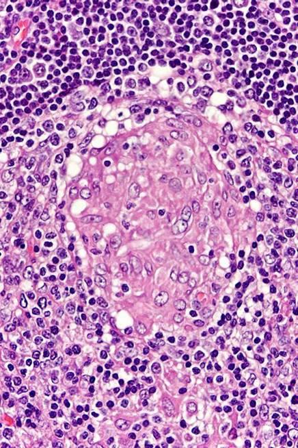
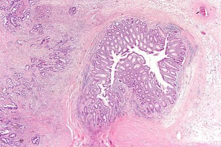
23 Appendix infections Bacterial infections Yersinia spp. Salmonella Shigella Campylobacter Actinomyces Viral infections Morbilli Parasitic infections Enterobius vermicularis
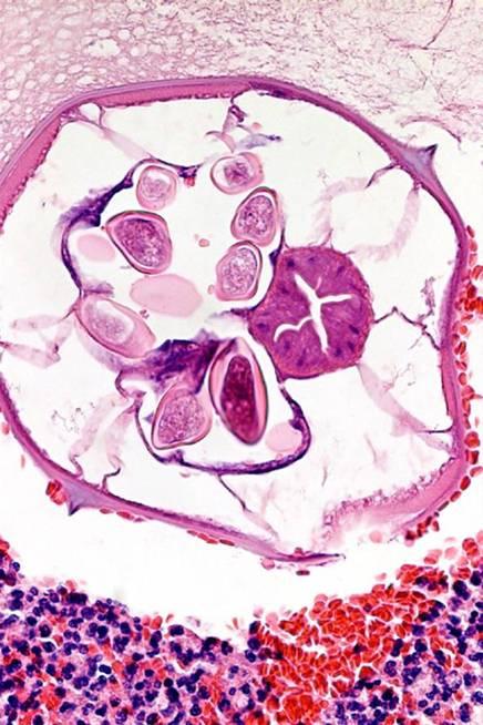
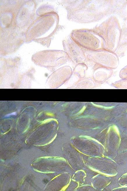
24 Appendiceal infections Enterobius
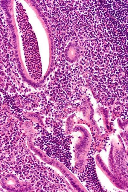
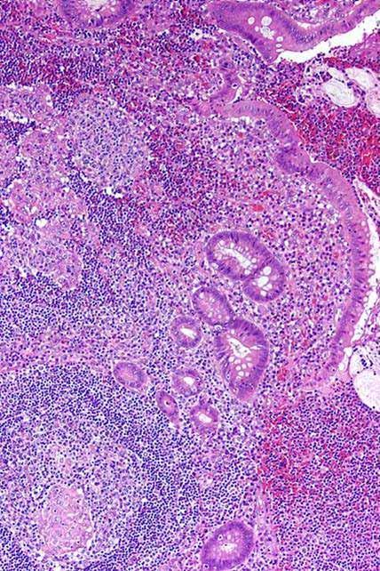
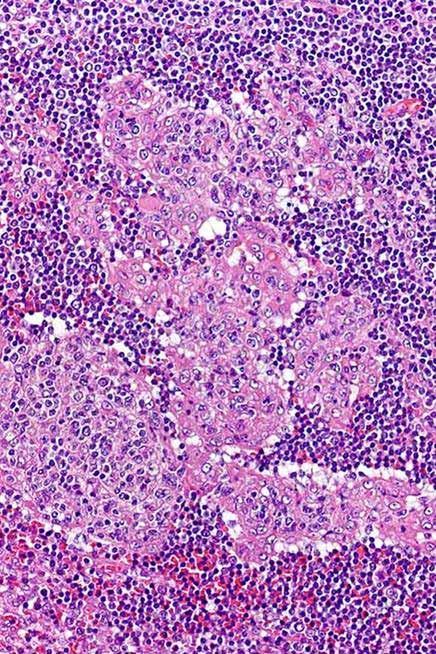
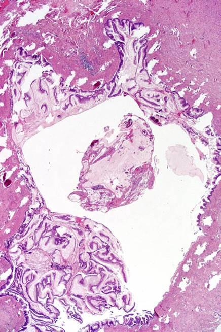
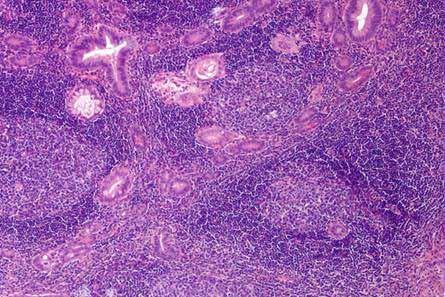
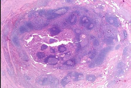
25 Appendiceal infections Yersiniosis
26 Appendiceal infections Yersiniosis
27 Appendiceal infections Yersiniosis

28 Appendix Chronic inflammatory bowel disease Crohn s disease 25-50% of Crohn s patients have granulomas in appendix <5% of granulomatous appendix have Crohn s disease Isolated appendiceal Crohn s disease dubious entity! Ulcerative colitis may appear without right colon involvement!
29 Appendix endometriosis & endosalpingiosis MLH1 MSH2


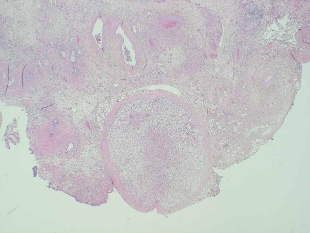
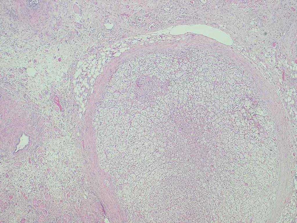
30 Hyperplastic polyp of appendix Varying from small to involving the whole mucosa Normal nuclei No stratification


31 Appendix tubular adenoma Rare Associated with colon adenomas and FAP

32 Appendix tubular adenoma
33 Appendix villous adenoma Female preponderance (20-) y Gross: Mucosal thickening, mucocele Histology: Diffuse mucosal involvement Cystadenoma Complication: Pseudomyxoma peritonei

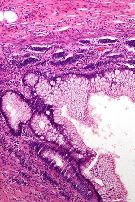
34 Appendix villous adenoma

35 Appendix villous adenoma & pseudomyxoma


36 Appendix villous adenoma & pseudomyxoma appendix serosa CK20 bladder wall

37 Appendix villous adenoma & pseudomyxoma

38 Appendix villous adenoma & pseudomyxoma

39 Appendix villous adenoma & pseudomyxoma

40 Appendix villous adenoma & pseudomyxoma
41 Appendix adenocarcinoma Classic colon like adenocarcinoma cystadenocarcinoma Goblet cell carcinoid = crypt cell carcinoma Signet ring cell carcinoma Complication: Pseudomyxoma peritonei

42 Appendix adenocarcinoma
43 Appendix adenocarcinoma MLH1 MSH2

44 Appendix mucocele Etiology Hyperplastic polyp Villous adenoma Adenocarcinoma


45 Appendix other neoplasms: B-cell lymphoma MLH1 T-cell rich B-cell lymphoma MSH2 IgKappa
46
47
48
49
Appendiceal diverticular disease
 Formosan Journal of Surgery (2013) 46, 4e9 Available online at www.sciencedirect.com journal homepage: www.e-fjs.com ORIGINAL ARTICLE Appendiceal diverticular disease Yun-Wu Deng a, Hsiao-Bai Yang b,e,
1. Esophageal diverticulum located above the upper esophageal sphincter is called
 Test Bank for Robbins Basic Pathology 9th Edition by Kumar Link full download: http://testbankair.com/download/test-bank-for-robbins-basic-pathology-9thedition-by-kumar/ Chapter 14: Oral Cavity and Gastrointestinal
Bowel obstruction and tumors
 Bowel obstruction and tumors Intestinal Obstruction Obstruction of the GI tract may occur at any level, but the small intestine is most often involved because of its relatively narrow lumen. Causes: Hernias
Joseph Misdraji, M.D. GI pathology Unit Massachusetts General Hospital
 Joseph Misdraji, M.D. GI pathology Unit Massachusetts General Hospital jmisdraji@partners.org Adenoma Low-grade appendiceal mucinous neoplasm High-grade appendiceal mucinous neoplasm Adenocarcinoma Serrated
Pitfalls in the Diagnosis of Inflammatory Bowel Disease
 Pitfalls in the Diagnosis of Inflammatory Bowel Disease Robert H Riddell MD Mt Sinai Hospital Toronto Prof of Lab. Medicine and Pathobiology University of Toronto Atypical gross / endoscopic distribution
Management of an Appendiceal Mass - Approach to acute presentation of appendiceal neoplasms
 Management of an Appendiceal Mass - Approach to acute presentation of appendiceal neoplasms Dr. Claudia LY WONG, Department of Surgery, Kwong Wah Hospital Joint Hospital Surgical Grand Round Presentation,
GASTROINTESTINAL IMAGING STUDY GUIDE
 GASTROINTESTINAL IMAGING STUDY GUIDE Pharynx Diverticula Foreign bodies Trauma o Motility Disorders Esophagus Diverticula Trauma Esophagitis Barrett esophagus Rings, webs, and strictures Varices Benign
Imaging Evaluation of Polyps. CT Colonography: Sessile Adenoma. Polyps, DALMs & Megacolon Objectives
 Polyps, DALMs & Megacolon: Pathology and Imaging of the Colon and Rectum Angela D. Levy and Leslie H. Sobin Washington, DC Drs. Levy and Sobin have indicated that they have no relationships which, in the
Bowel obstruction and tumors
 Bowel obstruction and tumors Intestinal Obstruction Obstruction of the GI tract may occur at any level, but the small intestine is most often involved because of its relatively narrow lumen. Causes: Hernias
Despite advances in our understanding of appendiceal. An Update on the Diagnosis, Grading, and Staging of Appendiceal Mucinous Neoplasms
 REVIEW ARTICLE An Update on the Diagnosis, Grading, and Staging of Appendiceal Mucinous Neoplasms Mark A. Valasek, MD, PhD* and Reetesh K. Pai, MD Abstract: Despite advances in our understanding of appendiceal
World Journal of Colorectal Surgery
 World Journal of Colorectal Surgery Volume 6, Issue 4 2016 Article 3 Laparoscopic Right Colectomy For Appendiceal Mucinous Cystadenoma: A Case Report Dion A. Putra Adianto Nugroho Ibrahim Basir University
Always keep it in the differential
 Acute Appendicitis Lissa C. Sakata and Lindsey Perea 2 Always keep it in the differential Learning Objectives 1. The learner should be able to describe the etiology of acute appendicitis. 2. The learner
GOBLET CELL CARCINOID. Hanlin L. Wang, MD, PhD University of California Los Angeles
 GOBLET CELL CARCINOID Hanlin L. Wang, MD, PhD University of California Los Angeles Disclosure of Relevant Financial Relationships USCAP requires that all planners (Education Committee) in a position to
GOBLET CELL CARCINOID
 GOBLET CELL CARCINOID Hanlin L. Wang, MD, PhD University of California Los Angeles Disclosure of Relevant Financial Relationships USCAP requires that all planners (Education Committee) in a position to
Chapter 6 Frozen Section Evaluation of the Appendix
 Chapter 6 Frozen Section Evaluation of the Appendix Abstract Appendiceal tumors are rarely diagnosed preoperatively, and their classification is both challenging and controversial owing to their tendency
Appendiceal Pathology. Prof Ray McMahon Histopathology Department Manchester Royal Infirmary Bryan Warren School Sarajevo November 2016
 Appendiceal Pathology Prof Ray McMahon Histopathology Department Manchester Royal Infirmary Bryan Warren School Sarajevo November 2016 Appendicitis Appendicitis Appendicitis Scattered groups of neutrophils
Appendiceal Adenocarcinoma with Suppurative Appendicitis: Case Report and Literature Review
 Appendiceal adenocarcinoma presenting with acute appendicitis 155 Appendiceal Adenocarcinoma with Suppurative Appendicitis: Case Report and Literature Review Va-Kei Kok 1, Teh-Kuang Wang 2, Hsi-Che Shen
Inflammatory Bowel Disease When is diarrhea not just diarrhea?
 Inflammatory Bowel Disease When is diarrhea not just diarrhea? Jackie Kazik, MA, PA C CME Resources CAPA Annual Conference, 2011 Inflammatory Bowel Disease Objectives Discuss what is known about the pathophysiology
Pitfalls in the CT diagnosis of appendicitis
 The British Journal of Radiology, 77 (2004), 792 799 DOI: 10.1259/bjr/95663370 E 2004 The British Institute of Radiology Pictorial review Pitfalls in the CT diagnosis of appendicitis 1 C D LEVINE, 2 O
Joseph Misdraji, M.D. GI pathology Unit Massachusetts General Hospital
 Joseph Misdraji, M.D. GI pathology Unit Massachusetts General Hospital jmisdraji@partners.org Low-grade appendiceal mucinous neoplasm (LAMN) High-grade appendiceal mucinous neoplasm (HAMN) Adenocarcinoma
CLINICAL VIGNETTE 2016; 2:1
 CLINICAL VIGNETTE 2016; 2:1 Editor-in-Chief: Olufemi E. Idowu. Neurological surgery Division, Department of Surgery, LASUCOM/LASUTH, Ikeja, Lagos, Nigeria. MANAGEMENT OF APPENDICITIS Ibrahim NA, Njokanma
Anatomy of the biliary tract
 Harvard-MIT Division of Health Sciences and Technology HST.121: Gastroenterology, Fall 2005 Instructors: Dr. Jonathan Glickman Anatomy of the biliary tract Figure removed due to copyright reasons. Biliary
-1- Pathology Department (code: 0605) Final Exam for Third year students Date: Time allowed: 2 hours. Paper II (75 marks).
 Final Exam for Third year students Date: Time allowed: 2 hours. Paper II (75 marks).") -1- BENHA UNIVERSITY FACULTY OF MEDICINE Pathology Department (code: 0605) Final Exam for Third year students Date: 28-5-2011 Time allowed: 2 hours. Paper II (75 marks). Please note that this question
-1- BENHA UNIVERSITY FACULTY OF MEDICINE Pathology Department (code: 0605) Final Exam for Third year students Date: 28-5-2011 Time allowed: 2 hours. Paper II (75 marks). Please note that this question
Mast cell profile in appendicitis
 Original Research Article DOI: 10.18231/2394-6792.2017.0120 Mast cell profile in appendicitis G. Patil Anuradha 1, AM Anita 2, Saini Kr. Seemant 3,*, S. Pratima 4 1 HOD, 2 Associate Professor, 3 PG Student,
PINDIGA UH, MALAMI SA, ADOGU IA, KEFAS JB, EMUAN TO
 CASE REPORT A RARE PATHOLOGICAL TRIAD UNMASKED BY TYPHOID ENTERITIS: REPORT OF A CASE OF ACUTE APPENDICITIS, APPENDICEAL SCHISTOSOMIASIS AND VILLOUS ADENOMA OF THE APPENDIX. 2 2 PINDIGA UH, MALAMI SA,
COLON AND RECTUM SOLID TUMOR RULES ABSTRACTORS TRAINING
 COLON AND RECTUM SOLID TUMOR RULES ABSTRACTORS TRAINING COLON AND RECTUM SOLID TUMOR RULES Separate sections for: Introduction Changes from 2007 MP/H rules Equivalent Terms Terms that are NOT Equivalent
하부위장관비종양성질환의 감별진단 주미인제의대일산백병원
 하부위장관비종양성질환의 감별진단 주미인제의대일산백병원 Solutions for diagnostic problems in Colitis : Please ask yourself five questions Normal or Inflamed? Acute or Chronic? IBD or Other chronic colitis? Ulcerative colitis or
Gastrointestinal Disorders. Disorders of the Esophagus 3/7/2013. Congenital Abnormalities. Achalasia. Not an easy repair. Types
 Gastrointestinal Disorders Congenital Abnormalities Disorders of the Esophagus Types Stenosis Atresia Fistula Newborn aspirates while feeding. Pneumonia Not an easy repair Achalasia Lack of relaxation
The Role of Ultrasound in the Assessment of Inflammatory Bowel Disease
 The Role of Ultrasound in the Assessment of Inflammatory Bowel Disease Dr. Richard A. Beable Consultant Gastrointestinal Radiologist Queen Alexandra Hospital Portsmouth Hospitals NHS Trust Topics for Discussion
Patho Basic Chronic Inflammatory Bowel Diseases. Jürg Vosbeck Pathology
 Patho Basic Chronic Inflammatory Bowel Diseases Jürg Vosbeck Pathology General Group of chronic relapsing diseases with chronic bloody or watery diarrhea Usually ulcerative colitis (UC) or Crohn s disease
Colon and Rectum: 2018 Solid Tumor Rules
 2018 SEER Solid Tumor Manual 2018 KCR SPRING TRAINING Colon and Rectum: 2018 Solid Tumor Rules 1 Colon and Rectum Solid Tumor Rules Separate sections for: Introduction Changes from 2007 MP/H rules Equivalent
APPENDICITIS AND ITS APPEARANCES ON CT
 APPENDICITIS AND ITS APPEARANCES ON CT APPENDICITIS Results from acute inflammation of the appendix. Most common abdominal surgical emergencies. Diagnosis usually clinical based on physical exam and lab
GUIDELINES FOR THE INITIAL BIOPSY DIAGNOSIS OF CHRONIC IDIOPATHIC INFLAMMATORY BOWEL DISEASE A STRUCTURED APPROACH TO COLORECTAL BIOPSY ASSESSMENT
 Guidelines for the Initial Biopsy Diagnosis of Chronic Idiopathic Inflammatory Bowel Disease 1 GUIDELINES FOR THE INITIAL BIOPSY DIAGNOSIS OF CHRONIC IDIOPATHIC INFLAMMATORY BOWEL DISEASE A STRUCTURED
Appendicitis. I. Background & Significance: Algorithm Definitions 1. CASE
 I. Background & Significance: Appendicitis Appendicitis is one of the most common acquired surgical conditions of childhood. Diagnosis of appendicitis remains difficult. Much work has been done on validation
Table 0: Description of Grading System for Anatomic Severity of Disease in Emergency. Local disease confined to the organ with minimal abnormality
 Table 0: of Grading System for Anatomic Severity of Disease in Emergency Local disease confined to the organ with minimal Local disease confined to the organ with severe Local extension Table 1: Universal
Unexpected Findings at Endoscopy
 The Endoscopic Incidentaloma: What to Tell Your Patient t with Unexpected Endoscopic Findings: Gastric Intestinal Metaplasia, Silent Ileitis, Carcinoid David Greenwald, MD Montefiore Medical Center Albert
Sonography of Gall Bladder
 Sonography of Gall Bladder Vikram Dogra,MD Professor of Radiology, Urology and BME Director of Ultrasound Associate Chair of Education and Research University of Rochester, NY Objectives Describe the Congenital
Polypectomy and Local Resections of the Colorectum Structured Pathology Reporting Proforma
 Polypectomy and Local Resections of the Colorectum Structured Pathology Reporting Proforma Mandatory questions (i.e. protocol standards) are in bold (e.g. S1.03). Family name Given name(s) Date of birth
Surgical Management of IBD. Val Jefford Grand Rounds October 14, 2003
 Surgical Management of IBD Val Jefford Grand Rounds October 14, 2003 Introduction Important Features Clinical Presentation Evaluation Medical Treatment Surgical Treatment Cases Overview Introduction Two
Emergency MDCT in case of right lower quadrant pain
 Emergency MDCT in case of right lower quadrant pain Poster No.: C-0563 Congress: ECR 2015 Type: Educational Exhibit Authors: M. Lisitskaya, V. Sinitsyn; Moscow/RU Keywords: Abdomen, Emergency, Gastrointestinal
Supplemental Digital Content 1. Endoscopic and histolological findings in INR and FR study subjects
 Supplemental Digital Content 1. Endoscopic and histolological findings in INR and FR study subjects Patient Group Macroscopic examination Ileum Histology Colon/rectum Histology 1 INR Normal Acute and chronic
Gastrointestinal Malignancies. Dr Rodney ITAKI Pathology Division, SMHS, UPNG Anatomical Pathology Discipline
 Gastrointestinal Malignancies Dr Rodney ITAKI Pathology Division, SMHS, UPNG Anatomical Pathology Discipline Esophagus normal anatomy Hollow tube 23-25cm long in adults Extends from pharynx to level of
Gastroenterology Tutorial
 Gastroenterology Tutorial Gastritis Poorly defined term that refers to inflammation of the stomach. Infection with H. pylori is the most common cause of gastritis. Most patients remain asymptomatic Some
Syllabus. Appendiceal GCC and LAMN Navigating the Alphabet Soup in the Appendix. Appendiceal tumors. Summary provided Complete presentation
 2016 Current Issues in Surgical Pathology Appendiceal GCC and LAMN Navigating the Alphabet Soup in the Appendix Syllabus Summary provided Complete presentation sanjay.kakar@ucsf.edu Sanjay Kakar, MD University
Gastrointestinal pathology 2018 lecture 4. Dr Heyam Awad FRCPath
 Gastrointestinal pathology 2018 lecture 4 Dr Heyam Awad FRCPath Topics to be covered Peptic ulcer disease Hiatal hernia Gastric neoplasms Peptic ulcer disease (PUD)= chronic gastric ulcer Causes H pylori
Rectal biopsy as an aid to cancer control in ulcerative colitis
 Rectal biopsy as an aid to cancer control in ulcerative colitis B. C. MORSON AND LILLIAN S. C. PANG From the Research Department, St. Mark's Hospital, London Gut, 1967, 8, 423 EDITORIAL COMMENT This is
12 Blueprints Q&A Step 2 Surgery
 12 Blueprints Q&A Step 2 Surgery 34. A 40-year-old female has been referred to you for a recent ER and hospital admission, from which she was given a diagnosis of acute diverticulitis. Treatment at that
Surveying the Colon; Polyps and Advances in Polypectomy
 Surveying the Colon; Polyps and Advances in Polypectomy Educational Objectives Identify classifications of polyps Describe several types of polyps Verbalize rationale for polypectomy Identify risk factors
Supplementary Appendix
 Supplementary Appendix This appendix has been provided by the authors to give readers additional information about their work. Supplement to: Kaminski MF, Regula J, Kraszewska E, et al. Quality indicators
Fig. 59 Malignant phaeochromocytoma, hepatic metastasis.
 Fig. 59 Malignant phaeochromocytoma, hepatic metastasis. X 120 Hyperte nsion Fig. 60 Malignant sympathetic paraganglioma, lymph node metastasis Primary in bladder. x 1 20 Hypertension Fig. 61 Malignant
Chromoendoscopy and Endomicroscopy for detecting colonic dysplasia
 Chromoendoscopy and Endomicroscopy for detecting colonic dysplasia Ralf Kiesslich I. Medical Department Johannes Gutenberg University Mainz, Germany Cumulative cancer risk in ulcerative colitis 0.5-1.0%
CT Evaluation of Bowel Wall Thickening. Dr: Adel El Badrawy; M.D. Lecturer of Radio Diagnosis Faculty of Medicine Mansoura University.
 CT Evaluation of Bowel Wall Thickening By Dr: Adel El Badrawy; M.D. Lecturer of Radio Diagnosis Faculty of Medicine Mansoura University. The CT findings of bowel wall thickening includes 1 Degree of thickening.
Alison Douglass Gillian Lieberman, MD. November. Colon Cancer. Alison Douglass, Harvard Medical School Year III Gillian Lieberman, MD
 November Colon Cancer Alison Douglass, Harvard Medical School Year III Our Patient Mr. K. is a 67 year old man with no prior medical problems other than hemorrhoids which have caused occasional rectal
Polyps in general: is a descriptive term of forming a mass that is exophytic & polypoid.
 ميحرلا نمحرلا هللا مسب Gastric Tumors: Benign tumours & tumor-like conditions: -Mucosal: Gastric polyps (they are uncommon) -Mesenchymal tumours: Leiomyoma & Lipoma (can occur anywhere in the body) Malignant:
Surveys and Anatomic Pathology Education Programs
 Surveys and Anatomic Pathology Education Programs Performance Improvement Program in Surgical Pathology PIP/PIPW-A 2018 Participant Summary 2018 College of American Pathologists. The College does not permit
Imaging in gastric cancer
 Imaging in gastric cancer Gastric cancer remains a deadly disease because of late diagnosis. Adenocarcinoma represents 90% of malignant tumors. Diagnosis is based on endoscopic examination with biopsies.
Acquired Megacolon: 1. Chaga s disease. 2. Organic obstruction by tumour or stricture. 3. Toxic megacolon in UC and CD. 4. Functional obstruction.
 GIT DISEASES : Small And Large Intestines: Meckel s Diverticulum: It results form failure of involution of omphalo-mesentric duct. It is a blind ended tubular protrusion up to 6 cm long, usually seen in
The term inflammatory bowel disease is used to designate two related inflammatory intestinal disorders:
 3. Disorders of the Small and Large Intestines a. Irritable Bowel Syndrome: The term irritable bowel syndrome is used to describe a functional gastrointestinal disorder characterized by a variable combination
Uncommon Findings in Appendicectomy Specimens
 IOSR Journal of Dental and Medical Sciences (IOSR-JDMS) e-issn: 2279-0853, p-issn: 2279-0861.Volume 14, Issue 5 Ver. VI (May. 2015), PP 01-06 www.iosrjournals.org Uncommon Findings in Appendicectomy Specimens
Review Article Cancers of the Appendix: Review of the Literatures
 International Scholarly Research Network ISRN Oncology Volume 2011, Article ID 728579, 6 pages doi:10.5402/2011/728579 Review Article Cancers of the Appendix: Review of the Literatures Carl Ruoff, 1 Louay
11/1/2017. Tetyana Mettler, MD Department of Laboratory Medicine and Pathology University of Minnesota. Cerilli & Greenson
 Tetyana Mettler, MD Department of Laboratory Medicine and Pathology University of Minnesota Acute infectious (self-limited) colitis Focal active colitis Pseudomembranous colitis Ischemic colitis Collagenous
South East England General Histopathology EQA Scheme
 South East England General Round i Final Case Analyses Cases 731 to 742 Circulated September December 2018 137 responses (86.16%) Prepared December 2018 Authorised by: Prof J Schofield Date: 14 th December
Icd 10 code for esophageal cancer stage 4
 Icd 10 code for esophageal cancer stage 4 Search Risk factors for developing esophageal cancer include.. 150. 4 Malignant neoplasm of middle third of esophagus convert 150. 4 to ICD - 10 -CM;. Free ICD
Accepted Article. Granulomatous appendicitis as an uncommon cause of abdominal pain. Description of a case
 Accepted Article Granulomatous appendicitis as an uncommon cause of abdominal pain. Description of a case Carmen Salvia López Ramos, Ana Fuentes Coronel, Santiago Rodríguez Gómez DOI: 10.17235/reed.2015.3763/2015
Circulation: V Case number: 301 Number of responses: 83 Date: 3 MAY 11
 Circulation: V Case number: 300 Number of responses: 73 Date: 3 MAY 11 Male, 58 years Right Renal Cell Carcinoma. Negative for Vimentin and Hales' Colloidal Iron Right Nephrectomry Right nephrectomy 432.0g
EDUCATIONAL CASES E1 & E2. Natasha Inglis 20/03/15
 EDUCATIONAL CASES E1 & E2 Natasha Inglis 20/03/15 CASE E1 79 year old female Rectum. Altemeier operation Histology Superficial erosions and mucosal congestion volcano lesion and pseudomembrane formation
Biopsy Evaluation of Non- Neoplastic Diseases of the Large Bowel: an algorithmic approach
 Biopsy Evaluation of Non- Neoplastic Diseases of the Large Bowel: an algorithmic approach Laura W. Lamps, M.D. Godfrey D. Stobbe Professor and Director of GI Pathology University of Michigan Health System
Granulomatous disease in the vermiform appendix
 J Clin Pathol 1983;36:632-638 Granulomatous disease in the vermiform appendix DC ALLEN, JD BIGGART From the Histopathology Laboratory, The Laboratories, Belfast City Hospital, Belfast SUMMARY In a twenty-year
elical CT plays an important role
 bdominal Imaging Yu et al. Helical CT of cute RLQ Pain Pictorial Essay Jinxing Yu 1 nn S. Fulcher Mary nn Turner Robert. Halvorsen Yu J, Fulcher S, Turner M, Halvorsen R Helical CT Evaluation of cute Right
What Every Pathologist Wants the GI Nurse to Know (and how you can help us help you)
") What Every Pathologist Wants the GI Nurse to Know (and how you can help us help you) Jonathan N. Glickman MD PhD Director, GI Pathology, Caris Diagnostics, Newton, MA Associate Professor of Pathology,
What Every Pathologist Wants the GI Nurse to Know (and how you can help us help you) Jonathan N. Glickman MD PhD Director, GI Pathology, Caris Diagnostics, Newton, MA Associate Professor of Pathology,
Gastrointestinal Tract. Anatomy of GI Tract. Anatomy of GI Tract. (Effective February 2007) (1%-5%)
 (1%-5%)") Gastrointestinal Tract (Effective February 2007) (1%-5%) Anatomy of GI Tract Esophagus bulls-eye or target EG junction seen on sagittal scan posterior to left lobe of liver and anterior to aorta Anatomy
Gastrointestinal Tract (Effective February 2007) (1%-5%) Anatomy of GI Tract Esophagus bulls-eye or target EG junction seen on sagittal scan posterior to left lobe of liver and anterior to aorta Anatomy
IBD. Crohn s. Outline. Ulcerative colitis versus Crohn s disease: is biopsy useful? UC vs. Crohn s? Is it easy? Biopsy settings 21/07/2017 IBD
 Outline Ulcerative colitis versus Crohn s disease: is biopsy useful? Roger Feakins Colorectal biopsies Ileal and upper GI biopsies Special situations New techniques Summary Inflammatory bowel disease (IBD)
Etiology. Extreme temperature, electric shock, ionization, physical injury, etc. Metabolic substances, acids, alkalis drugs, tissue necrosis
 INFLAMMATION Inflammation Protective response that is intended to eliminate the initial cause of injury Innate and acquired mechanisms Local or generalized (sepsis) processes Terminology: ~ itis ending
Histopathology: gastritis and peptic ulceration
 Histopathology: gastritis and peptic ulceration These presentations are to help you identify, and to test yourself on identifying, basic histopathological features. They do not contain the additional factual
NON OPERATIVE TREATMENT OF ACUTE APPENDICITIS - IS THIS APPROPRIATE IN SOUTH AFRICA O.D MONTWEDI
 NON OPERATIVE TREATMENT OF ACUTE APPENDICITIS - IS THIS APPROPRIATE IN SOUTH AFRICA O.D MONTWEDI Acute appendicitis is the commonest surgical emergency. About 300000 appendectomies are performed in the
ד"ר דוד ירדני המכון לגסטרואנטרולוגיה ומחלות כבד מרכז רפואי סורוקה
 ד"ר דוד ירדני המכון לגסטרואנטרולוגיה ומחלות כבד מרכז רפואי סורוקה Presentaion: S.A is 38 years old. Referred for rectal bleeding investigation. Describes several occasions of bleeding and abdominal pain.
Appendix 4: WHO Classification of Tumours of the pancreas 17
 S3.01 The WHO histological tumour type must be recorded. CS3.01a The histological type of the tumour should be recorded based on the current WHO classification 17 (refer to Appendices 4-7). Appendix 4:
Case presentation. Eran Zittan. MD Mount Sinai Hospital, Toronto, Canada. Emek Medical Center, Afula, Israel. March, 2016
 Case presentation Eran Zittan. MD Mount Sinai Hospital, Toronto, Canada. Emek Medical Center, Afula, Israel. March, 2016 60 y/o man with long standing UC+PSC. Last 10 years on clinical and endoscopic remission.
What do we need for diagnosis of IBD
 What do we need for diagnosis of IBD Kaichun Wu Dept. of Gastroenterology, Xijing Hospital Fourth Military Medical University Xi an an,, China In China UC 11.6/10 5,CD 1.4/10 5 Major cause of chronic diarrhea
Cross-sectional Imaging of Neuroendocrine Tumors of the Gastrointestinal Tract
 Cross-sectional Imaging of Neuroendocrine Tumors of the Gastrointestinal Tract Eric J. May 1, Shannon P. Sheedy 1, Joel G. Fletcher 1, Mark J. Truty 2, Thomas C. Smyrk 3, Jeff L. Fidler 1 1. Radiology,
How to differentiate Segmental Colitis Associated with Diverticulosis and Inflammatory Bowel Diseases?
 How to differentiate Segmental Colitis Associated with Diverticulosis and Inflammatory Bowel Diseases? Alessandro Armuzzi Lead IBD Unit Complesso Integrato Columbus Fondazione Policlinico Gemelli Università
665 " *** #$ %& #. / - $ ) # * + ",
 # * + ,") : 665 **! " *** #$ %& * #. / -$ )# * +," ( :'# & )" *8 * 673 +, ) /5!$. 0 12 3 @)8.;< = >0!$? 5 /$9 :.. # # "# 7 9 ' )" 3 =.$* < < ).3
: 665 **! " *** #$ %& * #. / -$ )# * +," ( :'# & )" *8 * 673 +, ) /5!$. 0 12 3 @)8.;< = >0!$? 5 /$9 :.. # # "# 7 9 ' )" 3 =.$* < < ).3
8. The polyp in the illustration can be described as (circle all that apply) a. Exophytic b. Pedunculated c. Sessile d. Frank
 a. Exophytic b. Pedunculated c. Sessile d. Frank") Quiz 1 Overview 1. Beginning with the cecum, which is the correct sequence of colon subsites? a. Cecum, ascending, splenic flexure, transverse, hepatic flexure, descending, sigmoid. b. Cecum, ascending,
Quiz 1 Overview 1. Beginning with the cecum, which is the correct sequence of colon subsites? a. Cecum, ascending, splenic flexure, transverse, hepatic flexure, descending, sigmoid. b. Cecum, ascending,
Appendicitis: When Simple Becomes not so Simple
 Wright State University CORE Scholar Department of Surgery Faculty Publications Surgery 1-26-2010 Appendicitis: When Simple Becomes not so Simple Elizabeth H. Ey Wright State University, elizabeth.ey@wright.edu
The Spectrum of IBD. Inflammatory Bowel Disease. Symptoms. Epidemiology. Tests for IBD. CD or UC? Inflamatory Bowel Disease. Fernando Vega, M.D.
 The Spectrum of IBD Inflammatory Bowel Disease Fernando Vega, M.D. Epidemiology CD and UC together 1:400 UC Prevalence 1:500 UC Incidence 6-12K/annum CD Prevalence 1:1000 CD Incidence 3-6K/annum Symptoms
UNDERSTANDING X-RAYS: ABDOMINAL IMAGING THE ABDOMEN
 UNDERSTANDING X-RAYS: ABDOMINAL IMAGING THE ABDOMEN Radiology Enterprises radiologyenterprises@gmail.com www.radiologyenterprises.com STOMACH AND SMALL BOWEL STOMACH AND SMALL BOWEL Swallowed air is a
Lahey Clinic Internal Medicine Residency Program: Curriculum for Gastroenterology
 Lahey Clinic Internal Medicine Residency Program: Curriculum for Gastroenterology Faculty representative: David L. Burns, MD, CNSP Resident representative: Tom Castiglione, MD Revision date: March 6, 2006
Summary and conclusions
 Summary and conclusions 7 Chapter 7 68 Summary and conclusions Chapter 1 provides a general introduction to this thesis focused on the use of ultrasound (US) in children with abdominal problems. The literature
Appendiceal Mucosal Schwann Cell Proliferation: A Putative Histologic Marker of Appendiceal Diverticular Disease
 494795IJSXXX10.1177/1066896913494795International Journal of Surgical PathologyStockl et al research-article2013 Original Article Appendiceal Mucosal Schwann Cell Proliferation: A Putative Histologic Marker
Colorectal Cancer Structured Pathology Reporting Proforma DD MM YYYY
 Colorectal Cancer Structured Pathology Reporting Proforma Mandatory questions (i.e. protocol standards) are in bold (e.g. S1.03). Family name Given name(s) Date of birth DD MM YYYY S1.02 Clinical details
IATROGENIC AND DRUG INDUCED PATHOLOGY IN THE SMALL INTESTINE. Claude Cuvelier N. Goormaghtigh Institute of Pathology
 IATROGENIC AND DRUG INDUCED PATHOLOGY IN THE SMALL INTESTINE Claude Cuvelier N. Goormaghtigh Institute of Pathology Content Iatrogenic pathology Radiation Chemotherapy Graft-Versus-Host disease Changes
Histological and immunological characteristics of colitis associated with anti-ctla 4 antibody therapy
 Histological and immunological characteristics of colitis associated with anti-ctla 4 antibody therapy M. Perdiki 2, G. Bamias 1, D. Pouloudi 2, H. Gogas 3, I. Delladetsima 2 1 Academic Dpt. of Gastroenterology,
Defective mismatch repair in the pathogenesis of low-grade appendiceal mucinous neoplasms and adenocarcinomas
 & 2004 USCAP, Inc All rights reserved 0893-3952/04 $30.00 www.modernpathology.org Defective mismatch repair in the pathogenesis of low-grade appendiceal mucinous neoplasms and adenocarcinomas Joseph Misdraji
Small Bowel Cases. Introduction. Introduction, Continued 12/7/2011. Lesions Found on endoscopic biopsies Just Like Signing Out
 Small Bowel Cases Lesions Found on endoscopic biopsies Just Like Signing Out Introduction Small intestinal biopsies have a few special pitfalls, for example: Neuroendocrine tumors are readily mistaken
The Morphologic Profile of Inflammatory Bowel Disease and the Diagnostic Problem of Crohn s Disease versus TB Colitis A Case Series
 OPEN ACCESS CASE REPORT The Morphologic Profile of Inflammatory Bowel Disease and the Diagnostic Problem of Crohn s Disease versus TB Colitis A Case Series Maria Lourdes Tilbe, Francia Victoria De Los
2015 복영증례 51/M C.C. Past Hx: DM, HTN (1998), Lab: WBC (11500/ μl ), CRP (0.71 mg/dl) 순천향서울병원황지영, 홍성숙 APCT (HAD #1) APCT (HAD#1) APCT (HAD #15)
, Lab: WBC (11500/ μl ), CRP (0.71 mg/dl) 순천향서울병원황지영, 홍성숙 APCT (HAD #1) APCT (HAD#1) APCT (HAD #15)") Case 1 2015 복영증례 순천향서울병원황지영, 홍성숙 51/M C.C Abdominal pain and chilling (1 week ago) Diarrhea (a month ago) Past Hx: DM, HTN (1998), Alcoholic liver disease (2008) Lab: WBC (11500/ μl ), CRP (0.71 mg/dl)
Case 1 2015 복영증례 순천향서울병원황지영, 홍성숙 51/M C.C Abdominal pain and chilling (1 week ago) Diarrhea (a month ago) Past Hx: DM, HTN (1998), Alcoholic liver disease (2008) Lab: WBC (11500/ μl ), CRP (0.71 mg/dl)
International Journal of Health Sciences and Research ISSN:
 International Journal of Health Sciences and Research www.ijhsr.org ISSN: 2249-9571 Original Research Article Role of Mast Cells in Appendicitis Dr. Jyoti Sharma 1*, Dr. Nitin Chaudhary 2*, Dr. Sunita
References. GI Biopsies. What Should Pathologists Assistants Know About Gastrointestinal Histopathology? James M Crawford, MD, PhD
 What Should Pathologists Assistants Know About Gastrointestinal Histopathology? James M Crawford, MD, PhD jcrawford1@nshs.edu Executive Director and Senior Vice President for Laboratory Services North
LOINC. Clinical information. RCPA code. Record if different to report header Operating surgeon name and contact details. Absent.
 Complete as narrative or use the structured format below 55752-0 17.02.28593 Clinical information 22027-7 17.02.30001 Record if different to report header Operating surgeon name and contact details 52101004
Icd 10 code for esophageal cancer stage 4
 Cari untuk: Cari Cari Icd 10 code for esophageal cancer stage 4 21-11-2016 There are two main types of lung cancer : non-small cell and small-cell. Learn about how these lung cancers are caused, your treatment