What s your diagnosis?
|
|
|
- Noah Henry
- 5 years ago
- Views:
Transcription

1 What s your diagnosis? Signalment: 9 year old MC 2.7 kg Papillion Presenting Complaint: Presented for work up of anorexia and vomiting History: He had presented to cardiology for work up of a grad IV/VI heart murmur and congestive heart failure diagnosed 7 days ago by the referring veterinarian. At that time he had a history of tracheal collapse, dental disease, mild exercise intolerance, and active anorexia and daily vomiting of foam and bile. Blood work from the referring veterinarian was unremarkable, and the referring veterinarian had prescribed Pimobendan 1.25 mg ½ tablet BID. He was diagnosed by the cardiologist on echocardiogram with stage B1 endocardiosis (myxomatous valvular disease) and it was recommended to monitor for heart enlargement, continue the Pimobendan at the same dose regimen, and further diagnostics for cause of anorexia and vomiting. Physical Exam Findings: BAR Mild bilateral lenticular opacity Moderate dental calculus on all arcades Grade IV/VI holosystolic murmur with strong, synchronous pulses Firm, round mass palpated in mid-cranial abdomen Blood Work Abnormalities: None Imaging and interpretations: R
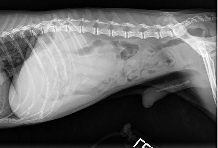
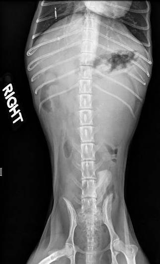
2

3 Radiographic Findings of 3 view abdomen: Musculoskeletal structures are normal. The abdominal serosal detail is adequate and the contour is normal. There is a round to ovoid, soft tissue opaque, well marginated mass in the left, cranial, dorsal abdomen causing ventral, right, and caudal displacement of the small intestines. It expands from cranial T13 to mid L4 in length and 3/4ths of the abdominal height going from the ventral vertebral bodies down to contact the splenic tail. There are numerous, granular, mineral opacities superimposed over the middle and ventral aspects of the mass. The bladder is not visualized. Radiographic Interpretations: The soft tissue mass with granular mineralization is most consistent with the left kidney with differential diagnosis of primarily neoplasia or lesser consideration to unilateral hydronephrosis, perinephric fluid, cystic disease, inflammatory disease, and hematoma. Recommend abdominal ultrasound with potential fine need aspiration of the mass if possible and thoracic radiographs.

4 Abdomen Ultrasound Findings: The gallbladder contained suspended and gravity dependent hyperechoic and echogenic debris. There was a heterogeneous, mixed echogenicity mass measuring 6.4 x 4.7 cm in dimension in the region of the left kidney. A normal left kidney was not identified. This mass did not have any identifiable normal renal architecture. There were multiple shadowing hyperechogenicities measuring up to 10.6 mm in length within the mass. The left adrenal gland measured 3.6 mm in thickness. The right adrenal gland measured 5.2 mm in thickness. The liver, spleen, right kidney, left and right adrenal glands, urinary bladder, prostate, gastrointestinal tract, and pancreas were within normal limits. Ultrasound Impressions: Left renal mass with mineralization is consistent with neoplasia. Gallbladder sludge. Ultrasound-guided procedure: Fine needle aspirates of the previous described left renal mass were obtained without immediate complication. Cytology of FNA of renal mass: Epithelial cell neoplasia -- given the presence of acinar-like structures cytologic findings are most suggestive of a nephroblastoma or renal adenocarcinoma. However, other carcinomas can't be ruled out. Histopathology is strongly recommended for confirmation.
5 Diagnostic Plan: This patient s renal mass was diagnosed as either a nephroblastoma or renal adenocarcinoma of the left kidney. This tumor may be the cause of his anorexia and vomiting due to abdominal discomfort and increased pressure on his other organs. I recommend thoracic radiographs to check for metastatic neoplasia, surgically removing the left kidney, and submitting it for histopathology for a definitive diagnosis and to plan further chemotherapy treatments. Carboplatin would be the chemotherapy agent of choice or secondarily toceranib (Palladia). The prognosis for patients with renal adenocarcinoma or nephroblastoma treated with surgery alone is overall good with a reported survival time of years after surgery. The specific prognosis for patients with metastatic renal adenocarcinoma is unknown since there are no published reports of response to chemotherapy alone.
Signalment: Gidget, 12 year old, female spayed, Scottish Terrier, 10.7 kg
 Signalment: Gidget, 12 year old, female spayed, Scottish Terrier, 10.7 kg Presenting Complaint: Gidget presented after having elevated liver enzymes, patchy alopecia and PU/PD. History: Gidget had been
What s Your Diagnosis?
 What s Your Diagnosis? Courtney S. Wait Signalment: 11 year old FS Labrador Retriever Presenting Complaint/History: The patient presented to the referring DVM for inappetance, vomiting, lethargy, and anorexia.
What s Your Diagnosis?
 What s Your Diagnosis? Signalment: 5 year old MC Belgian Malinois Presenting Complaint: Perineal hernia as well as not eating or defecating History: The patient presented to the KSU VHC on 7/28/2018 for
Abdominal Ultrasound
 Abdominal Ultrasound Imaging Control Buttons Depth The organ imaged should take up 3/4 of the screen Frequency = Penetration Use high frequencies (harmonics) for fluid filled and superficial structures
What is Your Diagnosis?
 What is Your Diagnosis? Izabela Ragan, Class of 2014 Signalment Species: Canine Breed: English Bulldog Sex: Male castrated Date of birth: 04/14/11 Presenting Complaint Dog was presented for vomiting and
What s Your Diagnosis? Sara Alves, Class of Signalment: 9-year-7-month old female spay American Miniature Eskimo dog
 What s Your Diagnosis? Sara Alves, Class of 2018 Signalment: 9-year-7-month old female spay American Miniature Eskimo dog Presenting Complaint: The patient presented on 5/30/17 with signs of lethargy and
What s Your Diagnosis??? Renée Fahrenholz, Class of 2012
 Renée Fahrenholz, Class of 2012 What s Your Diagnosis??? Signalment Emma, a 9 year old, Female, Spayed, Domestic Short Haired Feline Presenting Complaint Weight loss, vomited the morning of her visit,
What s Your Diagnosis? Allison Crow, Class of 2014
 What s Your Diagnosis? Allison Crow, Class of 2014 Signalment: 13 year old male castrated mixed breed dog History: The patient presented to the rdvm for pain in the hind end, weakness and neck stretching
What s Your Diagnosis? Signalment: Species: Canine Breed: Golden Retriever Sex: Female (spayed) Date of Birth: 04/01/99
 Date of Birth: 04/01/99") What s Your Diagnosis? Signalment: Species: Canine Breed: Golden Retriever Sex: Female (spayed) Date of Birth: 04/01/99 Presenting Complaint: Acute onset of lethargy Vomited twice (partially digested food)
What s Your Diagnosis? Signalment: Species: Canine Breed: Golden Retriever Sex: Female (spayed) Date of Birth: 04/01/99 Presenting Complaint: Acute onset of lethargy Vomited twice (partially digested food)
Abdominal ultrasound:
 Abdominal ultrasound: Non-traumatic acute abdomen Wittanee Na-ChiangMai, MD Department of Radiology ChiangMai University 26/04/2017 Contents Technique of examination Normal anatomy Emergency conditions
Principles of Surgical Oncology. Winnie Achilles Tierklinik Hollabrunn Lastenstrasse Hollabrunn
 Principles of Surgical Oncology Winnie Achilles Tierklinik Hollabrunn Lastenstrasse 2 2020 Hollabrunn boexi@gmx.de The first surgery provides the best chance for a cure in an animal with a tumor Clinical
Imaging the Urinary Tract
 Imaging the Urinary Tract Laura Armbrust, DVM, DACVR Gregory F. Grauer, DVM, MS, DACVIM Kansas State University Radiographic and ultrasound imaging in addition to history, physical examination, and clinicopathologic
My Patient Has Abdominal Pain PoCUS of the Biliary Tract and the Urinary Tract
 My Patient Has Abdominal Pain PoCUS of the Biliary Tract and the Urinary Tract Objectives PoCUS for Biliary Disease PoCUS for Renal Colic PoCUS for Urinary Retention Biliary Disease A patient presents
Close window to return to IVIS. in collaborazione con RICHIESTO ACCREDITAMENTO. organizzato da certificata ISO 9001:2000
 in collaborazione con Close window to return to IVIS RICHIESTO ACCREDITAMENTO SOCIETÀ CULTURALE ITALIANA VETERINARI PER ANIMALI DA COMPAGNIA SOCIETÀ FEDERATA ANMVI organizzato da certificata ISO 9001:2000
What s Your Diagnosis? Catherine Donewald, Class of 2016
 What s Your Diagnosis? Catherine Donewald, Class of 2016 Signalment: 9 ½ year old, male castrate Greyhound dog History: The patient presented to referring veterinarian with a history of decreased energy
GENERAL ABDOMINAL IMAGING PERITONEAL SPACE, PANCREAS, & SPLEEN. VMB 960 March 25, 2013
 GENERAL ABDOMINAL IMAGING PERITONEAL SPACE, PANCREAS, & SPLEEN VMB 960 March 25, 2013 REFERENCE Chapters 35-36 Pages 650-678 Chapter 37 Pages 694-701 Chapter 3 Pages 38-49 OBJECTIVES Radiography and Ultrasound
What s Your Diagnosis? Signalment: Species: Ferret, Mustela putorius furo Sex: Female Spayed Date of Birth: 03/01/02 History of Adrenal Disease
 What s Your Diagnosis? Signalment: Species: Ferret, Mustela putorius furo Sex: Female Spayed Date of Birth: 03/01/02 History of Adrenal Disease Presenting Complaint: Diarrhea; Acute Dyspnea. For a couple
ABDOMINAL RADIOLOGY UNDERSTANDING
 ABDOMINAL RADIOLOGY UNDERSTANDING CACVT 2017 SPRING CONFERENCE - GREENWOOD VILLAGE, CO Amy Newfield, CVT, VTS (ECC) BluePearl Massachusetts - Waltham, MA INTRODUCTION As a technician you will likely be
What s your diagnosis? Malori Marotz. Squirt, an 8month old mix breed puppy. History:
 What s your diagnosis? Malori Marotz Squirt, an 8month old mix breed puppy History: The owner obtained squirt at 12 weeks of age. The owner reported that Squirt was passing soft stools lately and he is
Case Scenario 1. Discharge Summary
 Case Scenario 1 Discharge Summary A 69-year-old woman was on vacation and noted that she was becoming jaundiced. Two months prior to leaving on that trip, she had had a workup that included an abdominal
Proceedings of the 34th World Small Animal Veterinary Congress WSAVA 2009
 www.ivis.org Proceedings of the 34th World Small Animal Veterinary Congress WSAVA 2009 São Paulo, Brazil - 2009 Next WSAVA Congress : Reprinted in IVIS with the permission of the Congress Organizers IMAGING
Imaging the Urogenital System
 maging the Urogenital System Tony Pease, DVM, MS, DACVR Assistant Professor of Radiology North Carolina State University Reading Thrall Chapters 42-46 Prostate Gland Not visible radiographically in normal
Calvin 9 year old NM DLH. Dr. Norman Ackerman Memorial Radiography Case Challenge
 September 2014 Dr. Norman Ackerman served the University of Florida, College of Veterinary Medicine with distinction as Professor of Radiology from 1979 to 1994. A concerned teacher of veterinary students
GENERAL ABDOMINAL IMAGING PERITONEAL SPACE, PANCREAS, & SPLEEN
 GENERAL ABDOMINAL IMAGING PERITONEAL SPACE, PANCREAS, & SPLEEN VMB 960 March 25, 2013 REFERENCE Chapters 35-36 Pages 650-678 Chapter 37 Pages 694-701 Chapter 3 Pages 38-49 OBJECTIVES Radiography and Ultrasound
Radiology Pathology Conference
 Radiology Pathology Conference Nadia F. Yusaf, M.D. PGY-3 1/29/2010 Presentation material is for education purposes only. All rights reserved. 2010 URMC Radiology Page 1 of 90 Case 1 60 year- old man presents
Pathology of the Hematopoietic System. Case studies
 Pathology of the Hematopoietic System Case studies Shannon Martinson, September 2015 Signalment: 9 yr-old MC cat Case Study 1 History: Cat had been anorexic and developed bleeding in the eyes Physical
GENERAL DIAGNOSTIC IMAGING IN SMALL ANIMAL ONCOLOGY
 GENERAL DIAGNOSTIC IMAGING IN SMALL ANIMAL ONCOLOGY Jantra Ngosuwan Suran, DVM, Dipl. ACVR, Cert Clin Res University of Pennsylvania, School of Veterinary Medicine 3900 Delancey St, Philadelphia, PA 19104
DIAGNOSTIC ULTRASOUND D R. E R I C A J O H N S O N
 DIAGNOSTIC ULTRASOUND D R. E R I C A J O H N S O N ULTRASOUND BASICS Medical ultrasound machines generate and receive ultrasound waves Ultrasound waves are emitted from the peizolectric crystals of the
Autosomal Dominant Polycystic Kidney Disease
 Case Studies [1] July 01, 2014 By Amar Udare, MBBS [2] Case History: 45-year-old female with vague pain in the abdomen. Case History: A 45-year-old female presented with vague pain in the abdomen. A USG
Case Scenario 1: Thyroid
 Case Scenario 1: Thyroid History and Physical Patient is an otherwise healthy 80 year old female with the complaint of a neck mass first noticed two weeks ago. The mass has increased in size and is palpable.
What s Your Diagnosis?
 Claire Legallet What s Your Diagnosis? Signalment: Species: Canine Breed: Catahoula Sex: Female Intact Age at presentation: 6 months Presenting Complaint: Chronic intermittent bloody diarrhea and vomiting
DIAGNOSTIC IMAGING: LIVER DISEASE
 Vet Times The website for the veterinary profession https://www.vettimes.co.uk DIAGNOSTIC IMAGING: LIVER DISEASE Author : Abby Caine Categories : Vets Date : February 1, 2010 ABBY CAINE reviews both established
What s Your Diagnosis? Jessica Eisenbarth. Signalment: Jazz is a female intact 2 year old German Shorthaired Pointer.
 What s Your Diagnosis? Jessica Eisenbarth Signalment: Jazz is a female intact 2 year old German Shorthaired Pointer. Presenting complaint: Jazz was presented to the K-State emergency service on August
Abdomen and Retroperitoneum Ultrasound Protocols
 Abdomen and Retroperitoneum Ultrasound Protocols Reviewed By: Anna Ellermeier, MD Last Reviewed: March 2018 Contact: (866) 761-4200, Option 1 **NOTE for all examinations: 1. If documenting possible flow
RELATIONSHIP BETWEEN PROSTATOMEGALY, PROSTATIC MINERALIZATION, AND CYTOLOGIC DIAGNOSIS
 RELATIONSHIP BETWEEN PROSTATOMEGALY, PROSTATIC MINERALIZATION, AND CYTOLOGIC DIAGNOSIS CHRISTINA A. BRADBURY, JODI L. WESTROPP, RACHEL E. POLLARD Canine prostatic disease is commonly evaluated with abdominal
Table of Contents. Preface xi. Acknowledgments xiii. Part I Overview of the Diagnostic Process 1. 1 Overview of Grading and Staging 3
 Table of Contents Preface xi Acknowledgments xiii Part I Overview of the Diagnostic Process 1 1 Overview of Grading and Staging 3 Identification of the process 3 Identification of tumor types 5 Grading
Case # nd Annual SEVPAC May 17, Kathy-Anne Clarke
 Case # 10 42 nd Annual SEVPAC May 17, 2014 Kathy-Anne Clarke Google images Babu Babu is 10 year old spayed female French Bulldog Chronic weight loss over 4 months Febrile and lethargic at the referring
Kidney Case 1 SURGICAL PATHOLOGY REPORT
 Kidney Case 1 Surgical Pathology Report February 9, 2007 Clinical History: This 45 year old woman was found to have a left renal mass. CT urography with reconstruction revealed a 2 cm medial mass which
Canine Cutaneous Melanoma
 Canine Cutaneous Melanoma By Elizabeth Downing Clinical Advisor: Dr. Angharad Waite, VMD Basic Science Advisor: Dr. Cheryl Balkman, DVM, DACVIM Senior Seminar Paper Cornell University College of Veterinary
The Child With An Abdominal Mass
 The Child With An Abdominal Mass Today we are going to talk about pediatric surgery, the abdominal masses in children. Firstly we have to take a full history and make a general, local and rectal examination
Case Discussion Splenic Abscess
 Case Discussion Splenic Abscess Personal Data Gender: male Birth Date: 1928/Mar/06th Allergy: Mefenamic Smoking: 0.5 PPD for 55 years Alcohol: negative (?) 4 Months Ago Abdominal pain: epigastric area
Ultrasonography of Peritoneal and Retroperitoneal Spaces and Abdominal Lymph Nodes
 IMAGING Ultrasonography of Peritoneal and Retroperitoneal Spaces and Abdominal Lymph Nodes Clifford R. Berry, DVM, DACVR; Elizabeth Huyhn, DVM; and Danielle Mauragis, CVT University of Florida Welcome
Case 2 Dwayne A. Williams CASE 2
 CASE 2 A 40- year- old male with no past medical history presents with bilateral flank pain and dark colored urine for 5 days. During family history taking, he states his father died from kidney failure
ASSESSING THE PLAIN ABDOMINAL RADIOGRAPH M A A M E F O S U A A M P O F O
 ASSESSING THE PLAIN ABDOMINAL RADIOGRAPH M A A M E F O S U A A M P O F O Introduction The abdomen (less formally called the belly, stomach, is that part of the body between the thorax (chest) and pelvis,
Guidelines, Policies and Statements D5 Statement on Abdominal Scanning
 Guidelines, Policies and Statements D5 Statement on Abdominal Scanning Disclaimer and Copyright The ASUM Standards of Practice Board have made every effort to ensure that this Guideline/Policy/Statement
Summary and conclusions
 Summary and conclusions 7 Chapter 7 68 Summary and conclusions Chapter 1 provides a general introduction to this thesis focused on the use of ultrasound (US) in children with abdominal problems. The literature
2 to 3% of All New Visceral Cancers Peak Incidence is 6th Decade M:F = 2:1 Grossly is a Bright Yellow, Necrotic Mass with a Pseudocapsule
 GENITOURINARY PATHOLOGY Kathleen M. O Toole, M.D. Renal Cell Carcinoma 2 to 3% of All New Visceral Cancers Peak Incidence is 6th Decade M:F = 2:1 Grossly is a Bright Yellow Necrotic Mass Grossly is a Bright
Welcome to ANAT 10A! What is Anatomy? Different levels of Anatomy The Language of Anatomy Pearson Education, Inc.
 Welcome to ANAT 10A! What is Anatomy? Different levels of Anatomy The Language of Anatomy Introduction Anatomy means to dissect: (ANAT 10A) The study of internal & external body structures The study of
Abdominal Ultrasound : Aorta, Kidneys, Bladder
 Abdominal Ultrasound : Aorta, Kidneys, Bladder Nilam J. Soni, MD, MSc Associate Professor of Medicine Divisions of Hospital Medicine and Pulmonary/Critical Care Medicine Department of Medicine University
Case Study: #3: Gallbladder Carcinoma?
 Case Study: #3: Gallbladder Carcinoma? By: Megan Wyatt K. SON Wyatt 225 2B1 RDMS, RVT Patient: Male 85 YOA Caucasian Indication: Elevated Alkaline Phosphatase History Annual physical showed elevated alkaline
Case Scenario 1. 4/19/13 Bone Scan: No scintigraphic findings to suggest skeletal metastases.
 Case Scenario 1 3/8/13 H&P 68 YR W/M presents w/elevated PSA. Patient is a non-smoker, current alcohol use. Physical Exam: On digital rectal exam the sphincter tone is normal and there is a 1 cm nodule
Radiographic Positioning. Small Animal Abdominal Radiography. Lecture Outline. Matthew Paek, VMD, MS, DACVR
 Small Animal Abdominal Radiography Matthew Paek, VMD, MS, DACVR Email: Matthew.Paek@SynergyVIP.com 7/30/2018 1 Lecture Outline Radiographic technique Introduction to systematic review and principles of
Birthday: 1952/07/31 Date of admission:1999/12/30 Age:48 y/o Past medication:esrd under regular HD for 5+ years; denied DM and HTN
 Birthday: 1952/07/31 Date of admission:1999/12/30 Age:48 y/o Past medication:esrd under regular HD for 5+ years; denied DM and HTN Chief Complaint : 1)intermittent LLQ cramping pain for 2 months 2) LGI
US in non-traumatic acute abdomen. Lalita, M.D. Radiologist Department of radiology Faculty of Medicine ChiangMai university
 US in non-traumatic acute abdomen Lalita, M.D. Radiologist Department of radiology Faculty of Medicine ChiangMai university Sagittal Orientation Transverse (Axial) Orientation Coronal Orientation Intercostal
LIFELONG CARE PLAN SMALL BREED
 LIFELONG CARE PLAN SMALL BREED Your pet is unique, and our veterinarian s treatment plan will be tailored to their specific needs. As your pet grows, however, there are certain health concerns associated
Angel 12 year old F Airedale Terrier
 December 2014 Dr. Norman Ackerman served the University of Florida, College of Veterinary Medicine with distinction as Professor of Radiology from 1979 to 1994. A concerned teacher of veterinary students
Sex: 女 Age: 51 Occupation: 無 Admission date:92/07/22
 Sex: 女 Age: 51 Occupation: 無 Admission date:92/07/22 Chief complaint Unknown fever for one month Hand tremor and left huge renal tumor was noted Present illness Suffered from fever for one month, hand
Lab Monitor Images Dissection of the Abdominal Vasculature + Lower Digestive System
 Lab Monitor Images Dissection of the Abdominal Vasculature + Lower Digestive System Stomach & Duodenum Frontal (AP) View Nasogastric tube 2 1 3 4 Stomach Pylorus Duodenum 1 Duodenum 2 Duodenum 3 Duodenum
SCOPE OF PRACTICE PGY-5
 Recognize normal cytomorphology of cells derived from the respiratory, gastrointestinal, and genitourinary tracts, and body fluid (Cerebrospinal fluid, pleural and peritoneal fluid) Recognize normal cytomorphology
Policies, Standards, and Guidelines. Guidelines for Abdominal Ultrasound Examination
 Policies, Standards, and Guidelines Guidelines for Abdominal Ultrasound Examination Approved by Council Feb 2018 Disclaimer and Copyright The ASUM Standards of Practice Board have made every effort to
The Focused Assessment with Sonography for Trauma, (FAST) procedure.
 procedure.") The Focused Assessment with Sonography for Trauma, (FAST) procedure. ROBERT H. WRIGLEY Professor Veterinary Diagnostic Imaging University of Sydney Veterinary Teaching Hospital Professor Emeritus Colorado
The Focused Assessment with Sonography for Trauma, (FAST) procedure. ROBERT H. WRIGLEY Professor Veterinary Diagnostic Imaging University of Sydney Veterinary Teaching Hospital Professor Emeritus Colorado
Case report Solid pseudopapillary tumor: a rare neoplasm of the pancreas
 Gastroenterology Report 2 (2014) 145 149, doi:10.1093/gastro/gou006 Advance access publication 28 February 2014 Case report Solid pseudopapillary tumor: a rare neoplasm of the pancreas Asim Shuja 1, *
Gastrointestinal & Genitourinary Emergencies. Lesson Goal. Learning Objectives 9/10/2012
 Gastrointestinal & Genitourinary Emergencies Lesson Goal Recognize, assess & provide care to patients with abdominal cavity injuries Learning Objectives Discuss different causes of nontraumatic abdominal
performed to help sway the clinician in what the appropriate diagnosis is, which can substantially alter the treatment of management.
 Hello, I am Maura Polansky at the University of Texas MD Anderson Cancer Center. I am a Physician Assistant in the Department of Gastrointestinal Medical Oncology and the Program Director for Physician
Bladder Case # 1. Principal Diagnosis: Bladder Tumor, Suspect Transitional Cell Carcinoma. Secondary Diagnoses: 1. Hypertension. 2. Hyperlipidemia.
 DISCHARGE SUMMARY Bladder Case # 1 Date: 04/22/2010 Principal Diagnosis: Bladder Tumor, Suspect Transitional Cell Carcinoma. Secondary Diagnoses: 1. Hypertension. 2. Hyperlipidemia. Hospital Course: Mr.
General Abdominal Radiography
 General Abdominal Radiography Tony Pease, DVM, MS Assistant Professor of Radiology North Carolina State University Objectives Acquisition of radiographs Abdominal radiographic anatomy Radiographic patterns
Abdomen and Pelvis CT (1) By the end of the lecture students should be able to:
 By the end of the lecture students should be able to:") RAD 451 Abdomen and Pelvis CT (1) By the end of the lecture students should be able to: State the common indications for Abdomen and pelvis CT exams Identify possible contra indications for Abdomen and
RAD 451 Abdomen and Pelvis CT (1) By the end of the lecture students should be able to: State the common indications for Abdomen and pelvis CT exams Identify possible contra indications for Abdomen and
MITRAL VALVE DISEASE IN CAVALIER KING CHARLES SPANIELS. Carroll Loyer, DVM, DACVIM
 MITRAL VALVE DISEASE IN CAVALIER KING CHARLES SPANIELS Carroll Loyer, DVM, DACVIM CAVIEPALOOZA!! MITRAL INSUFFICIENCY mitral regurgitation (MR) chronic degenerative mitral valve disease (CVD) myxomatous
Contrast Materials Patient Safety: What are contrast materials and how do they work?
 Contrast Materials Patient Safety: What are contrast materials and how do they work? Which imaging exams use contrast materials? How safe are contrast materials? How should I prepare for my imaging procedure
Basic Body Structure
 Basic Body Structure The Cell All life consists of microscopic living structures called cells. They perform various functions throughout the body. All cells are similar in structure, but not identical.
ULTRASOUND AND ABDOMINAL MASSES
 Med. J. Malaysia Vol. 37 No. I March 1982. ULTRASOUND AND ABDOMINAL MASSES AHMAD KAMAL BIN MD ALIF INTRODUCTION It is approximately 30 years since ultrasound was first introduced into the field of medicine,
AACE/ACE Advanced Endocrine Neck Ultrasound Training Course 2016
 AACE/ACE Advanced Endocrine Neck Ultrasound Training Course 2016 This 9mm left inferior nodule should remind us all why we re here! There is no absolute number of images required for documentation
Lab 9 Abdomen MUSCLES
 Lab 9 Abdomen MUSCLES External abdominal oblique continuous with the external intercostal muscle; its fibers point in a caudal direction as it moves anteriorly until it inserts on the linea alba via its
3 Circulatory Pathways
 40 Chapter 3 Circulatory Pathways Systemic Arteries -Arteries carry blood away from the heart to the various organs of the body. -The aorta is the longest artery in the body; it branches to give rise to
Appendix 5. EFSUMB Newsletter. Gastroenterological Ultrasound
 EFSUMB Newsletter 87 Examinations should encompass the full range of pathological conditions listed below A log book listing the types of examinations undertaken should be kept Training should usually
Obstetrics Content Outline Obstetrics - Fetal Abnormalities
 Obstetrics Content Outline Obstetrics - Fetal Abnormalities Effective February 2007 10 16% renal agenesis complete absence of the kidneys occurs when ureteric buds fail to develop Or degenerate before
Metachronic solitary breast metastasis from renal cell carcinoma: case report
 Metachronic solitary breast metastasis from renal cell carcinoma: case report Abstract We describe the case of a patient with solitary and metachronic breast metastasis, 3 years after nephrectomy for renal
RENAL CELL CARCINOMA 2 to 3% of All New Visceral Cancers Peak Incidence is 6th Decade M:F = 2:1 Grossly is a Bright Yellow, Necrotic Mass with a Pseud
 GENITOURINARY PATHOLOGY Kathleen M. O Toole Toole, M.D. RENAL CELL CARCINOMA 2 to 3% of All New Visceral Cancers Peak Incidence is 6th Decade M:F = 2:1 Grossly is a Bright Yellow, Necrotic Mass with a
Pancreas Case Scenario #1
 Pancreas Case Scenario #1 An 85 year old white female presented to her primary care physician with increasing abdominal pain. On 8/19 she had a CT scan of the abdomen and pelvis. This showed a 4.6 cm mass
Chief complaint. A mass at right chest
 Chief complaint A mass at right chest Present illness This 1-year-5-month-old girl had a mass at right side chest since one month ago. flat and not tender at first In the recent 2 days, the mass enlarged
Biology Human Anatomy Abdominal and Pelvic Cavities
 Biology 351 - Human Anatomy Abdominal and Pelvic Cavities You must answer all questions on this exam. Because statistics demonstrate that, on average, between 2-5 questions on every 100-point exam are
Human Anatomy Key Points Unit 1/ Study Guide
 Human Anatomy Key Points Unit 1/ Study Guide I. Anatomy and Physiology a. Anatomy 1. Means cutting apart (dissection) 2. Study of the body and the relationships of its parts to each other. 3. Dissection
Atrioventricular Valve Dysplasia
 Atrioventricular Valve Dysplasia How does the heart work? The heart is the organ responsible for pumping blood to and from all tissues of the body. The heart is divided into right and left sides. The job
Prostate Disease in Dogs
 Prostate Disease in Dogs An essential component of a complete physical examination for every male dog is an evaluation of the prostate, a walnut-sized gland located between the urinary bladder and the
Biology Human Anatomy Abdominal and Pelvic Cavities
 Biology 351 - Human Anatomy Abdominal and Pelvic Cavities Please place your name and I.D. number on the back of the last page of this exam. You must answer all questions on this exam. Because statistics
MHD I Session VIII Renal Disease November 6, 2013 STUDENT COPY
 MHD I, Session VIII, Student Copy Page 1 MHD I Session VIII Renal Disease November 6, 2013 STUDENT COPY MHD I, Session VIII, Student Copy Page 2 Case #1 Chief Complaint: I have been feeling just lousy
Guide to Small Animal Reproductive Imaging using the Vevo 770
 Guide to Small Animal Reproductive Imaging using the Vevo 770 Course Objectives: After completion of this module, the participant will be able to accomplish the following: Recognize reproductive female
APPROACH TO ABDOMINAL MASS
 Thomas Hong APPROACH TO ABDOMINAL MASS General Presentation An abdominal mass in a neonate, young child, or adolescent patient is something that every pediatrician needs to be wary of as these masses can
Endocrine Lab. Heather Fenton VPM 222 November
 Endocrine Lab Heather Fenton VPM 222 November 27 2012 Case 1: Nursery pig Case 1: Nursery pig Description: There are multifocal round (approximately 1cm diameter) firm lesions within the adrenal gland
Boot Camp Case Scenarios
 Boot Camp Case Scenarios Case Scenario 1 Patient is a 69-year-old white female. She presents with dyspnea on exertion, cough, and right rib pain. Patient is a smoker. 9/21/12 CT Chest FINDINGS: There is
Lymphoma. Types of Lymphoma. Clinical signs
 Lymphoma Lymphoma is a tumour originating from lymphoid tissue, either nodal (lymph ) or extranodal (thymus, spleen, mucosa, conjunctiva, or skin-associated lymphoid tissue). It is one of the most common
Ultrasonographic and Clinical Studies on Benign Prostatic Hyperplasia in Dogs
 Theriogenology Insight: 6(1): 67-72, April, 2016 DOI Number: 10.5958/2277-3371.2016.00009.7 Ultrasonographic and Clinical Studies on Benign Prostatic Hyperplasia in Dogs K. Rajkumar* and C. Ansarkamran
Pediatric Abdomen Trauma
 Pediatric Abdomen Trauma Susan D. John, MD, FACR Pediatric Trauma Trauma is leading cause of death and disability in children and adolescents Causes and effects vary between age groups Blunt trauma predominates
MECHANISMS OF HUMAN DISEASE: LABORATORY SESSION CYTOPATHOLOGY Monday, April 26, 2013 FACULTY COPY
 GOAL: MECHANISMS OF HUMAN DISEASE: LABORATORY SESSION CYTOPATHOLOGY Monday, April 26, 2013 FACULTY COPY 1. Understated the role of cytopathology in the clinical management of the patient and recognize
Radio-Pathologic Workup of a Retroperitoneal Abdominal Mass
 Radio-Pathologic Workup of a Retroperitoneal Abdominal Mass Joe Carlson Advanced Radiology Clerkship Harvard Medical School Year IV September 12, 2002 84 year old Male Presented to PCP With Abdominal Pain
for the Veterinary Technician
 An Overview of Abdominal Ultrasound for the Veterinary Technician Valerie Gates, CVT, VTS (ECC) Learning Objective: The reader should gain a basic understanding of ultrasound, including physics, terminology,
Body MRI from the Liver to the Bladder
 Body MRI from the Liver to the Bladder I Want You! Audience Participation Methodist Hospital Continuing Education Seminar Jordan Swensson, MD November 7, 2015 Objectives Observe the uses of MRI for organs
Imaging Guided Biopsy. Edited & Presented by ; Hussien A.B ALI DINAR. Msc Lecturer,Reporting Sonographer
 Imaging Guided Biopsy Edited & Presented by ; Hussien A.B ALI DINAR. Msc Lecturer,Reporting Sonographer Objective By the End of this lessons you should : Define what biopsy Justify Aim to perform biopsy
ISUOG Basic Training. Distinguishing between Normal & Abnormal Appearances of the Urinary Tract. Seshadri Suresh, India
 ISUOG Basic Training Distinguishing between Normal & Abnormal Appearances of the Urinary Tract Seshadri Suresh, India Learning objectives 13 & 14 At the end of the lecture you will be able to: describe