DIAGNOSTIC ULTRASOUND D R. E R I C A J O H N S O N
|
|
|
- Alvin Warren
- 5 years ago
- Views:
Transcription

1 DIAGNOSTIC ULTRASOUND D R. E R I C A J O H N S O N

2 ULTRASOUND BASICS Medical ultrasound machines generate and receive ultrasound waves Ultrasound waves are emitted from the peizolectric crystals of the transducer. The waves then will interact with objects of different densities and then white, black and varying degrees of gray are produced on the screen. The ultrasound is typically set in B-mode (brightness mode) which gives you a 2-D image
 Grays = soft tissue (liver,")
3 ULTRASOUND BASICS Ultrasound is made of mechanical waves that can transmit through different materials like fluids, soft tissues and solids. Liquid = black Gas = white Solids = bright white and depending on the solid can have a black shadowing beneath (stones) Grays = soft tissue (liver, spleen, kidney, etc)

4 ULTRASOUND PROBE OPTIONS There are numerous different types of transducers that we can use to get better images of different objects (organs vs. joints vs. ligaments vs. muscle)

5 ULTRASOUND USES Diagnostic tool for numerous illnesses Bladder stones Liver disease (gall bladder, masses, hepatophathy) Kidney disease IBD vs. lymphoma in cats Pancreatitis Echocardiograms for heart disease Diagnostic tool for musculature issues Iliopsoas tears/strains Hamstring injuries Diagnostic tool for joint issues CCL tears/meniscal tears Medial shoulder instability Lateral and medial collateral ligaments FAST scan for emergency situations Hemoabdomen Splenic masses

6 NORMAL ULTRASOUND FINDINGS Normal Liver and gallbladder


7 ABNORMAL LIVER AND GALLBLADDER Liver mass Gallbladder with sludge

8 NORMAL SPLEEN

9 ABNORMAL SPLEEN Splenic mas with hypoechoic pockets most likely Hemangiosarcoma
 NORMAL")
10 Right kidney is more cranial than the left kidney Middle portion is the renal pelvis Renal medulla with renal pyramids Renal cortex (outer layer) NORMAL KIDNEYS


11 ABNORMAL KIDNEYS Kidney stones Kidney mass

12 NORMAL URINARY BLADDER


13 ABNORMAL URINARY BLADDER Bladder mass Bladder stone


14 BABIES!!! Average Gestation Times Cats/Dogs = 61 days Rabbit = 31 days Horses = 336 days African Elephant = 675 days Opposum = 12 days Ultrasound can detect fetal heartbeats at days

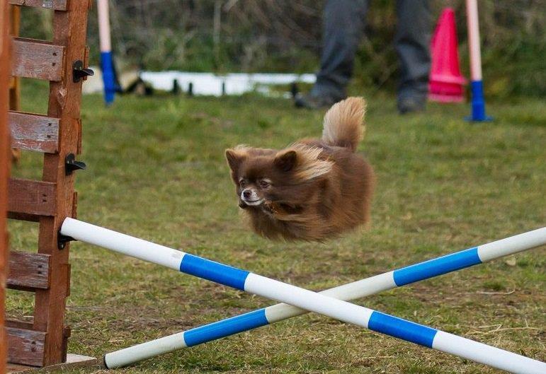
15 MAISIE!!!
 Reason we recommend spaying animals as this is an emergency surgery Best preventative = spay")
16 Pyometra = Infected uterus (pus filled) Open pyometra = drains (less dangerous) Closed pyometra = does not drain (emergency surgery) Reason we recommend spaying animals as this is an emergency surgery Best preventative = spay PYOMETRA

17 .THE REST Pancreas Prostate Adrenal Glands Lymph nodes GI Tract Cats with IBD vs. Lymphoma send out VDI

18 ECHOCARDIOGRAM Dr. Sayer referral or Dr. Tidwell to do in-house

19 NORMAL CARDIAC BLOOD FLOW Pulmonic valve Tricuspid valve Mitral Valve Aortic Valve


20 ECHOCARDIOGRAM


21 ABNORMAL ECHO S Pericardial Effusion DCM Feline Hypertrophic Cardiomyopathy


22 .SWITCHING GEARS

23 DIAGNOSTIC MUSCULOSKELETAL ULTRASOUND (MSK)
24 WHY IS ULTRASOUND NECESSARY? Agility dogs are the #1 subject for diagnostic ultrasound. Able to palpate muscular abnormalities but cannot see them with radiographs or even MRI/CT. Can see ligament and muscular tears and damage Can combine modalities such as stem cell and PRP therapy to inject into these areas to speed up the healing process
 Need to follow a")
25 ILIOPSOAS INJURY Most common injury we see here at Atrium and from our agility dogs Grade 1-3 strains noted Recovery time is about 9 months depending on the severity of the strain (faster with stem cells) Need to follow a rehab plan
26 STRAIN CHART Grade Grade 1 Grade 1-2 Grade 2 Grade 2-3 Grade 3 Description Mild strain, no loss of function, <5% muscle involvement, focal edema and hemorrhage Same as above but mild fascial tearing Moderate strain, decreased strength of musculotendinous unit, mild fiber rupture, increased hemorrhage and edema Same as above but with fascial tearing and mild fiber disruption Severe strain, significant fascial tearing, complete muscle fiber disruption, significant edema and hemorrhage

27 GRADE 2 ILIOPSOAS STRAIN Circle = Core lesion Arrow =Adductor muscle

28 GRADE 3 ILIOPSOAS STRAIN

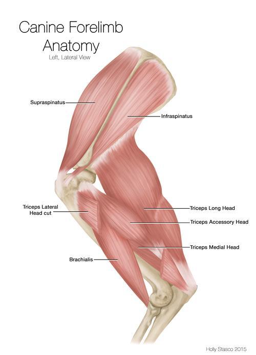


29 SHOULDER ANATOMY

30 SUPRASPINATUS TENDIONOPATHY Agility dogs!! Supraspinatus tendon can become inflammed and press on the bicipital bursa Ultrasound Findings: Hyperechoic: scar tissue or calcification Hypoechoic: edema/fluid active tear Mix: acute on chronic injury Clinical Signs = Pain with flexion of the shoulder and pain with direct palpation of the tendon and insertion Decreased range of motion Muscle atrophy (supraspinatus)


31 SHOULDER ULTRASOUND Supraspinatus muscle belly egg

32 STIFLE ULTRASOUND Meniscal tears (medial and lateral) Feel these through palpation and have a classic click with range of motion Can see hypoechoic tears in the meniscus with ultrasound A = Normal meniscus B= Abnormal hypoechoic meniscus

33 NORMAL CRANIAL CRUCIATE LIGAMENT


34 PARTIAL CRANIAL CRUCIATE LIGAMENT TEAR Cruciate tears Uneven CCL contour and hyperechoic stump Joint effusion and cranial displacement of fat pad

35 ACHILLES TENDON (COMMON CALCANEAL TENDON) Common Calcaneal Tendon (Achilles) Made of three separate muscles that come into one tendon and attach on the calcaneous Gastrocnemius Superficial digital flexor Common tendon (biceps femoris, gracilis and semitendinosus muscles)

36 ACHILLES ULTRASOUND


37 ACHILLES TENDON INJURY


38 OTHER INJURIES Hamstring Semitendinosus Semimebranosus Biceps femoris Collateral ligaments
39 STEM CELL/PRP USE Once we have located an injury/injuries we can use stem cells to inject into the area with ultrasound Some areas do not require ultrasound to be injected but you will need to use it for the tiny areas and areas of insertion Can be used for partial CCL tears vs. surgery Full CCL tears need surgery but use PRP s/stem cell with the TPLO to get the best recovery Stem cell injections will speed up healing by months For example: Iliopsoas tears can be healed in 3 months vs. 9+ months

40 REHAB POST-REGENERATIVE MEDICINE No therapeutic ultrasound, cryotherapy and no TENS K-laser therapy only 1-2 times per week for the first 8 weeks INTRA-ARTICULAR INJECTIONS -- HOLD LASER 2 WEEKS POST- INJECTION AS IT INCREASED BLOOD FLOW AND CAN MOVE THE CELLS OUT OF THE JOINT Must be on lower setting mW or less (go into settings and manually lower power for first 8 weeks 3B lasers are less than 1000mW so are safe

41 QUESTIONS?
Non Surgical Management of Soft Tissue Injuries. Megan LeFave, DVM cvma
 Non Surgical Management of Soft Tissue Injuries Megan LeFave, DVM cvma Non Surgical Management of Soft Tissue Injuries Biomechanical Principles Common front limb and hind limb injuries In hospital treatments
GENERAL ABDOMINAL IMAGING PERITONEAL SPACE, PANCREAS, & SPLEEN. VMB 960 March 25, 2013
 GENERAL ABDOMINAL IMAGING PERITONEAL SPACE, PANCREAS, & SPLEEN VMB 960 March 25, 2013 REFERENCE Chapters 35-36 Pages 650-678 Chapter 37 Pages 694-701 Chapter 3 Pages 38-49 OBJECTIVES Radiography and Ultrasound
Key words: Laser, sprain, strain, lameness, tendon
 MLS Master Class - Veterinary Imaging Presented by CelticSMR Ltd Free Phone (UK): 0800 279 9050 International: +44 (0) 1646 603150 AUTHOR DETAILS Carl Gorman BVSc MRCVS PUBLISHER DETAILS Mike Howe B Vet
Ultrasound of the Knee
 Ultrasound of the Knee Jon A. Jacobson, M.D. Professor of Radiology Director, Division of Musculoskeletal Radiology University of Michigan Disclosures: Consultant: Bioclinica Book Royalties: Elsevier Advisory
Abdominal Ultrasound
 Abdominal Ultrasound Imaging Control Buttons Depth The organ imaged should take up 3/4 of the screen Frequency = Penetration Use high frequencies (harmonics) for fluid filled and superficial structures
Biceps Femoris Muscle in Dogs Diana Powell 11/25/2016
 Biceps Femoris Muscle in Dogs Diana Powell 11/25/2016 The Biceps Femoris is the largest muscle in the muscle group that makes up the hamstring. The Biceps Femoris is covered only by fascia and skin and
GENERAL ABDOMINAL IMAGING PERITONEAL SPACE, PANCREAS, & SPLEEN
 GENERAL ABDOMINAL IMAGING PERITONEAL SPACE, PANCREAS, & SPLEEN VMB 960 March 25, 2013 REFERENCE Chapters 35-36 Pages 650-678 Chapter 37 Pages 694-701 Chapter 3 Pages 38-49 OBJECTIVES Radiography and Ultrasound
GASTROCNEMIUS TENDON REPAIR VETLIG USING THE STIF CAT 30 SOFT TISSUE INTERNAL FIXATION VETLIG
 VETLIG SOFT TISSUE INTERNAL FIXATION GASTROCNEMIUS TENDON REPAIR USING THE STIF CAT 30 VETLIG A R T I F I C I A L L I G A M E N T S F O R V E T E R I N A R Y U S E VETLIG MANAGEMENT OF CHRONIC GASTROCNEMIUS
As for the forelimb, treatment of condition of the hindlimb may be treated by both localised therapy, applying the laser
 MLS Master Class - Veterinary Imaging Presented by CelticSMR Ltd Free Phone (UK): 0800 279 9050 International: +44 (0) 1646 603150 AUTHOR DETAILS Carl Gorman BVSc MRCVS PUBLISHER DETAILS Mike Howe B Vet
Sonography of Knee and Calf Pain: the differential considerations
 Sonography of Knee and Calf Pain: the differential considerations Dr. Lisa L. S.Wong Consultant Radiologist St Paul s Hospital Outline Ultrasound techniques Common pathologies in calf and posterior knee
emoryhealthcare.org/ortho
 COMMON SOCCER INJURIES Oluseun A. Olufade, MD Assistant Professor, Department of Orthopedics and PM&R 1/7/18 GOALS Discuss top soccer injuries and treatment strategies Simplify hip and groin injuries in
1 st Ultrasound Masterclasses Live 2017
 Learning Tracks Learning Tracks plus Training Labs 09/14 09/15 09/16 09/17 Topics BCE Introduction to POCUS, Physics, Instrumentation, Doppler Physics, Basic Cardiac, etc. Master Cardiac Echo MCE Atypical
Case Studies. A. Kent Allen, DVM LAMENESS AND IMAGING IN THE SPORT HORSE
 Case Studies A. Kent Allen, DVM Author s address: Virginia Equine Imaging, 2716 Landmark School Road, The Plains, VA 20198; e-mail: vaequine@aol.com. 2007 AAEP. 1. Case Study #1: Medial Collateral Desmitis
Diagnostic Imaging
 www.fisiokinesiterapia.biz Diagnostic Imaging Diagnostic Imaging is no longer limited to radiography. Major technological advancements have lead to the use of new and improved imaging technologies. The
Pragmatic ultrasound in the diagnosis of soft tissue rheumatic pain. Plamen Todorov
 Pragmatic ultrasound in the diagnosis of soft tissue rheumatic pain Plamen Todorov INTRODUCTION Soft tissue rheumatism: nonsystemic, focal pathological syndromes involving the periarticular structures.
Knee, Ankle, and Foot: Normal and Abnormal Features with MRI and Ultrasound Correlation. Disclosures. Outline. Joint Effusion. Suprapatellar recess
 Knee, Ankle, and Foot: Normal and Abnormal Features with MRI and Ultrasound Correlation Jon A. Jacobson, M.D. Professor of Radiology Director, Division of Musculoskeletal Radiology University of Michigan
Basic of Ultrasound Physics E FAST & Renal Examination. Dr Muhammad Umer Ihsan MBBS,MD, DCH CCPU,DDU1,FACEM
 Basic of Ultrasound Physics E FAST & Renal Examination Dr Muhammad Umer Ihsan MBBS,MD, DCH CCPU,DDU1,FACEM What is Sound? Sound is Mechanical pressure waves What is Ultrasound? Ultrasounds are sound waves
Ultrasound Evaluation of Masses
 Ultrasound Evaluation of Masses Jon A. Jacobson, M.D. Professor of Radiology Director, Division of Musculoskeletal Radiology University of Michigan Disclosures: Consultant: Bioclinica Advisory Panel: GE,
Diagnostic Ultrasound. Sutiporn Khampunnip, M.D.
 Diagnostic Ultrasound Sutiporn Khampunnip, M.D. Definition of Ultrasound Ultrasound is simply sound waves, like audible sound. High-frequency sound and refers to mechanical vibrations above 20 khz. Human
What s your diagnosis?
 What s your diagnosis? Signalment: 9 year old MC 2.7 kg Papillion Presenting Complaint: Presented for work up of anorexia and vomiting History: He had presented to cardiology for work up of a grad IV/VI
10 million cells die in your body every minute of every day. Stem cells are what allow you to make new cells!
 Dr. Erica Johnson 10 million cells die in your body every minute of every day. Stem cells are what allow you to make new cells! They are the paramedics of cells They are primitive cells present in almost
Case study #11 Rt. knee
 The patient is a 55 year old female who presents with bilateral knee pain. Patient is a collegiate softball coach and has a very active lifestyle and career that is hampered by her chronic knee pain. She
Point of Care Ultrasound on the Field of Play K AT I E N ANOS, MD
 Point of Care Ultrasound on the Field of Play K AT I E N ANOS, MD H I GH P ERFORMANCE S PORTS MEDICINE P HYSI ATRIST, P R ACTICING S PORTS MEDI CINE No disclosures No disclosures Who am I? Objectives Over
CHAPTER 8: THE BIOMECHANICS OF THE HUMAN LOWER EXTREMITY
 CHAPTER 8: THE BIOMECHANICS OF THE HUMAN LOWER EXTREMITY _ 1. The hip joint is the articulation between the and the. A. femur, acetabulum B. femur, spine C. femur, tibia _ 2. Which of the following is
Joel S Sellers, DO, FAOASM CAQSM, RMSK
 Joel S Sellers, DO, FAOASM CAQSM, RMSK This is a sports slide of an Olympic wrestler Chris Taylor 1 This is a Sports Illustrated slide of jockey Johnny Sellers This is a slide of Coach Jim Sellers 2 This
Knee Joint Assessment and General View
 Knee Joint Assessment and General View Done by; Mshari S. Alghadier BSc Physical Therapy RHPT 366 m.alghadier@sau.edu.sa http://faculty.sau.edu.sa/m.alghadier/ Functional anatomy The knee is the largest
Ultrasound of the Knee Joint. Jun Sung Park,M.D. Bundang General Hospital Dept. of Rehabilitation Medicine
 Ultrasound of the Knee Joint Jun Sung Park,M.D. Bundang General Hospital Dept. of Rehabilitation Medicine Clinical History and P/E Chronic or Acute Symptoms Chronic Sx. : possible of systemic articular
Guidelines, Policies and Statements D5 Statement on Abdominal Scanning
 Guidelines, Policies and Statements D5 Statement on Abdominal Scanning Disclaimer and Copyright The ASUM Standards of Practice Board have made every effort to ensure that this Guideline/Policy/Statement
Prevention and Treatment of Injuries. The Femur. Quadriceps 12/11/2017
 Prevention and Treatment of Injuries The Thigh, Hip, Groin, and Pelvis Oak Ridge High School Conroe, Texas The Femur Is the longest and the second strongest bone in the body and is designed to permit maximum
Prevention and Treatment of Injuries. Anatomy. Anatomy. Chapter 20 The Knee Westfield High School Houston, Texas
 Prevention and Treatment of Injuries Chapter 20 The Knee Westfield High School Houston, Texas Anatomy MCL, Medial Collateral Ligament LCL, Lateral Collateral Ligament PCL, Posterior Cruciate Ligament ACL,
ELENI ANDIPA General Hospital of Athens G. Gennimatas
 ELENI ANDIPA General Hospital of Athens G. Gennimatas Technological advances over the last years have caused a dramatic improvement in ultrasound quality and resolution An established imaging modality
16) M. quadriceps femoris, m. tensor fasciae latae, m. sartorius
 M. quadriceps femoris, m. tensor fasciae latae, m. sartorius") Anatomy 1 - Lesson XIII: Myologia Part IV Objective: Students will examine the muscles of a canine in order to identify the musculature of the body.. Practical Tasks 15-18: 15) Medial muscles of the thigh,
Anatomy 1 - Lesson XIII: Myologia Part IV Objective: Students will examine the muscles of a canine in order to identify the musculature of the body.. Practical Tasks 15-18: 15) Medial muscles of the thigh,
Job Task Analysis for ARDMS Abdomen Data Collected: June 30, 2011
 Job Task Analysis for ARDMS Abdomen Data Collected: June 30, 2011 Reported: Analysis Summary for: Abdomen Examination Survey Dates 06/13/2011-06/26/2011 Invited Respondents 6,000 Surveys with Demographics
Basics of MR Imaging. Dynamic MRI. MRI Closed. The bed rotates from Upright to Recumbent, stopping at any angle in between.
 Basics of MR Imaging Dynamic MRI MRI Closed The bed rotates from Upright to Recumbent, stopping at any angle in between MRI Open Patient with Low Back Pain After Surgery Extremity MRI Sagittal T2 WI of
MUSCLES OF SHOULDER REGION
 Dr Jamila EL Medany OBJECTIVES At the end of the lecture, students should: List the name of muscles of the shoulder region. Describe the anatomy of muscles of shoulder region regarding: attachments of
What s Your Diagnosis?
 What s Your Diagnosis? Signalment: 5 year old MC Belgian Malinois Presenting Complaint: Perineal hernia as well as not eating or defecating History: The patient presented to the KSU VHC on 7/28/2018 for
Biology Human Anatomy Abdominal and Pelvic Cavities
 Biology 351 - Human Anatomy Abdominal and Pelvic Cavities Please place your name and I.D. number on the back of the last page of this exam. You must answer all questions on this exam. Because statistics
Abdominal Ultrasound : Aorta, Kidneys, Bladder
 Abdominal Ultrasound : Aorta, Kidneys, Bladder Nilam J. Soni, MD, MSc Associate Professor of Medicine Divisions of Hospital Medicine and Pulmonary/Critical Care Medicine Department of Medicine University
L o o k L i s t e n F e e l S c a n. Your Pocus Cards For Your Every Day Scanning.
 L o o k L i s t e n F e e l S c a n Your Pocus Cards For Your Every Day Scanning E-FAST Extended Focused Assessment by Sonography in Trauma Subcostal Heart View Pleural Sliding on M-mode (Sea-shore sign)
Chapter 14. Circulatory System Images. VT-122 Anatomy & Physiology II
 Chapter 14 Circulatory System Images VT-122 Anatomy & Physiology II The mediastinum Dog heart Dog heart Cat heart Dog heart ultrasound Can see pericardium as distinct bright line Pericardial effusion Fluid
Appendix 5. EFSUMB Newsletter. Gastroenterological Ultrasound
 EFSUMB Newsletter 87 Examinations should encompass the full range of pathological conditions listed below A log book listing the types of examinations undertaken should be kept Training should usually
The Essentials Tissue Characterization and Knobology
 The Essentials Tissue Characterization and Knobology Randy E. Moore, DC, RDMS RMSK No relevant financial relationships Ultrasound The New Standard of Care Musculoskeletal sonography has become the standard
MRI of the Shoulder What to look for and how to find it? Dr. Eric Handley Musculoskeletal Radiologist Cherry Creek Imaging
 MRI of the Shoulder What to look for and how to find it? Dr. Eric Handley Musculoskeletal Radiologist Cherry Creek Imaging MRI of the Shoulder Benefits of Ultrasound: * Dynamic * Interactive real time
Joints of the Lower Limb II
 Joints of the Lower Limb II Lecture Objectives Describe the components of the knee and ankle joint. List the ligaments associated with these joints and their attachments. List the muscles acting on these
This lab activity is aligned with Visible Body s Human Anatomy Atlas app. Learn more at visiblebody.com/professors
 1 This lab activity is aligned with Visible Body s Human Anatomy Atlas app. Learn more at visiblebody.com/professors 2 A. Digestive System Overview To Start: Go to the Views menu and scroll down to the
Musculoskeletal Ultrasound: Basics, Utility, and Clinical Applications
 Musculoskeletal Ultrasound: Basics, Utility, and Clinical Applications Andrew Lavigne, MD, FRCPC Physical Medicine and Rehabilitation CSCN Diplomat (EMG) Dip Sport Medicine Eugene Maida, MD, PGY-4 Resident
US finding of the shoulder (with live demonstration) 인제의대상계백병원 안재기
 인제의대상계백병원 안재기") US finding of the shoulder (with live demonstration) 인제의대상계백병원 안재기 Shoulder US Biceps tendon & Rotator Cuff Long Head of Biceps Tendon Subscapularis tendon Supraspinatus tendon Infraspinatus tendon Teres
US finding of the shoulder (with live demonstration) 인제의대상계백병원 안재기 Shoulder US Biceps tendon & Rotator Cuff Long Head of Biceps Tendon Subscapularis tendon Supraspinatus tendon Infraspinatus tendon Teres
What MRI has taught us about ultrasound. W. Michael Karlin DVM, MS, Dipl ACVS Mid-Atlantic Equine Medical Center
 What MRI has taught us about ultrasound W. Michael Karlin DVM, MS, Dipl ACVS Mid-Atlantic Equine Medical Center Overview Ultrasound uses MRI Experimentally induced injury How does understanding MRI help
Ligamentous and Meniscal Injuries: Diagnosis and Management
 Ligamentous and Meniscal Injuries: Diagnosis and Management Daniel K Williams, MD Franciscan Physician Network Orthopedic Specialists September 29, 2017 No Financial Disclosures INTRODUCTION Overview of
Principles of Ultrasound. Cara C. Prideaux, M.D. University of Utah PM&R Sports Medicine Fellow March 14, 2012
 Principles of Ultrasound Cara C. Prideaux, M.D. University of Utah PM&R Sports Medicine Fellow March 14, 2012 None Disclosures Outline Introduction Benefits and Limitations of US Ultrasound (US) Physics
Equine Lameness & Imaging Techniques
 Equine Lameness & Imaging Techniques Peter Heidmann DVM MPH Specialist in Equine Internal Medicine Montana Equine Medical & Surgical Center www.montanaequine.com 406-285-0123 Types of lameness Skeletal
for the Veterinary Technician
 An Overview of Abdominal Ultrasound for the Veterinary Technician Valerie Gates, CVT, VTS (ECC) Learning Objective: The reader should gain a basic understanding of ultrasound, including physics, terminology,
Office Orthopedics. No conflict of interest No financial disclosures 1/31/2018
 Office Orthopedics Amin Afsari DO Orthopedic Hand and Upper Extremity Surgery Orthopedic Institute of Wisconsin Midwest Orthopedic Specialty Hospital 1 No conflict of interest No financial disclosures
Chapter 2 Pitfalls in Musculoskeletal Ultrasound
 Chapter 2 Pitfalls in Musculoskeletal Ultrasound Violeta Maria Vlad MD, PhD Introduction Taking a good ultrasound (US) picture is an art. Interpreting it is a science. This is in fact everything US is
Objectives. Sprains, Strains, and Musculoskeletal Maladies. Sprains. Sprains. Sprains. Physical Exam 5/5/2010
 Objectives, Strains, and Musculoskeletal Maladies Robert Hosey, MD University of Kentucky Sports Medicine Define sprains and strains Systematically evaluate and manage joint / muscle injuries When to refer
Ultrasonography of Peritoneal and Retroperitoneal Spaces and Abdominal Lymph Nodes
 IMAGING Ultrasonography of Peritoneal and Retroperitoneal Spaces and Abdominal Lymph Nodes Clifford R. Berry, DVM, DACVR; Elizabeth Huyhn, DVM; and Danielle Mauragis, CVT University of Florida Welcome
RN(EC) ENC(C) GNC(C) MN ACNP *** MECHANISM OF INJURY.. MOST IMPORTANT *** - Useful in determining mechanism of injury / overuse
 ENC(C) GNC(C) MN ACNP *** MECHANISM OF INJURY.. MOST IMPORTANT *** - Useful in determining mechanism of injury / overuse") HISTORY *** MECHANISM OF INJURY.. MOST IMPORTANT *** Age of patient Sport / Occupation - Certain conditions are more prevalent in particular age groups (Osgood Schlaters in youth / Degenerative Joint Disease
HISTORY *** MECHANISM OF INJURY.. MOST IMPORTANT *** Age of patient Sport / Occupation - Certain conditions are more prevalent in particular age groups (Osgood Schlaters in youth / Degenerative Joint Disease
Case study # 6 Sharon P
 Patient is a morbidly obese 70 year old female presenting with left shoulder pain after a severe fall. Patient is in moderate to severe pain with extremely limited range of motion due to extensive shoulder
Chapter 3. Sonographic Image Interpretation
 Chapter 3 Sonographic Image Interpretation Sonograms are two-dimensional gray-scale images that allow assessment and diagnosis of many anatomic and pathologic changes that can occur in the human body.
Muscles to know. Lab 21. Muscles of the Pelvis and Lower Limbs. Muscles that Position the Lower Limbs. Generally. Muscles that Move the Thigh
 Muscles to know Lab 21 Muscles of the Pelvis, Leg and Foot psoas major iliacus gluteus maximus gluteus medius sartorius quadriceps femoris (4) gracilus adductor longus biceps femoris semitendinosis semimembranosus
RADPrimer Curriculum Breast Topics Covered Basic Intermediate 225
 Breast Anatomy & Normal Variants 11 Breast Imaging Modalities 13 BI RADS Lexicon 3 Mammography: Masses 9 Mammography: Calcifications 17 Mammography: Additional Findings 8 Ultrasound Features 10 Ultrasound
The Elbow 3/5/2015. The Elbow Scanning Sequence. * Anterior Joint (The anterior Pyramid ) * Lateral Epicondyle * Medial Epicondyle * Posterior Joint
 * Lateral Epicondyle * Medial Epicondyle * Posterior Joint") Scanning Sequence * Anterior Joint (The anterior Pyramid ) * Lateral Epicondyle * Medial Epicondyle * Posterior Joint Anterior Elbow Pyramid Courtesy of Jay Smith, MD. Vice chair PMR Mayo Clinic Rochester,
Scanning Sequence * Anterior Joint (The anterior Pyramid ) * Lateral Epicondyle * Medial Epicondyle * Posterior Joint Anterior Elbow Pyramid Courtesy of Jay Smith, MD. Vice chair PMR Mayo Clinic Rochester,
Abdominal ultrasound:
 Abdominal ultrasound: Non-traumatic acute abdomen Wittanee Na-ChiangMai, MD Department of Radiology ChiangMai University 26/04/2017 Contents Technique of examination Normal anatomy Emergency conditions
The Upper Limb II. Anatomy RHS 241 Lecture 11 Dr. Einas Al-Eisa
 The Upper Limb II Anatomy RHS 241 Lecture 11 Dr. Einas Al-Eisa Sternoclavicular joint Double joint.? Each side separated by intercalating articular disc Grasp the mid-portion of your clavicle on one side
Gluteal region DR. GITANJALI KHORWAL
 Gluteal region DR. GITANJALI KHORWAL Gluteal region The transitional area between the trunk and the lower extremity. The gluteal region includes the rounded, posterior buttocks and the laterally placed
Musculoskeletal Examination
 Musculoskeletal Examination Statement of Goals Know how to perform a complete musculoskeletal examination. Learning Objectives A. Describe the anatomy of the musculoskeletal system including the bony structures,
Appendix 9: Endoscopic Ultrasound in Gastroenterology
 Appendix 9: Endoscopic Ultrasound in Gastroenterology This curriculum is intended for clinicians who perform endoscopic ultrasonography (EUS) in gastroenterology. It includes standards for theoretical
The Kidneys. (L., ren; Gk, nephros; hence the adjectives renal and nephric) & Suprarenal (Adrenal) Glands. Dr Maan Al-Abbasi PhD, MBChB
 & Suprarenal (Adrenal) Glands. Dr Maan Al-Abbasi PhD, MBChB") The Kidneys (L., ren; Gk, nephros; hence the adjectives renal and nephric) & Suprarenal (Adrenal) Glands Dr Maan Al-Abbasi PhD, MBChB Functions of Urinary System Regulate electrolytes (K+, Na+, etc) Regulate
The Kidneys (L., ren; Gk, nephros; hence the adjectives renal and nephric) & Suprarenal (Adrenal) Glands Dr Maan Al-Abbasi PhD, MBChB Functions of Urinary System Regulate electrolytes (K+, Na+, etc) Regulate
The faculty will include physicians with international reputations as outstanding ultrasound educators.
 Ultrasound Courses Course Description Whether you re a beginner or a seasoned sonographer, this year s AAEM pre-conference ultrasound course will be worth your time. We will be offering a half day course
General Ultrasound. What is General Ultrasound Imaging?
 Scan for mobile link. General Ultrasound What is General Ultrasound Imaging? Ultrasound is safe and painless, and produces pictures of the inside of the body using sound waves. Ultrasound imaging, also
General Ultrasound. What is General Ultrasound Imaging?
 Scan for mobile link. General Ultrasound Ultrasound imaging uses sound waves to produce pictures of the inside of the body. It is used to help diagnose the causes of pain, swelling and infection in the
Focused Musculoskeletal Ultrasound
 Focused Musculoskeletal Ultrasound David Lewis Consultant Emergency Medicine Ipswich (Club Doctor, Ipswich Town FC) Advanced Emergency Ultrasound Objectives! General principles! Musculoskeletal anatomy!
 http://vanat.cvm.umn.edu/ http://www.cvmbs.colostate.edu/vetneuro/vca3/vca.html Arthrology 1 Where a bone joins Synchondrosis 2 basic types of joint structure classification Without a cavity (2 types)
The Elbow. The Elbow. The Elbow 12/11/2017. Oak Ridge High School Conroe, Texas. Compose of three bones. Ligaments of the Elbow
 Oak Ridge High School Conroe, Texas Compose of three bones The humerus The radius The ulna Ligaments of the Elbow Ulnar collateral ligament Radial collateral ligament Annular ligament 1 The elbow is considered
Contents. Pig Dissection. Contents. External Features Sex Determination Mouth and Maxillary Nerve Muscles Index Internal Systems Index
 Pig Dissection External Features Sex Determination Mouth and Maxillary Nerve Muscles Index Internal Systems Index External features Sex determination Male Female Male to External anatomy 1. Pinna 2. External
The posterior abdominal wall. Prof. Oluwadiya KS
 The posterior abdominal wall Prof. Oluwadiya KS www.oluwadiya.sitesled.com Posterior Abdominal Wall Lumbar vertebrae and discs. Muscles opsoas, quadratus lumborum, iliacus, transverse, abdominal wall
Radiology of the abdomen Lecture -1-
 Radiology of the abdomen Lecture -1- Objectives To know radiology modalities used in abdomen imaging mainly GI tract. To know advantages and disadvantages of each modality. To know indications and contraindications
In-Depth Foundations: Anatomy Terms to Know
 Be familiar with / able to identify and define all the following parts. The Spine Cranium Vertebrae Cervical, Thoracic, Lumbar Sacrum Coccyx Bones of Upper Body Cranium Mastoid process; Occipital condyle,
Overview Ligament Injuries. Anatomy. Epidemiology Very commonly injured joint. ACL Injury 20/06/2016. Meniscus Tears. Patellofemoral Problems
 Overview Ligament Injuries Meniscus Tears Pankaj Sharma MBBS, FRCS (Tr & Orth) Consultant Orthopaedic Surgeon Manchester Royal Infirmary Patellofemoral Problems Knee Examination Anatomy Epidemiology Very
What I Will Cover. Shock Wave Therapy. What are shock waves? What are shock waves? What are shock waves? What are shock waves?
 Shock Wave Therapy Sarah Matyjaszek Large Animal Surgery University of Florida April 25 th, 2009 What I Will Cover How they work Applications Research Case example Potential complications The future A
HUMAN BODY COURSE LOWER LIMB NERVES AND VESSELS
 HUMAN BODY COURSE LOWER LIMB NERVES AND VESSELS October 22, 2010 D. LOWER LIMB MUSCLES 2. Lower limb compartments ANTERIOR THIGH COMPARTMENT General lfunction: Hip flexion, knee extension, other motions
Practical 1 Worksheet
 Practical 1 Worksheet ANATOMICAL TERMS 1. Use the word bank to fill in the missing words. reference side stand body arms palms anatomical forward All anatomical terms have a(n) point which is called the
Policies, Standards, and Guidelines. Guidelines for Abdominal Ultrasound Examination
 Policies, Standards, and Guidelines Guidelines for Abdominal Ultrasound Examination Approved by Council Feb 2018 Disclaimer and Copyright The ASUM Standards of Practice Board have made every effort to
Original Report. The Reverse Segond Fracture: Association with a Tear of the Posterior Cruciate Ligament and Medial Meniscus
 Eva M. Escobedo 1 William J. Mills 2 John. Hunter 1 Received July 10, 2001; accepted after revision October 1, 2001. 1 Department of Radiology, University of Washington Harborview Medical enter, 325 Ninth
Newsletter CALCANEAL TENDON TEARS IN DOGS. Contents. View from the floor! 2012 Mar-Apr: Vol 1, Issue 1
 Newsletter CALCANEAL TENDON TEARS IN DOGS 2012 Mar-Apr: Vol 1, Issue 1 Contents Page 2 Page 3 Page 4 Page 5 Calcaneal tendon morphology & biomechanical properties Retrospective study of Achilles mechanism
Proceedings of the 10th International Congress of World Equine Veterinary Association
 www.ivis.org Proceedings of the 10th International Congress of World Equine Veterinary Association Jan. 28 Feb. 1, 2008 - Moscow, Russia Next Congress: Reprinted in IVIS with the permission of the Conference
May 2011, Issue 31. In addition to our regular ER hours, AMVS is providing emergency and critical care services to your patients: Fridays, all day
 Page 1 of 5 Having Trouble Viewing this Email? Click Here You're receiving this email because of your relationship with Aspen Meadow Veterinary Specialists. Please confirm your continued interest in receiving
Common Applications for Sonography and Guided Intervention: Shoulder
 Common Applications for Sonography and Guided Intervention: Shoulder Jon A. Jacobson, M.D. Professor of Radiology Director, Division of Musculoskeletal Radiology University of Michigan Disclosures: Consultant:
EFSUMB EUROPEAN FEDERATION OF SOCIETIES FOR ULTRASOUND IN MEDICINE AND BIOLOGY Building a European Ultrasound Community
 MINIMUM TRAINING REQUIREMENTS FOR THE PRACTICE OF MEDICAL ULTRASOUND IN EUROPE Appendix 9: Endoscopic Ultrasound in Gastroenterology This curriculum is intended for clinicians who perform endoscopic ultrasonography
US in non-traumatic acute abdomen. Lalita, M.D. Radiologist Department of radiology Faculty of Medicine ChiangMai university
 US in non-traumatic acute abdomen Lalita, M.D. Radiologist Department of radiology Faculty of Medicine ChiangMai university Sagittal Orientation Transverse (Axial) Orientation Coronal Orientation Intercostal
MR DIAGNOSTICS OF MUSCLE TRAUMA. Ivo Nikolov, M.D., Radiologist - Spectar Imaging Centre, Sofia
 MR DIAGNOSTICS OF MUSCLE TRAUMA Ivo Nikolov, M.D., Radiologist - Spectar Imaging Centre, Sofia Мyofibrils Сonnective tissue Fibers - Endomysium Fascicle - Permysium Мuscle - Еpimysium Question of the
ICCUME All Domain recommendations Montreal, October 14,
 Domain 1 (Scope) ICCUME All Domain recommendations Montreal, October 14, 2017 10 1.1 Goal: The ICC will produce consensus recommendations on An integrated ultrasound curriculum for undergraduate medical
SIMITRI STABLE IN STRIDE SURGICAL PROCEDURE
 Copyright 2016 NGD. All rights reserved Neil Embleton, B.Sc., DVM and Veronica Barkowski, DVM Helivet Mobile Surgical Services, Sundre, AB, Canada July 2016 SIMITRI STABLE IN STRIDE SURGICAL PROCEDURE
Hamstring Injuries and Avulsions. Charles A. Bush-Joseph, MD Rush University Medical Center Team Physician, Chicago White Sox Chicago, IL
 Hamstring Injuries and Avulsions Charles A. Bush-Joseph, MD Rush University Medical Center Team Physician, Chicago White Sox Chicago, IL Disclosures No personal disclosures on this topic Institutional
1-Muscles: 2-Blood supply: Branches of the profunda femoris artery. 3-Nerve supply: Sciatic nerve
 1-Muscles: B i c e p s f e m o r i s S e m i t e n d i n o s u s S e m i m e m b r a n o s u s a small part of the adductor magnus (h a m s t r i n g p a r t o r i s c h i a l p a r t ) 2-Blood supply:
Annex III. Amendments to relevant sections of the product information
 Annex III Amendments to relevant sections of the product information Note: These amendments to the relevant sections of the product information are the outcome of the referral procedure. The product information
Table of contents. Foreword. Preface. 1 Introduction Historical Perspective 00
 Table of contents Foreword Preface 1 Introduction 00 1.1 Historical Perspective 00 2 Fundamentals of musculoskeletal ultrasound 00 2.1 Frequency and wavelength 00 2.2 Generating ultrasound waves 00 2.3
Comparative study of high resolusion ultrasonography and magnetic resonance imaging in diagnosing traumatic knee injuries & pathologies
 Original article: Comparative study of high resolusion ultrasonography and magnetic resonance imaging in diagnosing traumatic knee injuries & pathologies Dr. Rakesh Gujjar*, Dr. R. P. Bansal, Dr. Sandeep
MUSCLES OF THE LOWER LIMBS
 MUSCLES OF THE LOWER LIMBS Naming, location and general function Dr. Nabil khouri ROLES THAT SHOULD NOT BE FORGOTTEN Most anterior compartment muscles of the hip and thigh Flexor of the femur at the hip
and K n e e J o i n t Is the most complicated joint in the body!!!!
 K n e e J o i n t K n e e J o i n t Is the most complicated joint in the body!!!! 1-Consists of two condylar joints between: A-The medial and lateral condyles of the femur and The condyles of the tibia