Success in FNAC Service minded - availability Sampling by trained cytopathologist Quick staining to avoid insufficient material Application of ancilla
|
|
|
- Brian Gilbert
- 5 years ago
- Views:
Transcription


1
2 Success in FNAC Service minded - availability Sampling by trained cytopathologist Quick staining to avoid insufficient material Application of ancillary technique Close contact with clinicians Triple diagnosis Multidisciplinary meetings pre-post operative and therapy conferences


3 Breast FNAC FNAC is a widely accepted technique in the diagnosis and management of palpable and non-palpable breast lesions due to its simplicity, accuracy and utility for avoidance more invasive procedures.
4
5
6 Most countries adopted a triple assessment approach. FNAC is the first-line pathological investigation in both screening and symptomatic populations, with the exception of microcalcifications. Cytopathologists are best qualified to collect and interpret FNA samples.
7 The majority of European countries use similar reporting systems for breast FNA (C1-C5) in keeping with European Guidelines for QA in Breast Cancer Screening and Diagnosis, although some still prefer descriptive reporting only. The use of CNB has increased, although not always for evidence-based reasons. CNB and FNAC are not mutually exclusive. FNAC should be used in diagnosis of benign, symptomatic lesions and CNB in microcalcifications, suspicious FNAC findings and malignancies where radiology cannot guarantee stromal invasion.
8 Pre-operative diagnostic procedure The choice of sampling method in any centre should be determined by: the sensitivity and specificity of the technique in the centre. the diagnostic information required for malignant lesions. patient comfort and costs. the availability of staff skilled and experienced in using the procedures, particularly FNAC sampling and interpretation.
9 Breast FNAC How not to be washed away? Doing the technique with quality Coupled with image. Solving specific problems
10 Accuracy of FNAC The accuracy of FNAC depends on three main factors: a sample that is adequate and representative of the lesion. suitable processing and staining without artifact. accurate interpretation of the cytological material with a clear report conveyed to the rest of the clinical team.
11 FNAC Multistep technique Clinical examination Image-guided (US) Aspiration Slide preparation Fixation and staining Cytological interpretation

12 Breast FNAC It would be difficult to think of a contemporary medical procedure that is less HIGH TECH than Fine Needle Aspiration Cytology

13 Breast FNAC A successful marriage of low tech and high tech occurs when modern radiographic techniques are used to guide a needle to a non-palpable lesion


14 TRIPLE ASSESSMENT APPROACH Clinical Imaging BBB: 98% benign follow up MMM: 1% error surgery Other: biopsy Cytology


15 FNAC Clinic IPATIMUP Material 23Gx30mm 25Gx16mm 10 ml


16 FNAC Clinic IPATIMUP US-Guided Needle

17 Aspiration

18 Slide preparation Material obtained with a fine needle is expelled onto appropriately labelled glass slide. This is usually performed by using a 10-ml syringe filled with air, attaching the needle to do it and pushing the contents out of the needle.


19 Slide preparation Sometimes, if the hub of the needle is full, it is possible to tap the hub against the glass and obtain the material directly from there.

20 Smears: one step

21 Smears: two steps

22 Quality of the smear
23 CYTOLOGICAL INTERPRETATION

24
25 BREAST FNAC: solving problems

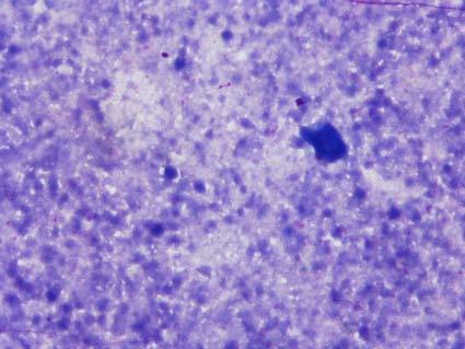
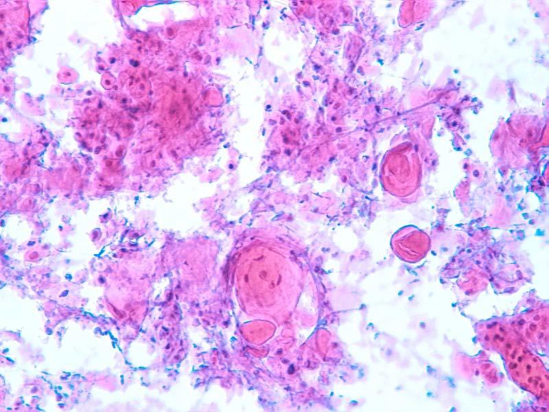
26 CYTOLOGICAL CRITERIA OF BENIGN LESIONS BENIGN* Low to moderate cellularity Cohesive epithelial groups without or with mild nuclear overlapping and presence of myoepithelial cells Background: naked nuclei * Adequate sample without evidence of malignancy.

27 CYTOLOGICAL CRITERIA OF MALIGNANCY MALIGNANT* High cellularity Loss of cohesiveness Nuclear pleomorphism Dirty background Absence of naked nuclei * Adequate smear containing cells with unequivocal characteristics of malignancy.
28 BREAST FNAC: solving problems Current evidence indicates that the use of nonoperative diagnosis substantially reduces the number of unnecessary operations performed both for benign disease and for malignancy, with reduced discomfort and inconvenience to the patient. What is the role of breast FNAC in benign lesions, malignant lesions and gray zone lesions?

29 BREAST FNAC: solving problems Benign Lesions 35-year old female presented with a 25 mm well-defined nodule in the right breast. Mammography and US are compatible with fibroadenoma. There is no family history of breast cancer.


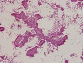
30
31 BREAST FNAC: solving problems Benign Lesions FNAC is a useful and reliable tool in the evaluation and management of benign breast lesions, such as: Inflammatory conditions Cysts Fibroadenoma

32 CYTOLOGICAL INTERPRETATION Inflammatory diseases BENIGN SUBAREOLAR ABSCESS A high yield of inflammatory cells and multinucleated giant cells. Keratin and squamous metaplastic cells. The identification of giant cells with keratin at cytoplasm is an important clue for the diagnosis. Reactive epithelial cells.


33 CYTOLOGICAL INTERPRETATION Inflammatory diseases BENIGN FAT NECROSIS Moderate to high cellularity. Foam cells (always present), sometimes multinucleated. Collapsed fat cells. Inflammatory cells. Sometimes, presence of worrisome nuclei atypical


34 CYTOLOGICAL INTERPRETATION Inflammatory diseases BENIGN GRANULOMATOUS MASTITIS


35 CYTOLOGICAL INTERPRETATION BENIGN - CYSTS

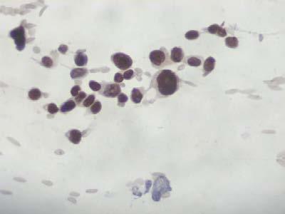
36 RULES TO EVALUATE CYSTS After complete aspiration of the cyst, it is especially important to re-evaluate the area (US) to determine if a residual breast mass is present. If a residual mass is found, a second aspiration should be performed. Be careful with apocrine changes
, sometimes multifocal and/or bilateral.")
 is similar to the")
37 CYTOLOGICAL INTERPRETATION Fibroadenoma It is the most common benign tumour of the breast. Solitary nodule (most), sometimes multifocal and/or bilateral. Usually are non-tender wellcircumscribed nodules. Biphasic proliferative lesion (epithelial and stromal elements) is similar to the structures of the terminal lobular-ductal unit (TLDU).

38 CYTOLOGICAL INTERPRETATION Fibroadenoma A high cell yield. The three characteristic components are: large branching, monolayered sheets of uniform epithelial cells; numerous single, bare bipolar nuclei (myoepithelial cells) fragments of fibromyxoid stroma.


39 CYTOLOGICAL INTERPRETATION Benign epithelial proliferative lesion Moderate to high cellularity. Cohesive epithelial groups without or with mild nuclear overlapping and presence of myoepithelial cells. Heterogeneous cell population: mild variation in the size and shape of the nuclei (oval, round or spindle). Bipolar naked nuclei in the background. Apocrine and foam cells.

40 BREAST FNAC: solving problems Malignant Lesions 33-year old female presented with a 15 mm ill-defined nodule in the right breast. Mammography and US are compatible with carcinoma.

41 Benign Granular cell tumour Imaging typically shows a dense mass with stellate margin, simulating malignancy. High cellular yield. Cells showing moderate atypia with intact, abundant and granular cytoplasm.
42 BREAST FNAC: solving problems Malignant Lesions Definitive surgery for carcinoma can be planned preoperatively using the triple approach or radiological imaging, clinical examination and FNAC (or CNB). This permits treatment for many malignant lesions in a one-stage operation.

43 CYTOLOGICAL INTERPRETATION Invasive ductal carcinoma Cellular smear Variable cell pattern, sometimes plasmocytoid appearance Nuclear pleomorphism Loss of cohesion


44


45 Diagnostic Cytopathology, 2009
. In some cases very poor cell yield.")

46 CYTOLOGICAL INTERPRETATION Invasive lobular carcinoma Variable cellularity (moderate to intense). In some cases very poor cell yield. Cells single and in small clusters, short single files common. Epithelial cells have small dark nuclei with scanty cytoplasm. The lack of pleomorphism can be cause of a falsenegative diagnosis. Intracytoplasmic lumina/vacuoles.

47 CYTOLOGICAL INTERPRETATION Invasive lobular carcinoma A most valuable clue on ILC is the tendency to form small chains of cells in the aspirates
.")

48 CYTOLOGICAL INTERPRETATION Mucinous carcinoma Well defined and circumscribed tumour (similar to fibroadenoma). Abundant background mucinous, atypical cells in small solid aggregates, single files or isolated. The mucin stains violet to blue with MGG or pink on HE staining.


49 CYTOLOGICAL INTERPRETATION Tubular carcinoma Variable cellularity (moderate to intense). At low magnification a pattern somewhat similar to fibroadenoma. Cells arranged mostly in tubular structures with comma-like pattern. Epithelial cells are uniform and bland. The lack of pleomorphism can be cause of a false-negative diagnosis. Bare nuclei are present in rare cases.


50 CYTOLOGICAL INTERPRETATION Medullary carcinoma Well-circumscribed mass (similar to fibroadenoma) with fibro-elastic consistency. Highly cellular smears with irregular cell groups and single atypical cells. The neoplastic cells are large, pleomorphic with prominent nucleoli. Background rich in lymphocytes. Definitive diagnosis requires the demonstration of well defined borders. They are frequently associated with germinal mutation of BRCA-1.


51 CYTOLOGICAL INTERPRETATION Apocrine carcinoma Malignant cells have a large dense eosinophilic granular cytoplasm with large nuclei with prominent nucleoli. Neoplastic cells are isolated, sometimes without cytoplasm and in small aggregates. Necrosis is frequent.

52 CYTOLOGICAL INTERPRETATION Metaplastic carcinoma Is an invasive ductal carcinoma with metaplastic changes: squamous cells, spindle cells, osteoid or chondroid. Smears can show different cell types: ductal epithelial, spindle or squamous. Sometimes we can observe multinucleated giant cells and myxoid material. Can be cystic at aspiration and with necrotic material.


53


54 P63 expression in sarcomatoid/metaplastic carcinomas of the breast Reis Filho JS & Schmitt FC. Histopathology 42: 92-99, 2003


55 CYTOLOGICAL INTERPRETATION Invasive micropapillary carcinoma Highly cellular smears composed by angulated small groups of cohesive cells with papillary configurations without fibro vascular cores. Cells showing nuclear atypical, irregular nuclear contours and prominent nucleoli. Cytoplasm vacuoles are rarely seen. Background is clean with rare isolated neoplastic cells.


56 CYTOLOGICAL INTERPRETATION Breast carcinoma with osteoclast-like giant cells Cellular smears composed by cohesive groups of epithelial cells, with low grade of atypia. Groups of plump spindle cells as well as isolated atypical epithelial cells. Presence of osteoclast-like multinucleated cells at periphery of the epithelial cells or in the background of the smears.


57 CYTOLOGICAL INTERPRETATION Adenoid cystic carcinoma Highly cellular Pattern of large tissue fragments, consisting of sheets of cells with poorly defined cytoplasm with minimal cytological atypia and myoepithelial cells, or crowded atypical cells without biphasic appearance and moderate nuclear atypia Background may have dispersed bare nuclei and/or dispersed intact atypical cells Hyaline spherules, varying from mucinous to collagenous Invagination of collagenous material into invasive epithelial sheets Scattered true lumens within epithelial proliferation


58 CYTOLOGICAL INTERPRETATION Breast carcinoma with signet-ring cells


59 CYTOLOGICAL INTERPRETATION Other special subtypes of breast carcinoma SECRETORY CARCINOMA ACINIC CELL CARCINOMA


60 CYTOLOGICAL INTERPRETATION Metastatic malignancy METASTATIC MELANOMA


61 CYTOLOGICAL INTERPRETATION Other malignancy NON-HODGKIN LYMPHOMA

62 CYTOLOGICAL INTERPRETATION Inflammatory diseases BENIGN INTRAMAMMARY LYMPH NODE
63 BREAST FNAC: solving problems Gray zone Conditions in which there is a risk of making a false-positive diagnosis Papillary lesions Atypical hyperplasia Complex sclerosing lesion Fibroadenoma Regenerative epithelial atypia Pregnancy and lactation Skin adnexal tumours Specific cytological problems Tubular and lobular carcinoma Low grade DCIS vs benign lesions High grade DCIS vs invasive ca Conditions in which there is a risk of a false-negative diagnosis Tumours with necrosis /fibrosis Small carcinomas associated with benign lesions Complex proliferative lesions with foci of carcinoma Low grade in situ or invasive carcinoma Lobular carcinoma


64 CYTOLOGICAL INTERPRETATION Papillary Lesions Cellular smears. Papillary three-dimensional arrangements. Complex folded and branching sheets of epithelial cells. Columnar cells in rows, palisades and single. Variable nuclear atypical Epithelial cells with cytoplasm vacuoles Others: macrophages, apocrine metaphase, bare oval nuclei.

 Tall columnar cells are frequent.")
65 CYTOLOGICAL INTERPRETATION Papillary carcinoma Is it possible to distinguish benign and malignant Papillary breast tumours on FNA? Cytological findings favouring malignant Higher cellularity Papillary three-dimensional arrangements without a central fibro vascular core (cell balls) Tall columnar cells are frequent. Isolated cells with cytoplasm. Absence of bare nuclei, apocrine metaplasia and rare macrophages.


66 PAPILLARY LESIONS: CNB helps?


67 European guidelines on breast cancer screening


68 CYTOLOGICAL INTERPRETATION Epithelial proliferative lesions Moderate to high cellularity. Epithelial cell groups with nuclear overlapping and without or with few myoepithelial cells. Bipolar naked nuclei in the background absent or in few numbers. Less cell cohesively in the borders of the cell groups with occasional isolated epithelial cells with preserved cytoplasm. 20% are malignant at biopsy


69 DCIS CCL RADIAL SCAR LOW GRADE INVASIVE CA
 similar to fibroadenoma but with predominance of the stroma over the epithelium")

70 CYTOLOGICAL INTERPRETATION Fibroepithelial lesions PHYLLODES TUMOUR Biphasic proliferative lesion (epithelial and stromal elements) similar to fibroadenoma but with predominance of the stroma over the epithelium Fibromyxoid stromal fragments are larger than those seen in fibroadenomas and are highly cellular with fibroblastic spindle cells. The presence of isolated stromal cells with spindle nuclei and abundant pale cytoplasm is suggestive of PT.


71


72 CYTOLOGICAL INTERPRETATION Fibroepithelial lesions Fibroadenoma Myxoid changes Atypia
73 Breast FNAC How not to be washed away? QUALITY QUALITY QUALITY


74 Accredited Laboratory of CAP in Pathology Certified Laboratory by ISO
75 NHSBSP, 2011
76 Technical factors False-positives Bad quality of smears Fixation artefacts False-negatives Operator dependent Characteristics of the lesion: Size of the lesion Size of the breast Location Histological type


77 Scanty material Inadequate smearing Artifacts of fixation Thick smear

78 BREAST FNAC X CNB
79 Core-needle biopsy Advantages More specific diagnosis of benign lesions Avoid the problem of atypias Allow to distinguish carcinoma in situ from invasive Diminish the inadequate rate Allow study of prognostic and predictive factors Needs less diagnostic expertise
80 Core-needle biopsy Advantages More specific diagnosis of benign lesions Avoid the problem of atypias Allow to distinguish carcinoma in situ from invasive Diminish the inadequate rate Allow study of prognostic and predictive factors Needs less diagnostic expertise


81 ATYPIA ON FNA 8.0 a 9.5% 60% are malignant ATYPIA ON CNB 3.6 a 9.0% 56% are malignant Connoly et al., USCAP 2002
82 Core-needle biopsy Advantages More specific diagnosis of benign lesions Avoid the problem of atypias Allow to distinguish carcinoma in situ from invasive Diminish the inadequate rate Allow study of prognostic and predictive factors Needs less diagnostic expertise


83 Core-needle biopsy Do not exclude invasion with only DCIS is present Until 30% of DCIS cases have invasion on the surgical specimen. Predicts invasion in 92% of the cases Connoly et al., USCAP 2002
84 Core-needle biopsy Advantages More specific diagnosis of benign lesions Avoid the problem of atypias Allow to distinguish carcinoma in situ from invasive Diminish the inadequate rate Allow study of prognostic and predictive factors Needs less diagnostic expertise
85 RATES OF UNSATISFACTORY MATERIAL ON FNA Type of lesion (microcalcification) Histological type (> benign lesion) Cytopathologist Type of system used to guide the needle. Variable on literature: 3 a 80%(!) Acceptable until 25%in non-palpable lesions
86 Core-needle biopsy Advantages More specific diagnosis of benign lesions Avoid the problem of atypias Allow to distinguish carcinoma in situ from invasive Diminish the inadequate rate Allow study of prognostic and predictive factors Needs less diagnostic expertise


87 ER/PR ASSESSMENT IN BREAST FNAs


88 Who should performs? The success of FNAC is directly related with the ability and experience of the aspirator. There is strong evidence that the procedure is better when performed by a well-trained staff.
89 Core-needle biopsy Disadvantages Sampling error (loss malignancy in complex benign lesions) Can not give immediate diagnosis. More expensive and traumatic than FNA Also have problematic lesions : papillary. mucinous, etc Dissemination of malignant cells?
90 Breast FNAC How not to be washed away? Aspiration should be direct to a define target. FNAC is a multi-step procedure and to obtain a good material is essential for the diagnosis. The cytological diagnosis should be done only with the knowledge of the clinical context and preferential in a multidisciplinary environment. Negative results can not solve the patient problem.


91
92


93 FOUNDED IN BRUSSELS, Belgium 1957 AS SUPRANATIONAL ORGANIZATION: To encourage cooperation; To foster international exchange; To stimulate development; To encourage research.

94 The official journal of the International Academy of Cytology Editor-in-Chief Marluce Bibbo, Philadelphia Associate Editors R. Marshall Austin, Pittsburgh Thomas A. Bonfiglio, Rochester Lukas Bubendorf, Basel Yener S. Erozan, Baltimore David B. Kaminsky, Palm Springs Harubumi Kato, Tokyo Leopold G. Koss, New York, N.Y. Gladwyn Leiman, Burlington, Vt. Shahla Masood, Jacksonville, Fla. Robert Y. Osamura, Tokyo Ibrahim Ramzy, Irvine, Calif. Fernando C. Schmitt, Porto Volker Schneider, Freiburg Mark E. Sherman, Rockville Diane Solomon, Rockville Alain P. Verhest, Brussels Philippe Vielh, Villejuif

95 INTERNATIONAL CONGRESS OF CYTOLOGY SINCE 1961: Paris, May 26 30,2013


96 February 24-27, 2012 Hong Kong IAC Tutorial on Gynecologic and Non Gynecologic Cytology A comprehensive course on gynecologic and non-gynecologic cytology


97 REGISTRY FOR CYTOTECHNOLOGISTS SINCE 1972:
98 INTERNATIONAL BOARD OF CYTOPATHOLOGY SINCE 1974: Comprehensive, one-day examination for medical members of the Academy to document proficiency in cytopathology

99 Asia: Medical Membership, IAC

100 FURTHER INFORMATION ON MEMBERSHIP:
101 ANY INFORMATION THAT YOU NEED:
The role of the cytologist in breast cancer screening
 The role of the cytologist in breast cancer screening I.Seili-Bekafigo, MD, PhD Clinical cytologist KBC Rijeka Croatian Society for Clinical Cytology Fine needle aspiration (FNA, FNAB, FNAC) Fine needle
IBCM 2, April 2009, Sarajevo, Bosnia and Herzegovina
 Preoperative diagnosis and treatment planning in breast cancer The pathologist s perspective L. Mazzucchelli Istituto Cantonale di Patologia Locarno, Switzerland IBCM 2, 23-25 April 2009, Sarajevo, Bosnia
Diseases of the breast (1 of 2)
") Diseases of the breast (1 of 2) Introduction A histology introduction Normal ducts and lobules of the breast are lined by two layers of cells a layer of luminal cells overlying a second layer of myoepithelial
Diseases of the breast (1 of 2) Introduction A histology introduction Normal ducts and lobules of the breast are lined by two layers of cells a layer of luminal cells overlying a second layer of myoepithelial
Breast pathology. 2nd Department of Pathology Semmelweis University
 Breast pathology 2nd Department of Pathology Semmelweis University Breast pathology - Summary - Benign lesions - Acute mastitis - Plasma cell mastitis / duct ectasia - Fat necrosis - Fibrocystic change/
CLINICAL SIGNIFICANCE OF BENIGN EPITHELIAL CHANGES
 Papillomas. Papillomas are composed of multiple branching fibrovascular cores, each having a connective tissue axis lined by luminal and myoepithelial cells ( Fig. 23-11 ). Growth occurs within a dilated
Cytyc Corporation - Case Presentation Archive - March 2002
 FirstCyte Ductal Lavage History: 68 Year Old Female Gail Index: Unknown Clinical History: Negative Mammogram in 1995 6 yrs. later presents with bloody nipple discharge Subsequent suspicious mammogram Suspicious
Lesion Imaging Characteristics Mass, Favoring Benign Circumscribed Margins Intramammary Lymph Node
 Lesion Imaging Characteristics Mass, Favoring Benign Circumscribed Margins Intramammary Lymph Node Oil Cyst Mass, Intermediate Concern Microlobulated Margins Obscured Margins Mass, Favoring Malignant Indistinct
Papillary Lesions of the breast
 Papillary Lesions of the breast Emad Rakha Professor of Breast Pathology The University of Nottingham Papillary lesions of the breast are a heterogeneous group of disease, which are characterised by neoplastic
Salivary Glands 3/7/2017
 Salivary Glands 3/7/2017 Goals and objectives Focus on the entities unique to H&N Common board type facts Information for your future practice Salivary Glands Salivary Glands Major gland. Paratid. Submandibular.
04/10/2018. Intraductal Papillary Neoplasms Of Breast INTRADUCTAL PAPILLOMA
 Intraductal Papillary Neoplasms Of Breast Savitri Krishnamurthy MD Professor of Pathology Deputy Division Head The University of Texas MD Anderson Cancer Center 25 th Annual Seminar in Pathology Pittsburgh,
1 NORMAL HISTOLOGY AND METAPLASIAS
 1 NORMAL HISTOLOGY AND METAPLASIAS, MD Anatomy and Histology 1 Metaplasias 2 ANATOMY AND HISTOLOGY The female breast is composed of a branching duct system, which begins at the nipple with the major lactiferous
FNA of Thyroid. Toward a Uniform Terminology With Management Guidelines. NCI NCI Thyroid FNA State of the Science Conference
 FNA of Thyroid NCI NCI Thyroid FNA State of the Science Conference Toward a Uniform Terminology With Management Guidelines Thyroid Thyroid FNA Cytomorphology NCI Thyroid FNA State of the Science Conference
Breast Pathology. Breast Development
 Breast Pathology Lecturer: Hanina Hibshoosh, M.D. Reading: Kumar, Cotran, Robbins, Basic Pathology, 6th Edition, pages 623-635 Breast Development 5th week - thickening of the epidermis - milk line 5th
PAAF vs Core Biopsy en Lesiones Mamarias Case #1
 5/19/2014 PAAF vs Core Biopsy en Lesiones Mamarias Case #1 Fine Needle Aspiration Cytology of Breast: Correlation with Needle Core Biopsy 64-year-old woman Mass in breast Syed Hoda, MD CD31 Post-Radiation
BREAST PATHOLOGY. Fibrocystic Changes
 BREAST PATHOLOGY Lesions of the breast are very common, and they present as palpable, sometimes painful, nodules or masses. Most of these lesions are benign. Breast cancer is the 2 nd most common cause
Interpretation of Breast Pathology in the Era of Minimally Invasive Procedures
 Shahla Masood, M.D. Professor and Chair Department of Pathology and Laboratory Medicine University of Florida College of Medicine Jacksonville Medical Director, UF Health Breast Center Chief of Pathology
Salivary Gland Cytology
 Salivary Gland Cytology Diagnostic challenges and potential pitfalls Tarik M. Elsheikh, MD Professor and Medical Director Anatomic Pathology Cleveland Clinic FNA Salivary Gland Lesions Indications Distinguish
Basement membrane in lobule.
 Bahram Memar, MD Basement membrane in lobule. Normal lobule-luteal phase Normal lobule-follicular phase Lactating breast Greater than 95% are adenocarcinomas in situ carcinomas and invasive carcinomas.
Thyroid follicular neoplasms in cytology. Ulrika Klopčič Institute of Oncology, Department of Cytopathology, Ljubljana, Slovenia
 Thyroid follicular neoplasms in cytology Ulrika Klopčič Institute of Oncology, Department of Cytopathology, Ljubljana, Slovenia Lecture overview importance of FNAB in assessing thyroid lesions follicular
Papillary Lesions of the Breast A Practical Approach to Diagnosis. (Arch Pathol Lab Med. 2016;140: ; doi: /arpa.
 Papillary Lesions of the Breast A Practical Approach to Diagnosis (Arch Pathol Lab Med. 2016;140:1052 1059; doi: 10.5858/arpa.2016-0219-RA) Papillary lesions of the breast Span the spectrum of benign,
CPC 4 Breast Cancer. Rochelle Harwood, a 35 year old sales assistant, presents to her GP because she has noticed a painless lump in her left breast.
 CPC 4 Breast Cancer Rochelle Harwood, a 35 year old sales assistant, presents to her GP because she has noticed a painless lump in her left breast. 1. What are the most likely diagnoses of this lump? Fibroadenoma
CURRICULUM FOR THE BREAST PATHOLOGY ROTATION UNIVERSITY OF FLORIDA DEPARTMENT OF PATHOLOGY
 CURRICULUM FOR THE BREAST PATHOLOGY ROTATION UNIVERSITY OF FLORIDA DEPARTMENT OF PATHOLOGY JULY, 2003 The following is a conceptual curriculum and set of guidelines for Pathology Residents on the Breast
Protocol for the Examination of Biopsy Specimens From Patients With Invasive Carcinoma of the Breast
 Protocol for the Examination of Specimens From Patients With Invasive Carcinoma of the Breast Version: BreastInvasive 1.0.0.0 Protocol Posting Date: February 2019 Accreditation Requirements The use of
Abid Irshad, MD Director Breast Imaging. Medical University of South Carolina Charleston
 Abid Irshad, MD Director Breast Imaging Medical University of South Carolina Charleston Cases Financial disclosure: I or my family have no financial interest related to the material discussed in this presentation
FNA OF SALIVARY GLANDS: A PRACTICAL APPROACH
 FNA OF SALIVARY GLANDS: A PRACTICAL APPROACH FNA of Salivary Glands: Challenges Wide range of neoplastic and non-neoplastic lesions Cytological overlap between the different benign and malignant tumors
DOWNLOAD ENTIRE DOCUMENT FROM
 PREVIEW ONLY 1 Atlas on Bethesda system for reporting Thyroid Cytology PREVIEW ONLY 2 OVERVIEW 1. Indications and goal of thyroid FNA 2. Contraindications 3. Procurement of cell sample 4. Staining methods
Benign, Reactive and Inflammatory Lesions of the Breast
 Benign, Reactive and Inflammatory Lesions of the Breast Marilin Rosa, MD Associate Member Section Head of Breast Pathology Department of Anatomic Pathology Program Director, Breast Pathology Fellowship
Thyroid master class. Thyroid Fine needle aspiration cytology and liquid-based techniques: Hologic and Becton Dickinson
 Thyroid master class Thyroid Fine needle aspiration cytology and liquid-based techniques: Hologic and Becton Dickinson Principle of LBC Collection of cells in liquid medium Immediate fixation Processor-prepared
INDEX. in this web service Cambridge University Press
 abscess. See also subareolar abscess acute mastitis, 44 lactational/puerperal mastitis, 55 mammary tuberculosis, 42 tuberculous, 43 adeno gastric, 198, 200 invasive, 157 lung, 197, 200 prostatic, 199 200
LYMPHATIC DRAINAGE AXILLARY (MOSTLY) INTERNAL MAMMARY SUPRACLAVICULAR
 INTERNAL MAMMARY SUPRACLAVICULAR") BREAST LYMPHATIC DRAINAGE AXILLARY (MOSTLY) INTERNAL MAMMARY SUPRACLAVICULAR HISTOLOGY LOBE: (10 in whole breast) LOBULE: (many per lobe) ACINUS/I, aka ALVEOLUS/I: (many per lobule) DUCT(S): INTRA- or
BREAST LYMPHATIC DRAINAGE AXILLARY (MOSTLY) INTERNAL MAMMARY SUPRACLAVICULAR HISTOLOGY LOBE: (10 in whole breast) LOBULE: (many per lobe) ACINUS/I, aka ALVEOLUS/I: (many per lobule) DUCT(S): INTRA- or
Pancreatitis: A Potential Pitfall in Endoscopic Ultrasound Guided Pancreatic FNA
 Pancreatitis: A Potential Pitfall in Endoscopic Ultrasound Guided Pancreatic FNA Jack Yang, MD Department of Pathology, Medical University of South Carolina Objectives Understand the indication of EUS
Outline 11/2/2017. Pancreatic EUS-FNA general aspects. Cytomorphologic features of solid neoplasms/lesions of the pancreas
 ENDOSCOPIC ULTRASOUND GUIDED-FINE NEEDLE ASPIRATION CYTOLOGY OF PANCREAS Khalid Amin M.D. Assistant Professor Department of Laboratory Medicine and Pathology University of Minnesota Outline Pancreatic
40th European Congress of Cytology Liverpool, UK, 2-5 th October 2016
 40th European Congress of Cytology Liverpool, UK, 2-5 th October 2016 EUS FNA of abdominal organs: An approach to reporting and triage for ancillary testing Date and time: Sunday 2 nd October 2016 15.00-16.30
Treatment options for the precancerous Atypical Breast lesions. Prof. YOUNG-JIN SUH The Catholic University of Korea
 Treatment options for the precancerous Atypical Breast lesions Prof. YOUNG-JIN SUH The Catholic University of Korea Not so benign lesions? Imaging abnormalities(10% recall) lead to diagnostic evaluation,
Scoring system of fine needle aspiration cytology samples for the detection of non high grade ductal breast carcinoma
 Oncocytology ;:. ORIGINAL ARTICLE OPEN ACCESS Scoring system of fine needle aspiration cytology samples for the detection of non high grade ductal breast carcinoma Ayumi Ryu, Akemi Takenaka, Shigenori
Diagnostic Value of Imprint Cytology During Image-Guided Core Biopsy in Improving Breast Health Care
 Available online at www.annclinlabsci.org 8 Annals of Clinical & Laboratory Science, vol. 41, no. 1, 2011 Diagnostic Value of Imprint Cytology During Image-Guided Core Biopsy in Improving Breast Health
Objectives. Salivary Gland FNA: The Milan System. Role of Salivary Gland FNA 04/26/2018
 Salivary Gland FNA: The Milan System Dr. Jennifer Brainard Section Head Cytopathology Cleveland Clinic Objectives Introduce the Milan System for reporting salivary gland cytopathology Define cytologic
Carcinoma mammario: le istologie non frequenti. Valentina Guarneri Università di Padova IOV-IRCCS
 Carcinoma mammario: le istologie non frequenti Valentina Guarneri Università di Padova IOV-IRCCS Histological diversity of breast adenocarcinomas Different histological types are defined according to specific
Case study 1. Rie Horii, M.D., Ph.D. Division of Pathology Cancer Institute Hospital, Japanese Foundation for Cancer Research
 NCCN/JCCNB Seminar in Japan April 15, 2012 Case study 1 Rie Horii, M.D., Ph.D. Division of Pathology Cancer Institute Hospital, Japanese Foundation for Cancer Research Present illness: A 50y.o.premenopausal
CORRELATION OF FINE NEEDLE ASPIRATION CYTOLOGY AND ITS HISTOPATHOLOGY IN DIAGNOSIS OF BREAST LUMPS
 wjpmr, 2017,3(9), 221-226 SJIF Impact Factor: 4.103 WORLD JOURNAL OF PHARMACEUTICAL AND MEDICAL RESEARCH www.wjpmr.com Research Article ISSN 2455-3301 WJPMR CORRELATION OF FINE NEEDLE ASPIRATION CYTOLOGY
Imaging in breast cancer. Mammography and Ultrasound Donya Farrokh.MD Radiologist Mashhad University of Medical Since
 Imaging in breast cancer Mammography and Ultrasound Donya Farrokh.MD Radiologist Mashhad University of Medical Since A mammogram report is a key component of the breast cancer diagnostic process. A mammogram
Objectives. Atypical Glandular Cells. Atypical Endocervical Cells. Reactive Endocervical Cells
 2013 California Society of Pathologists 66 th Annual Meeting San Francisco, CA Atypical Glandular Cells to Early Invasive Adenocarcinoma: Cervical Cytology and Histology Christina S. Kong, MD Associate
Fine-Needle Aspiration Cytology of Low-Grade Cribriform Cystadenocarcinoma with Many Psammoma Bodies of the Salivary Gland
 The Korean Journal of Pathology 2013; 47: 481-485 CASE STUDY Fine-Needle Aspiration Cytology of Low-Grade Cribriform Cystadenocarcinoma with Many Psammoma Bodies of the Salivary Gland Ji Yun Jeong Dongbin
ARIZONA SOCIETY OF PATHOLOGISTS 13 TH APRIL 2013 HEAD AND NECK CYTOPATHOLOGY. F ZAHRA ALY, MD, PhD
 ARIZONA SOCIETY OF PATHOLOGISTS 13 TH APRIL 2013 HEAD AND NECK CYTOPATHOLOGY F ZAHRA ALY, MD, PhD The main areas sites amenable for cytopathology include lymph nodes, thyroid, major salivary glands especially
Role of fine needle aspiration cytology in male breast lesion: 4 year observational study
 International Journal of Research in Medical Sciences Chide PM et al. Int J Res Med Sci. 2016 Sep;4(9):3945-3950 www.msjonline.org pissn 2320-6071 eissn 2320-6012 Research Article DOI: http://dx.doi.org/10.18203/2320-6012.ijrms20162913
Proliferative Epithelial lesions of the Breast. Sami Shousha, MD, FRCPath Charing Cross Hospital & Imperial College, London
 Proliferative Epithelial lesions of the Breast Sami Shousha, MD, FRCPath Charing Cross Hospital & Imperial College, London Amman, November2013 Proliferative Epithelial Lesions of the Breast Usual type
Salivary gland cytology. Salivary gland cytology. Triage helps the clinician. Salivary gland tumors. Diagnostic difficulties
 Salivary gland cytology Salivary Gland Cytology Pınar Fırat, MD Professor of Pathology İ.U. İstanbul Faculty of Medicine Çapa, İstanbul It is a reliable diagnostic test However, definitive subclassification
Minimizing Errors in Diagnostic Pathology
 Shahla Masood, M.D. Professor and Chair Department of Pathology and Laboratory Medicine University of Florida College of Medicine-Jacksonville Medical Director, Shands Jacksonville Breast Health Center
Presentation material is for education purposes only. All rights reserved URMC Radiology Page 1 of 98
 Presentation material is for education purposes only. All rights reserved. 2011 URMC Radiology Page 1 of 98 Radiology / Pathology Conference February 2011 Brooke Koltz, Cytopathology Resident Presentation
Salivary gland tumor cytologic and histologic correlation: Algorithmic and risk stratification based approaches
 Salivary gland tumor cytologic and histologic correlation: Algorithmic and risk stratification based approaches Christopher C. Griffith, MD, PhD Raja R. Seethala, MD 1. Salivary gland tumor cytology: A
Case 1. Slide 1 History: 65 year old male presents with bilateral pleural effusions, a 40 pack year smoking history and peripheral and hilar lung
 Case 1. Slide 1 History: 65 year old male presents with bilateral pleural effusions, a 40 pack year smoking history and peripheral and hilar lung masses. Specimen shown is from a tap of the pleural effusion.
Almost any suspected tumor can be aspirated easily and safely. Some masses are more risky to aspirate including:
 DOES THIS PATIENT HAVE CANCER? USING IN-HOUSE CYTOLOGY TO HELP YOU MAKE THIS DIAGNOSIS. Joyce Obradovich, DVM, Diplomate, ACVIM (Oncology) Animal Cancer & Imaging Center, Canton, Michigan Almost every
Utility of Fine Needle Aspiration Cytology in Evaluation of Breast Lesions.
 Utility of Fine Needle Aspiration Cytology in Evaluation of Breast Lesions. Komal Joshi 1*, Nandita Mehta 2, Hansa Goswami 3 1 2 nd Year Resident, Ahmedabad. 2 Professor, 3 Professor & Head, Pathology
Gross appearance of nodular hyperplasia in material obtained from suprapubic prostatectomy. Note the multinodular appearance and the admixture of
 Tiền liệt tuyến Tiền liệt tuyến Gross appearance of nodular hyperplasia in material obtained from suprapubic prostatectomy. Note the multinodular appearance and the admixture of solid and microcystic areas.
Introduction. 23 rd Annual Seminar in Pathology. FLUIDS, Part 1. Pittsburgh, PA Gladwyn Leiman UVMMC, VT
 23 rd Annual Seminar in Pathology Pittsburgh, PA Gladwyn Leiman UVMMC, VT FLUIDS, Part 1 "Blue walls", Claudia Hansen, 2009 Introduction o Challenging to everyone o Almost any benign or malignant process
Gynecologic Cytopathology: Glandular lesions
 Gynecologic Cytopathology: Glandular lesions Lin Wai Fung (MSc, MPH, CMIAC) 17/4/2014 Glandular lesions of the uterus Endocervix Endometrium Normal endocervical cells Sheets, strips well-preserved architecture:
Case year female. Routine Pap smear
 Case 1 57 year female Routine Pap smear Diagnosis? 1. Atypical glandular cells of unknown significance (AGUS) 2. Endocervical AIS 3. Endocervical adenocarcinoma 4. Endometrial adenocarcinoma 5. Adenocarcinoma
BOSNIAN-TURKISH CYTOPATHOLOGY SCHOOL June 18-19, 2016 Sarajevo. Case Discussions. 60 year old woman Routine gynecologic control LBC
 BOSNIAN-TURKISH CYTOPATHOLOGY SCHOOL June 18-19, 2016 Sarajevo Case Discussions Prof Dr Sıtkı Tuzlalı Tuzlalı Pathology Laboratory 60 year old woman Routine gynecologic control LBC 1 2 Endometrial thickening
Palpable Breast Lesions Cytomorphological Analysis and Scoring System with Histopatholgical Correlation
 IOSR Journal of Dental and Medical Sciences (IOSR-JDMS) e-issn: 2279-0853, p-issn: 2279-0861.Volume 15, Issue 10 Ver. III (October. 2016), PP 25-29 www.iosrjournals.org Palpable Breast Lesions Cytomorphological
Benign Mimics of Malignancy in Breast Pathology
 Arthur Purdy Stout Society of Surgical Pathologists Companion Meeting Benign Mimics of Malignancy in Breast Pathology Stuart J. Schnitt, M.D. Beth Israel Deaconess Medical Center and Harvard Medical School,
LGM International, Inc.
 Liqui-PREP TM Cytology Atlas Preface The following pictures are examples with descriptions of cytology slides processed with the Liqui-PREP TM System.. The descriptions are reviewed by Pathologists. It
Cytological study of palpable breast lumps with their histological correlation in a tertiary care hospital
 Original Research Article Cytological study of palpable breast lumps with their histological correlation in a tertiary care hospital Sunita Mistry 1*, Jignasha Patel 2, Kamlesh Shah 3, Ajit Patel 4 1 Assistant
TBSRTC 1- Probabilistic approach and Relationship to Clinical Algorithms
 The Benefits of a Uniform Reporting System for Thyroid Cytopathology BETHESDA REPORTING SYSTEM Prof. Fernando Schmitt Department of Pathology and Oncology, Medical Faculty of Porto University Head of Molecular
Proliferative Breast Disease: implications of core biopsy diagnosis. Proliferative Breast Disease
 Proliferative Breast Disease: implications of core biopsy diagnosis Jean F. Simpson, M.D. Breast Pathology Consultants, Inc. Nashville, TN Proliferative Breast Disease Must be interpreted in clinical and
Update on Thyroid FNA The Bethesda System. Shikha Bose M.D. Associate Professor Cedars Sinai Medical Center
 Update on Thyroid FNA The Bethesda System Shikha Bose M.D. Associate Professor Cedars Sinai Medical Center Thyroid Nodules Frequent occurrence Palpable: 4-7% of adults Ultrasound: 10-31% Majority benign
Ultrasound of the Breast BASICS FOR THE ORDERING CLINICIAN
 Ultrasound of the Breast BASICS FOR THE ORDERING CLINICIAN Breast Ultrasound Anatomy Skin Breast Parenchyma Pectoralis Fascia Pectoralis Breast Ultrasound Anatomy Indications for Breast Ultrasound Palpable
Cytology for the Endocrinologist. Nicole Massoll M.D
 Cytology for the Endocrinologist Nicole Massoll M.D Objectives Discuss slide preperation Definitions of adequacy ROSE (Rapid On-Site Evaluation) Thyroid Cytology Adequacy Nicole Massoll M.D. University
Can True Papillary Neoplasms of Breast and Their Mimickers Be Accurately Classified by Cytology?
 92 CANCER CYTOPATHOLOGY Can True Papillary Neoplasms of Breast and Their Mimickers Be Accurately Classified by Cytology? Claire W. Michael, M.D. 1 Bruce Buschmann, C.T. 2 1 University of Michigan, Department
Columnar Cell Lesions
 Columnar Cell Lesions Laura C. Collins, M.D. Department of Pathology Beth Israel Deaconess Medical Center and Harvard Medical School Boston, MA Question? Columnar cell lesions are: a) Annoying lesions
6/3/2010. Outline of Talk. Lobular Breast Cancer: Definition of lobular differentiation. Common Problems in Diagnosing LCIS in Core Biopsies
 Outline of Talk Lobular Breast Cancer: Common Problems in Diagnosing LCIS in Core Biopsies Definition of lobular differentiation Variants of LCIS that: carry risk for unsampled invasive cancer mimic DCIS
Case # year old man with a 2 cm right kidney mass
 Case # 4. 52 year old man with a 2 cm right kidney mass Figure 1 Figure 2 Figure 3 Figure 4 Diagnosis: Negative/Non-diagnostic Normal kidney tissue Fine needle aspiration (FNA) of the kidney is performed
Diagnostic Cytology of Cancer Cases
 Diagnostic Cytology of Cancer Cases Somporn Techangamsuwan Companion Animal Cancer Research Unit (CAC-RU) Department of Pathology, Faculty of Veterinary Science, Chulalongkorn University 1 Tumor or Non-tumor
Surgical Pathology Issues of Practical Importance
 Surgical Pathology Issues of Practical Importance Anne Moore, MD Medical Oncology Syed Hoda, MD Surgical Pathology The pathologist is central to the team approach needed to manage the patient with breast
Recent advances in breast cancers
 Recent advances in breast cancers Breast cancer is a hetrogenous disease due to distinct genetic alterations. Similar morphological subtypes show variation in clinical behaviour especially in response
Mody. AIS vs. Invasive Adenocarcinoma of the Cervix
 Common Problems in Gynecologic Pathology Michael T. Deavers, M.D. Houston Methodist Hospital, Houston, Texas Common Problems in Gynecologic Pathology Adenocarcinoma in-situ (AIS) of the Cervix vs. Invasive
Histological Type. Morphological and Molecular Typing of breast Cancer. Nottingham Tenovus Primary Breast Cancer Study. Survival (%) Ian Ellis
 Ian Ellis") Morphological and Molecular Typing of breast Cancer Ian Ellis Molecular Medical Sciences, University of Nottingham Department of Histopathology, Nottingham University Hospitals NHS Trust Histological Type
Morphological and Molecular Typing of breast Cancer Ian Ellis Molecular Medical Sciences, University of Nottingham Department of Histopathology, Nottingham University Hospitals NHS Trust Histological Type
Maram Abdaljaleel, MD Dermatopathologist and Neuropathologist University of Jordan, School of Medicine
 Maram Abdaljaleel, MD Dermatopathologist and Neuropathologist University of Jordan, School of Medicine The most common non-skin malignancy of women 2 nd most common cause of cancer deaths in women, following
Disclosures. Parathyroid Pathology. Objectives. The normal parathyroid 11/10/2012
 Disclosures Parathyroid Pathology I have nothing to disclose Annemieke van Zante MD/PhD Assistant Professor of Clinical Pathology Associate Chief of Cytopathology Objectives 1. Review the pathologic features
SIGNIFICANCE OF SILVER STAINING OF NULCEAR ORGANIZER REGIONS IN FINE NEEDLE ASPIRATION SMEARS OF BREAST LESIONS
 ORIGINAL ARTICLE SIGNIFICANCE OF SILVER STAINING OF NULCEAR ORGANIZER REGIONS IN FINE NEEDLE ASPIRATION SMEARS OF BREAST LESIONS Dimple H Darad 1, Aditi D Dholakia 2, Savitri Chauhan 1 1 Associate professor;
Oncocytic-Appearing Salivary Gland Tumors. Oncocytic, Cystic, Mucinous, and High Grade Salivary Gland Tumors SALIVARY GLAND FNA: PART II
 William C. Faquin, MD, PhD Professor of Pathology Harvard Medical School Director of Head and Neck Pathology Massachusetts Eye and Ear Massachusetts General Hospital SALIVARY GLAND FNA: PART II Oncocytic,
Guidance on the management of B3 lesions
 Guidance on the management of B3 lesions Lesion diagnosed on 14g or vacuumassisted biopsy (VAB) Risk of upgrade Recommended investigation Suggested approach for follow-up if no malignancy on VAE awaiting
BREAST PATHOLOGY MCQS
 BREAST PATHOLOGY MCQS 1) :The most important factor in breast enlargement during pregnancy is A. stromal edema B. secretion of chorionic gonadotropin C. glandular hyperplasia D. proliferation of stroma
04/10/2018 HIGH RISK BREAST LESIONS. Pathology Perspectives of High Risk Breast Lesions ELEVATED RISK OF BREAST CANCER HISTORICAL PERSPECTIVES
 Pathology Perspectives of High Risk Breast Lesions Savitri Krishnamurthy MD Professor of Pathology Deputy Division Head Director of Clinical Trials, Research and Development The University of Texas MD
University Journal of Pre and Para Clinical Sciences
 ISSN 2455 2879 Volume 2 Issue 1 2016 Metaplastic carcinoma breast a rare case report Abstract : Metaplastic carcinoma of the breast is a rare malignancy with two distinct cell lines described as a breast
Mammographic imaging of nonpalpable breast lesions. Malai Muttarak, MD Department of Radiology Chiang Mai University Chiang Mai, Thailand
 Mammographic imaging of nonpalpable breast lesions Malai Muttarak, MD Department of Radiology Chiang Mai University Chiang Mai, Thailand Introduction Contents Mammographic signs of nonpalpable breast cancer
Papillary Lesions of the Breast
 Papillary Lesions of the Breast Laura C. Collins, M.D. Associate Professor of Pathology Associate Director, Division of Anatomic Pathology Beth Israel Deaconess Medical Center and Harvard Medical School
NODULAR CYSTIC HIDRADENOMA OVER THE GLUTEAL REGION: A RARE CYTOMORPHOLOGICAL DIAGNOSIS
 NODULAR CYSTIC HIDRADENOMA OVER THE GLUTEAL REGION: A RARE CYTOMORPHOLOGICAL DIAGNOSIS Abstract: The primary as well as metastatic tumours of the skin can be diagnosed by fine needle aspiration cytology
RE-AUDIT OF THYROID FNA USING THE THY GRADING SYSTEM AND HISTOLOGY AT SUNDERLAND ROYAL HOSPITAL, 2011
 Audit: RE-AUDIT OF THYROID FNA USING THE THY GRADING SYSTEM AND HISTOLOGY AT SUNDERLAND ROYAL HOSPITAL, 2011 Auditors: Dr Lena Wilkinson SpR Histopathology Dr. Debra Milne Consultant Histocytopathologist
Cytyc Corporation - Case Presentation Archive - October 2001
 ThinPrep Pap Test History: 82 Year Old Female Specimen Type: Peritoneal Washings Case provided by Dr. Berle Stratton, Southwest Washington Medical Center, Vancouver, Washington. *The images, analysis and
Respiratory Tract Cytology
 Respiratory Tract Cytology 40 th European Congress of Cytology Liverpool, UK Momin T. Siddiqui M.D. Professor of Pathology and Laboratory Medicine Director of Cytopathology Emory University Hospital, Atlanta,
The Hot Topic for today is a biopsy from a 58-year-old woman who had worrisome mammographic calcifications on screening.
 The Hot Topic for today is a biopsy from a 58-year-old woman who had worrisome mammographic calcifications on screening. 1 My name is Dan Visscher; I am a consultant in the Division of Anatomic Pathology
Quality ID #263: Preoperative Diagnosis of Breast Cancer National Quality Strategy Domain: Effective Clinical Care
 Quality ID #263: Preoperative Diagnosis of Breast Cancer National Quality Strategy Domain: Effective Clinical Care 2018 OPTIONS FOR INDIVIDUAL MEASURES: REGISTRY ONLY MEASURE TYPE: Process DESCRIPTION:
Criteria of Malignancy. Evaluation Score
 30 5 Diagnostic Criteria Criteria of Malignancy Table 5.2 lists criteria in contrast-enhancing MR mammography that strongly indicate the presence of malignancy or are unspecific. Unifactorial evaluation
Salivary gland Workshop Trondheim 31th may 2012
 Salivary gland Workshop Trondheim 31th may 2012 Peter Jebsen cytopathologist Oslo University Hospital Rikshospitalet Anna Bofin ass. Professor St. Olavs Hospital, Trondheim Drying artifacts Lymfocytes
Enterprise Interest None
 Enterprise Interest None What are triple negative breast cancers? A synopsis of their histological patterns Ian Ellis Molecular Medical Sciences, University of Nottingham Department of Histopathology,
CINtec p16 INK4a Staining Atlas
 CINtec p16 INK4a Staining Atlas Rating Rating Positive The rating positive will be assigned if the p16 INK4a -stained slide shows a continuous staining of cells of the basal and parabasal cell layers of
Pleomorphic adenoma of breast - a case report and distinction with metaplastic carcinoma D Gupta, S Agrawal, N Trivedi, A Tewari
 of breast - a case report and distinction with metaplastic carcinoma D Gupta, S Agrawal, N Trivedi, A Tewari Introduction, also known as mixed tumour, is a benign tumour which typically presents as a painless,
57th Annual HSCP Spring Symposium 4/16/2016
 An Unusual Malignant Spindle Cell Lesion to Involve the Breast Erinn Downs-Kelly, D.O. Associate Professor of Pathology University of Utah & ARUP Laboratories No disclosures Case 39 y/o female with no
Prepared By Jocelyn Palao and Layla Faqih
 Prepared By Jocelyn Palao and Layla Faqih The structure of the suspected atypical cell should always be compared to the structure of other similar, benign, cells which are present in the smears. The diagnosis
MALIGNANT PHYLLODES TUMOUR OF BREAST WITH LIPOSARCOMATOUS DIFFERENTIATION PRESENTING CLINICALLY AS FIBROADENOMA REPORT OF A RARE CASE
 IJCRR Vol 06 issue 10 Section: Healthcare Category: Case Report Received on: 13/04/14 Revised on: 28/04/14 Accepted on: 13/05/14 MALIGNANT PHYLLODES TUMOUR OF BREAST WITH LIPOSARCOMATOUS DIFFERENTIATION