Anatomy Made Easy MSS
|
|
|
- Derek Dalton
- 5 years ago
- Views:
Transcription


1 Anatomy Made Easy MSS part #1 هذا الملف يشمل تفريغ المحاضرة الثانية لعون بدءا من الصفحة 11 وحتى األخير Done By :MohamedA. Diabat Edited by Awn Academic team
2 The Axial Skeleton The axial skeleton consist of 80 Bones, segregated to :- 1. The Skull 2. Vertebral coloumn 3. Thorax

3 The Skull
4 1.The Skull Body s most complex bony structure Consist mainly of two groups of bones : 1. The cranium bones :- More posterior and superior,designed for the protection of the brain and site of attachment for head and neck muscles. Cranial bones are thin flat bones,remarkibly strong for their weight. 2. Facial bones :- more anterior and inferior

5
6 The cranial bones consist of :- 2 Parietal bones 2Temporal bones one Frontal bones one Occipital bone :- can be seen from lateral and posterior aspect,but most of it is seen from superior and inferior aspect of the base of the skull. Sphenoid bone,can be seen from the superior & inferior aspects of the base of the skull, Laterally, we can only see the greater wing of sphenoid Ethmoid bone :- can be seen from the anterior aspect of the skull and from the superior when we look from the inferior aspect of the base of skull.

7

8

9
10
11 Facial bones 1.Supply the framework of the face, the sense organs, and the teeth 2. Provide openings for the passage of air and food. 3. Anchor the facial muscles of expression.
12 Neonatal Skull Is the skull formed during intrauterine life and persists till the age of 3-4 years. C haracterized by fontanels which are gaps between the bones of the skull covered by connective tissue. In the course of delivery,these fontanels will allow easier delivery by overlapping. *Has the same number of bone in adult skull, but not mature yet.
13 Neonatal fontanel In the lateral view of neonatal skull,you can see: The sphenoidal fontanel :- Occurs at the junction of frontal bone,the squamous part of the temporal bone,the parietal bone and the greater wing of sphenoid bone. Mastoid fontanel :- occur at the junction between occipital bone,the parietal bone and the mastoid portion of the temporal bone.

14

15
16 At the superior view of the skull there is :- Anterior Fontanel :- At the junction between the frontal bone and 2 parietal bones. Posterior fontanel :- which occurs at the junction between the two parietal bones & the occipital bone.

17

18
19 The importance of fontanels in some clinical aspects Normally,they must be flat. If you see a bulging fontanel,then this means that this baby has an increase in the intra-cranial pressure which might be caused by the accumulation of the intra-cranial fluid. Whereas,if you see a depressed fontanel,then this means loss of fluid and dehydration.
20 All of these fontanels will close after birth but in different periods, as follows: 1. Usually the posterior and mastoid fontanels close at the age of 6 months. 2. The sphenoidal fontanel usually closes at the age of 9 months. 3. The anterior fontanel usually closes after 1 year and sometimes persists until 2 years old and thus It s the biggest and major fontanel of the neonatal skull.

21
22 After 3-4 years,the bones will ligate together and form immovable joints called sutures,and they are like intercalated bones. Coronal suture Between parietal bones and frontal bone. Sagittal suture Between right and left parietal bones. Lambdoid suture Between parietal bones and occipital bone. Squamous suture Between parietal and temporal bones.
23 Also, we have : 1- a mastoid suture 2- a sphenoid suture or a sphenotemporal suture (between sphenoid bone and the squamous portion of temporal bone).

24 In addition to the previous sutures, there are very small sutures called Sphenoid sutures.

25
26 Frontal bone Frontal bone & occipital bone are L-shaped bones. Articulates posterior with the parietal bones via the coronal suture Major markings include the supra-orbital margins, the anterior cranial fosse, and the frontal sinuses (internal and lateral to the glabella) The roof of the orbital cavity is made of the horizontal aspect of the frontal bone. Forms the anterior portion of the cranium (the roof ). Also, We can't see it from the inferior border of the skull

27
28 Frontal bone is mainly two parts: Frontal squama of the frontal bone. An indented part(horizantel) near the top of the orbital cavity. Has two important Process :- Zygomatic process, that goes to Zygomatic bone, and it s lateral. Maxillary process, goes to the maxilla and it s medial. There is a suture between frontal bone and maxilla called frontomaxillary or frontonasal suture

29 REMEMBER It is important to differentiate between the zygomatic process of the frontal bone & the frontal process of the zygomatic bone. The first one is a part of the frontal bone whereas the second one is a part of the zygomatic bone.but both of them are ligated to each other. The same goes for the maxillary process of the frontal bone and the frontal process of maxilla.
30 Supraorbital margin is important clinically, WHY?! Because it has the Supraorbital notch and there is a Supraorbital nerve passes through this notch.this nerve is the most superficial nerve in the body. This nerver is important in HYS syndrome (hysteria),in this syndrome the patient ( usually females) come performing the act of unconsciousness.so you press on the nerve and suddenly he will feel the pain and awake.

31
32 there s also a Supraorbital foramen in which usually a Supraorbital artery passes through. Sometimes, the supraorbital nerve goes through the supraorbital foramen. But most of the times, it goes through the notch not the foramen.
33 Glabella is a smooth and depressed area in the middle aspect between the two maxillary processes of the frontal bone. From the superior view of Frontal bone,you can see that the transverse portion of the frontal bone is divided by the presence of the ethmoid bone (cribriform plate and crista galli) in the middle. You can see that the cribriform plate is perforated,why?! Because Post-ganglionic axons of the olfactory nerves go through the cribriform plate and perforate it

34
35 LateralView of the skull The greater wing of the sphenoid bone is seen in the lateral view of the skull and is a very important part,because medial to it there is an artery called middle meningeal artery. Accidents in this side of skull is very dangerous,because its relatively thin and fractures may lead to cut in the Middle MeningealArtery which may lead to intracranial hemorrhage.

36

37
38 Sphenoid bone is called the keystone bone of the cranium By its greater wing, it ligates the frontal, parietal and squamous portion of the temporal bone. And in the middle,it ligates the frontal, occipital and temporal bone.

39

40
41 Parietal bones Parietal bones are two flat bones that :- Ligated to each other in the middle by Sagittal suture. Ligated to Frontal bone anteriorly by coronal suture. Ligated to Occipital bones posteriorly by Lambdoid suture. Ligated to Temporal bone laterally by Squamous suture Also, we have Parietosphenoidal suture with sphenoid
42 Each Parietal bone usually has one line or two (pointed by arrows in next picture). These lines provide the insertion for Temporalis muscle and the lateral aspect of fronto-occipitalis muscle.

43

44 Temporal Bone In the lateral view,you can see the ear which is made of the temporal bone. Temporal bone has three parts :- 1. Squama of the temporal bone,thin and flat. 2. Mastoid Portion, posterior and inferior. Within this portion, there s a process called Mastoid process which is characterized by the presence of air cells.
45 Air cells which are found in the mastoid process are emptiness through the bone and have two main functions: 1. To reduce the weight of the skull. 2. Important for the resonance of the voice and thus clarifying the voice. Anterior to mastoid process there is Styloid process,(pen-shaped process) which is important for the insertion of many muscles such as stylohyoid muscle.
46 3. Petrous part of the temporal bone (The arrow in 2 ND picture) - this portion cannot be seen laterally. It is a triangular elevation of the temporal bone that goes toward the medial line.

47

48

49

50
51 External auditory meatus (external acoustic meatus) An opening between mastoid process and styloid process. The portal to the middle ear. Ends in what s called the tympanic membrane.from there on, there will be the middle and the inner ear. This portion of the bone continues toward the medial line and there it s called the petrous part of the temporal bone. External ear is made of a cartilaginous structure and external auditory meatus.
52 To see the middle and the inner ear you have to remove the petrous portion of the temporal bone. The temporomandibular joint is formed by the temporal bone and the mandible. The temporal bone provides what s called mandibular fossa. Anterior to temperomandibular joint.you can see the zygmoatic process of the temporal bone which goes to articulate with the temporal process of the zygomatic bone to form the zygomatic arch.

53 Zygomatic arch forms the cheeks which are beauty characteristic of humans.
54 Posterior view of the Skull The posterior view is occupied by the parietal bones. T he occipital bone is an L-shaped bone like the frontal bone.i.e.,it has a portion that s horizontal and another one that s posterior. The posterior potion is smooth except in the inferior aspect of this portion, you can see two lines which are more prominent than the lines present in the lateral aspect of the parietal bone.these lines are the superior and inferior nuchal lines. They provide insertions for the muscles of the neck and back.

55
56 These lines goes to the middle and then they form a protuberance (a smooth elevation) called external occipital protuberance. This protuberance is an important landmark for the middle of spinous processes of the cervical bones and used in anesthesia,surgery & dissection.

57
58 The two horizontal portions of the frontal bone are not fused << This allows the ethmoid bone to be seen from the superior aspect. Actually,you will not be able to see the cribriform plate in real life,because lateral to crista galli,you will see the olfactory bulbs.after you remove the olfactory bulbs and all their innervations, you can see the perforated cribriform plate.
59
60 Crista galli is the upper portion of the perpendicular plate of ethmoid bone which goes down to form the nasal septum.

61

62
63
64

65
66
Bones of the skull & face
 Bones of the skull & face Cranium= brain case or helmet Copyright The McGraw-Hill Companies, Inc. Permission required for reproduction or display. The cranium is composed of eight bones : frontal Occipital
Cranium Facial bones. Sternum Rib
 Figure 7.1 The human skeleton. Skull Thoracic cage (ribs and sternum) Cranium Facial bones Sternum Rib Bones of pectoral girdle Vertebral column Sacrum Vertebra Bones of pelvic girdle (a) Anterior view
Chapter 7 Part A The Skeleton
 Chapter 7 Part A The Skeleton Why This Matters Understanding the anatomy of the skeleton enables you to anticipate problems such as pelvic dimensions that may affect labor and delivery The Skeleton The
Biology 218 Human Anatomy. Adapted from Martini Human Anatomy 7th ed. Chapter 6 The Skeletal System: Axial Division
 Adapted from Martini Human Anatomy 7th ed. Chapter 6 The Skeletal System: Axial Division Introduction The axial skeleton: Composed of bones along the central axis of the body Divided into three regions:
Skeletal System -Axial System. Chapter 7 Part A
 Skeletal System -Axial System Chapter 7 Part A Skeleton Learn: Names of the s. Identify specific landmarks that allow: Bones to fit into each other, Organs to fit into the cavities, Muscles to attach,
Anatomy and Physiology. Bones, Sutures, Teeth, Processes and Foramina of the Human Skull
 Anatomy and Physiology Chapter 6 DRO Bones, Sutures, Teeth, Processes and Foramina of the Human Skull Name: Period: Bones of the Human Skull Bones of the Cranium: Frontal bone: forms the forehead and the
SKULL AS A WHOLE + ANTERIOR CRANIAL FOSSA
 SKULL AS A WHOLE + ANTERIOR CRANIAL FOSSA LEARNING OBJECTIVES At the end of this lecture, the student should be able to know: Parts of skeleton (axial and appendicular) Parts of skull Sutures of skull
Chapter 7. Skeletal System
 Chapter 7 Skeletal System 1 Skull A. The skull is made up of 22 bones: 8 cranial bones, 13 facial bones, and the mandible. B. The Cranium encloses and protects the brain, provides attachments for muscles,
APPENDICULAR SKELETON 126 AXIAL SKELETON SKELETAL SYSTEM. Cranium. Skull. Face. Skull and associated bones. Auditory ossicles. Associated bones.
 SKELETAL SYSTEM 206 AXIAL SKELETON 80 APPENDICULAR SKELETON 26 Skull Skull and associated s 29 Cranium Face Auditory ossicles 8 4 6 Associated s Hyoid Thoracic cage 25 Sternum Ribs 24 Vertebrae 24 column
AXIAL SKELETON SKULL
 AXIAL SKELETON SKULL CRANIAL BONES (8 total flat bones w/ 2 paired) 1. Frontal forms forehead & upper portion of eyesocket (orbital) 2. Parietal paired bones; form superior & lateral walls of cranium 3.
Structure Location Function
 Frontal Bone Cranium forms the forehead and roof of the orbits Occipital Bone Cranium forms posterior and inferior portions of the cranium Temporal Bone Cranium inferior to the parietal bone forms the
Chapter 7: Head & Neck
 Chapter 7: Head & Neck Osteology I. Overview A. Skull The cranium is composed of irregularly shaped bones that are fused together at unique joints called sutures The skull provides durable protection from
Skull-2. Norma Basalis Interna Norma Basalis Externa. Dr. Heba Kalbouneh Associate Professor of Anatomy and Histology
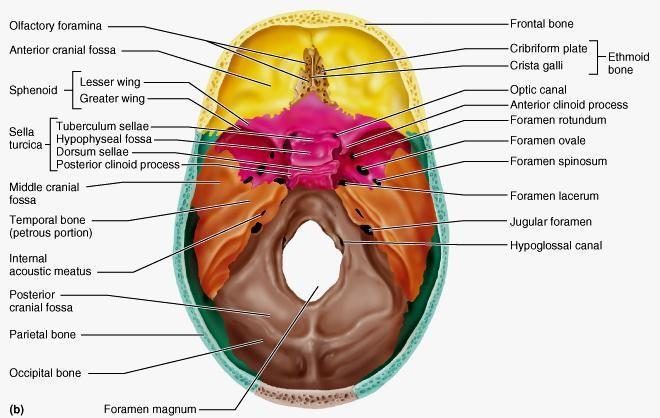
 Skull-2 Norma Basalis Interna Norma Basalis Externa Dr. Heba Kalbouneh Associate Professor of Anatomy and Histology Norma basalis interna Base of the skull- superior view The interior of the base of the
THE SKELETAL SYSTEM. Focus on the Skull
 THE SKELETAL SYSTEM Focus on the Skull Review Anatomical Terms Anterior/Posterior Dorsal/Ventral Medial/Lateral Superior/Inferior Bone Markings - Review Projections for attachment of muscles, ligaments
Dr. Sami Zaqout, IUG Medical School
 The skull The skull is composed of several separate bones united at immobile joints called sutures. Exceptions? Frontal bone Occipital bone Vault Cranium Sphenoid bone Zygomatic bones Base Ethmoid bone
ANATOMY & PHYSIOLOGY I Laboratory Version B Name Section. REVIEW SHEET Exercise 10 Axial Skeleton
 ANATOMY & PHYSIOLOGY I Laboratory Version B Name Section REVIEW SHEET Exercise 10 Axial Skeleton 1 POINT EACH. THE SKULL MULTIPLE CHOICE 1. The major components of the axial skeleton include the 7. The
ACTIVITY 3: AXIAL SKELETON AND LONG BONE DISSECTION COW BONE DISSECTION
 ACTIVITY 3: AXIAL SKELETON AND LONG BONE DISSECTION Objectives: 1) How to get ready: Read Chapter 7, McKinley et al., Human Anatomy, 4e. All text references are for this textbook. Learning the meanings
The Axial Skeleton. C h a p t e r. PowerPoint Lecture Slides prepared by Jason LaPres Lone Star College - North Harris
 C h a p t e r 7 The Axial Skeleton PowerPoint Lecture Slides prepared by Jason LaPres Lone Star College - North Harris Copyright 2009 Pearson Education, Inc., publishing as Pearson Benjamin Cummings An
YOU MUST BRING YOUR OWN GLOVES FOR THIS ACTIVITY.
 ACTIVITY 3: AXIAL SKELETON AND LONG BONE DISSECTION Objectives: 1) How to get ready: Read Chapter 7, McKinley et al., Human Anatomy, 5e. All text references are for this textbook. Learning the meanings
Skeletal system. Prof. Abdulameer Al-Nuaimi. E. mail:
 Skeletal system Prof. Abdulameer Al-Nuaimi E-mail: a.al-nuaimi@sheffield.ac.uk E. mail: abdulameerh@yahoo.com Functions of Bone and The Skeletal System Support: The skeleton serves as the structural framework
View of a Skull, 1489 by Leonardo Da Vinci. Kaan Yücel M.D., Ph.D Tuesday
 View of a Skull, 1489 by Leonardo Da Vinci Kaan Yücel M.D., Ph.D. 26.11.2013 Tuesday 1.SKULL skeleton of the head cranium 22 bones excluding ossicles of the ear 1.SKULL Mandible Lower jaw bone Neurocranium
Exercise 10. The Axial Skeleton
 Exercise 10 The Axial Skeleton The Axial Skeleton Consists of the skeletal structures found along the midline of the body. Includes the skull, hyoid, vertebrae, ribs, sternum, and sacrum. The cartilages
Human Anatomy and Physiology - Problem Drill 07: The Skeletal System Axial Skeleton
 Human Anatomy and Physiology - Problem Drill 07: The Skeletal System Axial Skeleton Question No. 1 of 10 Which of the following statements about the axial skeleton is correct? Question #01 A. The axial
Anatomy images for MSS practical exam- 2019
 Anatomy images for MSS practical exam- 2019 Ilium Ischium Pubis Acetabulaum Iliac crest Iliac tubercle ASIS (muscle and ligament attached) AIIS (muscle attached) PSIS PIIS Ischial spine Ischial tuberosity
Anatomy and Physiology 1 Chapter 7 self quiz Pro, Dima Darwish,MD.
 Anatomy and Physiology 1 Chapter 7 self quiz Pro, Dima Darwish,MD. 1) How many bones make up the axial skeleton? A) 50 B) 60 C) 70 D) 80 E) 90 2) Which of the following is a function of the axial skeleton?
Introduction to Local Anesthesia and Review of Anatomy
 5-Sep Introduction and Anatomy Review 12-Sep Neurophysiology and Pain 19-Sep Physiology and Pharmacology part 1 26-Sep Physiology and Pharmacology part 2 Introduction to Local Anesthesia and Review of
o Diaphysis o Area where red marrow is found o Area where yellow marrow is found o Epiphyseal plate AXIAL SKELETON Skull
 64 Anatomy & Physiology Coloring Workbook 7. Figure 5-2A is a midlevel, cross-sectional view of the diaphysis of the femur. Label the membrane that lines the cavity and the membrane that covers the outside
Skull-2. Norma Basalis Interna. Dr. Heba Kalbouneh Assistant Professor of Anatomy and Histology
 Skull-2 Norma Basalis Interna Dr. Heba Kalbouneh Assistant Professor of Anatomy and Histology Norma basalis interna Base of the skull- superior view The interior of the base of the skull is divided into
Chapter 7: Skeletal System: Gross Anatomy
 Chapter 7: Skeletal System: Gross Anatomy I. General Considerations A. How many bones in an average adult skeleton? B. Anatomic features of bones are based on II. Axial Skeleton A. Skull 1. Functionally
Skull basic structures. Neurocranium
 Assoc. Prof. Květuše Lovásová, M.V.D., PhD. Skull basic structures Skull consists of two groups of bones: neurocranium (bones forming the brain box) splanchnocranium (bones forming the facial skeleton)
BONE CHALLENGE DANIL HAMMOUDI.MD
 BONE CHALLENGE DANIL HAMMOUDI.MD Bone Basic functions? A. support B. protection C. movement assistance in D. RBC formation-hemopoiesis E. mineral homeostasis +importance of calcium F. energy supply -yellow
University of Palestine. Midterm Exam 2013/2014 Total Grade:
 Course No: DNTS2208 Course Title: Head and Neck Anatomy Date: 09/11/2013 No. of Questions: (50) Time: 1hour Using Calculator (No) University of Palestine Midterm Exam 2013/2014 Total Grade: Instructor
Skeletal System: Skull.
 Skeletal System: Skull www.fisiokinesiterapia.biz Bones of the Skull SPLANCHNOCRANIUM Nasal (2) Maxilla (2) Lacrimal (2) Zygomatic (2) Palatine (2) Inferior concha (2) Vomer Mandible NEUROCRANIUM Frontal
SKULL / CRANIUM BONES OF THE NEUROCRANIUM (7) Occipital bone (1) Sphenoid bone (1) Temporal bone (2) Frontal bone (1) Parietal bone (2)
 Occipital bone (1) Sphenoid bone (1) Temporal bone (2) Frontal bone (1) Parietal bone (2)") Important! 1. Memorizing these pages only does not guarantee the succesfull passing of the midterm test or the semifinal exam. 2. The handout has not been supervised, and I can not guarantee, that these
Important! 1. Memorizing these pages only does not guarantee the succesfull passing of the midterm test or the semifinal exam. 2. The handout has not been supervised, and I can not guarantee, that these
Dr.Noor Hashem Mohammad Lecture (5)
") Dr.Noor Hashem Mohammad Lecture (5) 2016-2017 If the mandible is discarded, the anterior part of this aspect of the skull is seen to be formed by the hard palate. The palatal processes of the maxillae
Dr.Noor Hashem Mohammad Lecture (5) 2016-2017 If the mandible is discarded, the anterior part of this aspect of the skull is seen to be formed by the hard palate. The palatal processes of the maxillae
The cribriform plate. ethmoid bone. Ethmoid bone consists from: 1) A horizontal cribriform plate. 2) A perpendicular plate. 3) Two lateral labyrinths.
 A horizontal cribriform plate. 2) A perpendicular plate. 3) Two lateral labyrinths.") ethmoid bone Ethmoid bone consists from: 1) A horizontal cribriform plate. 2) A perpendicular plate. 3) Two lateral labyrinths. The cribriform plate 1) Connect the two labyrinths to the perpendicular plate.
ethmoid bone Ethmoid bone consists from: 1) A horizontal cribriform plate. 2) A perpendicular plate. 3) Two lateral labyrinths. The cribriform plate 1) Connect the two labyrinths to the perpendicular plate.
Biology 2401 The Skeletal System
 Biology 2401 The Skeletal System Purpose: The lab will describe the microscopic and gross anatomy of bone, identify bones of the body, and identify important bone markings. I. Overview of the Skeleton
TEST YOURSELF- Chapter 7
 TEST YOURSELF- Chapter 7 Cranial Bones 1. Give the name of the bone for each of the following markings. Some of the markings are found on more than one bone. List all that apply. Cranium a. Frontal squama:
Human Anatomy & Physiology I Dr. Sullivan Unit VIIIa The Axial Skeleton Chapter 8 (Sections )
") Human Anatomy & Physiology I Dr. Sullivan Unit VIIIa The Axial Skeleton Chapter 8 (Sections 8.1-8.3) I. Divisions of the skeletal system a) An adult human skeleton has 206 named bones b) Most are paired
Human Anatomy & Physiology I Dr. Sullivan Unit VIIIa The Axial Skeleton Chapter 8 (Sections 8.1-8.3) I. Divisions of the skeletal system a) An adult human skeleton has 206 named bones b) Most are paired
An Introduction to the Axial Skeleton. Copyright 2009 Pearson Education, Inc., publishing as Pearson Benjamin Cummings
 An Introduction to the Axial Skeleton Copyright 2009 Pearson Education, Inc., publishing as Pearson Benjamin Cummings Terms: Structures of Bones Articulations: Contacts with other bones Landmarks (Bone
Crafton Hills College Human Anatomy & Physiology Axial Skeleton
 A. Major Divisions Crafton Hills College Human Anatomy & Physiology Axial keleton 1. Axial: Part of skeleton lies along long axis of body 2. Appendicular: Bones & features of the appendages B. AXIAL KELETON
External Acoustic Meatus. Mastoid Process. Zygomatic Process. Temporal Bone
 Bone lab review 1. Frontal Bone 2. Supra-Orbital Foramen 3. Orbit (Orbital Cavity) 4. Superior Orbital Fissure 5. Inferior Orbital Fissure 6. Zygomatic Bone 7. Infra-Orbital Foramen 8. Maxilla 9. Mandible
Anatomy and Physiology II. Review Spine and Neck
 Anatomy and Physiology II Review Spine and Neck Spine regions How many cervical vertibrae are there? 7 The curvature is the cervical region posterior? Concave posterior How many thoracic? And curvature?
Bones Ethmoid bone Inferior nasal concha Lacrimal bone Maxilla Nasal bone Palatine bone Vomer Zygomatic bone Mandible
 splanchnocranium - Consists of part of skull that is derived from branchial arches - The facial bones are the bones of the anterior and lower human skull Bones Ethmoid bone Inferior nasal concha Lacrimal
Bones of the Skull Lateral View
 Bones of the Skull Lateral View Frontal Bone Parietal Bone Occipital Bone Temporal Bone Sphenoid Bone Pterion Sutures of the Skull Lateral View Coronal Suture Lambdoid Suture Squamous Suture Sutures of
The Skeletal System: Axial Skeleton
 The Skeletal System: Axial Skeleton The Big Idea The Axial Skeleton & Homeostasis The bones of the axial skeleton contribute to homeostasis by protecting many of the body s organs such as the brain, spinal
Tikrit University collage of dentistry Dr.Ban I.S. head & neck anatomy 2 nd y. Lec [5] / Temporal fossa :
![Tikrit University collage of dentistry Dr.Ban I.S. head & neck anatomy 2 nd y. Lec [5] / Temporal fossa :](/thumbs/88/115294566.jpg "Tikrit University collage of dentistry Dr.Ban I.S. head & neck anatomy 2 nd y. Lec [5] / Temporal fossa :") Lec [5] / Temporal fossa : Borders of the Temporal Fossa: Superior: Superior temporal line. Inferior: gap between zygomatic arch and infratemporal crest of sphenoid bone. Anterior: Frontal process of the
Lec [5] / Temporal fossa : Borders of the Temporal Fossa: Superior: Superior temporal line. Inferior: gap between zygomatic arch and infratemporal crest of sphenoid bone. Anterior: Frontal process of the
University of Palestine. Midterm Exam 2013/2014 Total Grade:
 [ Course No: DNTS2208 Course Title: Head and Neck Anatomy Date: 17/11/1024 No. of Questions: (52) Time: 2hours Using Calculator (No) University of Palestine Midterm Exam 2013/2014 Total Grade: Instructor
The Skull DANIL HAMMOUDI.MD
 The Skull DANIL HAMMOUDI.MD summary of bones/structures in Chapter 15 of the manual need tp be print as soon as possible http://www.mnsu.edu/emuseum/biology/humananatomy/skeletal/skul l/frontal/frontal.html
Nasal region. cartilages: septal cartilage (l); lateral nasal cartilage (2); greater alar cartilages (2); lesser alar cartilages (?
; lateral nasal cartilage (2); greater alar cartilages (2); lesser alar cartilages (?") Nasal region skull bones: nasal and frontal processes of maxilla cartilages: septal cartilage (l); lateral nasal cartilage (2); greater alar cartilages (2); lesser alar cartilages (?) 1 Nasal cavity Roof
Nasal region skull bones: nasal and frontal processes of maxilla cartilages: septal cartilage (l); lateral nasal cartilage (2); greater alar cartilages (2); lesser alar cartilages (?) 1 Nasal cavity Roof
Chapter 8A. The Skeletal System: The Axial Skeleton. The Skeletal System: The Axial Skeleton. Types of Bones. Types of Bones
 Chapter 8A The Skeletal System: The Axial Skeleton The Skeletal System: The Axial Skeleton 206 named bones Axial Skeleton 80 bones lie along longitudinal axis skull, hyoid, vertebrae, ribs, sternum, ear
in compact bone, large vertical canals carrying blood vessels and nerves. in compact bone, large horizontal canals carrying blood vessels and nerves.
 Carl Christensen, PhD Skeletal System (Bones`) Bio. 2304 Human Anatomy 1. Identify a term for each of the following: shaft of a long bone ends of a long bone ossified remnant of the "growth plate" connective
Spring Written By: J. E. Sutton. Contents: I. Overview of the Skeleton: II. Appendicular Skeleton III. Axial Skeleton IV.
 Spring 2012 Written By: J. E. Sutton Contents: I. Overview of the Skeleton: II. Appendicular Skeleton III. Axial Skeleton IV. Articulations Overview of the Skeleton: I. Orientation to Human Skeleton: a.
Bone Flashcards for 10a
 Bone Flashcards for 0a CLAVICLE (collar bone). Sternal extremity (end) flat end. Acromial extremity (end) rounded end. SCAPULA (shoulder blade). Right or left scapula?. Superior border (superior margin).
CHAPTER 7, PART II (BONES)
") Anatomy Name: CHAPTER 7, PART II (BONES) Entry #: INSTRUCTIONS: 1) READ Chapter 7, pg. 140-161. 2) Using the outline, make a note card for each underlined bone name or phrase. 3) On each note card, put
Anatomy Name: CHAPTER 7, PART II (BONES) Entry #: INSTRUCTIONS: 1) READ Chapter 7, pg. 140-161. 2) Using the outline, make a note card for each underlined bone name or phrase. 3) On each note card, put
Infratemporal fossa: Tikrit University college of Dentistry Dr.Ban I.S. head & neck Anatomy 2 nd y.
 Infratemporal fossa: This is a space lying beneath the base of the skull between the lateral wall of the pharynx and the ramus of the mandible. It is also referred to as the parapharyngeal or lateral pharyngeal
THIEME. Scalp and Superficial Temporal Region
 CHAPTER 2 Scalp and Superficial Temporal Region Scalp Learning Objectives At the end of the dissection of the scalp, you should be able to identify, understand and correlate the clinical aspects: Layers
Musculoskeletal System (Part A-1) Module 7 -Chapter 10 Overview. Functions
 Module 7 -Chapter 10 Overview. Functions") Musculoskeletal System (Part A-1) Module 7 -Chapter 10 Overview Susie Turner, M.D. 1/8/13 Muscles Attachments Bones Bone types Surface features of bones Divisions of the skeletal system Joints or Articulations
Musculoskeletal System (Part A-1) Module 7 -Chapter 10 Overview Susie Turner, M.D. 1/8/13 Muscles Attachments Bones Bone types Surface features of bones Divisions of the skeletal system Joints or Articulations
LESSON ASSIGNMENT. Positioning for Exams of the Cranium, Sinuses, and Mandible. After completing this lesson, you should be able to:
 LESSON ASSIGNMENT LESSON 5 Positioning for Exams of the Cranium, Sinuses, and Mandible. LESSON ASSIGNMENT Paragraphs 5-1 through 5-9. LESSON OBJECTIVES After completing this lesson, you should be able
Major Anatomic Components of the Orbit
 Major Anatomic Components of the Orbit 1. Osseous Framework 2. Globe 3. Optic nerve and sheath 4. Extraocular muscles Bony Orbit Seven Bones Frontal bone Zygomatic bone Maxillary bone Ethmoid bone Sphenoid
BIO 137 AXIAL SKELETON BONE STUDY THE HUMAN SKELETON
 BIO 137 THE AXIAL SKELETON MARY CATHERINE FLATH, Ph.D. THE HUMAN SKELETON AXIAL SKULL HYOID THORACIC CAGE VERTEBRAL COLUMN APPENDICULAR PECTORAL GIRDLE UPPER LIMBS PELVIC GIRDLE LOWER LIMBS AXIAL SKELETON
Superior View of the Skull (Norma Verticalis) Anteriorly the frontal bone articulates with the two parietal bones AT THE CORONAL SUTURE
 Anteriorly the frontal bone articulates with the two parietal bones AT THE CORONAL SUTURE") Superior View of the Skull (Norma Verticalis) Anteriorly the frontal bone articulates with the two parietal bones AT THE CORONAL SUTURE 1 The two parietal bones articulate in the midline AT THE SAGITTAL
Superior View of the Skull (Norma Verticalis) Anteriorly the frontal bone articulates with the two parietal bones AT THE CORONAL SUTURE 1 The two parietal bones articulate in the midline AT THE SAGITTAL
Skull and Axial Skeleton
 Published on Second Faculty of Medicine, Charles University (http://www.lf2.cuni.cz ) Skull and Axial Skeleton Description of the test The examination of the skull skeleton is in oral format. It consists
SKELETON FUNCTIONS OF BONE:
 SKELETON FUNCTIONS OF BONE: SKELETON: 1. Performs a mechanical function in forming the skeletal support of the body and in forming a leverage system whereby work and movement are possible. 2. Serves as
Dr. Sami Zaqout Faculty of Medicine IUG
 The Nose External Nose Nasal Cavity External Nose Blood and Nerve Supplies of the External Nose Blood Supply of the External Nose The skin of the external nose Branches of the ophthalmic and the maxillary
Perpendicular Plate Zygomatic Bone. Mental Foramen Mandible
 Glabella Frontal Middle Nasal Concha Nasal Lacrimal Perpendicular Plate Zygomatic Inferior Nasal Concha Maxilla Mental Mandible Skull (anterior view) Squamosal Suture Coronal Suture Frontal Parietal Nasal
Axial skeleton bones and markings
 Axial skeleton bones and markings Skull Cranial bones Frontal x 1 Supraorbital foramen Occipital x 1 Foramen magnum Occipital condyles Superior nuchal line Inferior nuchal line Anterior cranial fossa External
Unit 18: Cranial Cavity and Contents
 Unit 18: Cranial Cavity and Contents Dissection Instructions: The calvaria is to be removed without damage to the dura mater which is attached to the inner surface of the calvaria. Cut through the outer
Dr.Ban I.S. head & neck anatomy 2 nd y. جامعة تكريت كلية طب االسنان مادة التشريح املرحلة الثانية أ.م.د. بان امساعيل صديق 6102/6102
 جامعة تكريت كلية طب االسنان مادة التشريح املرحلة الثانية أ.م.د. بان امساعيل صديق 6102/6102 The scalp The scalp extends from the supraorbital margins anteriorly to the nuchal lines at the back of the skull
The Skull and Temporomandibular joint II Prof. Abdulameer Al-Nuaimi. E. mail:
 The Skull and Temporomandibular joint II Prof. Abdulameer Al-Nuaimi E-mail: a.al-nuaimi@sheffield.ac.uk E. mail: abdulameerh@yahoo.com Temporal fossa The temporal fossa is a depression on the temporal
11/25/2012. Chapter 7 Part 2: Bones! Skeletal Organization. The Skull. Skull Bones to Know Cranium
 Chapter 7 Part 2: Bones! 5) Distinguish between the axial and appendicular skeletons and name the major parts of each 6) Locate and identify the bones and the major features of the bones that compose the
Omran Saeed. Luma Taweel. Mohammad Almohtaseb. 1 P a g e
 2 Omran Saeed Luma Taweel Mohammad Almohtaseb 1 P a g e I didn t include all the photos in this sheet in order to keep it as small as possible so if you need more clarification please refer to slides In
locomotice system Plastinated specimensⅠ: Silicone specimens Regional specimens and organs
 locomotice system Plastinated specimensⅠ: Silicone specimens Regional specimens and organs Art-No. Name Description The locomotor system SL001 Two hundred pieces of plastinated bones (without six The bones
Important Parts of Bones
 Important Parts of Bones For 2015 Know: Humerus (posterior) Clavical Femur (Anterior) Foot Hand Mandible Os Coxa Scapula Skull (Anterior, Inferior, Lateral) Sternum Humerus (posterior) A. olecranon fossa
Tikrit University College of Dentistry Dr.Ban I.S. head & neck anatomy 2 nd y.
 Lec [3]/The scalp The scalp extends from the supraorbital margins anteriorly to the nuchal lines at the back of the skull and down to the temporal lines at the sides. The forehead, from eyebrows to hairline,
Biology 210 Chapter 8: Skeletal Tissues Supplement 1
 Biology 210 Chapter 8: Skeletal Tissues Supplement 1 By John McGill Material contributed by Beth Wyatt & Jack Bagwell DIVISIONS OF THE SKELETAL SYSTEM AXIAL SKELETON (80 BONES) Bones of the Head, Neck,
Skeletal system overview. Classification of Bones
 Skeletal system overview BIOL241 Lab #9 Classification of Bones Bone are identified by: shape internal tissues bone markings 1 1. Flat bones 2. Long bones 3. Short bones 4. Irregular bones 5. Sutural bones
Parotid Gland. Parotid Gland. Largest of 3 paired salivary glands (submandibular; sublingual) Ramus of Mandible. Medial pterygoid.
 Ramus of Mandible. Medial pterygoid.") Parotid region Parotid Gland Largest of 3 paired salivary glands (submandibular; sublingual) Ramus of Mandible Medial pterygoid Cross section of mandible Masseter D S SCM Parotid Gland Mastoid Process
Parotid region Parotid Gland Largest of 3 paired salivary glands (submandibular; sublingual) Ramus of Mandible Medial pterygoid Cross section of mandible Masseter D S SCM Parotid Gland Mastoid Process
Labs 9 and 10. Classification of Bones. Bone Shapes 1/05/13. Skeletal system overview. Bone are identified by:
 Labs 9 and 10 Skeletal system overview Classification of Bones Bone are identified by: shape internal tissues bone markings 1. Flat bones 2. Long bones 3. Short bones 4. Irregular bones 5. Sutural bones
The orbit-1. Dr. Heba Kalbouneh Assistant Professor of Anatomy and Histology
 The orbit-1 Dr. Heba Kalbouneh Assistant Professor of Anatomy and Histology Orbital plate of frontal bone Orbital plate of ethmoid bone Lesser wing of sphenoid Greater wing of sphenoid Lacrimal bone Orbital
Skull. Sphenoid and Ethmoid bones
 Skull. Sphenoid and Ethmoid bones PhD., Dr. David Lendvai Department of Anatomy, Histology and Embriology Semmelweis University, Faculty of Medicine 2018. Skeletal system Structure of the skull Border
Lab 6, 7, 8: Skeletal System
 107 Lab 6, 7, 8: Skeletal System Adult Skull Bony orbit (FLEZMS) Frontal bone supraorbital foramen frontal sinus Lacrimal bone Ethmoid bone perpendicular plate of ethmoid middle nasal conchae cribriform
Lab Exercise #04 The Skeletal System Student Performance Objectives
 Lab Exercise #04 The Skeletal System Student Performance Objectives The material that you are required to learn in this exercise can be found in either the lecture text or the supplemental materials provided
PTERYGOPALATINE FOSSA
 PTERYGOPALATINE FOSSA Outline Anatomical Structure and Boundaries Foramina and Communications with other spaces and cavities Contents Pterygopalatine Ganglion Especial emphasis on certain arteries and
Parotid Gland, Temporomandibular Joint and Infratemporal Fossa
 M1 - Anatomy Parotid Gland, Temporomandibular Joint and Infratemporal Fossa Jeff Dupree Sanger 9-057 jldupree@vcu.edu Parotid gland: wraps around the mandible positioned between the mandible and the sphenoid
UNIT 4 - SKELETAL SYSTEM LECTURE NOTES
 UNIT 4 - SKELETAL SYSTEM LECTURE NOTES 4.01 FUNCTIONS OF THE SKELETAL SYSTEM A. Support 1. Provides a framework for the body 2. Supports soft tissue 3. Serves as a point of attachment for ligaments, tendons,
the Skeletal System provided by Academic Web Services Grand Canyon University
 Anatomy Resource Center Study Guides the Skeletal System HEAD & NECK REGIONAL VIEW SKULL BONES CRANIUM FACE SKULL LANDMARKS ANTERIOR SIDE SUPERIOR/INFERIOR VERTEBRAL COLUMN VERTEBRAL REGIONS CERVICAL C1
Thickened and thinner parts of the skull = important base for understanding of the functional structure of the skull - the transmission of masticatory
 Functional structure of the skull and Fractures of the skull Thickened and thinner parts of the skull = important base for understanding of the functional structure of the skull - the transmission of masticatory
3. The Jaw and Related Structures
 Overview and objectives of this dissection 3. The Jaw and Related Structures The goal of this dissection is to observe the muscles of jaw raising. You will also have the opportunity to observe several
Bisection of Head & Nasal Cavity 頭部對切以及鼻腔. 解剖學科馮琮涵副教授 分機
 Bisection of Head & Nasal Cavity 頭部對切以及鼻腔 解剖學科馮琮涵副教授 分機 3250 E-mail: thfong@tmu.edu.tw Outline: The structure of nose The concha and meatus in nasal cavity The openings of paranasal sinuses Canals, foramens
*in general the blood supply of the nose comes from branches of the internal and external carotid arteries.
 In the previous lecture we talked about the anatomy of the nasal cavity, today we will talk about its blood supply, venous drainage, innervations, and finally about the paranasal sinuses. When we describe
Bio 5/6 5 The Skeletal System Study Guide
 Name: THE SKELETAL SYSTEM: 5 The Skeletal System Study Guide Period: The skeleton is constructed of two of the most supportive tissues found in the human body - cartilage and bone. Besides supporting and
Anatomy Skull and Spinal Cord
 1 Anatomy Skull and Spinal Cord 1. Skull The skull is anterior to the spinal column and is the bony structure that encases the brain. Its purpose is to protect the brain and allow attachments for the facial
Mohammad Hisham Al-Mohtaseb. Lina Mansour. Reyad Jabiri. 0 P a g e
 2 Mohammad Hisham Al-Mohtaseb Lina Mansour Reyad Jabiri 0 P a g e This is only correction for the last year sheet according to our record. If you already studied this sheet just read the yellow notes which
Temporal fossa Infratemporal fossa Pterygopalatine fossa Terminal branches of external carotid artery Pterygoid venous plexus
 Outline of content Temporal fossa Infratemporal fossa Pterygopalatine fossa Terminal branches of external carotid artery Pterygoid venous plexus Boundary Content Communication Mandibular division of trigeminal
Anatomic Relations Summary. Done by: Sohayyla Yasin Dababseh
 Anatomic Relations Summary Done by: Sohayyla Yasin Dababseh Anatomic Relations Lecture 1 Part-1 - The medial wall of the nose is the septum. - The vestibule lies directly inside the nostrils (Nares). -
NEUROCRANIUM VISCEROCRANIUM VISCEROCRANIUM VISCEROCRANIUM
 LECTURE 4 SKULL NEUROCRANIUM VISCEROCRANIUM VISCEROCRANIUM VISCEROCRANIUM CRANIUM NEUROCRANIUM (protective case around brain) VISCEROCRANIUM (skeleton of face) NASOMAXILLARY COMPLEX MANDIBLE (DESMOCRANIUM)
Labs 6, 7, 8: Skeletal System
 153 Labs 6, 7, 8: Skeletal System Unit 6: Skeletal System: Bone tissue, Bones and Joints (p. 105-152) Ex. 6-1: Histology of Osseous Tissue, p. 113 Model: Osteon Tiss Lamella Osteocyte Lacunae Canaliculi