Head and Neck Image 頭頸部放射影像學
|
|
|
- Annabella Cobb
- 5 years ago
- Views:
Transcription
1 Head and Neck Image 頭頸部放射影像學 陳家媛 台北醫學大學 - 市立萬芳醫院 cychen@wanfang.gov.tw

2

3
4 Normal Suprahyoid neck: the old way Nasopharynx Oropharynx Oral cavity Staging of SCC

5

6
7 Spaces of Suprahyoid Neck: a New Way Deep cervical fascia Superficial layer (investing fascia) Middle layer (buccopharyngeal fascia) Deep layer (prevertebral fascia)
8 Approach to Suprahyoid Mass What space is mass in? What important neurovascular structures are in the vicinity of mass? What is the likely pathological diagnosis?
9 Spaces of Suprahyoid Neck Parapharyngeal space not fascia-enclosed Pharyngeal mucosal space not fascia-enclosed Masticator space Parotid space Carotid space Retropharyngeal space Perivertebral space
10 Parapharyngeal Space Central space Fatty tube from skull base to hyoid bone level as elevator shaft Few lesions primarily arise in this space

11
12 Contents of Parapharyngeal Space Fat Branch of CNV3 Internal maxillary artery Ascending pharyngeal artery Pharyngeal venous plexus
13 Primary Mass of PPS Fat surrounding the entire circumference of lesion No point of attachment to adjacent space, especially the deep lobe of parotid

14
15 DDx of Parapharyngeal Space Lesion Abscess from adjacent space Direct spread of malignant tumor Salivary gland rest lesion Lipoma

16
17 Pharyngeal Mucosal Space Area of nasopharynx and oropharynx Airway side of middle layer of deep cervical fascia SCC is the most common and important lesions


18
19 Contents of Pharyngeal Mucosal Space Lymphoid tissue Superior and middle constrictor muscle Salpingopharyngeal muscle Pharyngobasilar fascia Levator palatini muscle Torus tubarius aspect of E tube
20 Primary Mass of PMS Medial to parapharyngeal fat Invading PPS medially to laterally, displacing PPS fat laterally Disrupting normal mucosal and submucosal architecture of PMS

21
22 DDx of Pharyngeal Mucosal Space Lesion Inflammation Tonsillitis, pharyngitis Benign tumor Mixed tumor Malignant tumor SCC, lymphoma, minor salivary gland malignacy Tornwaldt s cyst


23

24
25 Masticator Space Enclosed by superficial layer of deep cervical fascia Mandible to skull base and temporal fossa Foramen ovale and CN 5th


26
27 Contents of Mastictor Space Lateral pterygoid muscle Medial pterygoid muscle Masseter muscle Temporalis muscle Inferior alveolar nerve, artery and vein Ramus and body of mandible
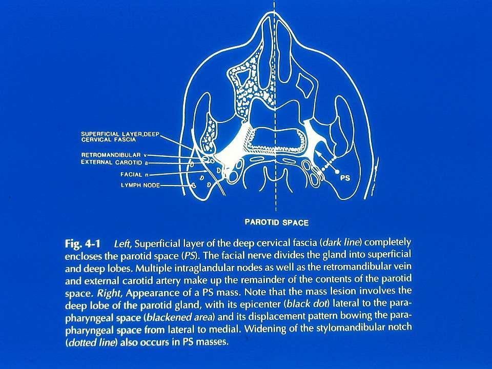
28 Primary Mass of MS Center of mass anterior to PPS fat within the muscle of mastication or the mandible Mass invading PPS fat from anterior to posterior, displacing PPS fat posteriorly

29
30 DDx of Masticator Space Lesion Odontogenic abscess Most common Sarcoma Soft tissue sa, chondrosa, osteosa Lymphoma SCC from oropharynx Rhabdomyosarcoma Pediatric population

31
32 Perineural Spreading of Tumor along Cranial Nerve V3 Along masticator or inferior alveolar nerves to CN V3 Mental foramen- mandibular foramen- foramen ovale- Meckel s cave MRI is more sensitive to perineural spreading

33

34
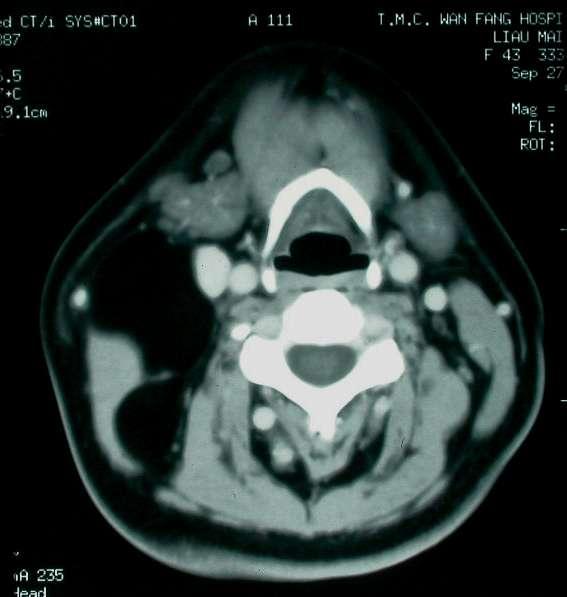
35 Parotid Space Enclosed by superficial layer of deep cervical fascia Facial nerve A plane between stylomastoid foramen and lateral wall of retromandibular vein Late encapsulation with LNs within space

36
37 Contents of Parotid Space Parotid gland Facial nerve Retromandibular vein External carotid artery Internal maxillary arteries Intraparotid LNs
38 Primary Mass of PS Center of mass lateral to PPS fat Mass invading PPS fat laterally to medially, displacing PPS fat medially Widening the distance between mandibular angle and styloid process
39
40 DDx of Parotid Space Lesion Benign mixed tumor (pleomorphic adenoma) 80% of benign tumor Warthin s tumor Mucoepidermoid ca Adenoid cystic ca Skin SCC with parotid LAP Melanoma with parotid LAP
41


42 62 y/o Female, mixed tumor


43 39y/o male, abscess 76y/o female, Undifferentiated ca
44 Carotid Space Carotid sheath All three layers of deep cervical fascia Nasopharyngeal, oropharyngeal, cervical and mediastinal segments Highest deep cervical chain as jugulodigastric nodes Jugular foramen and basal cistern

45

46
47 Contents of Carotid Space Common and internal carotid arteries Internal jugular vein Cranial nerves 9th, 10th, 11th, 12th Sympathetic plexus Deep cervical LNs
48 Primary Mass of CS Center of mass within the area of ICA or internal jugular vein Mass invading PPS fat from posterior to anterior, displacing PPS fat anteriorly Styloid process anteriolateral displacement ICA draped over anterior margin of mass

49
50 DDx of Carotid Space Lesion Vein thrombosis, thrombophlebitis ICA thrombosis, mural thrombus, aneurysm, pseudoaneurysm Paraganglioma Neurogenic tumor Neurilemmoma, neurofibroma SCC with LN metastasis

51 26y/0 male, paraganglioma
52 Retropharyngeal Space Between deep and middle layers of deep cervical fascias and alar fascia No LN below hyoid level Skull base to T4 vertebral body level
53

54
55 Contents of Suprahyoid Retropharyngeal Space Fat Lateral retropharyngeal LNs Medial retropharyngeal LNs
56 Primary Mass of Suprahyoid RPS Center of mass posteromedial to PPS fat and directly medial to carotid space Mass invading PPS fat posteromedially to anterolaterally Mass anterior to prevertebral muscle Styloid process is usually not displaced

57
58 Primary Mass to Infrahyoid RPS Bow-tie or oval shape in the posterior midline Anterior to prevertebral muscle

59
60 DDx of Retropharyngeal Space Lesion Inflammation of retropharyngeal nodes and absces Child: Waldeyer s ring infection Adult: Vertebral osteomyelitis, spine Sx Malignancy SCC of head and neck (NPC) Lymphoma Direct invasion of SCC Edema fluid or lymph

61

62
63 Perivertebral Space Enclosed by deep layer of deep cervical fascia Separate into prevertebral and paraspinal portions Skull base to T4 level Tenacious fascia resisting violation by infection or tumor
64
65 Contents of Perivertebral Space Prevertebral component: Prevertebral muscle, vertebral a. and V. Scalene muscle, branchial plexus Phrenic nerve, vertebral body Paraspinal component: Paraspinal muscle Posterior elements of vertebral body
66 Primary Mass in Prevertebral PVS Center of mass within prevertebral muscle or vertebral body Mass lifting prevertebral muscle anteriorly

67
68 Primary Mass of Paraspinal PVS Center of mass within paraspinal muscles Mass bowing posterior cervical space fat away from posterior elements of spine

69
70 DDx of Perivertebral Space Lesion Vertebral body osteophyte Vertebral body osteomyelitis Chordoma Vertebral body or epidural metastasis from lung, breast, prostate or lymphoma

71

72

73
74 Infrahyoid Neck Hyoid bone to clavicle Triangle: surgical and gross anatomy Space: imaging approach to anatomy
75 Infrahyoid Neck: Five Spaces Visceral space: unique to infrahyoid space Posterior cervical space Carotid space Retropharyngeal space Perivertebral space

76 Visceral Space
77 Contents of Visceral Space Thyroid gland/ parathyroid gland Larynx Trachea Hypopharynx Esophagus Recurrent laryngeal nerve Paratracheal LNs
78 DDx of Visceral Space Lesion Congenital: thyroglossal duct cyst Larynx: SCC, laryngocele, chondroma Thyroid: colloid cyst, goiter, adenoma, ca, lymphoma, meta, thyroiditis Parathyroid: adenoma, cyst Esophagus: Zenker diverticulum, ca LAP Inflammation/abscess


79

80
81 Posterior Cervical Space Between superficial and deep layers of deep cervical fascia Posterior cervical triangle

82
83 Centents of Posterior Cervical Space Fat Spinal accessory nerve Spinal accessory chains Pre-axillary brachial plexus

84
85 DDx of Posterior Cervical Space Lesion Congenital: cystic hygroma/lymphangioma, hemangioma Inflammatory: abscess, lymphadenitis Benign tumor: lipoma, neurogenic tumor Malignant tumor: metastatic LAP, lymphoma, liposa
86 lymphangioma
87

88
89 Summary Normal Suprahyoid neck Spaces of Suprahyoid Neck Approach to Suprahyoid Mass Parapharyngeal Space Pharyngeal Mucosal Space Masticator Space Parotid Space Carotid Space Retropharyngeal Space Perivertebral Space Infrahyoid Neck Posterior Cervical Space
"The Space Between Us:" A Radiographic Review of Common and Uncommon Pathologic Findings within the Deep Spaces of the Neck
 "The Space Between Us:" A Radiographic Review of Common and Uncommon Pathologic Findings within the Deep Spaces of the Neck Poster No.: C-2457 Congress: ECR 2015 Type: Educational Exhibit Authors: A. K.
Suprahyoid and Infrahyoid Neck Overview
 10 Imaging Approaches & Indications Neither CT nor MR is a perfect modality for imaging the extracranial H&N. MR is most useful in the suprahyoid neck (SHN) because it is less affected by oral cavity dental
The Neck the lower margin of the mandible above the suprasternal notch and the upper border of the clavicle
 The Neck is the region of the body that lies between the lower margin of the mandible above and the suprasternal notch and the upper border of the clavicle below Nerves of the neck Cervical Plexus Is formed
Neck-2. Dr. Heba Kalbouneh Associate Professor of Anatomy and Histology
 Neck-2 ` Dr. Heba Kalbouneh Associate Professor of Anatomy and Histology Triangles of the neck Side of the neck Midline Lower border of mandible Line between angle of mandible and mastoid Superior nuchal
Veins of the Face and the Neck
 Veins of the Face and the Neck Facial Vein The facial vein is formed at the medial angle of the eye by the union of the supraorbital and supratrochlear veins. connected through the ophthalmic veins with
Prevertebral Region, Pharynx and Soft Palate
 Unit 20: Prevertebral Region, Pharynx and Soft Palate Dissection Instructions: Step1 Step 2 Step 1: Insert your fingers posterior to the sternocleidomastoid muscle, vagus nerve, internal jugular vein,
The Neck. BY: Lina Abdullah & Rahaf Jreisat
 The Neck BY: Lina Abdullah & Rahaf Jreisat Boundaries of the Neck: generally from base of the skull to root of the neck Superior margin :From superior nuchal line of occipital bone up to mastoid process
Head&Neck Imaging. ssregypt.com. Parapharyngeal Spaces. Mamdouh mahfouz MD
 Head&Neck Imaging Parapharyngeal Spaces ssregypt.com Mamdouh mahfouz MD mamdouh.m5@gmail.com Definitio n Fat filled triangular space lateral the pharynx Extends from the skull base to the oropharynx Parapharyngeal
Anatomy: head and Neck (6 questions) 1. Prevertebral Flexor Musculature (lying in front of the vertebrae) include all, EXCEPT: Longus Colli.
 1. Prevertebral Flexor Musculature (lying in front of the vertebrae) include all, EXCEPT: Longus Colli.") Anatomy: head and Neck (6 questions) 1. Prevertebral Flexor Musculature (lying in front of the vertebrae) include all, EXCEPT: Longus Colli. Rectus Capitis Anterior. Rectus Capitis Lateralis. Rectus Capitis
Anatomy: head and Neck (6 questions) 1. Prevertebral Flexor Musculature (lying in front of the vertebrae) include all, EXCEPT: Longus Colli. Rectus Capitis Anterior. Rectus Capitis Lateralis. Rectus Capitis
C. Douglas Phillips, MD FACR Director of Head and Neck Imaging Weill Cornell Medical Center NewYork Presbyterian Hospital
 C. Douglas Phillips, MD FACR Director of Head and Neck Imaging Weill Cornell Medical Center NewYork Presbyterian Hospital Objectives Review basics of head and neck imaging Discuss our spatial approach
The Pharynx. Dr. Nabil Khouri MD. MSc, Ph.D
 The Pharynx Dr. Nabil Khouri MD. MSc, Ph.D Introduction The pharynx is the Musculo-fascial halfcylinder that links the oral and nasal cavities in the head to the larynx and esophagus in the neck Common
Downloaded from by on 02/06/18 from IP address Copyright ARRS. For personal use only; all rights reserved
 Downloaded from www.ajronline.org by 46.3.205.55 on 02/06/18 from IP address 46.3.205.55. opyright RRS. For personal use only; all rights reserved Imaging of Parapharyngeal Space Lesions: Focus on the
Clinical evaluation. Imaging Surgical treatment
 Parapharyngeal Space Khalid adhussain AL-Qahtani a MD,MSc,FRCS(c) Assistant Professor Consultant of Otolaryngology Advance Head & Neck Oncology, Thyroid & Parathyroid,Microvascular Reconstruction, ti and
THYROID & PARATHYROID. By Prof. Saeed Abuel Makarem & Dr. Sanaa Al-Sharawy
 THYROID & PARATHYROID By Prof. Saeed Abuel Makarem & Dr. Sanaa Al-Sharawy 1 OBJECTIVES By the end of the lecture, the student should be able to: Describe the shape, position, relations and structure of
Tikrit University collage of dentistry Dr.Ban I.S. head & neck anatomy 2 nd y. Lec [5] / Temporal fossa :
![Tikrit University collage of dentistry Dr.Ban I.S. head & neck anatomy 2 nd y. Lec [5] / Temporal fossa :](/thumbs/88/115294566.jpg "Tikrit University collage of dentistry Dr.Ban I.S. head & neck anatomy 2 nd y. Lec [5] / Temporal fossa :") Lec [5] / Temporal fossa : Borders of the Temporal Fossa: Superior: Superior temporal line. Inferior: gap between zygomatic arch and infratemporal crest of sphenoid bone. Anterior: Frontal process of the
Lec [5] / Temporal fossa : Borders of the Temporal Fossa: Superior: Superior temporal line. Inferior: gap between zygomatic arch and infratemporal crest of sphenoid bone. Anterior: Frontal process of the
Head & Neck Contouring
 Head & Neck Contouring Presented by James Wheeler, MD Center for Cancer Care Goshen, IN 46526 September 12, 2014 Special Thanks to: Spencer Boulter, Director of Operations (AAMD) Adam Moore, RT(T), CMD
SCHOOL OF ANATOMICAL SCIENCES Mock Run Questions. 4 May 2012
 SCHOOL OF ANATOMICAL SCIENCES Mock Run Questions 4 May 2012 1. With regard to the muscles of the neck: a. the platysma muscle is supplied by the accessory nerve. b. the stylohyoid muscle is supplied by
AJCC Cancer Staging 8 th edition. Lip and Oral Cavity Oropharynx (p16 -) and Hypopharynx Larynx
 and Hypopharynx Larynx") AJCC Cancer Staging 8 th edition Lip and Oral Cavity Oropharynx (p16 -) and Hypopharynx Larynx AJCC 7 th edition Lip and Oral cavity Pharynx Larynx KEY CHANGES Skin of head and neck (Vermilion of the lip)
AJCC Cancer Staging 8 th edition Lip and Oral Cavity Oropharynx (p16 -) and Hypopharynx Larynx AJCC 7 th edition Lip and Oral cavity Pharynx Larynx KEY CHANGES Skin of head and neck (Vermilion of the lip)
Anterior triangle of neck
 Anterior triangle of neck Dept. of Anatomy Zhou Hong Ying Outline boundary and subdivisions of ant. triangle contents of the triangle Muscles: suprahyoid muscles, infrahyoid muscles Nerves: CNⅩ, CNⅪ, CNⅫ,
For the following questions, indicate the letter that corresponds to the SINGLE MOST APPROPRIATE ANSWER
 GROSS ANATOMY EXAMINATION May 15, 2000 For the following questions, indicate the letter that corresponds to the SINGLE MOST APPROPRIATE ANSWER 1. Pain associated with an infection limited to the middle
Thyroid and Parathyroid Glands
 Thyroid and Parathyroid Glands Please view our Editing File before studying this lecture to check for any changes. Color Code Important Doctors Notes Notes/ explanation Objectives: By the end of the lecture,
Infratemporal fossa: Tikrit University college of Dentistry Dr.Ban I.S. head & neck Anatomy 2 nd y.
 Infratemporal fossa: This is a space lying beneath the base of the skull between the lateral wall of the pharynx and the ramus of the mandible. It is also referred to as the parapharyngeal or lateral pharyngeal
Alexander C Vlantis. Selective Neck Dissection 33
 05 Modified Radical Neck Dissection Type II Alexander C Vlantis Selective Neck Dissection 33 Modified Radical Neck Dissection Type II INCISION Various incisions can be used for a neck dissection. The incision
Thyroid INTRODUCTION ANATOMY SUMMARY OF CHANGES
 AJC 7/14/06 1:19 PM Page 67 Thyroid C73.9 Thyroid gland SUMMARY OF CHANGES Tumor staging (T) has been revised and the categories redefined. T4 is now divided into T4a and T4b. Nodal staging (N) has been
A pictorial review of the Benign and Malignant Conditions involving the Salivary Glands and Oral Cavity.
 A pictorial review of the Benign and Malignant Conditions involving the Salivary Glands and Oral Cavity. Poster No.: C-2266 Congress: ECR 2013 Type: Educational Exhibit Authors: L. Lavelle, J. F. Gerstenmaier,
Tympanic Bulla Temporal Bone. Digastric Muscle. Masseter Muscle
 Superior view Hyoid Bone The hyoid bone does not articulate with any other bones. It is held in place by ligaments to the styloid process of the temporal bone and the thyroid cartilage of the larynx. It
AJCC Staging of Head & Neck Cancer (7 th edition, 2010) -LIP & ORAL CAVITY-
 -LIP & ORAL CAVITY-") TX: primary tumor cannot be assessed T0: no evidence of primary tumor Tis: carcinoma in situ. T1: tumor is 2 cm or smaller AJCC Staging of Head & Neck Cancer (7 th edition, 2010) -LIP & ORAL CAVITY- T2:
TX: primary tumor cannot be assessed T0: no evidence of primary tumor Tis: carcinoma in situ. T1: tumor is 2 cm or smaller AJCC Staging of Head & Neck Cancer (7 th edition, 2010) -LIP & ORAL CAVITY- T2:
ANTERIOR CERVICAL TRIANGLE (Fig. 2.1 )
") 2 Neck Anatomy ANTERIOR CERVICAL TRIANGLE (Fig. 2.1 ) The boundaries are: Lateral: sternocleidomastoid muscle Superior: inferior border of the mandible Medial: anterior midline of the neck This large triangle
2 Neck Anatomy ANTERIOR CERVICAL TRIANGLE (Fig. 2.1 ) The boundaries are: Lateral: sternocleidomastoid muscle Superior: inferior border of the mandible Medial: anterior midline of the neck This large triangle
OBJECTIVE: To obtain a fundamental knowledge of the root of the neck with respect to structure and function
 The root of the neck Jeff Dupree, Ph.D. e mail: jldupree@vcu.edu OBJECTIVE: To obtain a fundamental knowledge of the root of the neck with respect to structure and function READING ASSIGNMENT: Moore and
Tumours of Parapharyngeal space
 Tumours of Parapharyngeal space The parapharyngeal spaces (PPS), as the name implies, lie laterally on either side of the pharynx. They are potential spaces, filled with fat and areolar tissue containing
Objectives. Thoracic Inlet. Thoracic Inlet Boundaries. Thoracic Inlet Sagittal View ANTERIOR SCALENE ANTERIOR SCALENE
 Objectives Thoracic Inlet Deborah L. Reede M.D. SUNY Downstate Medical Center Learn the anatomy of the thoracic inlet (TI) Review the clinical and radiographic findings of common lesions encountered in
Anatomy of the Thyroid Gland
 Anatomy of the Thyroid Gland Introduction Nomenclature G, thyreos= shield, eidos= like Location Root of the neck ventrally (C5-T1) Function endocrine gland that secretes: Thyroxine (T4) T3 Calcitonin LWW,
Neckmasses in infancy and childhood: Clinical and radiological classification and imaging approaches M. Mearadji
 Neckmasses in infancy and childhood: Clinical and radiological classification and imaging approaches M. Mearadji International Foundation for Pediatric Imaging Aid Introduction Neck masses are a frequent
Surgical Anatomy of the Neck. M. J. Jurkiewicz, John Bostwick. Surgical Clinics of North America, Vol 54, No 6, December 1974.
 Surgical Anatomy of the Neck M. J. Jurkiewicz, John Bostwick Surgical Clinics of North America, Vol 54, No 6, December 1974. The radical neck dissection is a safe, effective therapeutic procedure for eradication
Posterior Triangle of the Neck By Prof. Dr. Muhammad Imran Qureshi
 Posterior Triangle of the Neck By Prof. Dr. Muhammad Imran Qureshi For the purpose of anatomical description the neck is sub divided into two major triangles, the Anterior and the Posterior by muscle bellies
Surgical anatomy of thyroid and parathyroid glands
 Head & Neck Surgery Course Surgical anatomy of thyroid and parathyroid glands Dr Pierfrancesco PELLICCIA Pr Benjamin LALLEMANT Service ORL et CMF CHU de Nîmes CH de Arles Thyroid glands Dr Pierfrancesco
Dr.Ban I.S. head & neck anatomy 2 nd y. جامعة تكريت كلية طب االسنان املرحلة الثانية أ.م.د. بان امساعيل صديق 6102/6102
 جامعة تكريت كلية طب االسنان التشريح مادة املرحلة الثانية أ.م.د. بان امساعيل صديق 6102/6102 Parotid region The part of the face in front of the ear and below the zygomatic arch is the parotid region. The
Lecture 01. The Thyroid & Parathyroid Glands. By: Dr Farooq Khan PMC Date: 12 th March. 2018
 Lecture 01 The Thyroid & Parathyroid Glands By: Dr Farooq Khan PMC Date: 12 th March. 2018 INTRODUCTION LAYERS OF THE NECK The neck has four major compartments or layer which are enclosed by an outer musculofascial
CERVICAL LYMPH NODES
 CERVICAL LYMPH NODES (ANATOMY & EXAMINATION) Hemant (DTCD 1 st YEAR) 1. Lymphatic Tissues: A Type of connective tissue that contains large numbers of lymphocytes. 2. Lymphatic Vessels: Are Tubes that assist
Contents. Basic Ultrasound Principles and Terminology. Ultrasound Nodule Characteristics
 Contents Basic Ultrasound Principles and Terminology Basic Ultrasound Principles... 1 Ultrasound System... 2 Linear Transducer for Superficial Images and Ultrasound-Guided FNA... 3 Scanning Planes... 4
Parotid Gland, Temporomandibular Joint and Infratemporal Fossa
 M1 - Anatomy Parotid Gland, Temporomandibular Joint and Infratemporal Fossa Jeff Dupree Sanger 9-057 jldupree@vcu.edu Parotid gland: wraps around the mandible positioned between the mandible and the sphenoid
DISORDERS OF THE SALIVARY GLANDS Neoplasms Dr.M.Baskaran Selvapathy S IV
 DISORDERS OF THE SALIVARY GLANDS Neoplasms Dr.M.Baskaran Selvapathy S IV NEOPLASMS A) Epithelial I. Benign Pleomorphic adenoma( Mixed tumour) Adenolymphoma (Warthin s tumour) Oxyphil adenoma (Oncocytoma)
Lecture 07. Lymphatic's of Head & Neck. By: Dr Farooq Amanullah Khan PMC
 Lecture 07 Lymphatic's of Head & Neck By: Dr Farooq Amanullah Khan PMC Dated: 28.11.2017 Lymphatic Vessels Of the 800 lymph nodes in the human body, 300 are in the Head & neck region. The lymphatic vessels
Temporal region. temporal & infratemporal fossae. Zhou Hong Ying Dept. of Anatomy
 Temporal region temporal & infratemporal fossae Zhou Hong Ying Dept. of Anatomy Temporal region is divided by zygomatic arch into temporal & infratemporal fossae. Temporal Fossa Infratemporal fossa Temporal
Case Studies in the Skull Base
 Case Studies in the Skull Base Amy C Tsai, MD Neuroradiology Fellow Department of Radiology and Imaging Sciences University of Utah Health Sciences Center Salt Lake City, Utah, USA No disclosures related
The following images were all acquired using a CTI Biograph
 Positron Emission Tomography/ Computed Tomography Imaging of Head and Neck Tumors: An Atlas Michael M. Graham, MD, PhD, and Yusuf Menda, MD Department of Radiology, University of Iowa, Iowa City, IA. Address
Structure Location Function
 Frontal Bone Cranium forms the forehead and roof of the orbits Occipital Bone Cranium forms posterior and inferior portions of the cranium Temporal Bone Cranium inferior to the parietal bone forms the
Ultrasound Interpretation of Non-Thyroid Neck Pathology
 Ultrasound Interpretation of Non-Thyroid Neck Pathology Kevin T. Brumund, M.D., F.A.C.S. Associate Professor of Surgery Head and Neck Surgery University of California, San Diego Health Sciences VA Medical
PTERYGOPALATINE FOSSA
 PTERYGOPALATINE FOSSA Outline Anatomical Structure and Boundaries Foramina and Communications with other spaces and cavities Contents Pterygopalatine Ganglion Especial emphasis on certain arteries and
Q&A. Fabulous Prizes. Collecting Cancer Data: Pharynx 12/6/12. NAACCR Webinar Series Collecting Cancer Data Pharynx
 Collecting Cancer Data Pharynx NAACCR 2012 2013 Webinar Series Q&A Please submit all questions concerning webinar content through the Q&A panel. Reminder: If you have participants watching this webinar
Spinal nerves and cervical plexus Prof. Abdulameer Al Nuaimi. E mail: a.al E. mail:
 Spinal nerves and cervical plexus Prof. Abdulameer Al Nuaimi E mail: a.al nuaimi@sheffield.ac.uk E. mail: abdulameerh@yahoo.com Branches of ophthalmic artery Muscles of face A spinal nerve Spinal
Congenital Neck Masses C. Stefan Kénel-Pierre, MD
 Congenital Neck Masses C. Stefan Kénel-Pierre, MD SUNY-LICH Medical Center Department of Surgery Case Presentation xx year old male presents with sudden onset left lower neck swelling x 1 week Denies pain,
HEAD & NECK ANATOMY - MCQ HEAD & NECK ANATOMY
 . ' HEAD & NECK ANATOMY I. Deep investing layer of cervical fascia splits to enclose: A. Sternocleidomastoid B. Trapezius C. Parotid gland D. Omohyoid 2. Regarding the prevertebral fascia, the following
The PHARYNX. Dr. Nabil Khouri MD Ph.D
 The PHARYNX Dr. Nabil Khouri MD Ph.D PHARYNX Fibromuscular tube lined with mucous membrane extends from base of skull to lower border of cricoid cartilage (C-6). 12-14 cm long At the lower border of cricoid
Temporal fossa Infratemporal fossa Pterygopalatine fossa Terminal branches of external carotid artery Pterygoid venous plexus
 Outline of content Temporal fossa Infratemporal fossa Pterygopalatine fossa Terminal branches of external carotid artery Pterygoid venous plexus Boundary Content Communication Mandibular division of trigeminal
Anatomy 2. Parotid bed (V.imp): meaning that gland is sleeping on structures and they are:
: meaning that gland is sleeping on structures and they are:") Anatomy 2 Parotid Gland: "refer to previous sheet for extra details." Its pyramidal in shape, apex is toward pharynx. Its Medial surface is divided into Anterio-medial and posterio-medial and its posterio-medial
Anatomy 2 Parotid Gland: "refer to previous sheet for extra details." Its pyramidal in shape, apex is toward pharynx. Its Medial surface is divided into Anterio-medial and posterio-medial and its posterio-medial
Salivary Gland Imaging. Mary Scanlon MD FACR October 2016
 Salivary Gland Imaging Mary Scanlon MD FACR October 2016 Objectives Recognize normal and abnormal anatomy Discuss work up, management and differential diagnosis of commonly referred clinical scenarios
6 th Reprint Handbook Pages AJCC 7 th Edition
 6 th Reprint Handbook Pages AJCC 7 th Edition AJCC 7 th Edition Errata for 6 th Reprint Table 1 Handbook No Significant Staging Clarifications for 6 th Reprint AJCC 7 th Edition Errata for 6 th Reprint
Mohammad Mohtaseb. Nour Hussein. Faisal Nimri
 2 Mohammad Mohtaseb Nour Hussein Faisal Nimri Muscles of the tongue The tongue is a muscular organ and contains intrinsic and extrinsic muscles. The intrinsic muscle contains vertical, oblique, and transverse
Subdivided into Vestibule & Oral cavity proper
 Extends from the lips to the oropharyngeal isthmus The oropharyngeal isthmus: Is the junction of mouth and pharynx. Is bounded: Above by the soft palate and the palatoglossal folds Below by the dorsum
Face. Definition: The area between the two ears and from the chin to the eye brows. The muscles of the face
 Face Definition: The area between the two ears and from the chin to the eye brows. The muscles of the face The muscle of facial expression (include the muscle of the face and the scalp). All are derived
Management of unknown primary with neck node metastasis: Current evidence
 Management of unknown primary with neck node metastasis: Current evidence Dr. Pooja Nandwani Patel Associate Professor Dept. of Radiation Oncology GCRI, Ahmedabad Introduction- Approach to Topic What is
Lies in front and sides of the neck. Consists of two lobe connected anterior to the trachea by an isthmus.
 THYROID GLAND 1 Lies in front and sides of the neck. Consists of two lobe connected anterior to the trachea by an isthmus. A small pyramidal lobe projects upwards from the left lobe in 40% of cases. The
Oral cavity : consist of two parts: the oral vestibule and the oral cavity proper. Oral vestibule : is slit like space between.
 Oral cavity Oral cavity : consist of two parts: the oral vestibule and the oral cavity proper Oral vestibule : is slit like space between the teeth, buccal gingiva, lips, and cheeks 1 Oral cavity Oral
THE INTERIOR OF THE PHARYNX. By Dr. Muhammad Imran Qureshi
 THE INTERIOR OF THE PHARYNX By Dr. Muhammad Imran Qureshi The Cavity The cavity of the pharynx is divided into: 1. The Nasal part (called Nasopharynx) 2. The Oral part (called the Oropharynx), 3. And the
Dr. Weyrich G07: Superior and Posterior Mediastina. Reading: 1. Gray s Anatomy for Students, chapter 3
 Dr. Weyrich G07: Superior and Posterior Mediastina Reading: 1. Gray s Anatomy for Students, chapter 3 Objectives: 1. Subdivisions of mediastinum 2. Structures in Superior mediastinum 3. Structures in Posterior
Head & Neck Clinical Sub Group. Network Agreed Imaging Guidelines for UAT and Thyroid Cancer. Measure Nos: 11-1C-105i & 11-1C-106i
 Greater Manchester, Lancashire & South Cumbria Strategic Clinical Network & Senate Head & Neck Clinical Sub Group Network Agreed Imaging Guidelines for UAT and Thyroid Cancer Measure Nos: 11-1C-105i &
Evaluation of Head and Neck Masses in Adults
 Evaluation of Head and Neck Masses in Adults Kristi Chang, MD Associate Professor Department of Otolaryngology-Head and Neck Surgery University of Iowa Hospitals and Clinics Annual Refresher Course for
The Skull and Temporomandibular joint II Prof. Abdulameer Al-Nuaimi. E. mail:
 The Skull and Temporomandibular joint II Prof. Abdulameer Al-Nuaimi E-mail: a.al-nuaimi@sheffield.ac.uk E. mail: abdulameerh@yahoo.com Temporal fossa The temporal fossa is a depression on the temporal
OPEN ACCESS ATLAS OF OTOLARYNGOLOGY, HEAD & NECK OPERATIVE SURGERY
 OPEN ACCESS ATLAS OF OTOLARYNGOLOGY, HEAD & NECK OPERATIVE SURGERY BUCCINATOR MYOMUCOSAL FLAP The Buccinator Myomucosal Flap is an axial flap, based on the facial and/or buccal arteries. It is a flexible
Dr.Ban I.S. head & neck anatomy 2 nd y. جامعة تكريت كلية طب االسنان املرحلة الثانية
 جامعة تكريت كلية طب االسنان التشريح مادة املرحلة الثانية أ.م.د. بان امساعيل صديق 6102-6102 1 The Palate The palate forms the roof of the mouth and the floor of the nasal cavity. It is divided into two
Thyroid gland. importance. relations and connections. external laryngeal nerves. malformations.
 Thyroid gland 1. Recognize and understand the coverings of the thyroid gland and their clinical importance. 2. Recognize and understand the main parts of the thyroid gland and their locations, relations
Clinical Anatomy of the Thyroid and Adrenal Glands
 Clinical Anatomy of the Thyroid and Adrenal Glands Handout download: http://www.oucom.ohiou.edu/dbms-witmer/gs-rpac.htm 28 October 2003 Lawrence M. Witmer, PhD Department of Biomedical Sciences College
Chapter 28: The neck. Fascia of the neck
 Chapter 28: The neck Fascia of the neck The superficial fascia is a fatty areolar layer between the skin and the more obvious deep fascia. It contains the platysma muscles and the external jugular veins
Perineural Tumor Spread. In Head & Neck Cancer
 Head and Neck Imaging Conference University of Perineural Tumor Spread In Head & Neck Cancer Philip Chapman MD University of Alabama, Birmingham OBJECTIVES: 1. Define (PNTS) 2. Distinguish from pathologic
3-Deep fascia: is absent (except over the parotid gland & buccopharngeal fascia covering the buccinator muscle)
") The Face 1-Skin of the Face The skin of the face is: Elastic Vascular (bleed profusely however heal rapidly) Rich in sweat and sebaceous glands (can cause acne in adults) It is connected to the underlying
The Face 1-Skin of the Face The skin of the face is: Elastic Vascular (bleed profusely however heal rapidly) Rich in sweat and sebaceous glands (can cause acne in adults) It is connected to the underlying
Mohammad Hisham Al-Mohtaseb. Lina Mansour. Reyad Jabiri. 0 P a g e
 2 Mohammad Hisham Al-Mohtaseb Lina Mansour Reyad Jabiri 0 P a g e This is only correction for the last year sheet according to our record. If you already studied this sheet just read the yellow notes which
Anatomy and Physiology. Bones, Sutures, Teeth, Processes and Foramina of the Human Skull
 Anatomy and Physiology Chapter 6 DRO Bones, Sutures, Teeth, Processes and Foramina of the Human Skull Name: Period: Bones of the Human Skull Bones of the Cranium: Frontal bone: forms the forehead and the
Lec [8]: Mandibular nerve:
![Lec [8]: Mandibular nerve:](/thumbs/94/121295776.jpg "Lec [8]: Mandibular nerve:") Lec [8]: Mandibular nerve: The mandibular branch from the trigeminal ganglion lies in the middle cranial fossa lateral to the cavernous sinus. With the motor root of the trigeminal nerve [motor roots lies
Lec [8]: Mandibular nerve: The mandibular branch from the trigeminal ganglion lies in the middle cranial fossa lateral to the cavernous sinus. With the motor root of the trigeminal nerve [motor roots lies
Surgery in Head and neck cancers.principles. Dr Diptendra K Sarkar MS,DNB,FRCS Consultant surgeon,ipgmer
 Surgery in Head and neck cancers.principles Dr Diptendra K Sarkar MS,DNB,FRCS Consultant surgeon,ipgmer Email:diptendrasarkar@yahoo.co.in HNC : common inclusives Challenges Anatomical preservation R0 Surgical
Gross Anatomy of the. TEMPORAL BONE, EXTERNAL EAR, and MIDDLE EAR
 Gross Anatomy of the TEMPORAL BONE, EXTERNAL EAR, and MIDDLE EAR M1 Gross and Developmental Anatomy 9:00 AM, December 11, 2008 Dr. Milton M. Sholley Professor of Anatomy and Neurobiology Assignment: Head
CT of Nasopharyngeal Carcinoma: Significance of Widening of the Preoccipital Soft Tissue on Axial Scans
 839 CT of Nasopharyngeal Carcinoma: Significance of Widening of the Preoccipital Soft Tissue on Axial Scans John Hoe 1 Axial CT scans of 60 patients with biopsy-proved nasopharyngeal carcinoma were reviewed
Anatomic Relations Summary. Done by: Sohayyla Yasin Dababseh
 Anatomic Relations Summary Done by: Sohayyla Yasin Dababseh Anatomic Relations Lecture 1 Part-1 - The medial wall of the nose is the septum. - The vestibule lies directly inside the nostrils (Nares). -
Tikrit University College of Dentistry Dr.Ban I.S. head & neck anatomy 2 nd y.
 Lec [3]/The scalp The scalp extends from the supraorbital margins anteriorly to the nuchal lines at the back of the skull and down to the temporal lines at the sides. The forehead, from eyebrows to hairline,
Anatomy and Physiology II. Review Spine and Neck
 Anatomy and Physiology II Review Spine and Neck Spine regions How many cervical vertibrae are there? 7 The curvature is the cervical region posterior? Concave posterior How many thoracic? And curvature?
Gross Anatomy of the. TEMPORAL BONE, EXTERNAL EAR, and MIDDLE EAR. Assignment: Head to Toe Temporomandibular Joint (TMJ)
") Gross Anatomy the TEMPORAL BONE, EXTERNAL EAR, and MIDDLE EAR M1 Gross and Developmental Anatomy 9:00 AM, December 11, 2008 Dr. Milton M. Sholley Pressor Anatomy and Neurobiology Assignment: Head to Toe
Gross Anatomy the TEMPORAL BONE, EXTERNAL EAR, and MIDDLE EAR M1 Gross and Developmental Anatomy 9:00 AM, December 11, 2008 Dr. Milton M. Sholley Pressor Anatomy and Neurobiology Assignment: Head to Toe
Parotid Gland. Parotid Gland. Largest of 3 paired salivary glands (submandibular; sublingual) Ramus of Mandible. Medial pterygoid.
 Ramus of Mandible. Medial pterygoid.") Parotid region Parotid Gland Largest of 3 paired salivary glands (submandibular; sublingual) Ramus of Mandible Medial pterygoid Cross section of mandible Masseter D S SCM Parotid Gland Mastoid Process
Parotid region Parotid Gland Largest of 3 paired salivary glands (submandibular; sublingual) Ramus of Mandible Medial pterygoid Cross section of mandible Masseter D S SCM Parotid Gland Mastoid Process
APRIL
 APRIL - 2003 OCTOBER - 2003 February 2009 [KU 652] Sub. Code : 4131 FIRST B.D.S DEGREE EXAMINATION (Modified Regulations III) Paper I HUMAN ANATOMY, HISTOLOGY AND EMBRYOLOGY Time : Three hours
University of Palestine. Midterm Exam 2013/2014 Total Grade:
 Course No: DNTS2208 Course Title: Head and Neck Anatomy Date: 09/11/2013 No. of Questions: (50) Time: 1hour Using Calculator (No) University of Palestine Midterm Exam 2013/2014 Total Grade: Instructor
Nasopharyngeal Cancer. Nasopharynx: Anatomy. Disclosures. Objectives. Head and Neck Cancer I. Thank you. Nasopharynx
 Head and Neck Cancer I Nasopharyngeal Cancer Nancy J. Fischbein, MD Neuroradiology Section Stanford Medical Center Disclosures None* Objectives To briefly review the anatomy of the nasopharynx To discuss
04 Development of the Face and Neck. Development of the Face Development of the neck
 04 Development of the Face and Neck Development of the Face Development of the neck Development of the face Overview of facial development The fourth week ~ the twelfth week of prenatal development Between
- Reem Akiely. -Wardeh Al-Swalmeh. - Mohammad Al-Muhtaseb. 1 P a g e
 -2 - Reem Akiely -Wardeh Al-Swalmeh - Mohammad Al-Muhtaseb 1 P a g e The palate: * Hard palate * Soft palate the Uvula: is a muscular structure present In the midline of the soft palate (اللهاة) The Hard
The Thoracic wall including the diaphragm. Prof Oluwadiya KS
 The Thoracic wall including the diaphragm Prof Oluwadiya KS www.oluwadiya.com Components of the thoracic wall Skin Superficial fascia Chest wall muscles (see upper limb slides) Skeletal framework Intercostal
Chapter 84: Surgical Anatomy. Raleigh E. Lingeman
 Chapter 84: Surgical Anatomy Raleigh E. Lingeman Surgeons doing head and neck surgery must first master the surgical anatomy and technique of doing neck dissection. Neck dissection is either the classic
PEDIATRICS WK 3 HEAD AND NECK ALISON WALLACE MD, PHD
 PEDIATRICS WK 3 HEAD AND NECK ALISON WALLACE MD, PHD Topics 1. Cervical lymphadenopathy 2. Lymphatic malformation 3. Thyroglossal duct cysts 4. Branchial cleft cysts 5. Thyroid masses CASE 1 Case 1 A 2
Dr. Sami Zaqout Faculty of Medicine IUG
 Auricle External Ear External auditory meatus The Ear Middle Ear (Tympanic Cavity) Auditory ossicles Internal Ear (Labyrinth) Bony labyrinth Membranous labyrinth External Ear Auricle External auditory