Adult Brain Tumours: an approach based on imaging findings
|
|
|
- Erick Young
- 5 years ago
- Views:
Transcription
1 Adult Brain Tumours: an approach based on imaging findings Robert J Sevick, MD, FRCPC, FACR Professor, Radiology and Clinical Neurosciences Cumming School of Medicine University of Calgary
2 Learning objectives: Learn imaging characteristics of adult brain tumours Formulate basic differential diagnoses based on these imaging features pearls of wisdom for image interpretation that can lead to specific diagnosis
3 Disclosures, acknowledgements No disclosures Many acknowledgements! Dr. Walter Montanera - St. Michael s Hospital, University of Toronto ( Faculty/fellows UCalgaryNeuroradiology, (particularly James Scott) UCSF Neuroradiology/Neuropathology

4 Primary brain tumours in adults Meningioma GBM ~1/2 of tumours in adults are metastatic! The Lancet , DOI: ( /S (11) ) Copyright 2012 Elsevier Ltd Terms and Conditions
5 Intra-axial vs. extra-axial CSF cleft Widened CSF space/cistern adjacent to lesion Intervening pial vessels Buckling of cortex +/- claw sign dural tail Bony changes Note: pituitary and pineal tumours are extra-axial (also cranial nerve schwannomas) Intraventricular masses distinct category or considered intra-axial

6
7
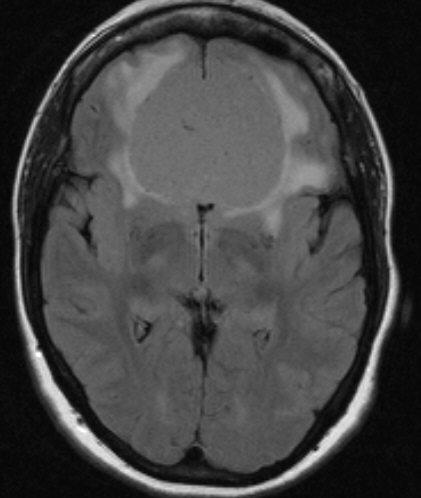
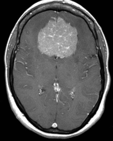
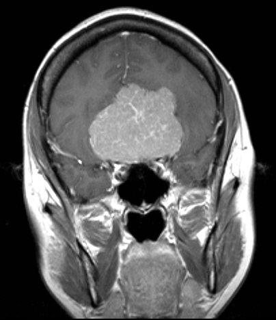
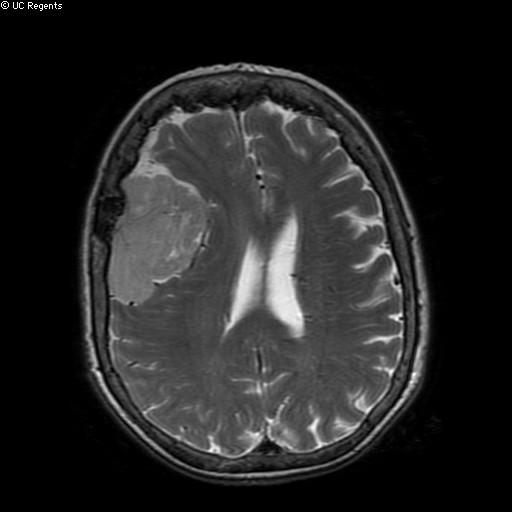
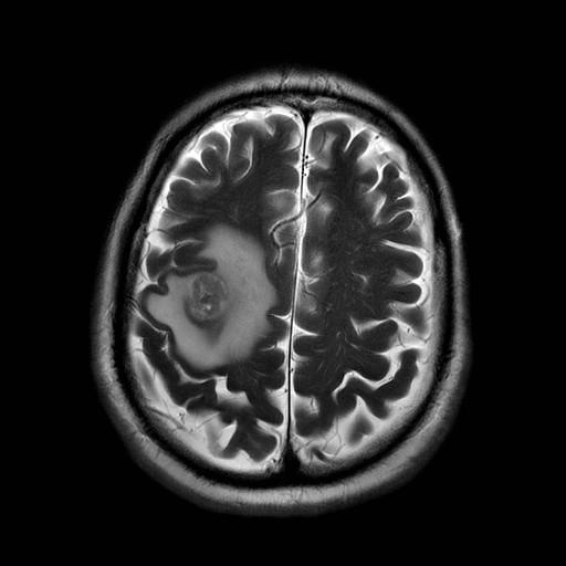
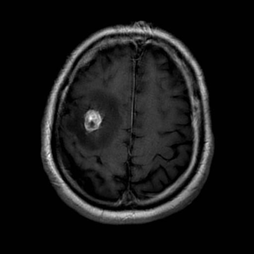
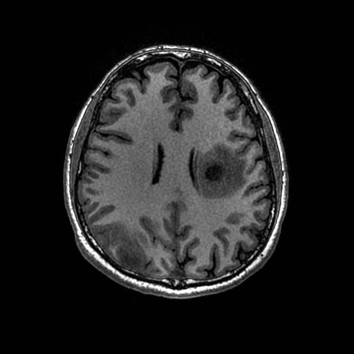
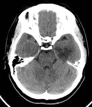
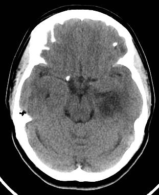
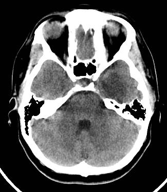
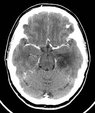
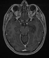
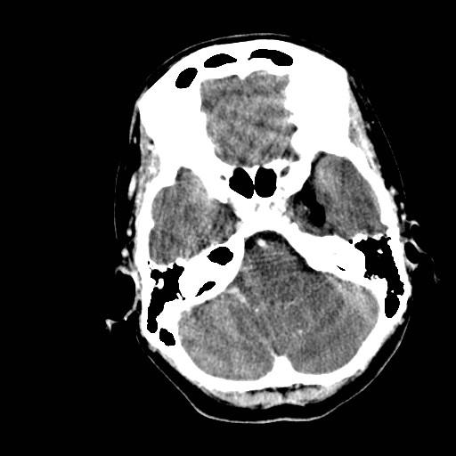
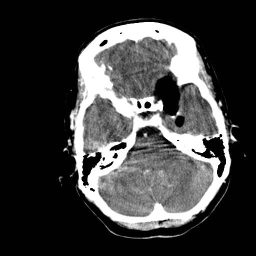
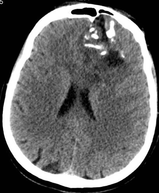
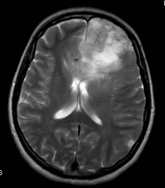
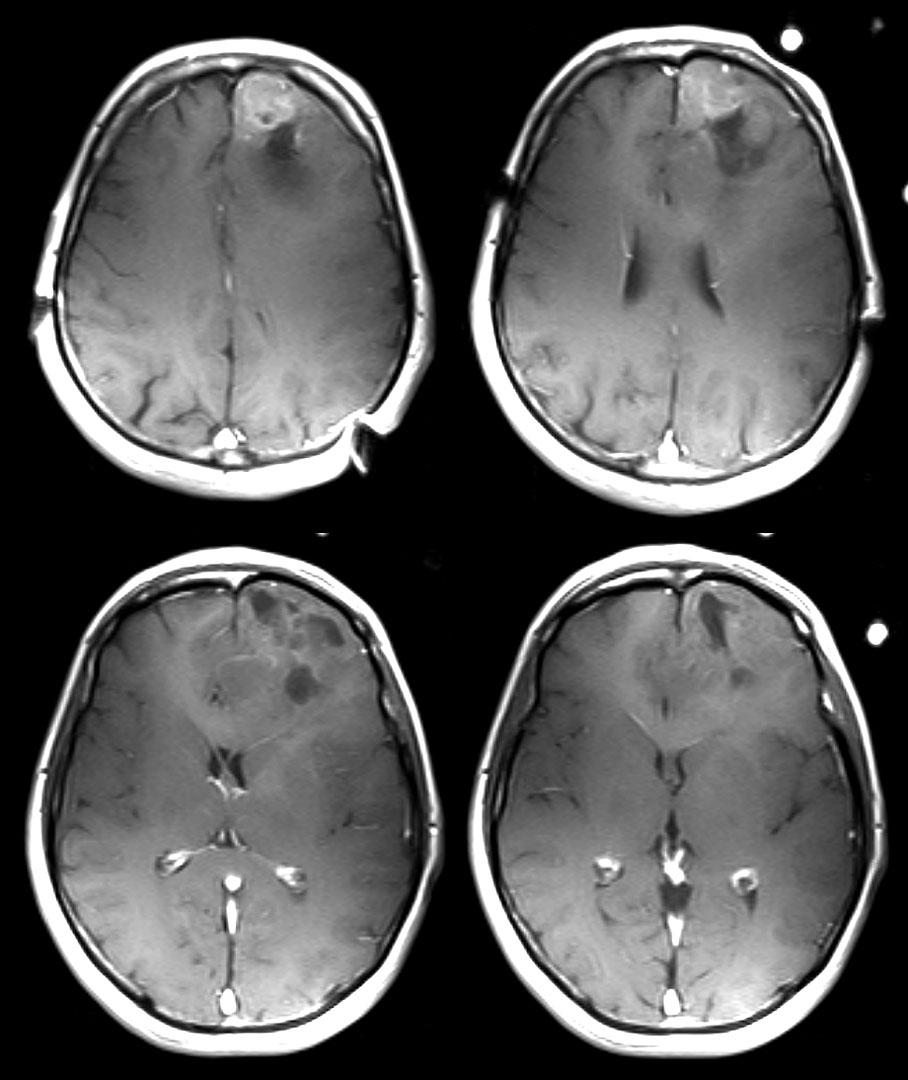
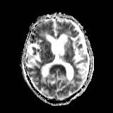
8 38 y female with severely increased headache, 1-2x/d x 1/12 + caffeine use (Pepsi - 2-3L/d x 1 yr).
9
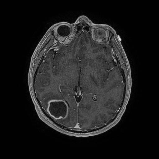
10
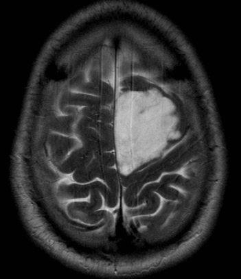
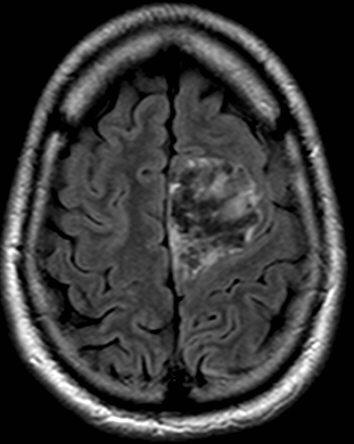
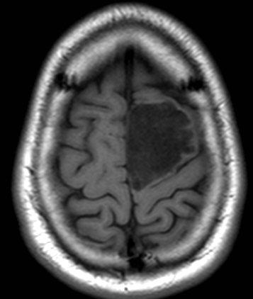
11 MR/CT imaging of meningiomas T1W iso-hypointense,t2w iso-hyperintense (hyperintensity may correlate with softer tumor) Assess for brain invasion, brain edema Intense enhancement Dural tail neoplastic infiltration vs. non-neoplastic meningothelial proliferation, hyper-vascularity CT for bone changes infiltration, hyperostosis
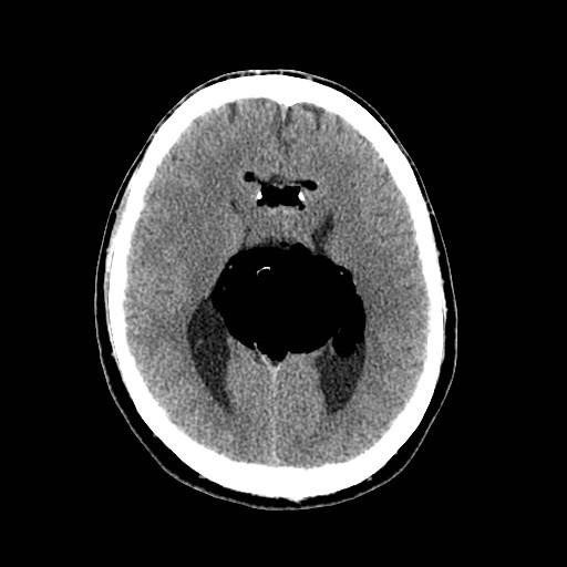
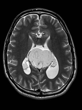
12 Local tumour spread in the brain Astrocytoma Infiltrative White matter tracts Do not respect lobar boundaries Ependymoma plastic spread through ventricular system
13 Huge tumour, little mass effect Low grade glioma/gliomatosis

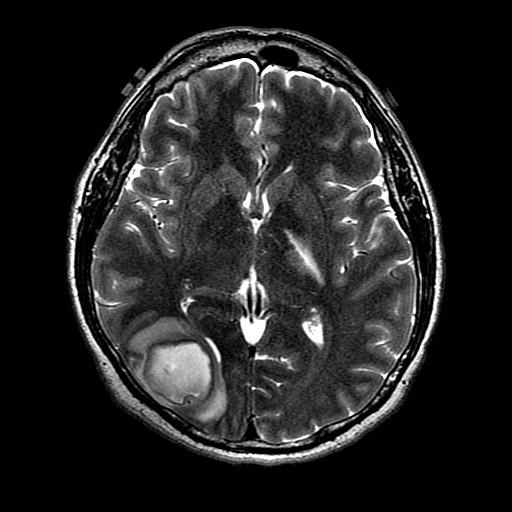
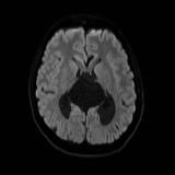
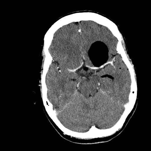
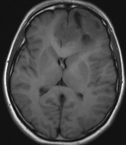
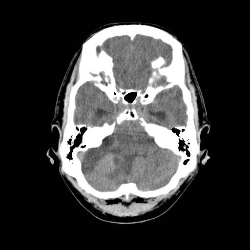
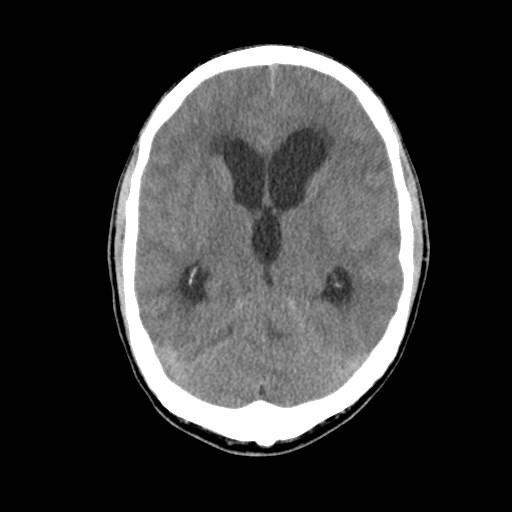
14 46F, rule out lesion
15
16 WHO 2016 Gliomatosis cerebri deleted as a distinct entity It is a growth pattern, found in many different gliomas Growth pattern = three or more cerebral lobes, frequently bilateral, infratentorial extension
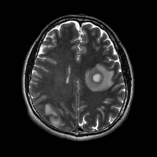
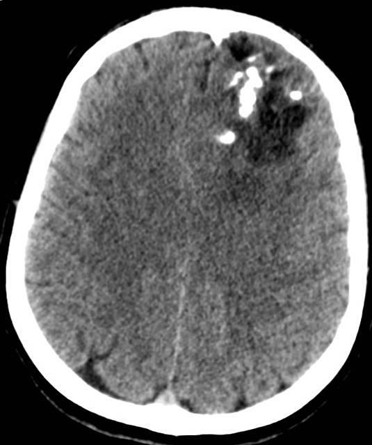
17 Small tumour with lots of mass effect/edema Metastatic (but make sure it s not an abscess)
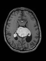
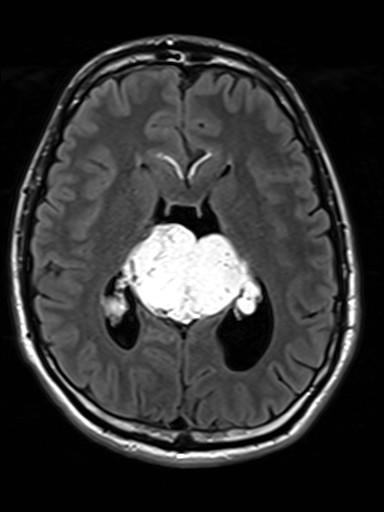
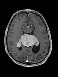
18 Metastatic adenocarcinoma
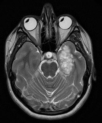
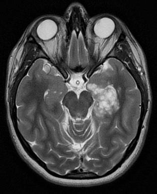
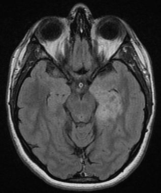
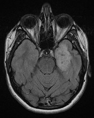
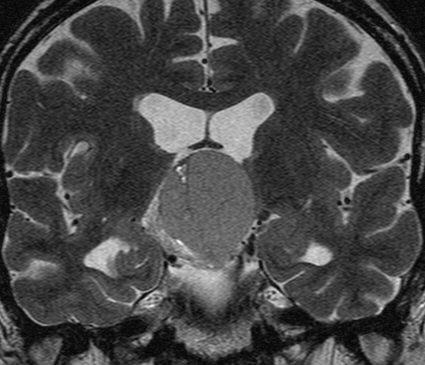
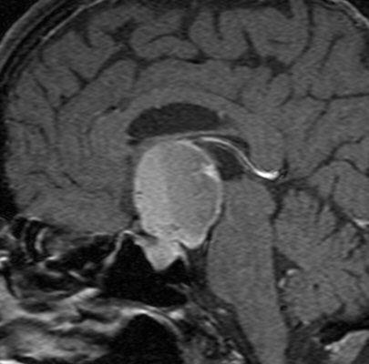
19

20 Restricted diffusion, pyogenic abscess
21 Tumour spread Full anatomical extent Perineural spread of head and neck tumours Leptomeningeal metastases


")

22 CN V2 Infraorbital Nerve Perineural Tumor Spread 74 y.o. male, left CN V(I & II) complete sensory loss, remote cheek SCCa
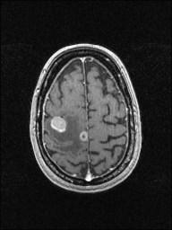
23 Subarachnoid spread of tumours Metastases from non-cns primaries GBM Lymphoma Ependymoma Choroid plexus tumours PNET (medulloblastoma, pineoblastoma)

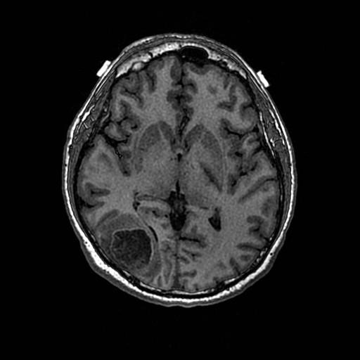
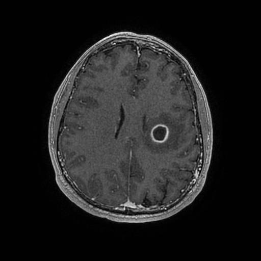
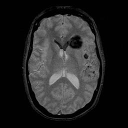
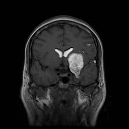
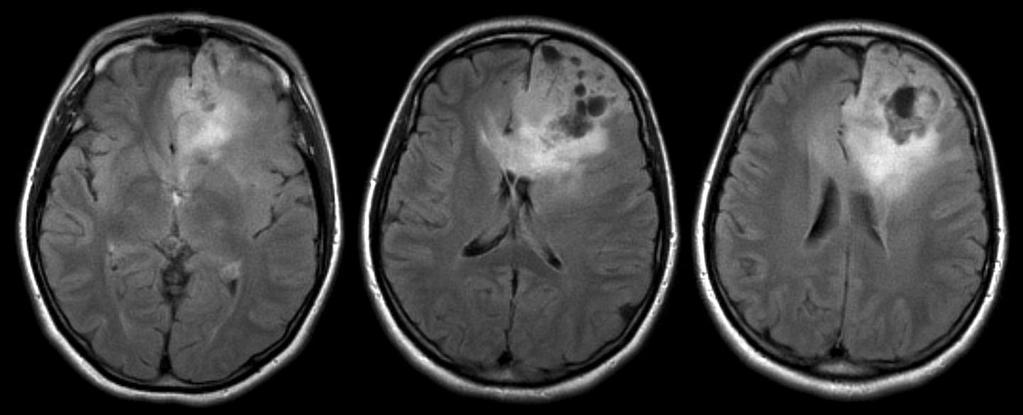
24 54F extensive leptomeningeal disease, breast cancer

25 plastic spread: Ependymoma


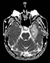
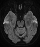
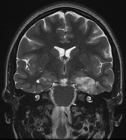
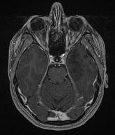
26 Tumour that spills : CRANIOPHARYNGIOMA
27 Tumours that cross the midline Meningioma GBM (radiation necrosis) Primary CNS lymphoma Epidermoid cyst (tumefactive MS)
28

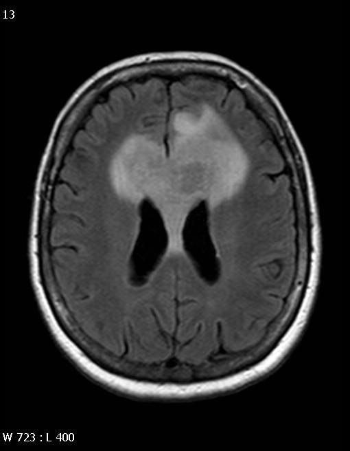
29 Glioblastoma Multiforme Rapidly enlarging malignant astrocytic tumor characterized by necrosis and neovascularity WHO grade IV Most common primary brain tumor Supratentorial white matter most common Frontal, temporal, parietal Occipital lobes relatively spared Cerebral hemispheres > brainstem > cerebellum Peak yrs Relentless progression (death in 9-12 months)

30 Glioblastoma Multiforme Neuroimaging: Thick, irregular-enhancing rind of neoplastic tissue surrounding necrotic core Tumor typically crosses WM tracts to involve contralateral hemisphere Rarely may be multifocal or multicentric Necrosis, cysts, hemorrhage, fluid/debris levels, flow voids MRS: NAA; myoinositol; choline, lipids & lactate Corpus callosum involvement may be seen in GBM, lymphoma, are rarely metastases & demyelination Viable tumor extends far beyond signal abnormalities
31 Solitary vs. multiple lesions Primary distinguishing characteristic for primary vs. metastatic lesions Primary brain tumours can be multicentric (high grade and low grade gliomas)

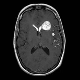
32 52F, breast cancer
33 Brain tumours in the phakomatoses NFI optic pathway gliomas, astrocytoma NFII meningiomas, ependymomas, schwannomas Tuberous sclerosis SEGA Von-Hippel Lindau multiple hemangioblastomas

34 26F, multiple cranial neuropathies
35 NF 2: IMAGING Bilateral enhancing CPA-IAC masses Ovoid when small; "ice cream on cone" when large enough to fill IAC & CPA CNS Calcifications: Choroid plexus, cerebellar hemispheres, & cerebral cortex Other meningiomas & schwannoma (CN3-12) Ependymomas > > gliomas Spine Meningiomas, schwannomas, & ependymomas
36 PATHOLOGY Autosomal dominant disorder Mutation of NF2 gene chromosome 22 50% result from new dominant gene mutation
37 Cortical based tumours Ganglioglioma DNET oligodendroglioma
38 20 yo female. Suspected absence seizures. of episodes.? organic etiology
39
40
41
42 DNET Dysembryoplastic neuroepithelial tumour Hx longstanding partial complex seizures in child/young adult Benign mixed glial-neuronal neoplasm Frequent association with cortical dysplasia Wedge shape, point toward ventricle Mesial temporal most common location Well demarcated, non-enhancing, little or no mass effect/edema Cystic, bubbly appearance on T2W Surgical resection usually curative
43 Fat, calcification and cysts Fat = very limited ddx Lipoma dermoid
44 37 year old male History of seizures
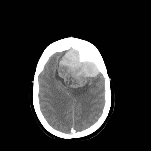
45
46
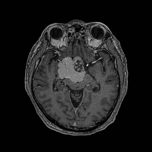
47 LIPOMA Mass of mature non-neoplastic adipose tissue Congenital malformation Midline location common, 80% supratentorial 40-50% interhemispheric fissure Two kinds of interhemispheric lipoma Curvilinear curves around CC, splenium Tubulonodular bulky mass, may calcify associated CC agensis/dysgenesis T1 hyperintense Chemical shift artefact on T2

48 DERMOID CYST (RUPTURED)

49 DERMOID CYST (RUPTURED)
50 calcification Intra-axial: Gliomas old elephants age gracefully oligodendroglioma (almost all), ependymoma, astrocytoma, GBM Other metastases, choroid plexus tumours, ganglioglioma Extra-axial: Meningioma Craniopharyngioma Chordoma Chondrosarcoma
51 Anaplastic Oligodendroglioma
52
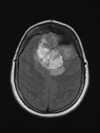
53 73M sleepy/tired, memory impairment
54
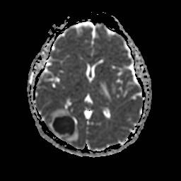
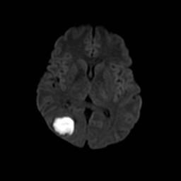
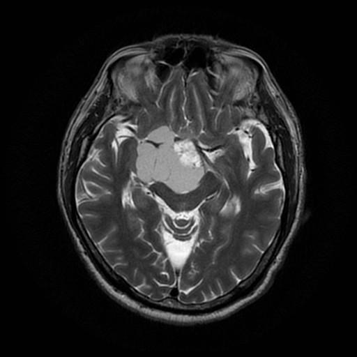
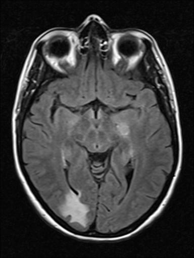
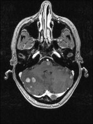
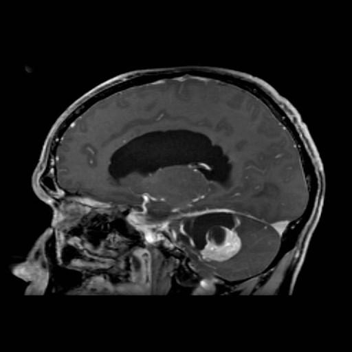
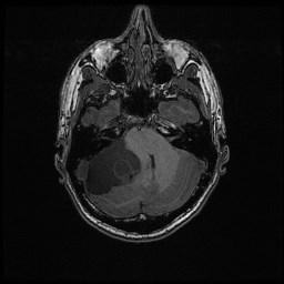
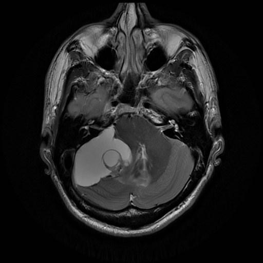
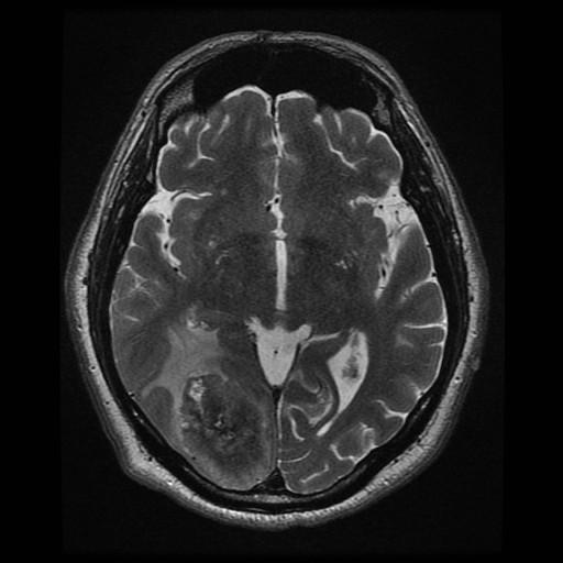
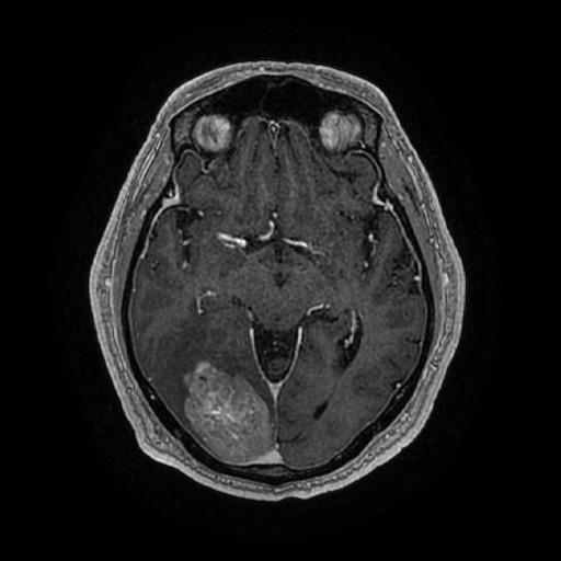
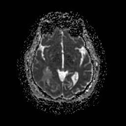
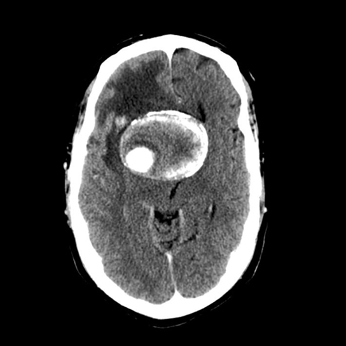
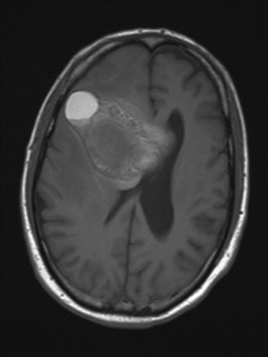
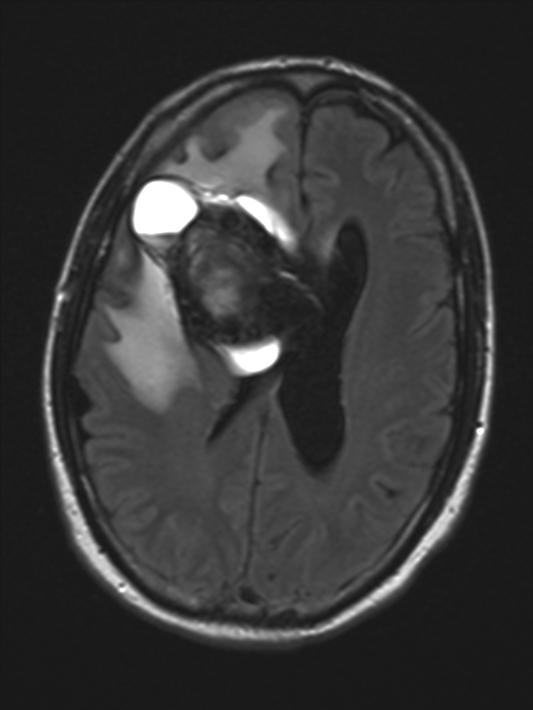
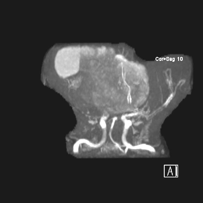
55 Craniopharyngioma Arises from remnants of Rathke s pouch Primarily suprasellar, can be quite large and spill out of the sellar region middle and anterior fossa, prepontine Adamantinomatous most common Cystic/solid but mostly cystic, machinery oil Bimodal = 5-10, years Visual symptoms Slow growing but tend to recur
56 Craniopharyngioma Imaging CT rule of 90 s 90% cystic/solid, 90% Ca++, 90% enhance MR appearance variable depending on cyst contents Multiple cysts common and may have different signal Hypo-hyperintense on T1, hyperintense on T2 Nodule often calcified and hypointense on T2 Cyst walls and nodules enhance
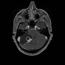
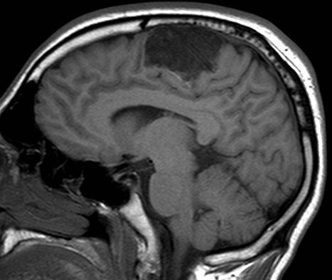
57 Relatively dense (CT)/hypointense T2 tumours Correspondence with reduced diffusion Lymphoma PNET Solid part of GBM

58 PRIMARY CNS LYMPHOMA

59 PRIMARY CNS LYMPHOMA
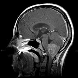
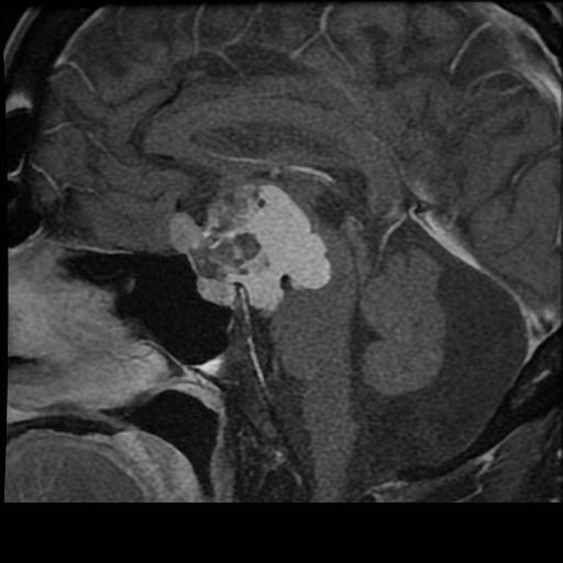
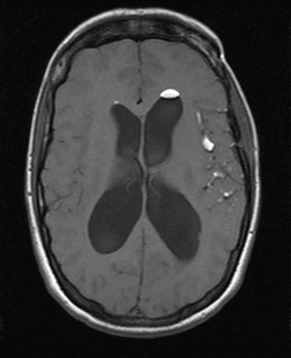
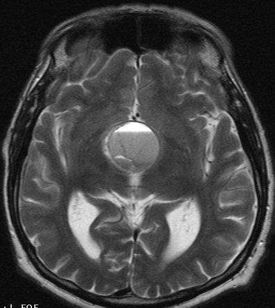
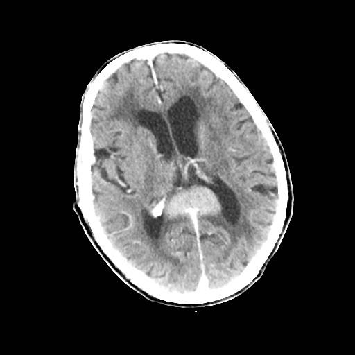
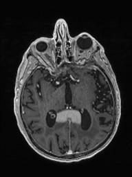
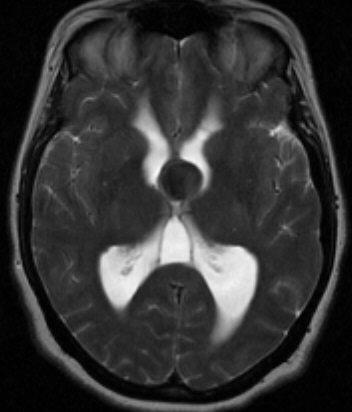
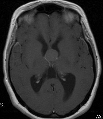
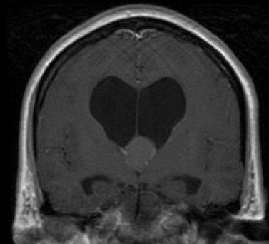
60 Tumours that characteristically have cystic components Intra-axial GBM Hemangioblastoma Ganglioglioma, DNET, PXA Extra-axial Craniopharyngioma Non-neoplastic Dermoid/epidermoid Beware of mimics!

61 45, headache
62
63 Hemangioblastoma Benign and slow growing Sporadic or multiple (VHL) Most in PF, nodule typically abuts pial surface Nodule with peri-tumoral non-neoplastic cyst most common They can be entirely solid can also have intratumoral neoplastic cyst both peritumoral non-neoplastic and intratumoral neoplastic cysts (as in this case)
64 Hyperintensity T1W (-contrast) Hemorrhage Calcification Proteinacious cyst melanin


65 38-year-old man with progressive decreased vision, ataxia, and pressure-like frontal headache.
66
67 Post-Gd
68 COLLOID CYST Mucin containing 3 rd ventricular cyst Hyperdense foramen of Monro mass on unenhanced CT <1% other sites (lateral & 4 th ventricles, extraaxial) 1/3 isointense on T1, 2/3 hyperintense on T1 Variable T2 signal No DWI restriction Enhancement unusual 90% stable, 10% enlarge Acute obstruction may lead to rapid onset hydrocephalus
69 Metastatic melanoma
70 Density/Signal intensity similar to CSF Epidermoid cyst Neurocysticercosis

71 Seizures 26M
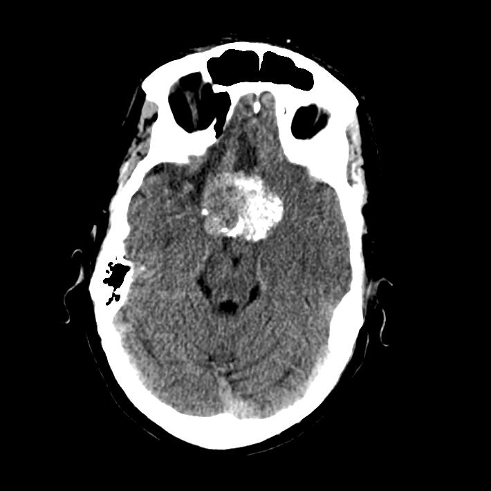
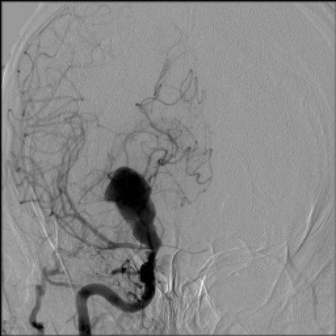
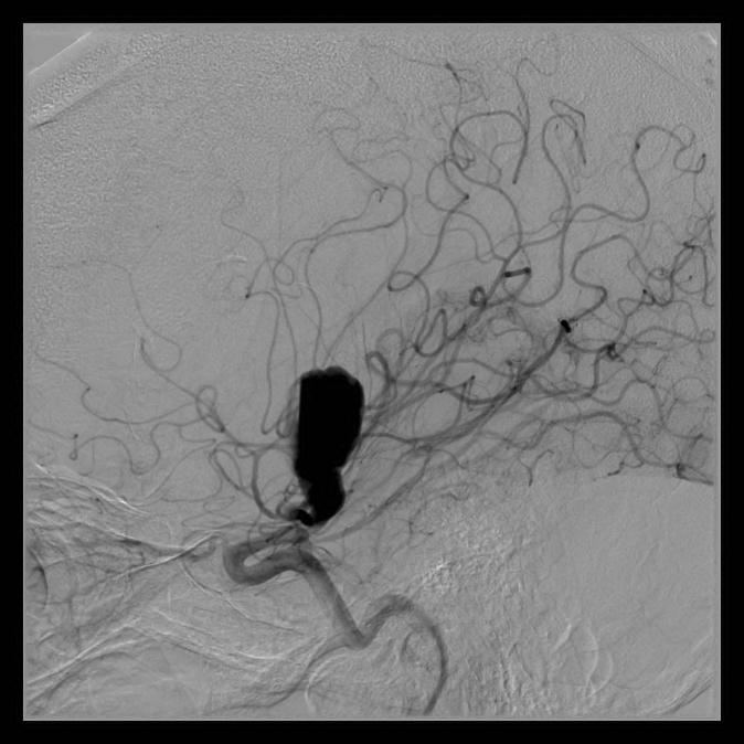
72

73 Pre Post-Gd
74 Epidermoid tumours Stratified squamous epithelium, contains keratinaceous debris and cholesterol CPA most common, middle fossa, parasellar Cerebral convexities less common CT hypodense, Ca++ in 25% Iso-slightly hyperintense relative to CSF on T1 and T2, slightly heterogeneous Do not suppress on FLAIR, restrict on DWI Insinuate in/around structures, burrow into brain
75 Test your skills! 45yr/M with h/o Increasing behavioural changes x 3 yrs
76
77
78 MRA
79 ANSWER: IT S NOT A TUMOUR! a. Central neurocytoma b. Meningioma c. CNS lymphoma d. IT S NOT A TUMOUR!!
80 Giant intracranial aneurysms. Def. : > 2.5 cm.. Types : Saccular fusiform.. Epidemiology and natural history - Comprise 3-5% of intracranial aneurysms - peak age of presentation yr - F:M, 3:1-2/3 rd Ant circulation 1/3 rd Post circulation
81 Slow growing with repeated internal hemorrhage Laminated thrombus of varying ages Symptoms commonly related to mass effect, lower rate of rupture Imaging: CT hyperdense mass with calcification. MRI-Mixed signal intensity mass with thrombus of various stages, Perianeurysm hematoma and edema
Peter Canoll MD. PhD.
 Tumors of the Nervous System Peter Canoll MD. PhD. What I want to cover What are the most common types of brain tumors? Who gets them? How do they ypresent? What do they look like? How do they behave?
Tumors of the Nervous System
 Tumors of the Nervous System Peter Canoll MD. PhD. What I want to cover What are the most common types of brain tumors? Who gets them? How do they present? What do they look like? How do they behave? 1
Brain Tumors. Medulloblastoma. Pilocytic astrocytoma: Ahmed Koriesh, MD. Pathological finding
 NeuroPathology Page 8 Brain Tumors Pathological finding Pseudorosette Rosenthal fibers Rosettes Wet Keratin Psammoma bodies Fried egg Tumor Ependymoma, SEGA Pilocytic astrocytoma Medulloblastoma Craniopharyngioma
CNS TUMORS. D r. Ali Eltayb ( U. of Omdurman. I ). M. Path (U. of Alexandria)
. M. Path (U. of Alexandria)") CNS TUMORS D r. Ali Eltayb ( U. of Omdurman. I ). M. Path (U. of Alexandria) CNS TUMORS The annual incidence of intracranial tumors of the CNS ISmore than intraspinal tumors May be Primary or Secondary
CNS TUMORS D r. Ali Eltayb ( U. of Omdurman. I ). M. Path (U. of Alexandria) CNS TUMORS The annual incidence of intracranial tumors of the CNS ISmore than intraspinal tumors May be Primary or Secondary
Pediatric CNS Tumors. Disclosures. Acknowledgements. Introduction. Introduction. Posterior Fossa Tumors. Whitney Finke, MD
 Pediatric CNS Tumors Disclosures Whitney Finke, MD Neuroradiology Fellow PGY-6 University of Utah Health Sciences Center Salt Lake City, Utah None Acknowledgements Introduction Nicholas A. Koontz, MD Luke
CT & MRI Evaluation of Brain Tumour & Tumour like Conditions
 CT & MRI Evaluation of Brain Tumour & Tumour like Conditions Dr. Anjana Trivedi 1, Dr. Jay Thakkar 2, Dr. Maulik Jethva 3, Dr. Ishita Virda 4 1 M.D. Radiology, Professor and Head, P.D.U. Medical College
PITUITARY PARASELLAR LESIONS. Kim Learned, MD
 PITUITARY PARASELLAR LESIONS Kim Learned, MD DIFFERENTIALS Pituitary Sella Clivus, Sphenoid Sinus Suprasellar Optic chiasm, Hypothalamus, Circle of Willis Parasellar Cavernous Sinus Case 1 17 YEAR-OLD
RINGS N THINGS: Imaging Patterns in Differential Diagnosis. Anne G. Osborn, M.D.
 RINGS N THINGS: Imaging Patterns in Differential Diagnosis Anne G. Osborn, M.D. ExpDDxs: Intra-axial (Parenchymal) Lesions Ring-enhancing lesions, solitary 1 Ring-enhancing lesion crossing corpus callosum
NEURORADIOLOGY DIL part 5
 NEURORADIOLOGY DIL part 5 Masses and tumors K. Agyem MD, G. Hall MD, D. Palathinkal MD, Alexandre Menard March/April 2015 OVERVIEW Introduction to Neuroimaging - DIL part 1 Basic Brain Anatomy - DIL part
Supra- and infratentorial brain tumors from childhood to maternity
 Supra- and infratentorial brain tumors from childhood to maternity What to expect? I am going to show you the characteristic imaging findings of following tumors: Thierry A.G.M. Huisman, MD, FICIS, EQNR
Posterior fossa tumors: clues to differential diagnosis with case-based review
 Posterior fossa tumors: clues to differential diagnosis with case-based review Poster No.: C-0323 Congress: ECR 2017 Type: Educational Exhibit Authors: H. A. Aboughalia, M. Abdelhady; Doha/QA Keywords:
Case Studies in Sella/Parasellar Region. Child thirsty, increased urination. Imaging. Suprasellar Germ Cell Tumor (Germinoma) No Disclosures
 No Disclosures") Case Studies in Sella/Parasellar Region No Disclosures 2018 Head and Neck Imaging Conference Child thirsty, increased urination Suprasellar Germ Cell Tumor (Germinoma) Midline Pineal >> Suprasellar > Other
Case Studies in Sella/Parasellar Region No Disclosures 2018 Head and Neck Imaging Conference Child thirsty, increased urination Suprasellar Germ Cell Tumor (Germinoma) Midline Pineal >> Suprasellar > Other
General: Brain tumors are lesions that have mass effect distorting the normal tissue and often result in increased intracranial pressure.
 1 Lecture Objectives Know the histologic features of the most common tumors of the CNS. Know the differences in behavior of the different tumor types. Be aware of the treatment modalities in the various
Pathologic Analysis of CNS Surgical Specimens
 2015 Kenneth M. Earle Memorial Neuropathology Review Pathologic Analysis of CNS Surgical Specimens Peter C. Burger, MD Interdisciplinary Quality Control Familiarity with entities Use of diagnostic algorithm
Case 7391 Intraventricular Lesion
 Case 7391 Intraventricular Lesion Bastos Lima P1, Marques C1, Cabrita F2, Barbosa M2, Rebelo O3, Rio F1. 1Neuroradiology, 2Neurosurgery, 3Neuropathology, Coimbra University Hospitals, Portugal. University
Cross sectional imaging of Intracranial cystic lesions Abdel Razek A
 Cross sectional imaging of Intracranial cystic lesions Abdel Razek A Department of Radiology. Mansoura Faculty of Medicine, Mansoura. Egypt. arazek@mans.edu.eg Introduction Intracranial cystic lesions
Neuroimaging Core Curriculum
 Neuroimaging Core Curriculum Program Content The purpose of the training program is to prepare the physician for the independent practice of neuroimaging. Neuroimaging is the subspecialty of Neurology
Tumors of the Central Nervous System
 Tumors of the Central Nervous System 1 Financial Disclosures I have NO SIGNIFICANT FINANCIAL, GENERAL, OR OBLIGATION INTERESTS TO REPORT Introduction General: Brain tumors are lesions that have mass effect
JMSCR Vol 05 Issue 08 Page August 2017
 www.jmscr.igmpublication.org Impact Factor 5.84 Index Copernicus Value: 83.27 ISSN (e)-2347-176x ISSN (p) 2455-0450 DOI: https://dx.doi.org/10.18535/jmscr/v5i8.19 Magnetic Resonance Imaging in Evaluation
Masses of the Corpus Callosum
 Masses of the Corpus Callosum Kesav Raghavan, HMS Year III Dr. Agenda Corpus Callosum Development and Anatomy Our Patient: Clinical Presentation Differential Diagnosis of Masses in the Corpus Callosum
Metastasis. 57 year old with progressive Headache and Right Sided Visual Loss
 Metastasis 1% of sellar/parasellar masses Usually occurs with known primary Can involve third ventricle, hypothalamus, infundibular stalk May be both supra-, intrasellar 57 year old with progressive Headache
Patients Treated with Leksell Gamma Knife
 Patients Treated with Leksell Gamma Knife 1968-2016 TREATMENTS REPORTED 2016 BY REGION AND INDICATION INDICATION Asia excl. Europe Latin Middle East & Africa North Grand Total Benign Tumors 12283 9778
Disclosures. Posterior Fossa Masses. I m from the Government. and I here to help! Differential Diagnosis
 Posterior Fossa Masses Differential Diagnosis James G. Smirniotopoulos, M.D. Radiology, Neurology, Biomedical Informatics Uniformed Services University Bethesda, Maryland http://rad.usuhs.edu http://medpix.usuhs.edu
Disclosure. + Outline. Case-based approach to neurological emergencies that might present to the ED
 Kathleen R. Fink, MD University of Washington 5 th Nordic Emergency Radiology Course May 21, 2015 Disclosure My spouse receives research salary support from: Bracco BayerHealthcare Guerbet Outline Case-based
Laurie A. Loevner, MD
 Laurie A. Loevner, MD Chief, Division of Neuroradiology UPHS Professor of Radiology, Otorhinolaryngology: Head & Neck Surgery, Neurosurgery, and Ophthalmology University of Pennsylvania Health System Disclosures
Imaging the Spinal Cord & Intradural Disease
 Department of Radiology University of California San Diego Imaging the Spinal Cord & Intradural Disease John R. Hesselink, M.D. Spinal Cord Diseases Tumors Syringohydromyelia Trauma Ischemia / Infarction
Differential diagnosis of intracranial cystic lesions.
 Differential diagnosis of intracranial cystic lesions. Poster No.: C-0215 Congress: ECR 2015 Type: Educational Exhibit Authors: S. P. G. Alandete, M. A. Meseguer, E. De la Via, D. Uceda, C. Poyatos; Valencia/ES
EXPERT DIFFERENTIAL DIAGNOSIS:
 EXPERT DIFFERENTIAL DIAGNOSIS: Sellar Region Anne G. Osborn, M.D. DISCLOSURE: Published RSNA 2008 SELLA, PITUITARY: Normal Gross, 3T Anatomy SELLA, PITUITARY: Anatomically-Based Differential Diagnoses
CNS pathology Third year medical students. Dr Heyam Awad 2018 Lecture 12: CNS tumours 2/3
 CNS pathology Third year medical students Dr Heyam Awad 2018 Lecture 12: CNS tumours 2/3 Pilocytic astrocytoma Relatively benign ( WHO grade 1) Occurs in children and young adults Mostly: in the cerebellum
H Haloes cautions, 57 neurocytomas, perinuclear, 56 Headache blue cell tumors, 147 cautions, 135, 147, 152 clinical history, 132, 144, 148
 Index A ADC. See Apparent diffusion coefficient Adult. See also Supratentorial mass, adult cerebral tumor, 1 headache and ataxia cysts, mural nodules, 118 sporadic tumors, 118 headaches and visual changes,
Brain tumors: tumor types
 Brain tumors: tumor types Tumor types There are more than 120 types of brain tumors. Today, most medical institutions use the World Health Organization (WHO) classification system to identify brain tumors.
S Alandete, M Meseguer, CR Poyatos, D Uceda, E de la Via, J Sales, J Vilar. H.U. Dr Peset, Valencia (Spain)
") S Alandete, M Meseguer, CR Poyatos, D Uceda, E de la Via, J Sales, J Vilar. H.U. Dr Peset, Valencia (Spain) Introduction Cystic lesions are usually a common finding in clinical practice and you can find
S Alandete, M Meseguer, CR Poyatos, D Uceda, E de la Via, J Sales, J Vilar. H.U. Dr Peset, Valencia (Spain) Introduction Cystic lesions are usually a common finding in clinical practice and you can find
Imaging of Hearing Loss
 Contemporary Imaging of Sensorineural Hearing Loss Imaging of Hearing Loss Discussion Outline (SNHL) Imaging Approaches Anatomic Relationships Lesions: SNHL KL Salzman, MD University of Utah School of
RING ENCHANCING LESION BY M.S. HEMHNATH
 RING ENCHANCING LESION BY M.S. HEMHNATH A 21 YRS FEMALE CAME WITH H/O HEADACHE AND SEIZURE FOR THE PAST ONE MONTH. NO OTHER FOCAL NEUROLOGICAL DEFICIT. DIFFERENTIAL DIAGNOSIS For this case are Neurocysticerosis
Joana Ramalho, MD C. Ryan Miller, MD, PhD
 Joana Ramalho, MD C. Ryan Miller, MD, PhD Case 1 3 month old baby girl Presented with new onset of seizures Newborn. Questionable blurring of the gray-white junction within the right occipital lobe. Findings
Essentials of Clinical MR, 2 nd edition. 51. Primary Neoplasms
 51. Primary Neoplasms As with spinal central canal neoplasms in other regions, those of the lumbar spine may be classified as extradural, intradural extramedullary, and medullary. If an extradural lesion
Table of Contents: SKULL AND BRAIN. Scalp, Skull. Anatomically Based Differentials. Skull Normal Variants. Scalp Mass, Child.
 Table of Contents: SKULL AND BRAIN Scalp, Skull Skull Normal Variants Scalp Mass, Child Scalp Mass, Adult Congenital Anomalies of Skull Base Sellar/Parasellar Mass With Skull Base Invasion "Hair on End"
Role of Diffusion weighted Imaging in the Evaluation of Intracranial Tumors
 IOSR Journal of Dental and Medical Sciences (IOSR-JDMS) e-issn: 2279-0853, p-issn: 2279-0861.Volume 15, Issue 12 Ver. IX (December. 2016), PP 99-104 www.iosrjournals.org Role of Diffusion weighted Imaging
Cerebro-vascular stroke
 Cerebro-vascular stroke CT Terminology Hypodense lesion = lesion of lower density than the normal brain tissue Hyperdense lesion = lesion of higher density than normal brain tissue Isodense lesion = lesion
This article appeared in a journal published by Elsevier. The attached copy is furnished to the author for internal non-commercial research and
 This article appeared in a journal published by Elsevier. The attached copy is furnished to the author for internal non-commercial research and education use, including for instruction at the authors institution
DISCLOSURES LEARNING OBJECTIVES WE WILL NOT DISCUSS. CSB: Birdseye View MESSAGE NAVIGATING THE SELLA AND CENTRAL SKULL BASE
 NAVIGATING THE SELLA AND CENTRAL SKULL BASE Christopher P. Hess, M.D., Ph.D. DISCLOSURES Research Support, General Electric SLIDES: http://www.radiology.ucsf.edu/research/meetings/rsna LEARNING OBJECTIVES
NEURORADIOLOGY-NEUROPATHOLOGY CONFERENCE
 THE UNIVERSITY OF NORTH CAROLINA at CHAPEL HILL SEPTEMBER 2013 NEURORADIOLOGY-NEUROPATHOLOGY CONFERENCE Claudia da Costa Leite, MD, PhD Thomas Bouldin, MD CASE 1 6 y-o female with headaches and vomiting
Structural and functional imaging for the characterization of CNS lymphomas
 Structural and functional imaging for the characterization of CNS lymphomas Cristina Besada Introduction A few decades ago, Primary Central Nervous System Lymphoma (PCNSL) was considered as an extremely
Oligodendroglioma: imaging findings, radio-pathological correlation and evolution
 Oligodendroglioma: imaging findings, radio-pathological correlation and evolution Poster No.: C-2104 Congress: ECR 2013 Type: Authors: Keywords: DOI: Scientific Exhibit A. Hernandez Castro, M. D. Monedero
For Emergency Doctors. Dr Suzanne Smallbane November 2011
 For Emergency Doctors Dr Suzanne Smallbane November 2011 A: Orbit B: Sphenoid Sinus C: Temporal Lobe D: EAC E: Mastoid air cells F: Cerebellar hemisphere A: Frontal lobe B: Frontal bone C: Dorsum sellae
Five Most Common Problems in Surgical Neuropathology
 Five Most Common Problems in Surgical Neuropathology If the brain were so simple that we could understand it, we would be so simple that we couldn t Emerson Pugh What is your greatest difficulty in neuropathology?
Astroblastoma: Radiologic-Pathologic Correlation and Distinction from Ependymoma
 AJNR Am J Neuroradiol 23:243 247, February 2002 Case Report Astroblastoma: Radiologic-Pathologic Correlation and Distinction from Ependymoma John D. Port, Daniel J. Brat, Peter C. Burger, and Martin G.
EEG IN FOCAL ENCEPHALOPATHIES: CEREBROVASCULAR DISEASE, NEOPLASMS, AND INFECTIONS
 246 Figure 8.7: FIRDA. The patient has a history of nonspecific cognitive decline and multiple small WM changes on imaging. oligodendrocytic tumors of the cerebral hemispheres (11,12). Electroencephalogram
Year 2003 Paper two: Questions supplied by Tricia
 question 43 A 42-year-old man presents with a two-year history of increasing right facial numbness. He has a history of intermittent unsteadiness, mild hearing loss and vertigo but has otherwise been well.
Pleomorphic Xanthoastrocytoma
 Pleomorphic Xanthoastrocytoma Christine E. Fuller Keywords Pleomorphic xanthoastrocytoma; Pleomorphic xanthoastrocytoma with anaplastic features 2.1 OVERVIEW Pleomorphic xanthoastrocytoma (PXA) is an uncommon
Imaging The Turkish Saddle. Russell Goodman, HMS III Dr. Gillian Lieberman
 Imaging The Turkish Saddle Russell Goodman, HMS III Dr. Gillian Lieberman Learning Objectives Review the anatomy of the sellar region Discuss the differential diagnosis of sellar masses Discuss typical
Case Studies in CPA/IAC
 Outline Case Studies in CPA/IAC Atul K Mallik MD PhD Department of Radiology and Imaging Sciences University of Utah Health Sciences Center Salt Lake City, Utah, USA Case based review of cerebellopontine
Pediatric Spine Tumors (and other masses)
") Pediatric Spine Tumors (and other masses) Francisco A Perez, MD, PhD Assistant Professor Neuroradiology and Pediatric Radiology Seattle Children s Hospital University of Washington, Seattle Commercial
Pediatric Spine Tumors (and other masses) Francisco A Perez, MD, PhD Assistant Professor Neuroradiology and Pediatric Radiology Seattle Children s Hospital University of Washington, Seattle Commercial
Spinal Neoplasms. First Things First!! Localize the Lesion!! Ependymomas. Common Intramedullary Lesions
 Acta Radiológica Portuguesa, Vol.XXIII, nº 90, pág. 101-114, Abr.-Jun., 2011 Spinal Neoplasms Bruno A Policeni University of Iowa Hospitals and Clinics Assistant Professor of Radiology Disclosure of Commercial
Index. aneurysm, 92 carotid occlusion, 94 ICA stenosis, 95 intracranial, 92 MCA, 94
 A ADC. See Apparent diffusion coefficient (ADC) Aneurysm cerebral artery aneurysm, 93 CT scan, 93 gadolinium, 93 Angiography, 13 Anoxic brain injury, 25 Apparent diffusion coefficient (ADC), 7 Arachnoid
Clinics in diagnostic imaging (175)
") Singapore Med J 2017; 58(3): 121-125 doi: 10.11622/smedj.2017017 CMEArticle Clinics in diagnostic imaging (175) Vijay Krishnan 1, MD, FRCR, Tze Chwan Lim 1, MBBS, FRCR, Francis Cho Hao Ho 2, MBBS, FRANZCR,
Singapore Med J 2017; 58(3): 121-125 doi: 10.11622/smedj.2017017 CMEArticle Clinics in diagnostic imaging (175) Vijay Krishnan 1, MD, FRCR, Tze Chwan Lim 1, MBBS, FRCR, Francis Cho Hao Ho 2, MBBS, FRANZCR,
MRI Findings Of An Atypical Cystic Meningioma A Rare Case
 ISPUB.COM The Internet Journal of Radiology Volume 14 Number 1 MRI Findings Of An Atypical Cystic Meningioma A Rare Case D Saxena, P Rout, K Pavan, B Philip Citation D Saxena, P Rout, K Pavan, B Philip.
Understanding general brain tumor pathology, Part I: The basics. Craig Horbinski, M.D., Ph.D. Department of Pathology University of Kentucky
 Understanding general brain tumor pathology, Part I: The basics Craig Horbinski, M.D., Ph.D. Department of Pathology University of Kentucky plan of attack what IS a pathologist, anyway? what s so special
Pediatric Brain Tumors Pre, Intra & Post Op Evaluation and Management. Timothy M. George, MD, FACS, FAAP
 Pediatric Brain Tumors Pre, Intra & Post Op Evaluation and Management Timothy M. George, MD, FACS, FAAP PEDIATRIC BRAIN TUMORS BACKGROUND: Incidence: Third most common pediatric tumor type (leukemia, neuroblastoma,
Brain Space Occupying Lesions by Magnetic Resonance Imaging: A Prospective Study
 Original Article DOI: 10.17354/ijss/2015/523 Brain Space Occupying Lesions by Magnetic Resonance Imaging: A Prospective Study Bulabai Karpagam 1, V Vadanika 2 1 Associate Professor, Department of Radiology,
Attenuation value in HU From -500 To HU From -10 To HU From 60 To 90 HU. From 200 HU and above
 Brain Imaging Common CT attenuation values Structure Air Fat Water Brain tissue Recent hematoma Calcifications Bone Brain edema and infarction Normal liver parenchyma Attenuation value in HU From -500
Dr. T. Venkat Kishan Asst. Prof Department of Radiodiagnosis
 Dr. T. Venkat Kishan Asst. Prof Department of Radiodiagnosis Schwannomas (also called neurinomas or neurilemmomas) constitute the most common primary cranial nerve tumors. They are benign slow-growing
Imaging in Epilepsy. Nucharin Supakul, MD Ramathibodi Hospital, Mahidol University August 22, 2015
 Imaging in Epilepsy Nucharin Supakul, MD Ramathibodi Hospital, Mahidol University August 22, 2015 Nothing to disclose Outline Role of Imaging and pitfalls Imaging protocol Case scenarios Clinical & Electrophysiologic
Neuro-oncology Update Andrew Kokkino, MD Medical Director, The Neurosciences Institute at Sacred Heart at Riverbend May 20, 2013
 Neuro-oncology Update 2013 Andrew Kokkino, MD Medical Director, The Neurosciences Institute at Sacred Heart at Riverbend May 20, 2013 Case 1 58 year old man with recent facial droop and HA s Thin, cachectic
Brain Pain Infections of the CNS
 FRIDAY, OCTOBER 28, 2016 Brain Pain Infections of the CNS Suyash Mohan MD, PDCC Assistant Professor of Radiology & Neurosurgery Division of Neuroradiology, Department of Radiology Perelman School of Medicine
An Introduction to Imaging the Brain. Dr Amy Davis
 An Introduction to Imaging the Brain Dr Amy Davis Common reasons for imaging: Clinical scenarios: - Trauma (NICE guidelines) - Stroke - Tumours - Seizure - Neurological degeneration memory, motor dysfunction,
Case 9 10/29/2018. CJD (Creutzfeldt -Jakob Disease) CJD (Creutzfeldt -Jakob Disease) CJD (Creutzfeldt -Jakob Disease)
 CJD (Creutzfeldt -Jakob Disease) CJD (Creutzfeldt -Jakob Disease)") CJD (Creutzfeldt -Jakob Disease) Rare fatal neurodegen dz caused by infectious protein Prion (lacks nucleic acid)- causes spongiform changes of the brain and neuronal death. 4 types: scjd- 85% of cases
CJD (Creutzfeldt -Jakob Disease) Rare fatal neurodegen dz caused by infectious protein Prion (lacks nucleic acid)- causes spongiform changes of the brain and neuronal death. 4 types: scjd- 85% of cases
TUMORS of nervous system
 TUMORS of nervous system By: Shifaa Alqa qa Done By : Ola Hijjawi CNS tumors : The annual incidence of CNS tumors ranges from 10 to 17 per 100,000 persons for intracranial tumors and 1 to 2 per 100,000
The central nervous system
 Sectc.qxd 29/06/99 09:42 Page 81 Section C The central nervous system CNS haemorrhage Subarachnoid haemorrhage Cerebral infarction Brain atrophy Ring enhancing lesions MRI of the pituitary Multiple sclerosis
Accuracy of intra-operative rapid diagnosis by Squash smear in CNS lesions An early institutional experience. KK Bansal,
 Accuracy of intra-operative rapid diagnosis by Squash smear in CNS lesions An early institutional experience. KK Bansal, Monika Bansal, Sanjeev Kishore, Anuradha K, Meena H, Dushyant G. Department of Neurosurgery
Histopathological Study and Categorisation of Brain Tumors
 Histopathological Study and Categorisation of Brain Tumors Ruchira Wadhwa 1*, Purvi Patel 2, Hansa Goswami 3 1 Third Year Resident, 2 Assistant Professor, 3 Professor and Head, Department of Pathology,
Where Has My Vision Gone? Evaluation of Sellar Lesions. Caleb Stowell,, HMS III Gillian Lieberman, MD November 2008
 Where Has My Vision Gone? Evaluation of Sellar Lesions Caleb Stowell,, HMS III Gillian Lieberman, MD November 2008 Objectives Present a case highlighting the clinical presentation and evaluation of a sellar
DelMarVa-DC Regional Cancer Registrar s Educational Meeting. Doordan Conference Center Anne Arundel Medical Center Annapolis, MD
 DelMarVa-DC Regional Cancer Registrar s Educational Meeting Doordan Conference Center Anne Arundel Medical Center Annapolis, MD TNM Transition Updates & News from SEER Peggy Adamo, RHIT, CTR NCI SEER adamom@mail.nih.gov
Pediatric Brain Tumors: Updates in Treatment and Care
 Pediatric Brain Tumors: Updates in Treatment and Care Writer Classroom Rishi R. Lulla, MD MS Objectives Introduce the common pediatric brain tumors Discuss current treatment strategies for pediatric brain
RADIOLOGY TEACHING CONFERENCE
 RADIOLOGY TEACHING CONFERENCE John Athas, MD Monica Tadros, MD Columbia University, College of Physicians & Surgeons Department of Otolaryngology- Head & Neck Surgery September 27, 2007 CT SCAN IMAGING
Benign brain lesions
 Benign brain lesions Diagnostic and Interventional Radiology Hung-Wen Kao Department of Radiology, Tri-Service General Hospital, National Defense Medical Center Computed tomography Hounsfield unit (HU)
Joana Ramalho, MD C. Ryan Miller, MD, PhD
 Joana Ramalho, MD C. Ryan Miller, MD, PhD Case 1 56 year old female Presented with: 3-4 weeks of visual symptoms (asymmetric vision loss, blurry & dark vision, photosensitivity & decreased peripheral vision)
Neuro - imaging. Sella. ssregypt.com
 Neuro - imaging Sella ssregypt.com Bony Sella AP diameter Depth Contents 16mm 14mm Pituitary gland, part of infundibular stalk, CSF CT Technique 5 mm slices Axial and coronal Contrast injection Bone and
NEURORADIOLOGY Angela Lignelli, MD
 Neuroradiology NEURORADIOLOGY Angela Lignelli, MD Plain radiographs CT MRI Cerebral Angiogram Myelograms Neuroradiology Computerized Axial Tomography (CT) CT without and with contrast CTA CT angiogram
MRS and Perfusion of Brain Tumors
 Department of Radiology University of California San Diego MRS and Perfusion of Brain Tumors John R. Hesselink, M.D. MRS & Perfusion of Brain Tumors Tumor histology Degree of malignancy Delineate tumor
CONTENTS. Section 1 Bilateral Predominantly Symmetric Abnormalities. Cases. Other Relevant Cases
 Edited by,, and List of contributors xi List of abbreviations xii Preface xv Section 1 Bilateral Predominantly Symmetric Abnormalities 1 Hepatic Encephalopathy 2 2 Neurofibromatosis Type 1 UBOs 4 3 Carbon
intracranial anomalies
 Chapter 5: Fetal Central Nervous System 84 intracranial anomalies Hydrocephaly Dilatation of ventricular system secondary to an increase in the amount of CSF. Effects of hydrocephalus include flattening
HEAD AND NECK IMAGING. James Chen (MS IV)
") HEAD AND NECK IMAGING James Chen (MS IV) Anatomy Course Johns Hopkins School of Medicine Sept. 27, 2011 OBJECTIVES Introduce cross sectional imaging of head and neck Computed tomography (CT) Review head
HEAD AND NECK IMAGING James Chen (MS IV) Anatomy Course Johns Hopkins School of Medicine Sept. 27, 2011 OBJECTIVES Introduce cross sectional imaging of head and neck Computed tomography (CT) Review head
Site Specific Coding Rules MALIGNANT CENTRAL NERVOUS SYSTEM TUMORS
 Multiple Primary and Histology Site Specific Coding Rules MALIGNANT CENTRAL NERVOUS SYSTEM TUMORS 1 Prerequisites 2 Completion of Multiple Primary and Histology General Coding Rules 3 There are many ways
brain MRI for neuropsychiatrists: what do you need to know
 brain MRI for neuropsychiatrists: what do you need to know Christoforos Stoupis, MD, PhD Department of Radiology, Spital Maennedorf, Zurich & Inselspital, University of Bern, Switzerland c.stoupis@spitalmaennedorf.ch
SURGICAL MANAGEMENT OF BRAIN TUMORS
 SURGICAL MANAGEMENT OF BRAIN TUMORS LIGIA TATARANU, MD, Ph D NEUROSURGICAL CLINIC, BAGDASAR ARSENI CLINICAL HOSPITAL BUCHAREST, ROMANIA SURGICAL INDICATIONS CONFIRMING HISTOLOGIC DIAGNOSIS REDUCING TUMOR
NEURORADIOLOGY Part I
 NEURORADIOLOGY Part I Vörös Erika University of Szeged Department of Radiology SZEGED DISEASES OF CNS BRAIN Developmental anomalies Cerebrovascular disorders Tumours Inflammatory diseases Trauma DISEASES
Neurocutaneous Syndromes. Phakomatoses
 Neurocutaneous Syndromes Phakomatoses Financial Disclosures I have NO SIGNIFICANT FINANCIAL, GENERAL, OR OBLIGATION INTERESTS TO REPORT Neurocutaneous Syndomes Definition Entities Diagnosis/ Presentation
Automated Identification of Neoplasia in Diagnostic Imaging text reports
 Automated Identification of Neoplasia in Diagnostic Imaging text reports "This work has been funded in whole or in part with Federal funds from the National Cancer Institute, National Institutes of Health,
General Identification. Name: 江 X X Age: 29 y/o Gender: Male Height:172cm, Weight: 65kg Date of admission:95/09/27
 General Identification Name: 江 X X Age: 29 y/o Gender: Male Height:172cm, Weight: 65kg Date of admission:95/09/27 Chief Complaint Sudden onset of seizure for several minutes Present illness This 29-year
Case Report Intracranial Capillary Hemangioma in the Posterior Fossa of an Adult Male
 Case Reports in Radiology Volume 2016, Article ID 6434623, 4 pages http://dx.doi.org/10.1155/2016/6434623 Case Report Intracranial Capillary Hemangioma in the Posterior Fossa of an Adult Male Jordan Nepute,
Diffusion-weighted imaging and ADC mapping in the differentiation of intraventricular brain tumors
 Diffusion-weighted imaging and ADC mapping in the differentiation of intraventricular brain tumors Poster No.: C-2652 Congress: ECR 2010 Type: Educational Exhibit Topic: Neuro Authors: M. Gavrilov, T.
NEURO IMAGING 2. Dr. Said Huwaijah Chairman of radiology Dep, Damascus Univercity
 NEURO IMAGING 2 Dr. Said Huwaijah Chairman of radiology Dep, Damascus Univercity I. EPIDURAL HEMATOMA (EDH) LOCATION Seventy to seventy-five percent occur in temporoparietal region. CAUSE Most likely caused
Spinal cord tumours Luc van den Hauwe et al.
 overview spinal cord tumours L. van den Hauwe 1,2, D. Balériaux 3, J.W. Van Goethem 2, C. Venstermans 2, F. De Belder 2, P.M. Parizel 2 introduction imaging spinal tumour classification spinal cord tumours
Intracranial Lesions: MRI Signs for Localization
 Intracranial Lesions: MRI Signs for Localization Poster No.: C-1574 Congress: ECR 2017 Type: Educational Exhibit Authors: M. Cucos, A. Puiu, S. Manole ; Cluj-Napoca/RO, Cluj napoca/ RO Keywords: Cerebrospinal
Detection of Leptomeningeal CNS Metastases in Children
 Detection of Leptomeningeal CNS Metastases in Children Noah D. Sabin, M.D. Julie H. Harreld M.D. Kathleen J. Helton M.D. Zoltan Patay M.D., Ph.D. St. Jude Children s Research Hospital Memphis, TN Leptomeningeal
Neuroradiology. J.Lisý
 Neuroradiology J.Lisý X-ray of skull/spine trauma (2 perpendicular projections) congenital developemental errors (scoliosis, spina bifida) Perimyelography (PMG) Lumbar puncture, isoosmolar iodine CM Dural
Primary Central Nervous System Lymphoma with Lateral Ventricle Involvement
 The Open Medical Imaging Journal, 2012, 6, 103-107 103 Open Access Primary Central Nervous System Lymphoma with Lateral Ventricle Involvement Yumi Oie 1,*, Kazuhiro Murayama 1, Shinya Nagahisa 2, Masato
STUDY OFPAEDIATRIC CNS TUMORS IN TERTIARY CARE CENTER
 IJCRR Section: Healthcare Sci. Journal Impact Factor 4.016 Original Article STUDY OFPAEDIATRIC CNS TUMORS IN TERTIARY CARE CENTER Grishma P. Jobanputra Tutor, Department of Pathology, B.J. Medical College,
A Journey Down The Canal
 A Journey Down The Canal Radiological Assessment of Spinal Cord Masses John Berry-Candelario HMS III Gillian Lieberman, MD BIDMC Objectives Patient review Anatomy of the spine Imaging techniques Classification