Basal Nuclei (Ganglia)
|
|
|
- Angelina Anderson
- 5 years ago
- Views:
Transcription
1 Doctor said he will not go deep within these slides because we will take them in physiology, so he will explain the anatomical structures, and he will go faster in the functions sheet in yellow Basal Nuclei (Ganglia) By : Zaid Al-Ghnaneem
2 Basal Ganglia The basal ganglia include the caudate, putamen, and globus pallidus and number of closely related nuclei and there is other nuclei we will take them They influence motor system primarily through projections to upper motor neurons Motor deficits depend on the specific nucleus damaged Understanding the neurochemistry of basal ganglia drives the development of clinical treatment remember the heirarchy in the last lecture the beggining of motor system, was in cerebellum and basal ganglia
3 Basal Ganglia The basal ganglia act as Brake against involuntary movement Switch to turn on a fixed action pattern important in smoothness of movement, so it is disrupted, there will be movement but it will not be smooth and it will be hard یعني مثال بدك تمسك كا س, بدایة الحركة من basal ganglia یعني لو انضربت رح یكون في تردد في البدایة Their major output is to the VA of the thalamus Projects primarily to area 6 (premotor & supplementary motor areas) there is other functions but we will focus on motor output mainly to the ventral anterior nucleus of the thalamus
4 they are all close to each other Basal Ganglia Terminology Striatum (neostriatum) = caudate + putamen Lentiform nucleus = putamen + globus pallidus Corpus striatum = caudate + lentiform Basal ganglia = corpus striatum + amygdala Globus pallidus = pallidum = paleostriatum Claustrum is some times included with the basal ganglia Basal ganglia is included by the extrapyramidal system
5 Basal Ganglia: Gross Anatomy parasaggital section Caudate nucleus Parts Location Relations Lateral ventricle Amygdaloid nucleus part of lateral wall of lateral ventricle near the end of the caudate nucleus (tail)

6 Lentiform nucleus Parts Putamen Globus pallidus Internal (GPi) Basal Ganglia: Gross Anatomy External (GPe) Shape Location Relations External & internal capsules medial to putamen medial to external one Claustrum after external capsule, close to insula external capsule is association fibers whereas internal capsule is projection fibers
 Pars compacta (SNc) Claustrum not visible in this pic caudate with putamen it is straitum, the most anterior part of this straitum called ventral striatum, it is the same \"nucleus")
7 inferior to thalamus, superior to midbrain, posterior to hypothalamus Basal Ganglia: Gross Anatomy Accessories to basal ganglia : Amygdaloid nucleus Subthalamic nucleus Substantia nigra Pars reticulata (SNr) Pars compacta (SNc) Claustrum not visible in this pic caudate with putamen it is straitum, the most anterior part of this straitum called ventral striatum, it is the same "nucleus accumbens"

8 we will take function quickly and more info will be taken in physiology Inputs Most inputs enter the striatum From cerebral cortex & thalamus These inputs are excitatory Outputs Most leave from Gpi & SNr Most go to VA nucleus of the thalamus, which projects to motor cortex The outputs are GABAergic and inhibitory VA excites motor cortex, leading to movements Increase basal ganglia output will inhibit the VA and reduce overall movements Basal Ganglia Circuitry globus pallidus internal & substantia nigra reticulata
9 Basal Ganglia Circuitry Intrinsic Circuits Large number of connections between components of the basal ganglia Can be grouped into Direct pathway Indirect pathway These pathways affect the VA activity and thus the motor cortex activity
10 From striatum to Gpi Uses GABA, which inhibits another GABAergic projection (Gpi to VA) Disinhibition Cortical activity direct pathway Gpi activity VA activity Activity in the direct pathway leads to increased motor cortex activity and increased movements The Direct Pathway
11 Goes from striatum to GPe (GABA) to the subthalamic nucleus (GABA) Subthalamic nucleus to Gpi (Glu) activity in the cortex activity of subthalamic nu. GPi VA activity motor cortex activity The Indirect Pathway
12 Basal Ganglia Circuitry The direct pathway increase movements The indirect pathway decrease movements Normal behavior requires a balance between the direct and indirect pathways All pathways are uncrossed Right basal ganglia modulate right cortex and affect movements on the left side of the body Acetylcholine is used by the interneurons in the striatum It affect the output of the direct and indirect pathways It s a target for drug therapy
13 In the striatum different cell types give rise to the direct and indirect pathways Both cell types receive dopaminergic input from SN pars compacta These cells have different receptors for DA For direct pathway, DA excites the striatal cells For indirect pathway, DA inhibits the striatal cells Thus the nigrostriatal pathway the activity of the VA and motor cortex PD leads to direct pathway activity indirect pathway activity activity of VA and motor cortex Nigrostriatal Pathway here in parkinson disease the opposite occur!!
14 Cerebellum
15 important in movements, mostly close in function to basal ganglia Cerebellum The cerebellum is essential for normal movements It affects motor behavior by affecting UMNs The cerebellum acts as a comparator Compares intended movements (data from cerebral cortex) to the actual movements (sensory data) Sends corrective signals into the descending motor pathways remember basal ganglia was initiator for movements but cerebellum for modification of movements یعني تدارك الحركات الي اصلا موجودة نرجع لمثال الكا س, بعد ما مسكتھ حسیت انھ بده یقع فالحركة الي عملتھا حتى ما یقع وتداركت الموقف ھاي جایة من ال cerebellum
16 Cerebellar Function it has close association with vestibular nuclei for balance during movements It affects all movements, it is important in: Balance Locomotion Simple & complex movements Eye movements, etc. Site of motor learning Important for learning new motor skills and adjusting movements to changing sensory inputs
17 Location. Relations.. The cerebellum consists of two hemispheres The hemispheres are connected by vermis similar to cerebrum, but here the fissure between the two hemisphers is narrow,, what is this fissure?? the falx cerebelli Cerebellar Anatomy Gross Anatomy cerebellum composed of cerebellar cortex on the periphery, inside there is white matter, and within the white matter there is also islands of grey matter
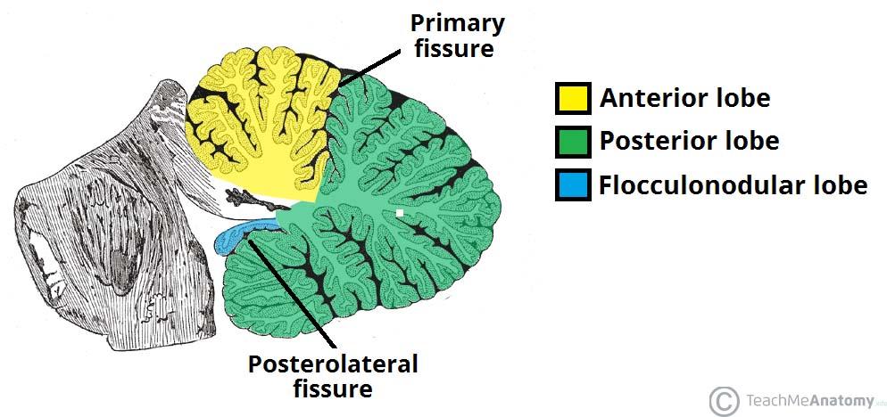
 Flocculonodular lobe between posterior and flocculonodular lobe")
18 Cerebellum: Gross Anatomy Three main lobes Anterior lobe Primary fissure Posterior lobe (middle lobe) Cerebellar tonsils between anterior and posterior lobe the largest lobe Posterolateral fissure (uvulonodular fissure) Flocculonodular lobe between posterior and flocculonodular lobe
19
20 Cerebellum: Gross Anatomy
 & central nuclei are")

21 Cerebellum: Internal Structure Content Cerebellar cortex (folia) & central nuclei are grey matter Arbor vitae = tree of life = white matter the white matter also called arbor vitae or tree of life

22 Cerebellar Anatomy Cerebellum includes a cortex & deep nuclei The deep nuclei are the major source of output from the cerebellum Four nuclei from medial to lateral Fastigial Globose Emboliform Dentate Interposed nuclei the white matter contains 4 nuclei, the fastigial which is the most medial one, Globose and Emboliform called interposed nuclei because they have the same function, and the most lateral one which is Dentate nucleus " Not dentate gyrus!!" these nuclei, mostly the efferent will exit from them
\" it is inhibitory so")
23 Cerebellar cortex includes 5 cell types in 3 layers Five cell types Inhibitory cells Purkinje, basket, Golgi, and stellate cells Excitatory cells Granule cells Three layers Molecular layer Purkinje cell layer Granule cell layer cerebellar cortex efferent is the purkinje, the other layers affect the purkinje Cerebellar Cortex purkinje cells " which is the output (efferent)" it is inhibitory so if you inhibit it, overall you are exciting :)
24 Climbing fibers directly affect purkinje. Mossy fibers affect other cells, where these other cells affect purkinje as we said. Inputs to the cerebellum Climbing fibers From inferior olivary complex (olivocerebellar fibers) Decussate Inferior cerebellar peduncle Mossy fibers All remaining inputs: spinal cord, vestibular n. & nuclei, & pontine nuclei Each type of input fibers branches Branch to deep nuclei Branch to cerebellar cortex Cerebellar Inputs
25 Cerebellar Circuit The basic cerebellar circuit includes Main excitatory loop Inhibitory cortical side loop
26 The Main Excitatory Loop Includes the input and the deep cerebellar nuclei Both the inputs & the cells of the deep nuclei are excitatory
27 The Inhibitory Cortical Side Loop Serves to modulate the activity in the deep cerebellar nuclei Mossy & climbing fibers are inputs to cerebellar cortex Climbing fibers contact Purkinje cells directly Mossy fibers contact granule cells Granule cells contact Purkinje cells Output of cerebellar cortex (Purkinje fibers) depend on the mossy & climbing fibers
28 The Inhibitory Cortical Side Loop Remaining cells (Golgi, basket & stellate) are inhibitory interneurons Alter granule & Purkinje cells Purkinje cells (cerebellar cortex output) are inhibitory Purkinje cells targets deep cerebellar nuclei & vestibular nuclei Thus cerebellar output is driven by the main excitatory loop and limited by the inhibitory cortical side loop
 Motor execution 3. Cerebrocerebellum Lateral hemispheres & dentate nu.")
29 Cerebellar Functional Divisions 1. Vestibulocerebellum Flocculonodular lobe & fastigial nu. Balance, eye movements 2. Spinocerebellum Vermis & paravermal parts of hemispheres & interposed nuclei (emboliform & globose) Motor execution 3. Cerebrocerebellum Lateral hemispheres & dentate nu. Motor planning cerebellum functionally divided to 3 parts
 Vestibular nuclei Reticular formation VL of thalamus Part of motor system targeted UMNs of medial pathway Major signs of damage Staggering or falling,")
30 Vestibulocerebellum Function Balance & eye movements Inputs Vestibular n. fibers Vestibular nuclei Inferior olive Deep nucleus Fastigial nucleus Outputs (From fastigial nu. & Purkinje cells) Vestibular nuclei Reticular formation VL of thalamus Part of motor system targeted UMNs of medial pathway Major signs of damage Staggering or falling, nystagmus

31 Function Spinocerebellum Execution of movement Compensates for changes in load, regulates muscle tone, guides limb movement, helps maintain posture Organized somatotopically Head & trunk vermis Limbs paravermal areas Inputs Spinal & trigeminal inputs Inferior olive Deep nucleus Fastigial & interposed nuclei Outputs Vermis Reticular formation & Vestibular nu. Paravermal Red nucleus, VL of thalamus & Inferior olive Part of motor system targeted UMNs of medial & lateral pathways Major signs of damage Staggering gait, intention tremor
 VL of thalamus Part of motor system targeted Motor cortex (via VL) ventrolateral nucleus Major signs of damage Decomposition of")
32 Cerebrocerebellum Function Coordination, planning of voluntary movements Inputs Pontine nuclei (relaying information from sensory & motor cerebral cortex) Inferior olive Deep nucleus Dentate nucleus Outputs Red nucleus (to inferior olive, back to cerebellum) VL of thalamus Part of motor system targeted Motor cortex (via VL) ventrolateral nucleus Major signs of damage Decomposition of movements
33 as we know there are 3 cerebellar peduncles, every one has it's own contents " inputs and outputs" Cerebellar Peduncles divided to 2 parts Peduncle Inferior Restiform body Juxtarestiform body Major inputs to cerebellum Fibers from: Inferior olive (climbing fibers) Dorsal spinocerebellar tract Cuneocerebellar tract Vestibular nerve Vestibular nuclei Major outputs from cerebellum Fibers to: Vestibular nuclei Middle (brachium pontis) Pontine nuclei (relay inputs from cerebral cortex) None Superior (brachium conjunctivum) Ventral spinocerebellar Rostral spinocerebellar Red nucleus VL thalamus Reticular formation Inferior olive
34 Cerebellar Circuitry Cerebra l Cortex Motor cortex SCP V A V L Thalamus Red nu. Inferior olive Retic. Form. Sup. Colliculus Vestibular Nu. UMNs Lateral system Medial system Spinal cord & Brainstem γ α Pontine nuclei MCP Deep nuclei Cortex + + Cerebellum ICP Vestibular nerve Intrafusal Extrafusal

35 Blood Supply of Cerebellum SCA Superior cerebellar hemispheres Superior vermis Dentate nucleus Most of white matter Superior cerebellar peduncle AICA Middle cerebellar peduncle Flocculus Anteroinferior surface of the cerebellum PICA Posteroinferior cerebellar hemispheres Inferior portion of the vermis Inferior cerebellar peduncle
36 Motor System Intralaminar nu. Of thalamus SN compacta Basal Ganglia Globus pallidus Cerebral Cortex Glu + Striatum GABA GABA GPe Gpi SNr GABA Glu Subthalamic nucleus Motor cortex Glu VA VL Dorsal thalamus Red nu. GABA To other UMNs: Retic. Form. & Sup. Coll. Upper Motor Neurons Spinal cord & Brainstem SCP Inferior olive Retic. Form. Sup. Colliculus Vestibular Nu. γ α Pontine nuclei MCP Deep nuclei Cortex + + Cerebellum ICP Vestibular nerve Intrafusal Extrafusal
Lecture : Basal ganglia & Cerebellum By : Zaid Al-Ghnaneem
 Lecture : Basal ganglia & Cerebellum By : Zaid Al-Ghnaneem Some notes in the beginning : #1 : there is a slides file contains the sheet info as notes for those who love slides more than word papers. #2
Motor System Hierarchy
 Motor Pathways Lectures Objectives Define the terms upper and lower motor neurons with examples. Describe the corticospinal (pyramidal) tract and the direct motor pathways from the cortex to the trunk
Located below tentorium cerebelli within posterior cranial fossa. Formed of 2 hemispheres connected by the vermis in midline.
 The Cerebellum Cerebellum Located below tentorium cerebelli within posterior cranial fossa. Formed of 2 hemispheres connected by the vermis in midline. Gray matter is external. White matter is internal,
Medial View of Cerebellum
 Meds 5371 System Neuroscience D. L. Oliver CEREBELLUM Anterior lobe (spinal) Posterior lobe (cerebral) Flocculonodular lobe (vestibular) Medial View of Cerebellum 1 Ventral View of Cerebellum Flocculus
Cerebellum. Steven McLoon Department of Neuroscience University of Minnesota
 Cerebellum Steven McLoon Department of Neuroscience University of Minnesota 1 Anatomy of the Cerebellum The cerebellum has approximately half of all the neurons in the central nervous system. The cerebellum
Cerebellum John T. Povlishock, Ph.D.
 Cerebellum John T. Povlishock, Ph.D. OBJECTIVES 1. To identify the major sources of afferent inputs to the cerebellum 2. To define the pre-cerebellar nuclei from which the mossy and climbing fiber systems
1/2/2019. Basal Ganglia & Cerebellum a quick overview. Outcomes you want to accomplish. MHD-Neuroanatomy Neuroscience Block. Basal ganglia review
 This power point is made available as an educational resource or study aid for your use only. This presentation may not be duplicated for others and should not be redistributed or posted anywhere on the
BASAL GANGLIA. Dr JAMILA EL MEDANY
 BASAL GANGLIA Dr JAMILA EL MEDANY OBJECTIVES At the end of the lecture, the student should be able to: Define basal ganglia and enumerate its components. Enumerate parts of Corpus Striatum and their important
The Cerebellum. Little Brain. Neuroscience Lecture. Dr. Laura Georgescu
 The Cerebellum Little Brain Neuroscience Lecture Dr. Laura Georgescu Learning Objectives 1. Describe functional anatomy of the cerebellum- its lobes, their input and output connections and their functions.
Teach-SHEET Basal Ganglia
 Teach-SHEET Basal Ganglia Purves D, et al. Neuroscience, 5 th Ed., Sinauer Associates, 2012 Common organizational principles Basic Circuits or Loops: Motor loop concerned with learned movements (scaling
The Cerebellum. The Little Brain. Neuroscience Lecture. PhD Candidate Dr. Laura Georgescu
 The Cerebellum The Little Brain Neuroscience Lecture PhD Candidate Dr. Laura Georgescu Learning Objectives 1. Describe functional anatomy of the cerebellum - its lobes, their input and output connections
Basal nuclei, cerebellum and movement
 Basal nuclei, cerebellum and movement MSTN121 - Neurophysiology Session 9 Department of Myotherapy Basal Nuclei (Ganglia) Basal Nuclei (Ganglia) Role: Predict the effects of various actions, then make
Unit VIII Problem 5 Physiology: Cerebellum
 Unit VIII Problem 5 Physiology: Cerebellum - The word cerebellum means: the small brain. Note that the cerebellum is not completely separated into 2 hemispheres (they are not clearly demarcated) the vermis
Voluntary Movement. Ch. 14: Supplemental Images
 Voluntary Movement Ch. 14: Supplemental Images Skeletal Motor Unit: The basics Upper motor neuron: Neurons that supply input to lower motor neurons. Lower motor neuron: neuron that innervates muscles,
Abdullah AlZibdeh. Dr. Maha ElBeltagy. Maha ElBeltagy
 19 Abdullah AlZibdeh Dr. Maha ElBeltagy Maha ElBeltagy Introduction In this sheet, we discuss the cerebellum; its lobes, fissures and deep nuclei. We also go into the tracts and connections in which the
For more information about how to cite these materials visit
 Author(s): Peter Hitchcock, PH.D., 2009 License: Unless otherwise noted, this material is made available under the terms of the Creative Commons Attribution Non-commercial Share Alike 3.0 License: http://creativecommons.org/licenses/by-nc-sa/3.0/
The Cerebellum. Outline. Lu Chen, Ph.D. MCB, UC Berkeley. Overview Structure Micro-circuitry of the cerebellum The cerebellum and motor learning
 The Cerebellum Lu Chen, Ph.D. MCB, UC Berkeley 1 Outline Overview Structure Micro-circuitry of the cerebellum The cerebellum and motor learning 2 Overview Little brain 10% of the total volume of the brain,
Connection of the cerebellum
 CEREBELLUM Connection of the cerebellum The cerebellum has external layer of gray matter (cerebellar cortex ), & inner white matter In the white matter, there are 3 deep nuclei : (a) dentate nucleus laterally
The Cerebellum. Outline. Overview Structure (external & internal) Micro-circuitry of the cerebellum Cerebellum and motor learning
 Micro-circuitry of the cerebellum Cerebellum and motor learning") The Cerebellum P.T Ji Jun Cheol Rehabilitation Center 1 HansarangAsan Hospital. Outline Overview Structure (external & internal) Micro-circuitry of the cerebellum Cerebellum and motor learning 2 1 Cerebellum
The Cerebellum P.T Ji Jun Cheol Rehabilitation Center 1 HansarangAsan Hospital. Outline Overview Structure (external & internal) Micro-circuitry of the cerebellum Cerebellum and motor learning 2 1 Cerebellum
A. General features of the basal ganglia, one of our 3 major motor control centers:
 Reading: Waxman pp. 141-146 are not very helpful! Computer Resources: HyperBrain, Chapter 12 Dental Neuroanatomy Suzanne S. Stensaas, Ph.D. April 22, 2010 THE BASAL GANGLIA Objectives: 1. What are the
CASE 48. What part of the cerebellum is responsible for planning and initiation of movement?
 CASE 48 A 34-year-old woman with a long-standing history of seizure disorder presents to her neurologist with difficulty walking and coordination. She has been on phenytoin for several days after having
MODULE 6: CEREBELLUM AND BASAL GANGLIA
 MODULE 6: CEREBELLUM AND BASAL GANGLIA This module will summarize the important neuroanatomical and key clinical concepts from Chapters 15 and 16 of the textbook for the course. The first part of this
A. General features of the basal ganglia, one of our 3 major motor control centers:
 Reading: Waxman pp. 141-146 are not very helpful! Computer Resources: HyperBrain, Chapter 12 Dental Neuroanatomy Suzanne S. Stensaas, Ph.D. March 1, 2012 THE BASAL GANGLIA Objectives: 1. What are the main
Developmental sequence of brain
 Cerebellum Developmental sequence of brain Fourth week Fifth week Location of cerebellum Lies above and behind the medullar and pons and occupies posterior cranial fossa Location of cerebellum External
Making Things Happen 2: Motor Disorders
 Making Things Happen 2: Motor Disorders How Your Brain Works Prof. Jan Schnupp wschnupp@cityu.edu.hk HowYourBrainWorks.net On the Menu in This Lecture In the previous lecture we saw how motor cortex and
Cerebellum: little brain. Cerebellum. gross divisions
 Cerebellum The anatomy of the cerebellum and its gross divisions Its principal input and output pathways The organization of the cerebellar cortex Role of climbing vs. mossy fibre input The parallel-fibre/
Strick Lecture 3 March 22, 2017 Page 1
 Strick Lecture 3 March 22, 2017 Page 1 Cerebellum OUTLINE I. External structure- Inputs and Outputs Cerebellum - (summary diagram) 2 components (cortex and deep nuclei)- (diagram) 3 Sagittal zones (vermal,
Cerebellum: little brain. Cerebellum. gross divisions
 Cerebellum The anatomy of the cerebellum and its gross divisions Its principal input and output pathways The organization of the cerebellar cortex Role of climbing vs. mossy fibre input The parallel-fibre/
COGNITIVE SCIENCE 107A. Motor Systems: Basal Ganglia. Jaime A. Pineda, Ph.D.
 COGNITIVE SCIENCE 107A Motor Systems: Basal Ganglia Jaime A. Pineda, Ph.D. Two major descending s Pyramidal vs. extrapyramidal Motor cortex Pyramidal system Pathway for voluntary movement Most fibers originate
FUNCTION: It COORDINATES movement HOW IT WORKS
 CEREBELLUM Chris Cohan, Ph.D. Dept. of Pathology/Anat Sci University at Buffalo Objectives: Describe the anatomy of the cerebellum, its 3 functions and associated regions Describe how the cerebellum influences
Ch 13: Central Nervous System Part 1: The Brain p 374
 Ch 13: Central Nervous System Part 1: The Brain p 374 Discuss the organization of the brain, including the major structures and how they relate to one another! Review the meninges of the spinal cord and
The Nervous System: Sensory and Motor Tracts of the Spinal Cord
 15 The Nervous System: Sensory and Motor Tracts of the Spinal Cord PowerPoint Lecture Presentations prepared by Steven Bassett Southeast Community College Lincoln, Nebraska Introduction Millions of sensory
For more information about how to cite these materials visit
 Author(s): Peter Hitchcock, PH.D., 2009 License: Unless otherwise noted, this material is made available under the terms of the Creative Commons Attribution Non-commercial Share Alike 3.0 License: http://creativecommons.org/licenses/by-nc-sa/3.0/
Copy Right- Hongqi ZHANG-Department of Anatomy-Fudan University. Systematic Anatomy. Nervous system Cerebellum. Dr.Hongqi Zhang ( 张红旗 )
") Systematic Anatomy Nervous system Cerebellum Dr.Hongqi Zhang ( 张红旗 ) Email: zhanghq58@126.com 1 The Cerebellum Cerebellum evolved and developed with the complication of animal movement. Key points about
Systematic Anatomy Nervous system Cerebellum Dr.Hongqi Zhang ( 张红旗 ) Email: zhanghq58@126.com 1 The Cerebellum Cerebellum evolved and developed with the complication of animal movement. Key points about
Brain anatomy and artificial intelligence. L. Andrew Coward Australian National University, Canberra, ACT 0200, Australia
 Brain anatomy and artificial intelligence L. Andrew Coward Australian National University, Canberra, ACT 0200, Australia The Fourth Conference on Artificial General Intelligence August 2011 Architectures
Biological Bases of Behavior. 8: Control of Movement
 Biological Bases of Behavior 8: Control of Movement m d Skeletal Muscle Movements of our body are accomplished by contraction of the skeletal muscles Flexion: contraction of a flexor muscle draws in a
Basal Ganglia George R. Leichnetz, Ph.D.
 Basal Ganglia George R. Leichnetz, Ph.D. OBJECTIVES 1. To understand the brain structures which constitute the basal ganglia, and their interconnections 2. To understand the consequences (clinical manifestations)
Computational cognitive neuroscience: 8. Motor Control and Reinforcement Learning
 1 Computational cognitive neuroscience: 8. Motor Control and Reinforcement Learning Lubica Beňušková Centre for Cognitive Science, FMFI Comenius University in Bratislava 2 Sensory-motor loop The essence
Damage on one side.. (Notes) Just remember: Unilateral damage to basal ganglia causes contralateral symptoms.
 Just remember: Unilateral damage to basal ganglia causes contralateral symptoms.") Lecture 20 - Basal Ganglia Basal Ganglia (Nolte 5 th Ed pp 464) Damage to the basal ganglia produces involuntary movements. Although the basal ganglia do not influence LMN directly (to cause this involuntary
Lecture 20 - Basal Ganglia Basal Ganglia (Nolte 5 th Ed pp 464) Damage to the basal ganglia produces involuntary movements. Although the basal ganglia do not influence LMN directly (to cause this involuntary
Anatomy of the basal ganglia. Dana Cohen Gonda Brain Research Center, room 410
 Anatomy of the basal ganglia Dana Cohen Gonda Brain Research Center, room 410 danacoh@gmail.com The basal ganglia The nuclei form a small minority of the brain s neuronal population. Little is known about
PETER PAZMANY CATHOLIC UNIVERSITY Consortium members SEMMELWEIS UNIVERSITY, DIALOG CAMPUS PUBLISHER
 PETER PAZMANY CATHOLIC UNIVERSITY SEMMELWEIS UNIVERSITY Development of Complex Curricula for Molecular Bionics and Infobionics Programs within a consortial* framework** Consortium leader PETER PAZMANY
Organization of Motor Functions 4.
 Organization of Motor Functions 4. Dr. Attila Nagy 2018 Sensory-motor system Limbic cortex Structure Subcortical Motivational sub areas Frontal cortex Task Motivation Sequence Plan Tim e Ascending system
THE CEREBELLUM SUDIVISIONS, STRUCTURE AND CONNECTIONS
 THE CEREBELLUM Damage to the cerebellum produces characteristic symptoms primarily with respect to the coordination of voluntary movements. The cerebellum receives information from the skin, joints, muscles,
A3.1.7 Motor Control. 10 November 2016 Institute of Psychiatry,Psychology and Neuroscience Marinela Vavla
 A3.1.7 Motor Control 10 November 2016 Institute of Psychiatry,Psychology and Neuroscience Marinela Vavla marinela.vavla@kcl.ac.uk Learning objectives Motor systems: components & organization Spinal cord
Brainstem. By Dr. Bhushan R. Kavimandan
 Brainstem By Dr. Bhushan R. Kavimandan Development Ventricles in brainstem Mesencephalon cerebral aqueduct Metencephalon 4 th ventricle Mylencephalon 4 th ventricle Corpus callosum Posterior commissure
Brainstem. Steven McLoon Department of Neuroscience University of Minnesota
 Brainstem Steven McLoon Department of Neuroscience University of Minnesota 1 Course News Change in Lab Sequence Week of Oct 2 Lab 5 Week of Oct 9 Lab 4 2 Goal Today Know the regions of the brainstem. Know
I: To describe the pyramidal and extrapyramidal tracts. II: To discuss the functions of the descending tracts.
 Descending Tracts I: To describe the pyramidal and extrapyramidal tracts. II: To discuss the functions of the descending tracts. III: To define the upper and the lower motor neurons. 1. The corticonuclear
Basal ganglia Sujata Sofat, class of 2009
 Basal ganglia Sujata Sofat, class of 2009 Basal ganglia Objectives Describe the function of the Basal Ganglia in movement Define the BG components and their locations Describe the motor loop of the BG
Faculty of Dental Medicine and Surgery. Sem 4 Cerebellum Dr. Abbas
 Faculty of Dental Medicine and Surgery Sem 4 Cerebellum Dr. Abbas Anatomy of the cerebellum Cerebellum Configurations External - located in posterior cranial fossa - communicate with other structure via
Basal Ganglia. Steven McLoon Department of Neuroscience University of Minnesota
 Basal Ganglia Steven McLoon Department of Neuroscience University of Minnesota 1 Course News Graduate School Discussion Wednesday, Nov 1, 11:00am MoosT 2-690 with Paul Mermelstein (invite your friends)
The Wonders of the Basal Ganglia
 Basal Ganglia The Wonders of the Basal Ganglia by Mackenzie Breton and Laura Strong /// https://kin450- neurophysiology.wikispaces.com/basal+ganglia Introduction The basal ganglia are a group of nuclei
Functional Distinctions
 Functional Distinctions FUNCTION COMPONENT DEFICITS Start Basal Ganglia Spontaneous Movements Move UMN/LMN Cerebral Cortex Brainstem, Spinal cord Roots/peripheral nerves Plan Cerebellum Ataxia Adjust Cerebellum
Chapter 8. Control of movement
 Chapter 8 Control of movement 1st Type: Skeletal Muscle Skeletal Muscle: Ones that moves us Muscles contract, limb flex Flexion: a movement of a limb that tends to bend its joints, contraction of a flexor
b. The groove between the two crests is called 2. The neural folds move toward each other & the fuse to create a
 Chapter 13: Brain and Cranial Nerves I. Development of the CNS A. The CNS begins as a flat plate called the B. The process proceeds as: 1. The lateral sides of the become elevated as waves called a. The
Basal Ganglia. Introduction. Basal Ganglia at a Glance. Role of the BG
 Basal Ganglia Shepherd (2004) Chapter 9 Charles J. Wilson Instructor: Yoonsuck Choe; CPSC 644 Cortical Networks Introduction A set of nuclei in the forebrain and midbrain area in mammals, birds, and reptiles.
THE CEREBELLUM. - anatomy of the cerebellum cerebellar nuclei cerebellar inputs and neuronal structure of the Purkinje cells outputs cerebellum
 CHAPTER THE CEREBELLUM Key Terms - anatomy of the cerebellum cerebellar nuclei cerebellar inputs and neuronal structure of the Purkinje cells outputs cerebellum cerebellar disorders Figure 14.9 For each
1. The cerebellum coordinates fine movement through interactions with the following motor-associated areas:
 DENT/OBHS 131 2009 Take-home test 4 Week 6: Take-home test (2/11/09 close 2/18/09) 1. The cerebellum coordinates fine movement through interactions with the following motor-associated areas: Hypothalamus
PETER PAZMANY CATHOLIC UNIVERSITY Consortium members SEMMELWEIS UNIVERSITY, DIALOG CAMPUS PUBLISHER
 PETER PAZMANY CATHOLIC UNIVERSITY SEMMELWEIS UNIVERSITY Development of Complex Curricula for Molecular Bionics and Infobionics Programs within a consortial* framework** Consortium leader PETER PAZMANY
CN V! touch! pain! Touch! P/T!
 CN V! touch! pain! Touch! P/T! Visual Pathways! L! R! B! A! C! D! LT! E! F! RT! G! hypothalamospinal! and! ALS! Vestibular Pathways! 1. Posture/Balance!!falling! 2. Head Position! 3. Eye-Head Movements
Cerebellum 1/20/2016. Outcomes you need to be able to demonstrate. MHD Neuroanatomy Module
 This power point is made available as an educational resource or study aid for your use only. This presentation may not be duplicated for others and should not be redistributed or posted anywhere on the
Brainstem: Midbrain. 1. Midbrain gross external anatomy 2. Internal structure of the midbrain:
 Brainstem: Midbrain 1. Midbrain gross external anatomy 2. Internal structure of the midbrain: cerebral peduncles tegmentum tectum (guadrigeminal plate) Midbrain Midbrain general features location between
Dr. Farah Nabil Abbas. MBChB, MSc, PhD
 Dr. Farah Nabil Abbas MBChB, MSc, PhD The Basal Ganglia *Functions in association with motor cortex and corticospinal pathways. *Regarded as accessory motor system besides cerebellum. *Receive most of
The Embryology and Anatomy of the Cerebellum
 The Embryology and Anatomy of the Cerebellum Maryam Rahimi Balaei, Niloufar Ashtari, and Hugo Bergen Abstract The cerebellum is an important structure in the central nervous system that controls and regulates
Anatomy and Physiology (Bio 220) The Brain Chapter 14 and select portions of Chapter 16
 The Brain Chapter 14 and select portions of Chapter 16") Anatomy and Physiology (Bio 220) The Brain Chapter 14 and select portions of Chapter 16 I. Introduction A. Appearance 1. physical 2. weight 3. relative weight B. Major parts of the brain 1. cerebrum 2.
Anatomy and Physiology (Bio 220) The Brain Chapter 14 and select portions of Chapter 16 I. Introduction A. Appearance 1. physical 2. weight 3. relative weight B. Major parts of the brain 1. cerebrum 2.
Introduction to the Central Nervous System: Internal Structure
 Introduction to the Central Nervous System: Internal Structure Objective To understand, in general terms, the internal organization of the brain and spinal cord. To understand the 3-dimensional organization
Connections of basal ganglia
 Connections of basal ganglia Introduction The basal ganglia, or basal nuclei, are areas of subcortical grey matter that play a prominent role in modulating movement, as well as cognitive and emotional
VL VA BASAL GANGLIA. FUNCTIONAl COMPONENTS. Function Component Deficits Start/initiation Basal Ganglia Spontan movements
 BASAL GANGLIA Chris Cohan, Ph.D. Dept. of Pathology/Anat Sci University at Buffalo I) Overview How do Basal Ganglia affect movement Basal ganglia enhance cortical motor activity and facilitate movement.
Brain Stem and cortical control of motor function. Dr Z Akbari
 Brain Stem and cortical control of motor function Dr Z Akbari Brain stem control of movement BS nuclear groups give rise to descending motor tracts that influence motor neurons and their associated interneurons
Nsci 2100: Human Neuroanatomy 2017 Examination 3
 Name KEY Lab Section Nsci 2100: Human Neuroanatomy 2017 Examination 3 On this page, write your name and lab section. On your bubble answer sheet, enter your name (last name, space, first name), internet
Brainstem. Amadi O. Ihunwo, PhD School of Anatomical Sciences
 Brainstem Amadi O. Ihunwo, PhD School of Anatomical Sciences Lecture Outline Constituents Basic general internal features of brainstem External and Internal features of Midbrain Pons Medulla Constituents
Motor systems III: Cerebellum April 16, 2007 Mu-ming Poo
 Motor systems III: Cerebellum April 16, 2007 Mu-ming Poo Population coding in the motor cortex Overview and structure of cerebellum Microcircuitry of cerebellum Function of cerebellum -- vestibulo-ocular
CNS consists of brain and spinal cord PNS consists of nerves
 CNS consists of brain and spinal cord PNS consists of nerves 1 As with sensory input, motor output is organized in central nervous system Peripheral Nervous system divides efferent signals somatotopically
Prof. Saeed Abuel Makarem & Dr.Sanaa Alshaarawy
 Prof. Saeed Abuel Makarem & Dr.Sanaa Alshaarawy 1 Objectives By the end of the lecture, you should be able to: Describe the anatomy and main functions of the thalamus. Name and identify different nuclei
skilled pathways: distal somatic muscles (fingers, hands) (brainstem, cortex) are giving excitatory signals to the descending pathway
 (brainstem, cortex) are giving excitatory signals to the descending pathway") L15 - Motor Cortex General - descending pathways: how we control our body - motor = somatic muscles and movement (it is a descending motor output pathway) - two types of movement: goal-driven/voluntary
L15 - Motor Cortex General - descending pathways: how we control our body - motor = somatic muscles and movement (it is a descending motor output pathway) - two types of movement: goal-driven/voluntary
General Sensory Pathways of the Trunk and Limbs
 General Sensory Pathways of the Trunk and Limbs Lecture Objectives Describe gracile and cuneate tracts and pathways for conscious proprioception, touch, pressure and vibration from the limbs and trunk.
I. Anatomy of the Brain A. Cranial Meninges and Ventricles of the Brain 1. Meninges a. Dura mater 1) Endosteal/Periosteal Layer - Outer 2) Meningeal
 Endosteal/Periosteal Layer - Outer 2) Meningeal") I. Anatomy of the Brain A. Cranial Meninges and Ventricles of the Brain 1. Meninges a. Dura mater 1) Endosteal/Periosteal Layer - Outer 2) Meningeal Layer - Inner 3) Falx cerebri a) Superior sagittal sinus
I. Anatomy of the Brain A. Cranial Meninges and Ventricles of the Brain 1. Meninges a. Dura mater 1) Endosteal/Periosteal Layer - Outer 2) Meningeal Layer - Inner 3) Falx cerebri a) Superior sagittal sinus
The basal forebrain: Questions, chapter 29:
 The basal forebrain: Questions, chapter 29: 7) What is the "basal forebrain", and what is its involvement in Alzheimer' s Disease? The acetylcholine-containing neurons of the nucleus basalis of Meynart
By Dr. Saeed Vohra & Dr. Sanaa Alshaarawy
 By Dr. Saeed Vohra & Dr. Sanaa Alshaarawy 1 By the end of the lecture, students will be able to : Distinguish the internal structure of the components of the brain stem in different levels and the specific
Basal Ganglia. Today s lecture is about Basal Ganglia and it covers:
 Basal Ganglia Motor system is complex interaction between Lower motor neurons (spinal cord and brainstem circuits) and Upper motor neurons (pyramidal and extrapyramidal tracts) plus two main regulators
 Spinal Cord Tracts DESCENDING SPINAL TRACTS: Are concerned with somatic motor function, modification of ms. tone, visceral innervation, segmental reflexes. Main tracts arise form cerebral cortex and others
The neurvous system senses, interprets, and responds to changes in the environment. Two types of cells makes this possible:
 NERVOUS SYSTEM The neurvous system senses, interprets, and responds to changes in the environment. Two types of cells makes this possible: the neuron and the supporting cells ("glial cells"). Neuron Neurons
GBME graduate course. Chapter 43. The Basal Ganglia
 GBME graduate course Chapter 43. The Basal Ganglia Basal ganglia in history Parkinson s disease Huntington s disease Parkinson s disease 1817 Parkinson's disease (PD) is a degenerative disorder of the
Basal Ganglia General Info
 Basal Ganglia General Info Neural clusters in peripheral nervous system are ganglia. In the central nervous system, they are called nuclei. Should be called Basal Nuclei but usually called Basal Ganglia.
Systems Neuroscience Dan Kiper. Today: Wolfger von der Behrens
 Systems Neuroscience Dan Kiper Today: Wolfger von der Behrens wolfger@ini.ethz.ch 18.9.2018 Neurons Pyramidal neuron by Santiago Ramón y Cajal (1852-1934, Nobel prize with Camillo Golgi in 1906) Neurons
神經解剖學 NEUROANATOMY BASAL NUCLEI 盧家鋒助理教授臺北醫學大學醫學系解剖學暨細胞生物學科臺北醫學大學醫學院轉譯影像研究中心.
 神經解剖學 NEUROANATOMY BASAL NUCLEI 盧家鋒助理教授臺北醫學大學醫學系解剖學暨細胞生物學科臺北醫學大學醫學院轉譯影像研究中心 http://www.ym.edu.tw/~cflu OUTLINE Components and Pathways of the Basal Nuclei Functions and Related Disorders of the Basal Nuclei
Motor Functions of Cerebral Cortex
 Motor Functions of Cerebral Cortex I: To list the functions of different cortical laminae II: To describe the four motor areas of the cerebral cortex. III: To discuss the functions and dysfunctions of
DEVELOPMENT OF BRAIN
 Ahmed Fathalla OBJECTIVES At the end of the lecture, students should: List the components of brain stem. Describe the site of brain stem. Describe the relations between components of brain stem & their
Biological Bases of Behavior. 3: Structure of the Nervous System
 Biological Bases of Behavior 3: Structure of the Nervous System Neuroanatomy Terms The neuraxis is an imaginary line drawn through the spinal cord up to the front of the brain Anatomical directions are
Neuroanatomy Dr. Maha ELBeltagy Assistant Professor of Anatomy Faculty of Medicine The University of Jordan 2018
 Neuroanatomy Dr. Maha ELBeltagy Assistant Professor of Anatomy Faculty of Medicine The University of Jordan 2018 Dr Maha ELbeltagy THE BRAIN STEM Dr Maha ELbeltagy It includes: Midbrain - Pons - Medulla
Gross Morphology of the Brain
 Gross Morphology of the Brain Done by : Marah Marahleh & Razan Krishan *slides in bold Principal Parts of the Brain Cerebrum : largest part of the brain Diencephalon Thalamus & hypothalamus Cerebellum
Strick Lecture 4 March 29, 2006 Page 1
 Strick Lecture 4 March 29, 2006 Page 1 Basal Ganglia OUTLINE- I. Structures included in the basal ganglia II. III. IV. Skeleton diagram of Basal Ganglia Loops with cortex Similarity with Cerebellar Loops
NS219: Basal Ganglia Anatomy
 NS219: Basal Ganglia Anatomy Human basal ganglia anatomy Analagous rodent basal ganglia nuclei Basal ganglia circuits: the classical model of direct and indirect pathways + Glutamate + - GABA - Gross anatomy
Psyc 311A, fall 2008 Conference week 3 TA: Jürgen Germann
 Psyc 311A, fall 2008 Conference week 3 TA: Jürgen Germann e-mail: jurgen.germann@mcgill.ca Overview: 1. Meninges 2. Cerebral cortex-cytoarchitecture 3. Diencephalon (thalamus/hypothalamus) (this replaces
SENSORY (ASCENDING) SPINAL TRACTS
 SPINAL TRACTS") SENSORY (ASCENDING) SPINAL TRACTS Dr. Jamila El-Medany Dr. Essam Eldin Salama OBJECTIVES By the end of the lecture, the student will be able to: Define the meaning of a tract. Distinguish between the different
SENSORY (ASCENDING) SPINAL TRACTS Dr. Jamila El-Medany Dr. Essam Eldin Salama OBJECTIVES By the end of the lecture, the student will be able to: Define the meaning of a tract. Distinguish between the different
Announcement. Danny to schedule a time if you are interested.
 Announcement If you need more experiments to participate in, contact Danny Sanchez (dsanchez@ucsd.edu) make sure to tell him that you are from LIGN171, so he will let me know about your credit (1 point).
Chapter 3. Structure and Function of the Nervous System. Copyright (c) Allyn and Bacon 2004
 Allyn and Bacon 2004") Chapter 3 Structure and Function of the Nervous System 1 Basic Features of the Nervous System Neuraxis: An imaginary line drawn through the center of the length of the central nervous system, from the
Chapter 3 Structure and Function of the Nervous System 1 Basic Features of the Nervous System Neuraxis: An imaginary line drawn through the center of the length of the central nervous system, from the
The Central Nervous System I. Chapter 12
 The Central Nervous System I Chapter 12 The Central Nervous System The Brain and Spinal Cord Contained within the Axial Skeleton Brain Regions and Organization Medical Scheme (4 regions) 1. Cerebral Hemispheres
Functions. Traditional view: Motor system. Co-ordination of movements Motor learning Eye movements. Modern view: Cognition
 The Cerebellum Involved in motor coordination and timing Is simple and well documented Only has one type of output cell (Purkinje) The cerebellum influences motor activity through inhibition The Cerebellum
Chapter 14: Integration of Nervous System Functions I. Sensation.
 Chapter 14: Integration of Nervous System Functions I. Sensation A. General Organization 1. General senses have receptors a. The somatic senses provide information about & 1. Somatic senses include: a.