Functional Imaging with Positron Emission Tomography. Jeih San Liow, PhD Molecular Imaging Branch National Institute Mental Health
|
|
|
- Merry Gray
- 5 years ago
- Views:
Transcription

1 Functional Imaging with Positron Emission Tomography Jeih San Liow, PhD Molecular Imaging Branch National Institute Mental Health
2 Outlines What is positron emission tomography (PET) Applications Imaging glucose metabolism Imaging transporter function at the blood brain barrier Imaging neuroimflammation Other targets
3 What is PET? Positron Emission Tomography Produce positron emitted radioactive isotopes Incorporate isotopes into compounds through chemical synthesis Inject compound into human body and image functionality

4 Positron Emission Tomography
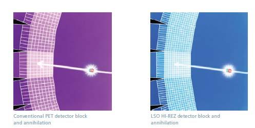
5 Coincidence Imaging

6 Cyclotron
7 Common PET Isotopes Half Life (Min) Compounds 15 O 2 Water 13 N 10 NH3 11 C 20 CO, CO 2, Acetate 18 F 110 FDG, FDOPA
8 Radioligand = Drug + Radioactivity Drug administered at tracer doses No pharm effects Labels <1% receptors Labeled subset reflects entire population Radioligand disposed like all drugs Metabolism & distribution Radiation exposure
9 PET vs. MRI Spatial Resolution PET MRI 2 6 mm << 1 mm Sensitivity M 10-4 M Temporal Resolution minutes <1 sec Radionuclide ( 11 C): high sensitivity Ligand (raclopride): high selectivity Radioligand [ 11 C]raclopride: high sensitivity & selectivity

10

11 2D vs. 3D Acquisition

12 Random, scatter and true coincidence Scatter coincidence True coincidence Random coincidence
13 PET Image Formation Correction for error sources in the projection data Detector Normalization Random Correction Scatter Correction Attenuation Correction Decay correction Reconstruction Filter Backprojection (FBP) Order-subset Maximum Likelihood EM (OSEM)
14 Purpose of a CT scan in the PET/CT examination To perform attenuation correction To provide anatomical reference for localizing target of interest

15 Filtered Backprojection Reconstruction

16 Qunatification of PET images through compartmental models Free + nonspecific Specific
 PET scanner")


17 Depth of interaction (DOI) PET scanner with 2 layer of detector rings Single Layer Phoswich FF BF FB BB Front layer Back layer
18 Time of Flight (TOF) 1 light nano sec 30 cm

")
19 Siemens mct Biograph (PET/CT)
 31.2 cm (transverse); 25.")
 # of blocks in a bank 9 (transverse) x 13 (axial) # of banks 8 # of detectors per")
 Slice thickness Resolution 1.")
20 HRRT PET Scanner (High Resolution Research Tomograph) Gantry Opening 35 cm Image field of view (FOV) 31.2 cm (transverse); 25.2 cm (axial) Crystal LSO (10 mm) + LYSO (10 mm) Block 8 x 8 elements (each 2.1 x 2.1 mm) # of blocks in a bank 9 (transverse) x 13 (axial) # of banks 8 # of detectors per plane 1152 Axial > 25 cm Total # of detectors Total # of line of response (LOR) 4.48 x 10 9 # of 2D plane 207 Linear/Angular sampling (sinogram) Slice thickness Resolution 1.2 mm / 0.6 degree (256 x 288) 1.2 mm 2.5 mm (transverse); 3 mm (axial)


21 Motion Correction: HRRT Polaris infrared tracking system * No correction Motion corrected


")

22 MicroPET scanner for small animals Focus 220 (nonhuman primate) Focus 120 (rodent) Axial field of view 7.5 cm Resolution 1.6 mm FWHM


23 Wearable PET scanner (Craig Woody, BNL) For conscious rat brain imaging Resolution 2 mm FWHM
24 Imaging glucose metabolism
![[ 18 F]Fluorodeoxyglucose (FDG) to](/docs-images/85/92902508/images/25-0.jpg "measure glucose metabolism X Irreversible")
25 [ 18 F]Fluorodeoxyglucose (FDG) to measure glucose metabolism X Irreversible binding
26 Typical Whole Body PET Procedure [ 18 F]FDG Injection of 5-10 mci of FDG Scan begins minutes after injection 7 position emission scans, 5 minutes each Whole body CT scan prior to injection ( < 1 minute) for attenuation correction and anatomical reference

27 Courtesy of University of Pittsburgh Medical Center

28

29 Lung cancer: Attenuation correction increases the contrast of deep structure thus provides more accurate quantification No attenuation corr with attenuation corr


30 Esophageal cancer: comparison of two different image reconstruction algorithms FBP OSEM Coronal Axial


31 Hodgkin s Lymphoma: Before and after chemotherapy Pretreatment Post trteatment

32 PET/CT image fusion CT PET PET/CT

33
34 Cardiac PET: Hibernation
35 Imaging transporter function at the blood brain barrier
 ABCC1 Multidrug resistant protein (MRP1, 20%) Influx P-gp ABCG2 MRP Active Efflux Drug uptake depends on BBB and affinity for transporters Clinical")

36 Transporters at the BBB control brain uptake 3 most common ABC transporters: ABCB1 P glycoprotein (P gp, 50%) ABCG2 Brest cancer resistant protein (BCRP, 30%) ABCC1 Multidrug resistant protein (MRP1, 20%) Influx P-gp ABCG2 MRP Active Efflux Drug uptake depends on BBB and affinity for transporters Clinical challenge to effective therapeutic interventions 98% of small molecule drugs do not pass through the BBB Plays a role in the pathophysiology of CNS diseases Pardridge, 2012, JCBFM, Loscher et al Nat Rev Neuro; Levin et al J. Med. Chem. 30 min after intravenous injection of radiolabeled histamine
37 P-glycoprotein removes lipophilic substrates directly from the plasma membrane substrate + OUTSIDE + P-gp MEMBRANE + + ATP ATP + INSIDE
![[ 11 C]dLop: brain uptake > 4 fold higher in P gp KO](/docs-images/85/92902508/images/38-0.jpg "than in wild type mice MRI WT P gp KO Conc Activity")
 Loperamide is an opioid agonist which does")

38 [ 11 C]dLop: brain uptake > 4 fold higher in P gp KO than in wild type mice MRI WT P gp KO Conc Activity (%SUV) P-gp KO WT Time (min) Loperamide is an opioid agonist which does not cross the BBB Desmethyl loperamide (dlop) is a metabolite of loperamide which also acts as a substrate of P gp

 Brain")
39 P gp blockade increases uptake of [ 11 C]dLop in monkey brain but not in pituitary Baseline P gp blockade (with DCPQ) Brain Pituitary gland
40 Brain uptake of [ 11 C]dLop increases after P-gp inhibition and is trapped Naloxone DCPQ Baseline DCPQ 16 mg/kg (P gp inhibitor) Naloxone 5 mg/kg (opioid antagonist)


![C]dLop in](/docs-images/85/92902508/images/41-2.jpg "healthy")


41 Minimal brain uptake of [ 11 C]dLop in healthy human brain PET Fused MRI 20 Pitiutary gland Conc radioactivity (%SUV) Time after injection (min)
![[ 11 C]dLop: 0-10](/docs-images/85/92902508/images/42-0.jpg "min no entry to")
42 [ 11 C]dLop: 0-10 min no entry to brain


43 Tariquidar a potent P gp inhibitor at 6 mg/kg increases [ 11 C]dLop uptake by 250% Baseline %SUV 400 B Tariquidar 6 mg/kg 0
44 Brain uptake of [ 11 C]dLop increases dose dependently after inhibition of P gp Brain uptake (%SUV min) Tariquidar dose (mg kg -1 )
45 [ 18 F]FCWAY: Brain uptake increases after P glycoprotein inhibition in human Conc radioactivity (SUV) Hippocampuc (TQD) Hippocampus (BL) Striatum (TQD) Striatum (BL) V T baseline tariquidar Time (min) 0 HP Insula Cin ST AMY CE Paired t test, one tailed, p < 0.02, n=8 FCWAY is a serotonin 1A receptor (5HT 1A ) antagonist a weak substrate of P gp at the blood brain barrier
46 Imaging neuroinflammation
47 Translocator Protein (TSPO) aka Peripheral Benzodiazepine Receptor 1. Mitochondrial protein transports cholesterol to enzyme that synthesizes pregnenolone 2. Highly expressed in macrophages, activated microglia, & reactive astrocytes 3. Marker for activation of the immune system in brain: neuroinflammation


48


![violet staining [ 11 C]PBR28 PET](/docs-images/85/92902508/images/49-2.jpg "Peri-ischemic core Contralateral")
49 Imaging Brain Infarction in Rats using [ 11 C]PBR28 Middle cerebral artery occlusion (MCAO) In vitro [ 3 H]PK11195 autoradiography Cresyl violet staining [ 11 C]PBR28 PET Peri-ischemic core Contralateral Ischemic core

 PET 6")


50 Incidental Stroke Original MRI (T1 ) PET 6 weeks after MRI Repeat MRI 8 weeks after PET Repeat MRI (FLAIR, edema)


51 TSPO identifies gliosis in epileptogenic focus
52 Genetical variability of binding affinity to [ 11 C]PBR28 in Brain and Periphery Lungs Normal Binding Heart Kidneys Spleen 2 min 26 min 103 min No Binding (~10% subjects)
 but not mild")


53 TSPO increased in Alzheimer s disease (AD) but not mild cognitive impairment (MCI) Control vs MCI vs mild AD Healthy volunteer Mild cognitive impairment Alzheimer s disease MMSE = 30 MMSE = 27 MMSE = 23 53
54 [ 11 C]PBR28 binding greater in Alzheimer s in target regions after correcting for TSPO genotype Inf parietal cortex Cerebellum p = p = p = p = [ 11 C]PBR28 binding (V T /f P ) p = AD MCI HC n=19 n=10 n=13 [ 11 C]PBR28 binding (V T /f P ) p = AD MCI HC
![[ 11 C]PBR28 binding correlates with clinical severity across Alzheimer s disease spectrum [ 11 C]PBR28](/docs-images/85/92902508/images/55-0.jpg "binding (standardized) r= 0.590 p = 0.")
55 [ 11 C]PBR28 binding correlates with clinical severity across Alzheimer s disease spectrum [ 11 C]PBR28 binding (standardized) r= p = Clinical Dementia Rating Scale Sum of Boxes score (standardized)
56 Early onset AD patients have greater [ 11 C]PBR28 binding p = EOAD LOAD [ 11 C]PBR28 binding (VT/fP) [ 11 C]PBR28 binding (standardized) Age of onset (standardized)
![[ 11 C]PBR: A rat inflammation model after injecting LPS into the right striatum Day 1 300 250](/docs-images/85/92902508/images/57-0.jpg "Activity (Bq/mL) 200 150 100 50 Ipsi Contra 0 0 20 40 60 80 100 Time (min) LPS:")
57 [ 11 C]PBR: A rat inflammation model after injecting LPS into the right striatum Day Activity (Bq/mL) Ipsi Contra Time (min) LPS: Lipopolysaccharide
58 PET Imaging of other targets


![F]FECNT: a dopamine](/docs-images/85/92902508/images/59-2.jpg "transporter probe)")

59 PET Imaging to Monitor Embryonic Stem Cell Treatment of Parkinson Disease in Rats ([ 18 F]FECNT: a dopamine transporter probe) Normal Unilateral Lesion Embryonic Stem Cells PET & MRI
![[ 18 F]FCWAY: Disulfiram decreases](/docs-images/85/92902508/images/60-0.jpg "Skull Activity & Increases Brain Uptake")

60 [ 18 F]FCWAY: Disulfiram decreases Skull Activity & Increases Brain Uptake Baseline Disulfiram Images at 2 h in same subject. Disulfiram 500 mg PO prior night
![[ 11 C]PIB & [ 18 F]FDG PET scans in Alzheimer s disease PIB:](/docs-images/85/92902508/images/61-0.jpg "Pittsburgh compound B, a biomarker for amyloid plaque commonly present")
61 [ 11 C]PIB & [ 18 F]FDG PET scans in Alzheimer s disease PIB: Pittsburgh compound B, a biomarker for amyloid plaque commonly present in AD brain
![[ 18 F]FDOPA uptake in the striatum](/docs-images/85/92902508/images/62-0.jpg "Normal Vs.")
62 [ 18 F]FDOPA uptake in the striatum Normal Vs. Parkinsonian Normal Parkinsonian
![[ 18 F]FIMX: high binding to](/docs-images/85/92902508/images/63-0.jpg "metabotropic glutamate type 1")


63 [ 18 F]FIMX: high binding to metabotropic glutamate type 1 receptor (mglur1) in the cerebellum and blocked by mglur1 antagonist Nonhuman primate SUV baseline SUV blocked Cerebellum Thalamus Caudate Caudate Thalamus Cerebellum min Human min


64 Oxytocin: The love receptor [ 11 C]Oxytocin scan in monkey show small specific binding Baseline Insel & Young, Nature Neuroscience, Feb Prairie vole: monogamous Montane vole: polygamous Blocked


65 [ 18 F]Fluoride: Bone scan of a rat (NIH ATLAS small animal PET scanner)
66 PET: Tool in Therapeutic Drug Development Determine dose and dosing interval Identify homogeneous group Biomarker for drug efficacy Monitor gene or stem cell therapy
67 Thank you
PET Imaging of P-gp: an efflux transporter at blood-brain barrier
 PET Imaging of P-gp: an efflux transporter at blood-brain barrier Robert B. Innis, MD, PhD Molecular Imaging Branch National Inst. Mental Health 1 Outline of Talk 1. Positron emission tomography (PET)
Positron Emission Tomography: Tool to Facilitate Drug Development and to Study Pharmacokinetics
 Positron Emission Tomography: Tool to Facilitate Drug Development and to Study Pharmacokinetics Robert B. Innis, MD, PhD Molecular Imaging Branch National Institute Mental Health 1 Outline of Talk 1. PET
PET Imaging of P-gp: an efflux transporter that protects brain but also causes drug resistance
 PET Imaging of P-gp: an efflux transporter that protects brain but also causes drug resistance Robert B. Innis, MD, PhD Molecular Imaging Branch National Inst. Mental Health 1 Outline of Talk 1. P-glycoprotein
Matthew D. Hall, NCI PCP 01/03/13
 P-glycoprotein at the blood-brain barrier Matthew D. Hall Clinical Pharmacology Permeability-glycoprotein (P-gp): Efflux Transporter 1. Transports drugs out of cells in many locations e.g., placenta, brain
Positron Emission Tomography of Translocator Protein 18 kda (TSPO) as a Biomarker of Neuroinflammation in Dementia and in Depression
 as a Biomarker of Neuroinflammation in Dementia and in Depression") Positron Emission Tomography of Translocator Protein 18 kda (TSPO) as a Biomarker of Neuroinflammation in Dementia and in Depression Robert B. Innis, MD, PhD Chief, Molecular Imaging Branch NIMH Translocator
Positron Emission Tomography of Translocator Protein 18 kda (TSPO) as a Biomarker of Neuroinflammation in Dementia and in Depression Robert B. Innis, MD, PhD Chief, Molecular Imaging Branch NIMH Translocator
ABC transporters at the blood-brain barrier
 ABC transporters at the blood-brain barrier Matthew D. Hall @cispt2 1 Permeability-glycoprotein (P-gp): Efflux Transporter 1. Transports drugs out of cells in many locations e.g., placenta, brain and testes
Neurotransmitter Systems Studied with Positron Emission Tomography. By Lauri Tuominen
 Neurotransmitter Systems Studied with Positron Emission Tomography By Lauri Tuominen Outline of talk 1. Neurotransmission what is it? 2. Basic principles 3. Examples of study designs Why PET? PET imaging
David J. Donnelly Sr. Research Investigator Bristol-Myers Squibb
 David J. Donnelly Sr. Research Investigator Bristol-Myers Squibb 1 Phosphodiesterases are a family of enzymes involved in the inactivation of the secondary messengers camp and cgmp These secondary messengers
Applications of PET in medicine and biomedical studies
 Lecture 21 Applications of PET in medicine and biomedical studies PET imaging is a technique for observing biological processes. Because disease is a biological process, molecular imaging is a sensitive
MRI and CT of the CNS
 MRI and CT of the CNS Dr.Maha ELBeltagy Assistant Professor of Anatomy Faculty of Medicine The University of Jordan 2018 Computed Tomography CT is used for the detection of intracranial lesions. CT relies
Typical PET Image. Elevated uptake of FDG (related to metabolism) Lung cancer example: But where exactly is it located?
 Lung cancer example: But where exactly is it located?") Typical PET Image Elevated uptake of FDG (related to metabolism) Lung cancer example: But where exactly is it located? PET/CT Oncology Imaging Anatometabolic fusion images are useful in the management
Typical PET Image Elevated uptake of FDG (related to metabolism) Lung cancer example: But where exactly is it located? PET/CT Oncology Imaging Anatometabolic fusion images are useful in the management
RADIOPHARMACEUTICALS FOR NEUROIMAGING. Prof. Cristina Maria Moriguchi Jeckel Brain Institute of Rio Grande do Sul, PUCRS
 RADIOPHARMACEUTICALS FOR NEUROIMAGING Prof. Cristina Maria Moriguchi Jeckel Brain Institute of Rio Grande do Sul, PUCRS Workshop on Quantitative SPECT and PET Brain Studies BRA6024 PUCRS, Porto Alegre,
CEREBRAL BLOOD FLOW AND METABOLISM
 Supported by: HURO/0901/069/2.3.1 HU-RO-DOCS CEREBRAL BLOOD FLOW AND METABOLISM Part 3 Modern imaging methods SPECT, PET, nmri History of Nuclear Medicine Starts with the invention of the X-ray 1946: radioactive
Nuclear Medicine and PET. D. J. McMahon rev cewood
 Nuclear Medicine and PET D. J. McMahon 150504 rev cewood 2018-02-15 Key Points Nuclear Medicine and PET: Imaging: Understand how Nuc Med & PET differ from Radiography & CT by the source of radiation. Be
Integrated PET/CT systems State of the art and Clinical Applications
 Integrated PET/CT systems State of the art and Clinical Applications V. Bettinardi Nuclear Medicine Dep. Scientific Institute San Raffaele Hospital Milan Italy The announcement of a new Diagnostic Imaging
Molecular Imaging and the Brain
 Molecular imaging technologies are playing an important role in neuroimaging, a branch of medical imaging, by providing a window into the living brain. Where CT and conventional MR imaging provide important
Nuclear neurology. Zámbó Katalin Department of Nuclear Medicine
 Nuclear neurology Zámbó Katalin Department of Nuclear Medicine To refresh your memory Brain has a high rate of oxidative metabolism. It has no reserves either of oxygen or of glucose and has a very limited
Yin-Hui Siow MD, FRCPC Director of Nuclear Medicine Southlake Regional Health Centre
 Yin-Hui Siow MD, FRCPC Director of Nuclear Medicine Southlake Regional Health Centre Today Introduction to CT Introduction to MRI Introduction to nuclear medicine Imaging the dementias The Brain ~ 1.5
Principles of nuclear metabolic imaging. Prof. Dr. Alex Maes AZ Groeninge Kortrijk and KULeuven Belgium
 Principles of nuclear metabolic imaging Prof. Dr. Alex Maes AZ Groeninge Kortrijk and KULeuven Belgium I. Molecular imaging probes A. Introduction - Chemical disturbances will precede anatomical abnormalities
NEUROIMAGING IN PANS/PANDAS
 NEUROIMAGING IN PANS/PANDAS Harry T. Chugani, M.D. Chief, Pediatric Neurology Nemours A.I. dupont Hospital for Children Wilmington, Delaware, USA Professor of Pediatrics and Neurology Thomas Jefferson
Identifying Image Artifacts, Their Causes and How to Fix Them: PET. Brad J Kemp, PhD Mayo Clinic, Rochester, MN
 Identifying Image Artifacts, Their Causes and How to Fix Them: PET Brad J Kemp, PhD Mayo Clinic, Rochester, MN Case 1 Can we scan with a defective block detector? Daily Quality Assurance Results Singles
Austin Radiological Association Nuclear Medicine Procedure PET SODIUM FLUORIDE BONE SCAN (F-18 NaF)
") Austin Radiological Association Nuclear Medicine Procedure PET SODIUM FLUORIDE BONE SCAN (F-18 NaF) Overview Indication Sodium Fluoride F18 injection is a radioactive diagnostic agent for positron emission
Austin Radiological Association Nuclear Medicine Procedure PET SODIUM FLUORIDE BONE SCAN (F-18 NaF) Overview Indication Sodium Fluoride F18 injection is a radioactive diagnostic agent for positron emission
Austin Radiological Association BRAIN AMYLOID STUDY (F-18-Florbetapir)
") Austin Radiological Association BRAIN AMYLOID STUDY (F-18-Florbetapir) Overview The Brain Amyloid Study with F-18-florbetapir depicts the extracellular deposition of B- amyloid (Aβ) peptides (or plaques
Austin Radiological Association BRAIN AMYLOID STUDY (F-18-Florbetapir) Overview The Brain Amyloid Study with F-18-florbetapir depicts the extracellular deposition of B- amyloid (Aβ) peptides (or plaques
Cardiac Imaging Tests
 Cardiac Imaging Tests http://www.medpagetoday.com/upload/2010/11/15/23347.jpg Standard imaging tests include echocardiography, chest x-ray, CT, MRI, and various radionuclide techniques. Standard CT and
Facility IBA 18/9 Cyclotron Accelerates p and d. Facility GMP Grade Clean Room Automated Dispenser Synthera Rig FDG 07/11/2014
 Dr Chris Marshall Director Introduction to PET Positron Emission Tomography Positron is anti matter equivalent of the electron Isotopes that are proton rich can decay by emission of a positron Positron
Challenges in (PET) tracer kinetic modelling. Dr Julian C. Matthews Division of Informatics, Imaging and Data Sciences, University of Manchester
 tracer kinetic modelling. Dr Julian C. Matthews Division of Informatics, Imaging and Data Sciences, University of Manchester") Challenges in (PET) tracer kinetic modelling Dr Julian C. Matthews Division of Informatics, Imaging and Data Sciences, University of Manchester Outline of presentation Give an overview of my perception
Challenges in (PET) tracer kinetic modelling Dr Julian C. Matthews Division of Informatics, Imaging and Data Sciences, University of Manchester Outline of presentation Give an overview of my perception
Supplementary Online Content
 Supplementary Online Content Gregg NM, Kim AE, Gurol ME, et al. Incidental cerebral microbleeds and cerebral blood flow in elderly individuals. JAMA Neurol. Published online July 13, 2015. doi:10.1001/jamaneurol.2015.1359.
Radionuclides in Medical Imaging. Danielle Wilson
 Radionuclides in Medical Imaging Danielle Wilson Outline Definitions History and development Radionuclide applications & techniques in imaging Conclusion Definition #1 : Radionuclide An unstable nucleus
Outline. Biological Psychology: Research Methods. Dr. Katherine Mickley Steinmetz
 Biological Psychology: Research Methods Dr. Katherine Mickley Steinmetz Outline Neuroscience Methods Histology Electrophysiological Recordings Lesion Neuroimaging Neuroanatomy Histology: Brain structure
Laura Tormoehlen, M.D. Neurology and EM-Toxicology Indiana University
 Laura Tormoehlen, M.D. Neurology and EM-Toxicology Indiana University Disclosures! No conflicts of interest to disclose Neuroimaging 101! Plain films! Computed tomography " Angiography " Perfusion! Magnetic
Austin Radiological Association Ga-68 NETSPOT (Ga-68 dotatate)
") Austin Radiological Association Ga-68 NETSPOT (Ga-68 dotatate) Overview Ga-68 dotatate binds to somatostatin receptors, with highest affinity for subtype 2 receptors (sstr2). It binds to cells that express
Austin Radiological Association Ga-68 NETSPOT (Ga-68 dotatate) Overview Ga-68 dotatate binds to somatostatin receptors, with highest affinity for subtype 2 receptors (sstr2). It binds to cells that express
Positron Emission Tomography (PET) Applications
 Applications") Principles of Neuroimaging Positron Emission Tomography (PET) Applications Edythe D. London, Ph.D. elondon@mednet.ucla.edu 310-825-0606 Semel Institute C8-831 Goals of Molecular Imaging Research: Figure
Principles of Neuroimaging Positron Emission Tomography (PET) Applications Edythe D. London, Ph.D. elondon@mednet.ucla.edu 310-825-0606 Semel Institute C8-831 Goals of Molecular Imaging Research: Figure
Pitfalls and Remedies in PET/CT imaging for RT planning
 Pitfalls and Remedies in PET/CT imaging for RT planning Tinsu Pan, Ph.D. M.D. Anderson Cancer Center The University of Texas Outlines Background Average CT (< 1 msv) to reduce mis-alignment of PET and
4D PET: promises and limitations
 4D PET: promises and limitations Tinsu Pan, Ph.D. M.D. Anderson Cancer Center The University of Texas Background Outlines Gating techniques: Deep inspiration breath hold 4D PET/CT Non-gating techniques
45 Hr PET Registry Review Course
 45 HR PET/CT REGISTRY REVIEW COURSE Course Control Document Timothy K. Marshel, MBA, R.T. (R), (N)(CT)(MR)(NCT)(PET)(CNMT) The PET/CT Training Institute, Inc. SNMMI-TS 028600-028632 45hr CEH s Voice Credits
Medical imaging X-ray, CT, MRI, scintigraphy, SPECT, PET Györgyi Műzes
 Medical imaging X-ray, CT, MRI, scintigraphy, SPECT, PET Györgyi Műzes Semmelweis University, 2nd Dept. of Medicine Medical imaging: definition technical process of creating visual representations about
X-Ray & CT Physics / Clinical CT
 Computed Tomography-Basic Principles and Good Practice X-Ray & CT Physics / Clinical CT INSTRUCTORS: Dane Franklin, MBA, RT (R) (CT) Office hours will be Tuesdays from 5pm to 6pm CLASSROOM: TIME: REQUIRED
Appendix 1: Regional Lymph Node Stations for Staging Esophageal Cancer
 Appendix 1: Regional Lymph Node Stations for Staging Esophageal Cancer Locoregional (N stage) disease was redefined in the seventh edition of the AJCC Cancer Staging Manual as any periesophageal lymph
Applicable Neuroradiology
 For the Clinical Neurology Clerkship LSU Medical School New Orleans Amy W Voigt, MD Clerkship Director Introduction The field of Radiology first developed following the discovery of X-Rays by Wilhelm Roentgen
An Overview of Clinical PET/CT
 1 INVITED ARTICLE An Overview of Clinical PET/CT Arman Rahmim 1* PhD and Richard L. Wahl 2 MD Department of Radiology, School of Medicine Johns Hopkins University, Baltimore MD, USA (Received 10 November
Assessment of the renal elimination of a radiolabelled fluoroquinolone antibiotic using combined PET/MR imaging
 Assessment of the renal elimination of a radiolabelled fluoroquinolone antibiotic using combined PET/MR imaging Wanek, Thomas; Kuntner, Claudia; Langer, Oliver; AIT Austrian Institute of Technology GmbH,
Molecular Imaging and Cancer
 Molecular Imaging and Cancer Cancer causes one in every four deaths in the United States, second only to heart disease. According to the U.S. Department of Health and Human Services, more than 512,000
Body control systems. Let s start at the top: the human brain. The Cerebrum. The human brain. What parts of your brain are you using right now?
 What parts of your brain are you using right now? Body control systems Quick Sends message directly to target organ Endocrine system Frontal lobe Parietal lobe Movement and conscious thought; Frontal speech
Round table: Moderator; Fereshteh Sedaghat, MD, PhD Brain Mapping in Dementias and Non-invasive Neurostimulation
 Round table: Moderator; Fereshteh Sedaghat, MD, PhD Brain Mapping in Dementias and Non-invasive Neurostimulation 1. Reflection of Mild Cognitive Impairment (MCI) and Dementias by Molecular Imaging, PET
PET Guidance of Therapy for BNCT and in vivo B-10 imaging
 INFN LNL Legnaro 17-19 Novembre 2009 Principles of Positron Emission Tomography and Radiopharmaceuticals PET Guidance of Therapy for BNCT and in vivo B-10 imaging Luca Menichetti, Ph.D C.N.R. Institute
Quantitation of Cerebral Glucose Utilization using the Arterial Input Function or the Standardized Uptake Value (SUV)
") Quantitation of Cerebral Glucose Utilization using the Arterial Input Function or the Standardized Uptake Value (SUV) Brian R. Moyer BRMoyer & Associates, LLC, Amherst, NH 03031 http://www.brmassocllc-org.com/
Quantitation of Cerebral Glucose Utilization using the Arterial Input Function or the Standardized Uptake Value (SUV) Brian R. Moyer BRMoyer & Associates, LLC, Amherst, NH 03031 http://www.brmassocllc-org.com/
POSITRON EMISSION TOMOGRAPHY PHANTOM STUDIES FOR RADIATION THERAPY TARGET DELINEATION MICHAEL VINTSON LAWRENCE
 POSITRON EMISSION TOMOGRAPHY PHANTOM STUDIES FOR RADIATION THERAPY TARGET DELINEATION BY MICHAEL VINTSON LAWRENCE A Dissertation Submitted to the Graduate Faculty of WAKE FOREST UNIVERSITY GRADUATE SCHOOL
Cardiac PET. John Buscombe
 Cardiac PET John Buscombe Why PET? Improved resolution-not really required in cardiology Improved sensitivity this may be important-financially as reduced acquisition time Improved attenuation correction-good
COGNITIVE SCIENCE 17. Peeking Inside The Head. Part 1. Jaime A. Pineda, Ph.D.
 COGNITIVE SCIENCE 17 Peeking Inside The Head Part 1 Jaime A. Pineda, Ph.D. Imaging The Living Brain! Computed Tomography (CT)! Magnetic Resonance Imaging (MRI)! Positron Emission Tomography (PET)! Functional
Photon Attenuation Correction in Misregistered Cardiac PET/CT
 Photon Attenuation Correction in Misregistered Cardiac PET/CT A. Martinez-Möller 1,2, N. Navab 2, M. Schwaiger 1, S. G. Nekolla 1 1 Nuklearmedizinische Klinik der TU München 2 Computer Assisted Medical
Microglial depletion and activation: A [ 11 C]PBR28 PET study in nonhuman primates
![Microglial depletion and activation: A [ 11 C]PBR28 PET study in nonhuman primates](/thumbs/88/115777370.jpg "Microglial depletion and activation: A [ 11 C]PBR28 PET study in nonhuman primates") Hillmer et al. EJNMMI Research (2017) 7:59 DOI 10.1186/s13550-017-0305-0 PRELIMINARY RESEARCH Microglial depletion and activation: A [ 11 C]PBR28 PET study in nonhuman primates Open Access Ansel T. Hillmer
Hillmer et al. EJNMMI Research (2017) 7:59 DOI 10.1186/s13550-017-0305-0 PRELIMINARY RESEARCH Microglial depletion and activation: A [ 11 C]PBR28 PET study in nonhuman primates Open Access Ansel T. Hillmer
FDOPA, C11Choline, C11 Methionine. Dr K.G.Kallur
 FDOPA, C11Choline, C11 Methionine Dr K.G.Kallur Why? 11C Methionine scan Had undergone resection Earlier. Post op recurrent hypercalcemia C11 Methionine Unable to see in Sestamibi scan Brain Tumor After
Molecular Imaging: - SPECT agents under development - Imaging challenges
 Molecular Imaging: - SPECT agents under development - Imaging challenges Jody Garrard, CNMT Gamma Camera Product Manager Philips Nuclear Medicine jody.garrard@philips.com When you reach turning points
Η Πυρηνική Καρδιολογία Το 2017 ΟΜΑΔΑ ΕΡΓΑΣΙΑΣ ΑΠΕΙΚΟΝΙΣΤΙΚΩΝ ΤΕΧΝΙΚΩΝ
 Η Πυρηνική Καρδιολογία Το 2017 ΟΜΑΔΑ ΕΡΓΑΣΙΑΣ ΑΠΕΙΚΟΝΙΣΤΙΚΩΝ ΤΕΧΝΙΚΩΝ huma human n Setting diagnosis of the early stages of chronic diseases (i.e cancer, neuropsychiatric, cardiovascular disorders), in
Parametric Imaging of [ 11 C]PIB Studies Using Spectral Analysis
![Parametric Imaging of [ 11 C]PIB Studies Using Spectral Analysis](/thumbs/88/115457967.jpg "Parametric Imaging of [ 11 C]PIB Studies Using Spectral Analysis") Parametric Imaging of [ 11 C]PIB Studies Using Spectral Analysis Rainer Hinz 1, Gunnar Blomquist 2, Paul Edison 3, David J. Brooks 1,3 1 Ltd., London, UK 2 AB,, Sweden 3 MRC Clinical Sciences Centre, London,
Parametric Imaging of [ 11 C]PIB Studies Using Spectral Analysis Rainer Hinz 1, Gunnar Blomquist 2, Paul Edison 3, David J. Brooks 1,3 1 Ltd., London, UK 2 AB,, Sweden 3 MRC Clinical Sciences Centre, London,
Name: Period: Chapter 2 Reading Guide The Biology of Mind
 Name: Period: Chapter 2 Reading Guide The Biology of Mind The Nervous System (pp. 55-58) 1. What are nerves? 2. Complete the diagram below with definitions of each part of the nervous system. Nervous System
María del Pilar Garrido Ruiz Teresa Mendoza Dobaño Cristian Jesús Lucena Morales
 María del Pilar Garrido Ruiz Teresa Mendoza Dobaño Cristian Jesús Lucena Morales Index 1. Introduction. 2. Physical principles: annihilation reaction. 3. PET image creation. 4. Advantages of PET use. 5.
Breast Cancer PET/CT Imaging Protocol
 Breast Cancer PET/CT Imaging Protocol Scanning Protocol: Patients are scanned from the top of the neck through the pelvis. Arms-up position is used to avoid beam-hardening artifact in the chest and abdomen.
The Role of PET / CT in Lung Cancer Staging
 July 2004 The Role of PET / CT in Lung Cancer Staging Vlad Vinarsky, Harvard Medical School Year IV Patient AM HPI: 81 yo F p/w hemoptysis x 1 month LLL lesion on CXR, not responsive to Abx 35 pack-year
Ø We used a standard dose of 5 instead of 3 mci in order to ensure optimal signal to noise in the data
 Differentiation of the effects of an AMPA potentiator on regional brain metabolism between working memory and control tasks: A functional PET (fpet) study. *G. V. WILLIAMS 1,3, B. M. ROBERTS 1,3, D. E.
To understand AD, it is important to
 To understand AD, it is important to know a bit about the brain. This part of Unraveling the Mystery gives an inside view of the normal brain, how it works, and what happens during aging. The brain is
Organization of the nervous system. [See Fig. 48.1]
![Organization of the nervous system. [See Fig. 48.1]](/thumbs/90/103926552.jpg "Organization of the nervous system. [See Fig. 48.1]") Nervous System [Note: This is the text version of this lecture file. To make the lecture notes downloadable over a slow connection (e.g. modem) the figures have been replaced with figure numbers as found
Nervous System [Note: This is the text version of this lecture file. To make the lecture notes downloadable over a slow connection (e.g. modem) the figures have been replaced with figure numbers as found
fmri (functional MRI)
") Lesion fmri (functional MRI) Electroencephalogram (EEG) Brainstem CT (computed tomography) Scan Medulla PET (positron emission tomography) Scan Reticular Formation MRI (magnetic resonance imaging) Thalamus
Lesion fmri (functional MRI) Electroencephalogram (EEG) Brainstem CT (computed tomography) Scan Medulla PET (positron emission tomography) Scan Reticular Formation MRI (magnetic resonance imaging) Thalamus
POSITRON EMISSION TOMOGRAPHY (PET)
") Status Active Medical and Behavioral Health Policy Section: Radiology Policy Number: V-27 Effective Date: 08/27/2014 Blue Cross and Blue Shield of Minnesota medical policies do not imply that members should
Status Active Medical and Behavioral Health Policy Section: Radiology Policy Number: V-27 Effective Date: 08/27/2014 Blue Cross and Blue Shield of Minnesota medical policies do not imply that members should
GUNDERSEN HEATLH SYSTEM NUCLEAR MEDICINE DEPARTMENT PROTOCOL MANUAL
 GUNDERSEN HEATLH SYSTEM NUCLEAR MEDICINE DEPARTMENT PROTOCOL MANUAL PROCEDURE: POSITRON EMISSION TOMOGRAPHY (PET) SECTION: PET 12.1 ORIGINAL DATE: 2 13 02 DATE REVISED: 6 14-17 REVIEWED: ANNUAL 1 Indications
From Vision to Seeing: Tracing Erich's role in bringing positron emission tomography to BC and Canada.
 Canada s National Laboratory for Particle and Nuclear Physics Laboratoire national canadien pour la recherche en physique nucléaire et en physique des particules From Vision to Seeing: Tracing Erich's
Use of molecular and functional imaging for treatment planning The Good, The Bad and The Ugly
 Use of molecular and functional imaging for treatment planning The Good, The Bad and The Ugly Robert Jeraj, PhD Associate Professor of Medical Physics, Human Oncology, Radiology and Biomedical Engineering
NMTCB Positron Emission Tomography Specialty Examination Content Outline
 NMTCB Positron Emission Tomography Specialty Examination Content Outline I. Diagnostic Procedures (~35%) [70 items] II. Instrumentation/Quality Control (~30%) [60 items] III. Radiation Protection (~10%)
SHORT COMMUNICATION. Key words: FBP, OSEM, PET, [ 11 C]raclopride, [ 11 C]DASB INTRODUCTION
![SHORT COMMUNICATION. Key words: FBP, OSEM, PET, [ 11 C]raclopride, [ 11 C]DASB INTRODUCTION](/thumbs/88/116245558.jpg "SHORT COMMUNICATION. Key words: FBP, OSEM, PET, [ 11 C]raclopride, [ 11 C]DASB INTRODUCTION") SHORT COMMUNICATION Annals of Nuclear Medicine Vol. 20, No. 3, 237 243, 2006 Effects of image reconstruction algorithm on neurotransmission PET studies in humans: comparison between filtered backprojection
SHORT COMMUNICATION Annals of Nuclear Medicine Vol. 20, No. 3, 237 243, 2006 Effects of image reconstruction algorithm on neurotransmission PET studies in humans: comparison between filtered backprojection
Nuclear Medicine in the Diabetic Foot
 26.11.2015, Uniklinik Balgrist Nuclear Medicine in the Diabetic Foot Martin Hüllner Nuklearmedizin und Neuroradiologie, USZ / UZH Outline A. Imaging modalities brief technical overview B. Nuclear medicine
Contact: Course outline: Contact for other times.
 Contact: kdelaney@uvic.ca Course outline: http://web.uvic.ca/~kdelaney/b367 Scheduled office hours: 1:00-3:00, M&Th Cunn. 259A Contact kdelaney@uvic.ca for other times. Quiz (0.5 hrs) midterm (1.4 hrs)
Introduction to the Course and the Techniques. Jeffry R. Alger, PhD Ahmanson-Lovelace Brain Mapping Center Department of Neurology
 Introduction to the Course and the Techniques Jeffry R. Alger, PhD Ahmanson-Lovelace Brain Mapping Center Department of Neurology (jralger@ucla.edu) CTSI Neuroimaging April 2014 Rationale for the Course
CARDIAC PET PERFUSION IMAGING with RUBIDIUM-82
 CARDIAC PET PERFUSION IMAGING with RUBIDIUM-82 Pr Denis AGOSTINI Président du Groupe de Cardiologie Nucléaire et IRM CHU Caen Bordeaux 2006 Cardiac Perfusion-Metabolism Mismatch with PET Cumulative Survival
biological psychology, p. 40 The study of the nervous system, especially the brain. neuroscience, p. 40
 biological psychology, p. 40 The specialized branch of psychology that studies the relationship between behavior and bodily processes and system; also called biopsychology or psychobiology. neuroscience,
SCINTIGRAPHY OF THE CENTRAL NERVOUS SYSTEM Part 1: Introduction and BBB studies
 SCINTIGRAPHY OF THE CENTRAL NERVOUS SYSTEM Part 1: Introduction and BBB studies George N. Sfakianakis MD Professor of Radiology and Pediatrics Director, Division of Nuclear Medicine October 2009 FIRST
Molecular Imaging of Coronary Plaques
 Molecular Imaging of Coronary Plaques Daniel S. Berman, MD Director, Cardiac Imaging Cedars-Sinai Heart Institute CSMC 20113 Professor of Medicine David Geffen School of Medicine at UCLA Zahi A. Fayad,
Methods of Visualizing the Living Human Brain
 Methods of Visualizing the Living Human Brain! Contrast X-rays! Computerized Tomography (CT)! Magnetic Resonance Imaging (MRI)! Positron Emission Tomography (PET)! Functional MRI! Magnetoencephalography
Quantitative Molecular Imaging Using PET/CT to Assess Response to Therapy
 Quantitative Molecular Imaging Using PET/CT to Assess Response to Therapy Paul Kinahan, PhD Director of PET/CT Physics Imaging Research Laboratory, Department of Radiology University of Washington, Seattle,
III. Studying The Brain and Other Structures
 III. Studying The Brain and Other Structures 1. Accidents (case study) In 1848, a railroad worker named Phineas Gage was involved in an accident that damaged the front part of his brain. Gage s doctor
Understanding L-dopa L and Metabolism in the Human Brain
 Understanding L-dopa L Transport and Metabolism in the Human Brain Final Presentation for REU program August 3, 2006 Megan Mary Mekarski Advisor: Professor Linninger Dr. Libin Zhang Laboratory for Product
8/10/2016. PET/CT for Tumor Response. Staging and restaging Early treatment response evaluation Guiding biopsy
 PET/CT for Tumor Response Evaluation August 4, 2016 Wei Lu, PhD Department of Medical Physics www.mskcc.org Department of Radiation Oncology www.umaryland.edu FDG PET/CT for Cancer Imaging Staging and
LYMPHATIC DRAINAGE IN THE HEAD & NECK
 LYMPHATIC DRAINAGE IN THE HEAD & NECK Like other parts of the body, the head and neck contains lymph nodes (commonly called glands). Which form part of the overall Lymphatic Drainage system of the body.
Medical Imaging. Alex Elliott Western Infirmary Glasgow
 Medical Imaging Alex Elliott Western Infirmary Glasgow History of medical imaging X-rays - Roentgen, 1895 Nuclear medicine - Cassen, 1951 Ultrasound Donald, 1962 SPECT - Kuhl, Edwards, 1963 PET Ter-Pogossian,
Positron Emission Tomography Computed Tomography (PET/CT)
") Positron Emission Tomography Computed Tomography (PET/CT) What is Positron Emission Tomography Computed Tomography (PET/CT) Scanning? What are some common uses of the procedure? How should I prepare for
Positron Emission Tomography Computed Tomography (PET/CT) What is Positron Emission Tomography Computed Tomography (PET/CT) Scanning? What are some common uses of the procedure? How should I prepare for
First Clinical Experience with
 First Clinical Experience with Discovery MI Digital PET/CT Martin Huellner Department of Nuclear Medicine University Hospital Zurich / University of Zurich Switzerland Agenda BelNuc GE Symposium 1. Digital
Studying tissue pharmacokinetics by Positron Emission Tomography (PET)
") BIOMATH M263 Clinical Pharmacology Lecture Studying tissue pharmacokinetics by Positron Emission Tomography (PET) Imaging tumor responses to therapy Caius G. Radu, M.D. Ahmanson Translational Imaging Division
BIOMATH M263 Clinical Pharmacology Lecture Studying tissue pharmacokinetics by Positron Emission Tomography (PET) Imaging tumor responses to therapy Caius G. Radu, M.D. Ahmanson Translational Imaging Division
PET-CT for radiotherapy planning in lung cancer: current recommendations and future directions
 PET-CT for radiotherapy planning in lung cancer: current recommendations and future directions Gerry Hanna Centre for Cancer Research and Cell Biology Queen s University of Belfast @gerryhanna Talk Outline
Myocardial viability testing. What we knew and what is new
 Myocardial viability testing. What we knew and what is new Dr B K S Sastry, MD, DM. CARE Hospitals, Hyderabad What is Viability Viability Dysfunctional myocardium subtended by diseased coronary arteries
The Nervous System. Biological School. Neuroanatomy. How does a Neuron fire? Acetylcholine (ACH) TYPES OF NEUROTRANSMITTERS
 TYPES OF NEUROTRANSMITTERS") Biological School The Nervous System It is all about the body!!!! It starts with an individual nerve cell called a NEURON. Synapse Neuroanatomy Neurotransmitters (chemicals held in terminal buttons that
Biological School The Nervous System It is all about the body!!!! It starts with an individual nerve cell called a NEURON. Synapse Neuroanatomy Neurotransmitters (chemicals held in terminal buttons that
Molecular Imaging of Alzheimer s Disease Using PET
 Molecular Imaging of Alzheimer s Disease Using PET Research indicates that positron emission tomography (PET) aids in assisting specialists in the early and differential diagnosis of Alzheimer s disease
Multimodality Imaging in Cardiac Stem Cell Research
 Multimodality Imaging in Cardiac Stem Cell Research IL SUK SOHN, MD, PhD Department of Cardiology Kyung Hee University Hospital at Gangdong Kyung Hee University School of Medicine, Seoul, Korea Stem Cell
Case Reports: Tumor Detection by Diffusion-Weighted MRI and ADC-Mapping with Correlation to PET/CT Results
 Case Reports: Tumor Detection by Diffusion-Weighted MRI and ADC-Mapping with Correlation to PET/CT Results Matthias Philipp Lichy, M.D.; Philip Aschoff, M.D.; Christina Pfannenberg, M.D.; Schlemmer Heinz-Peter,
Biases affecting tumor uptake measurements in FDG-PET
 Biases affecting tumor uptake measurements in FDG-PET M. Soret, C. Riddell, S. Hapdey, and I. Buvat Abstract-- The influence of tumor diameter, tumor-tobackground activity ratio, attenuation, spatial resolution,
Using Neuroimaging to Explore Addiction in Animal Models
 Using Neuroimaging to Explore Addiction in Animal Models NHPs in neuroimaging can be used for Radiotracer development / Occupancy studies Modeling human drug addiction and other brain diseases Radiotracer
Cardiac CT - Coronary Calcium Basics Workshop II (Basic)
") Cardiac CT - Coronary Calcium Basics Workshop II (Basic) J. Jeffrey Carr, MD, MSCE Dept. of Radiology & Public Health Sciences Wake Forest University School of Medicine Winston-Salem, NC USA No significant
Cardiac CT - Coronary Calcium Basics Workshop II (Basic) J. Jeffrey Carr, MD, MSCE Dept. of Radiology & Public Health Sciences Wake Forest University School of Medicine Winston-Salem, NC USA No significant
Towards Clinical Implementation of Dynamic Positron Emission Tomography in Neurodegenerative Diseases
 Digital Comprehensive Summaries of Uppsala Dissertations from the Faculty of Medicine 1429 Towards Clinical Implementation of Dynamic Positron Emission Tomography in Neurodegenerative Diseases MY JONASSON
Dr Lucy Garvey. Imperial College Healthcare NHS Trust, London. 18 th Annual Conference of the British HIV Association (BHIVA)
") 18 th Annual Conference of the British HIV Association (BHIVA) Dr Lucy Garvey Imperial College Healthcare NHS Trust, London 18-20 April 2012, The International Convention Centre, Birmingham Microglial
18 th Annual Conference of the British HIV Association (BHIVA) Dr Lucy Garvey Imperial College Healthcare NHS Trust, London 18-20 April 2012, The International Convention Centre, Birmingham Microglial
A Snapshot on Nuclear Cardiac Imaging
 Editorial A Snapshot on Nuclear Cardiac Imaging Khalil, M. Department of Physics, Faculty of Science, Helwan University. There is no doubt that nuclear medicine scanning devices are essential tool in the
Achieva 7.0T put into action in Alzheimer s disease research
 Publication for the Philips MRI Community Issue 43 MaY 2011 Achieva 7.0T put into action in Alzheimer s disease research With its exceptionally high SNR, 7.0T MR is used in research on Alzheimer s disease