Endochondral growth defect and deployment of transient chondrocyte behaviours underlie osteoarthritis onset in a natural murine model
|
|
|
- Mercy Robertson
- 5 years ago
- Views:
Transcription

1 Edinburgh Research Explorer Endochondral growth defect and deployment of transient chondrocyte behaviours underlie osteoarthritis onset in a natural murine model Citation for published version: Staines, K, Madi, K, Mirczuk, SM, Parker, S, Burleigh, A, Poulet, B, Hopkinson, M, Bodey, AJ, Fowkes, RC, Farquharson, C, Lee, PD & Pitsillides, AA 2016, 'Endochondral growth defect and deployment of transient chondrocyte behaviours underlie osteoarthritis onset in a natural murine model', vol. 68, no. 4, pp DOI: /art Digital Object Identifier (DOI): /art Link: Link to publication record in Edinburgh Research Explorer Document Version: Peer reviewed version Published In: General rights Copyright for the publications made accessible via the Edinburgh Research Explorer is retained by the author(s) and / or other copyright owners and it is a condition of accessing these publications that users recognise and abide by the legal requirements associated with these rights. Take down policy The University of Edinburgh has made every reasonable effort to ensure that Edinburgh Research Explorer content complies with UK legislation. If you believe that the public display of this file breaches copyright please contact openaccess@ed.ac.uk providing details, and we will remove access to the work immediately and investigate your claim. Download date: 01. Jan. 2019
2 Clinical Image Endochondral growth defect and deployment of transient chondrocyte behaviours underlie osteoarthritis onset in a natural murine model DOI /art KA Staines 1, K Madi 2, SM Mirczuk 1, S Parker 1, A Burleigh 1, B Poulet 3, M Hopkinson 1, AJ Bodey 4, RC Fowkes 1, C Farquharson 5, PD Lee 2, AA Pitsillides 1 1 Comparative Biomedical Sciences, The Royal Veterinary College, Royal College Street, London, UK NW1 0TU; 2 Manchester X-ray Imaging Facility, School of Materials, University of Manchester, UK, M13 9PL; 3 University College London, Centre for Rheumatology and Connective Tissue Diseases, UCL Medical School, Rowland hill street, London, UK NW3 2PF; 4 I13 Diamond Light Source, Harwell Science and Innovation Campus, Fermi Ave, Didcot, Oxfordshire UK OX11 0QX 5 Roslin Institute and R(D)SVS, University of Edinburgh, Easter Bush, Midlothian UK EH25 9RG Corresponding author: Andrew Pitsillides Royal Veterinary College, Royal College Street, London NW1 0TU apitsillides@rvc.ac.uk Tel: Financial support: This work was funded by Arthritis Research UK (18768) and was made possible by the facilities and support provided by the Manchester x-ray Imaging Facility, Diamond Light Source and the Research Complex at Harwell, funded in part by the EPSRC (EP/I02249X/1). This article has been accepted for publication and undergone full peer review but has not been through the copyediting, typesetting, pagination and proofreading process which may lead to differences between this version and the Version of Record. Please cite this article as an Accepted Article, doi: /art American College of Rheumatology Received: Sep 02, 2015; Revised: Oct 02, 2015; Accepted: Nov 03, 2015
3 Page 2 of 42 Abstract Objective To explore whether aberrant transient chondrocyte behaviours occur in STR/Ort mouse joints (spontaneously osteoarthritis) and whether this is attributable to an endochondral growth defect. Methods STR/Ort mouse knee joints at advancing osteoarthritis stages and age-matched CBA (control) joints were examined, by affymetrix microarray profiling, multiplex PCR analysis and immunohistochemical labelling of endochondral markers including sclerostin and MEPE. The endochondral phenotype of STR/Ort mice was analysed by histology, microct and ex vivo organ culture. A novel protocol for quantifying bony bridges across the murine epiphysis (growth-plate fusion) using synchrotron x-ray computed microtomography was developed and applied. Results Meta-analysis of transcriptional profiles revealed significant elevation in functions linked with endochondral ossification in STR/Ort mice (in comparison to CBA; P<0.05). Consistent with this, immunolabelling revealed increased MMP13 and collagen type-x expression in STR/Ort joints and multiplex RT-qPCR showed differential expression of known mineralisation regulators suggesting an inherent chondrocyte defect. Support for an endochondral defect was provided by accelerated growth, increased zone of growth-plate proliferative chondrocytes (P<0.05) and widespread collagen type-x/mmp13 labelling beyond the expected hypertrophic zone distribution. We found osteoarthritis development involved concomitant focal suppression of sclerostin/mepe in STR/Ort mice. Our novel 2
4 Page 3 of 42 synchrotron radiation microtomography method revealed increased numbers (P<0.001) and mean areal growth plate bridge densities (P<0.01) in young and aged STR/Ort compared to age-matched CBA mice. Conclusion Our data collectively supports an inherent endochondral defect that is linked to growth dynamics and subject to regulation by the MEPE/sclerostin axis that may represent an underpinning mechanism in pathological ossification in osteoarthritis. Key Words: osteoarthritis, growth plate, mineralisation, chondrocyte, STR/Ort 3
5 Page 4 of 42 Osteoarthritis (OA) is a degenerative joint disease and a world-wide healthcare burden. Characterised by articular cartilage loss, subchondral bone thickening and osteophyte formation, the OA joint afflicts much pain and disability. Its underpinning molecular mechanisms are, nevertheless, not fully understood; indeed it is even still a matter of debate as to which is the precipitating pathology. As such, there is an ever growing need for an effective disease modifying treatment. Canine hip dysplasia is a hereditary predisposition to the development of degenerative OA and this is more common in certain breeds, in particular those larger breeds which tend to grow more rapidly (1). Whilst no direct link has been made between growth dynamics and osteoarthritis, recent murine and human studies have prompted speculation that the articular cartilage chondrocytes may undergo a switching from their inherently stable phenotype to a more transient one characteristic of the chondrocytes in the growth plate (2-9). The epiphyseal growth plates are responsible for long bone development (endochondral ossification) and growth. This is secured by growth plate chondrocytes undergoing differentiation, maturation, hypertrophy and death, resulting in mineralisation of the cartilage matrix (10-13). Transience of growth plate cartilage chondrocytes is thus a crucial attribute. However, this could hardly contrast more than with the inherent stability of articular cartilage chondrocytes, in which these dynamic events must be restricted to assure life-long articular integrity and joint function. Interlinks between these apparently discordant phenotypes are not fully understood and whether switching in these behaviours may contribute to the structural demise of articular cartilage in OA joints is not yet established (13-15). However, based on the common embryology of cartilage and bone, along with the recent evidence supporting uncommon origins for growth plate and articular cartilage chondrocytes, it is not surprising that this hypothesis has been controversial (16-18). Regardless, an exploration of 4
6 Page 5 of 42 the mechanisms controlling changes that chondrocytes undergo during their transition through the various stages of endochondral ossification may help to decipher those which underpin pathological ossification in OA. The STR/Ort mouse is a well-established, natural OA model, resembling the human condition. Mice develop articular cartilage lesions on the medial tibia plateau, with subchondral bone thickening and expected degenerative changes in other joint tissues from ~18 weeks of age, coincident with attainment of skeletal maturity (19-22). CBA mice, the closest available parental strain show, in contrast, very low spontaneous OA susceptibility (21, 23). We therefore aimed to establish whether an aberrant deployment of the transient chondrocyte phenotype is observed in STR/Ort mouse joints and whether this can be attributed to modified growth dynamics underpinned by an inherent endochondral growth defect. Methods Animals. Male STR/Ort (bred in-house), and CBA (Charles River, UK) mice were used in all experiments. All procedures complied with Animals (Scientific Procedures) Act 1986 and local ethics committee. Meta-analysis of microarray data. Gene ontology classification was performed using DAVID ( on Affymetrix mouse gene microarray profiling of articular cartilage performed by us previously (22, 24). RNA extraction. RNA was extracted from knee joint articular cartilage from STR/Ort and CBA joints (n=3 joints/sample for each strain at each age) at ages 8-10, and 40+ weeks of age, as described (22). 5
7 Page 6 of 42 Multiplex RT-qPCR. A GeXP multiplex RT-qPCR assay was designed for gene targets; Ank, Dmp1, Enpp1, Mepe, Opn (Spp1), Phex, and Sost (Suppl. Table 3). Target-specific reverse transcription was performed as previously described using 50ng of total RNA (25, 26). Immunohistochemistry. Immunohistochemistry was performed on 6µm coronal sections using anti-sclerostin (1/100; R&D systems), anti-mmp13 (1/200; Abcam), anti-col10a1 (1/500; Prof. Boot-Handford, University of Manchester) or anti-mepe antibodies (1/200; Prof. Rowe, KUMC). Articular cartilage and growth plate zone analysis. Multiple toluidine blue stained coronal sections (n>6) from the joints of 4 individual mice (at each age) were used to measure the width of: (i) joint compartments (ii) growth plate zones based on established cell morphology (27). Joint imaging by micro-computed tomography (µct). Joints were scanned using a laboratory source for 5 micron voxel size and at a synchrotron for 1 micron voxel size. The laboratory scans were performed with an 1172 x-ray microtomograph (Skyscan, Belgium) to evaluate the cortical and trabecular bone geometry. The synchrotron radiation microtomography was performed at Diamond Light Source on the Diamond-Manchester Branchline I13-2 with projections being reconstructed and a procedure developed to characterise each individual bridge and map their location on the tibial joint surface (Supplementary methods) (28-30). Metatarsal organ cultures. Metatarsal bones (E15) were cultured for up to 7 days (31, 32). The total length of the bone through the centre of the mineralising zone, and the length of the central mineralisation zone were determined using Image J analysis software. 6
8 Page 7 of 42 Sclerostin ELISA. Serum sclerostin levels in CBA and STR/Ort mice at 8-10 weeks, weeks, and 40+ weeks (n=4 for each strain at each age) were measured using a mouse/rat sclerostin ELISA kit (R&D systems). Statistics. Data were analysed by one-way analysis of variance (ANOVA), the Student's t- test, or a suitable non-parametric test using GraphPad prism 6 and following normality checks (La Jolla, USA). All data are expressed as the mean ± SEM. Results Retention of calcified cartilage thickness despite articular cartilage loss and subchondral bone thickening in STR/Ort mice. We first sought to determine temporospatial patterns of changing joint architecture in the STR/Ort mice. Consistent with previous findings (33) we show that young STR/Ort mice have thicker medial tibia articular cartilage than age-matched CBA controls (P<0.001). As STR/Ort mice age, this becomes thinner with concomitant thickening of subchondral bone, neither of which occur in CBA mice (P<0.001) (Fig. 1A&B). Despite this, there is no change in calcified cartilage thickness (Fig. 1B) and further analysis discloses similar changes in the medial femur (Suppl. Fig. 1). Lateral condyles in STR/Ort mice were unaffected by such age-related structural modifications. The lateral tibia also exhibited greater articular cartilage thickness, maybe in compensation to altered mechanical loads (Suppl. Fig. 1). Articular cartilage in STR/Ort mice deploys transient chondrocyte behaviours prior to OA onset. We next examined whether this predisposition to age-related subchondral bone thickening and loss of articular cartilage in STR/Ort OA was linked to the expression of markers of the transient chondrocyte phenotype. Initially, we used the DAVID functional annotation clustering tool to identify biological functions enriched within differentially expressed genes in articular cartilage from OA affected mice (STR/Ort >18weeks) compared 7
9 Page 8 of 42 to unaffected mice (CBA 8-40 weeks, STR/Ort at 8-10 weeks) (22). Within the major gene ontology classifications, there was significant up-regulation of endochondral bone growth (P<0.01) (Table 1, Suppl. Table 1). No gene ontology classifications associated with skeletal development and ossification were found to be significantly down-regulated. These endochondral ossification gene ontologies in STR/Ort OA articular cartilage led us to examine protein expression of well-established chondrocyte hypertrophy markers in OA development. Immunohistochemistry revealed positive MMP13 labelling in both superficial and deep articular chondrocytes in the joints of STR/Ort mice prior to OA onset (Fig. 1C&D). In accord with previous findings, an expected pattern of collagen type X expression was observed in the unaffected condyles of STR/Ort mouse joints, with immunolabelling restricted to hypertrophic chondrocytes (Fig. 1F). Intriguingly, collagen type X immunolabelling was observed throughout the medial condylar articular cartilage matrix in STR/Ort mice aged 8-10weeks, before histologically detectable OA (Fig. 1E). Multiplex gene analysis of genes associated with matrix mineralization confirmed significant elevation in Enpp1 and Ank mrna isolated from 8-10 week-old STR/Ort mice articular cartilage, compared to CBA mice (P<0.001, Fig. 1G&H). Interestingly, these increases were diminished upon OA onset (P<0.001, at 18-20weeks, Fig. 1G&H). These findings suggest that OA-prone regions of the STR/Ort mouse cartilage exhibit an inherent instability defect in articular chondrocytes. STR/Ort mice exhibit accelerated growth, dysfunctional growth plate morphology and matrix mineralisation. To more fully define this inherent endochondral chondrocyte defect, we examined growth trajectories of 1-8 week old STR/Ort and CBA mice. We found that STR/Ort mice weigh less (P<0.05) until 4-5 weeks of age, corresponding with the attainment of sexual maturity, when they overtake the CBA mice (Fig. 2A). Consistent with this, analysis of embryonic longitudinal growth and mineralisation in vitro revealed aberrant rates 8
10 Page 9 of 42 of growth and endochondral ossification in STR/Ort mouse metatarsals (31, 32) (Fig. 2C-F), involving both a slowing of metatarsal growth (Fig. 2E) and marked reductions in the mineralised portion of the element (Fig. 2F). Consistent with this accelerated growth phenotype, STR/Ort mice also have shorter tibiae at 3 weeks of age (P<0.05), which is seemingly revised as tibia length at 6 weeks of age is not significantly different (Fig. 2B). This is further endorsed by comparisons of 8-week-old mice when the STR/Ort tibia is longer than in age matched CBA mice (P<0.01, Fig. 2B). MicroCT analysis revealed significantly enhanced cortical and trabecular parameters in 6 week old STR/Ort tibia, with higher % differences in BV/TV (12%; P<0.01), cortical thickness (23%; P<0.05), cortical area (23%; P<0.001), polar moment of inertia (46%; P<0.05), trabecular pattern factor (27%; P<0.05) and SMI (13%, P<0.05) compared to age-matched CBA mice (Suppl. Table 2). In comparison, there were no significant differences at 3 weeks of age (Suppl. Table 2). Growth plate zone analysis revealed a significantly enlarged proliferating zone of chondrocytes in both 3- and 6-week-old STR/Ort mice (P<0.001 and P<0.05, respectively; Fig. 2G). This was not apparent in 8-week-old STR/Ort mice when compared with agematched CBA (Fig. 2G). Despite the lack of any differences in the size of hypertrophic chondrocyte zones, immunolabelling for collagen type X disclosed the expected localisation in CBA mouse growth plates, limited exclusively to the hypertrophic zone and underlying metaphyseal bone at both 3- and 6-weeks of age (Fig. 2H&I). In contrast, STR/Ort mice at both ages showed considerably greater and more widely dispersed collagen type X expression which extended additionally into the proliferative chondrocyte zone (Fig. 2J&K). This disrupted distribution of growth plate zone markers was also evident for MMP13 in the growth plates of STR/Ort mice (Fig. 2L-O). 9
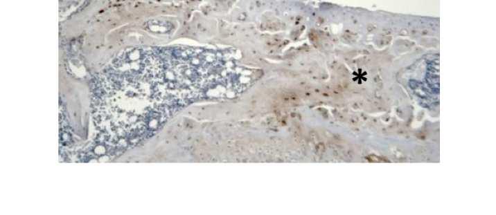
11 Page 10 of 42 STR/Ort mouse OA is linked to modifications in the MEPE/sclerostin axis. Molecular mechanisms controlling endochondral ossification may help identify those involved in OA. We have previously shown MEPE, a member of the SIBLING family, to be a negative regulator of growth plate chondrocyte matrix mineralisation (31). Examination of MEPE expression by multiplex analysis found significantly higher Mepe mrna in STR/Ort mouse articular cartilage than CBA mice (P<0.001) (Fig. 3A). Immunolabelling for MEPE showed differential expression across the tibia of the STR/Ort mouse joints, with a distinct lack of positive MEPE protein labelling in the medial (affected) aspects both prior to and during OA progression (Fig. 3C). This is in contrast to the lateral (unaffected) aspect of the STR/Ort joints and throughout all aspects of CBA joints where labelling for MEPE is observed throughout the depth of the articular cartilage tissue (Fig. 3C&D). Assessment of the MEPE regulator, Phex, revealing elevated mrna in 8-10 week STR/Ort articular cartilage compared to age-matched CBA mice (P<0.001, Fig. 3B). In STR/Ort mice, this expression significantly decreased with OA onset (P<0.001, Fig. 4B). Identical agerelated expression patterns were found for Dmp1 mrna, another SIBLING family member (P<0.001, Fig. 3E). Analysis of mrna levels of osteopontin (Opn), a SIBLING with shared roles in biomineralisation, revealed significantly higher levels in aged STR/Ort compared to young STR/Ort mice (P<0.05, Fig. 3F) and age-matched CBA mice, resembling patterns of Mepe expression (P<0.05, Fig. 3F). Taken together, this suggests a regulatory role for the SIBLING family of proteins in OA development in these mice. We next sought to examine the temporal expression of another important regulator of MEPE expression, the Wnt signalling inhibitor sclerostin (Sost) (34). Our analyses showed greater Sost mrna levels in articular cartilage of 8-10 week STR/Ort than in age-matched CBA mice (P<0.05, Fig. 4A), which significantly decreased with OA onset (P<0.01, Fig. 4A). Despite this, no differences were observed in circulating serum sclerostin concentrations in 10
12 Page 11 of 42 these mice at any age, supportive of solely local effects (Fig. 4B). Consistent with this, sclerostin immunolabelling disclosed a clear enrichment in cells at the osteochondral interface in unaffected regions of STR/Ort mouse joints (arrows, Fig. 4C). In contrast, STR/Ort mice with OA showed suppression in positive sclerostin labelling in regions of subchondral bone-thickening underlying those with compromised articular cartilage integrity (Fig. 4D). Premature growth plate closure in STR/Ort mice linked to OA development. To directly test whether longitudinal growth, growth plate fusion and osteoarthritis exhibit interrelationships in STR/Ort mice we developed a novel protocol for quantifying bony bridges formed across the entire murine tibia epiphysis during growth plate fusion (Suppl. Methods, Fig. 5A-C). Applying this novel method to examine growth plate closure in STR/Ort and CBA mice at 8 and 40+ weeks of age revealed a dramatically (10-fold) greater total number of bridges in 8-week-old STR/Ort (137 ± 10) than in CBA mice (14 ± 10; Fig. 5D, E&H; P<0.001; Suppl. Fig. 2). This enriched growth plate bridging was apparent in all aspects of STR/Ort mouse tibiae (P<0.05; Fig. 5H). Although still evident in aged STR/Ort mice (40+ weeks), this enriched bone bridging was much more discrete (STR/Ort: 295 ± 72, CBA: 266 ± 53; Fig. 5F, G&I; Suppl. Fig. 2). Mean areal bridge densities were also greater in STR/Ort mice at both ages (P<0.01, Fig. 5J). These exciting data reveal an accelerated cartilage-bone transition in the growth plate which, together with our aforementioned data, support an inherent endochondral defect in both the articular and growth plate cartilage in STR/Ort mice. Discussion Our data reveal changes in the articular cartilage of STR/Ort mouse knee joints consistent with an aberrant deployment of endochondral processes. This is associated with inherent longitudinal growth modifications, disrupted growth plate morphology, premature growth 11
13 Page 12 of 42 plate fusion, and aberrant bone formation and matrix mineralisation prior to OA onset. These data indicate, at least in the spontaneous human-like OA in STR/Ort mice, that growthrelated endochondral ossification abnormalities may forecast mechanisms of OA development in articular cartilage. There is certainly some tantalising previously published data exploring expression of endochondral ossification markers which agrees with this notion (14). Collagen type X is a marker of chondrocyte hypertrophy, usually found in the growth plate and unique to the calcified cartilage in normal joints (35). Expression of collagen type X mrna transcripts, as examined by in situ hybridization, have however been observed throughout articular cartilage in both young (9 week) and older (41+ weeks) STR/Ort mice (36). Here we provide evidence for the first time for associated collagen type X protein expression in these mice. In agreement with our studies, an additional marker of chondrocyte hypertrophy, MMP13 has also been detected in the calcified cartilage chondrocytes of STR/Ort mice at both young and old ages, at levels greater than in age-matched CBA (37). Similarly, higher expression levels of several other MMPs (MMP2, MMP3, MMP7, MMP9, MT1-MMP) were observed in the tibial articular chondrocytes of the STR/Ort mouse (37). Indeed, many of these MMPs were also shown to be significantly increased in our previous microarray study, further highlighting their likely role as key players in cartilage degradation in OA (22). The STR/Ort mouse growth plate has remained relatively unexamined with, to our knowledge, only one paper describing phenotypic changes associated with ageing. Chambers et al describe collagen type X mrna expression localized to hypertrophic chondrocytes, as expected, in both young CBA and STR/Ort mice. However in the aged mice, there was no expression of collagen type X mrna observed despite the preservation of collagen type II mrna throughout the depth of the thinned growth plate cartilage (36). Our results now reveal aberrant expression of collagen type X and MMP13 additionally in the growth plate of young 12
14 Page 13 of 42 STR/Ort mice. STR/Ort mice also display an increased zone of proliferative chondrocytes, based on well-established cell morphological features (27). As such, these results may seem counterintuitive and just highlight that there is clearly an inherent endochondral defect in the STR/Ort mice which may also precipitate OA pathogenesis. Molecular mechanisms controlling endochondral ossification may help identify those involved in OA. Effective control of the Wnt signalling pathway is certainly proving critical in regulating both the extent of OA joint pathology (38) and growth plate chondrocyte behaviour, and our data here corroborates this. Genetic and microarray analyses have been performed in STR/Ort mice in order to better elucidate their OA aetiology (39-42). Jaeger and colleagues identified a QTL associated with articular cartilage degeneration on chromosome 8 of the STR/Ort mouse (39). This however was not corroborated in a more recent QTL analysis in which STR/Ort mice were backcrossed to the C57BL/6N strain (43). This QTL was therefore suggested to be a recessive trait, as originally implicated by Walton and colleagues, among the polygenetic factors in STR/Ort OA (19, 43). Instead the authors identify a QTL for the OA phenotype that is mapped to chromosome 4 (43). Chromosome 8 was however revisited and fine mapping of the OA-QTL in a more recent study revealed Wnt related genes associated with altered chondrogenesis, including dickkopf 4 (Dkk4), secreted frizzled related protein 1 (Sfrp1) and fibroblast growth factor 1 (Fgfr1) (38, 42). Whilst a number of genes, including Wnt related genes, have been implicated in OA by association studies in human populations, there is a distinct lack of functional data to support a causative link between these associated genes and OA. Pasold et al., attempted to find such link and identified 23 polymorphic changes in the Sfrp1 gene in STR/Ort in comparison to C57BL/6 mice (42). Further immunohistochemical studies 13
15 Page 14 of 42 revealed the expression of sfrp1 was reduced in articular chondrocytes of young STR/Ort mice, and this was confirmed by in vitro analysis of STR/Ort mesenchymal stem cells (42). Our meta-analysis of previous STR/Ort microarray data herein did not reveal any significant changes in the gene expression of Dkk4, Sfrp1 or Fgfr1. Instead we provide evidence for the role of the Wnt inhibitor sclerostin, in OA development in STR/Ort mice. This is consistent with other studies which have shown expression of sclerostin in articular cartilage of various species including mice and sheep (44, 45). Whilst we observed no differences in serum sclerostin levels in STR/Ort mice, in humans an inverse relationship with radiographic knee OA severity has been observed, therefore implicating sclerostin as a potential biochemical marker (46). This does not mean that the role of sclerostin in OA is uncontroversial. Recent studies examining OA in aged sclerostin-deficient mice and in rats treated with sclerostin neutralising antibodies following surgical induction (45), concluded that genetic ablation of sclerostin does not alter spontaneous age-dependent murine OA development, nor does pharmacological inhibition of sclerostin in a surgical model of OA (45). This therefore highlights the growing need for further investigations into the precise role of sclerostin in this debilitating disease. This may come from examining the role of its downstream pathways; here we were interested in identifying the role of one such novel pathway involving the SIBLING protein MEPE (34). MEPE has been shown to be a critical regulator of osteoblast and chondrocyte matrix mineralisation (12, 31, 47, 48). The similarity in differential patterns of MEPE and sclerostin expression we observe in STR/Ort mice implies a novel mechanism by which sclerostin may function in OA. Alterations in this pathway support the case for abnormal Wnt/β-catenin signalling, as has been reported in many OA studies including in the STR/Ort mouse (42). 14
16 Page 15 of 42 In endochondral growth, the Wnt pathway is known to play an intricate and yet critical role with cartilage specific β-catenin deficient mice lacking typical growth plate zones and exhibiting delayed endochondral ossification (49). It has been shown that appropriate control of Wnt signalling in the growth plate is essential in regulating proliferation, alignment, differentiation, hypertrophy and replacement of calcified matrix with bone (38). During chondrogenesis, Wnt signalling is thought to influence the cell-cell and cell-extracellular matrix interactions upon which this fundamental process depends. Various Wnt factors, including Wnt3a, Wnt6, Wnt7a and many more, have been implicated as inhibitors of chondrogenesis, whilst a similar number, including Wnt5a and Wnt5b, have stimulatory roles (38). Similar ambiguity applies to the role of Wnt signalling in chondrocyte hypertrophic differentiation (38). In particular, it has been revealed that Wnt signalling regulates the parathyroid related peptide (PTHrP), Indian hedgehog (ihh) and transforming growth factor (TGF)-β feedback loop (49). Chondrocytes undergoing hypertrophy secrete Ihh which acts upon the proliferating chondrocytes to maintain their proliferative state and to restrict hypertrophy. Ihh also stimulates TGF-β production which in turn upregulates PTHrP. This acts on the pre-hypertrophic chondrocytes to prevent their further differentiation and thus Ihh production (50). This pathway has also been implicated in OA with increased Ihh expression reported in OA cartilage and selective Ihh deletion protecting against surgical OA progression (51, 52). Whilst this pathway was not examined here, it would be interesting to examine whether sclerostin/mepe regulates the PTHrP/Ihh pathway in the STR/Ort mouse and whether this contributes to their OA pathology. Our data do however strengthen the intimacy of the relationship between molecular dysregulation of the Wnt/β-catenin pathway and endochondral growth defects. 15
17 Page 16 of 42 Whether the results presented here are a unique feature of OA in the STR/Ort mouse or of OA in general is an interesting consideration and one that should be deliberated upon, as OA is being more widely accepted as a clinicopathological syndrome with multiple aetiologies. An elegant and comprehensive microarray study by Bateman and colleagues (53) in which gene expression profiling was performed in cartilage from wild-type mice with surgically induced OA (destabilisation of the medial meniscus, DMM) at 1, 2, and 6 weeks postsurgery, details the full list of differentially expressed genes between DMM-operated and sham-operated mice. Bateman et al., found that the marker of hypertrophy MMP13, was unchanged in DMM-operated mice in comparison to sham-operated mice at all stages postsurgery (53). This contrasts with our data in STR/Ort mice where elevated MMP13 expression levels were found prior to and during OA. In accord with our data, Bateman et al., found Col10a1 to be significantly increased in DMM-operated mice in comparison to shamoperated mice at 1 and 2 weeks post-surgery and the matrix mineralisation regulators Enpp1 and Ank to be increased at all time points post-surgery (53). The differential expression of these matrix and mineralisation markers at early time points post-surgery suggests their involvement in the initial OA processes in the DMM model. This is in accord with our data which shows similar changes prior to OA development in STR/Ort mice. Together this suggests a point of integration with these endochondral pathways at which the different OA subtypes, surgical (DMM) and natural (STR/Ort), may converge. In this manuscript we highlight the MEPE/sclerostin pathway as a potential pathway for future investigation in OA research. Our data shows differential expression of MEPE and sclerostin in the STR/Ort mouse, along with the MEPE regulator PHEX and other members of the SIBLING family of proteins, DMP1 and osteopontin. In the DMM model, none of these genes of interest were changed (53). This therefore identifies this subset of genes as selective to the STR/Ort mouse OA and our identification of this molecular phenotype will 16
18 Page 17 of 42 not only aid understanding into this diverse human condition, but also suggests that we may be able to identify specific gene signatures within particular at-risk human patients. Our reports of an inherent endochondral defect in STR/Ort mice is further strengthened by our data acquired using synchrotron x-ray computed microtomography that shows premature growth plate closure in STR/Ort mice. This novel method for 3D quantification of bony bridging will no doubt advance understanding of growth plate closure mechanisms, and our unique data revealing the complex internal topographies of the growth plate cartilage layer in CBA and STR/Ort mice (Fig. 5B&D) may also yield more insights into the micro-mechanical environment of the cells in the growth plate (54). This method has revealed that OA-prone STR/Ort and healthy CBA mice both display overt bone bridges prior to growth cessation. More specifically, spatial localisation of these bridges has shown greater clustering in STR/Ort mice, suggesting that their formation is driven by local factors, likely altered mechanical loading. The idea that STR/Ort mouse OA is driven by loading has certainly been supported by previous reports suggesting an association with medial patellar dislocation (55) and by those showing accelerated OA following mechanical joint loading (33). There have also been direct links made between growth plate function and mechanical loads, but whether this extends to the regulation of growth plate closure has yet to be explored (56). Regardless, it remains clear that these studies have, for the first time, shown that early growth plate closure is indicative of modified growth trajectory in STR/Ort mice and that growth ceases sooner in the lifetime of these OA-prone mice than in the closelyrelated parental CBA strain which shows healthy joint ageing. These data may also be interpreted as evidence that the OA in STR/Ort mice is secondary to a chondrodysplasia. Data indicating internal tibia rotation of the tibia of STR/Ort mice may offer support for such a contribution (57). However, the linked deployment of transient chondrocyte behaviours in the articular cartilage prior to overt OA development that we observe suggests, instead, that 17
19 Page 18 of 42 this reflects an inherent chondrocyte defect. Nonetheless, the likelihood that such relationships with modified growth trajectories are also prevalent in human OA is yet to be defined and longitudinal studies examining associations between growth plate dynamics and OA development in human patients would be particularly informative in our understanding of OA in general. Despite their predictable disease development with characteristics resembling those seen in human OA, including osteophytes, subchondral bone sclerosis, and synovial hyperplasia (19, 20, 58) the insights into the aetiology of OA provided by our data from this STR/Ort mouse model are limited. For instance, the aetiology of this OA is not yet known, despite extensive genetic and microarray analyses (39-41). As such, there are potential factors which may confound interpretation of our data, and it is vital to highlight that our findings define only the distinct pathophysiological mechanisms important in this subset of OA. Concurrent with this, the molecular phenotype we describe is unmodified in CBA mice suggesting that it is disease and indeed STR/Ort OA specific as opposed to any result of ageing. It is still however important to consider the molecular phenotype of this particular OA as it is becoming more widely accepted that generalisation of the OA disease is somewhat distracting and flawed in our pursuit of a disease modifying treatment (59). In conclusion, our findings show that aberrant deployment of transient chondrocyte behaviours, consistent with re-initiation of endochondral processes, occurs in joints of spontaneously OA STR/Ort mice. Articular cartilage transcriptional profiles, labelling for endochondral markers and age-related growth plate dynamics, both in vivo and in vitro, support that OA in STR/Ort mice is allied to an inherent endochondral growth defect that is subject to regulation by the MEPE/sclerostin axis. Further investigation will determine whether this is an underpinning mechanism in some or all forms of pathological ossification in OA. 18
20 Page 19 of 42 Acknowledgements We are grateful to the Arthritis Research UK for funding this research (18768). This work was made possible by the facilities and support provided by the Manchester x-ray Imaging Facility, Diamond Light Source and the Research Complex at Harwell, funded in part by the EPSRC (EP/I02249X/1). We remain indebted to Prof. Roger Mason (Imperial College London, UK) for providing us with our original STR/Ort mice and for his advice in their use. We would like to thank Prof. Boot-Handford (University of Manchester) and Prof. Rowe (University of Kansas Medical Centre) for providing antibodies used. References 1. Comhaire FH & Snaps F (2008) Comparison of two canine registry databases on the prevalence of hip dysplasia by breed and the relationship of dysplasia with body weight and height. American journal of veterinary research 69(3): Henrotin Y, et al. (2013) Early decrease of serum biomarkers of type II collagen degradation (Coll2-1) and joint inflammation (Coll2-1 NO2 ) by hyaluronic acid intra-articular injections in patients with knee osteoarthritis: A research study part of the Biovisco study. Journal of orthopaedic research : official publication of the Orthopaedic Research Society 31(6): Helminen HJ, et al. (1993) An inbred line of transgenic mice expressing an internally deleted gene for type II procollagen (COL2A1). Young mice have a variable phenotype of a chondrodysplasia and older mice have osteoarthritic changes in joints. The Journal of clinical investigation 92(2): Garnero P, et al. (2002) Uncoupling of type II collagen synthesis and degradation predicts progression of joint damage in patients with knee osteoarthritis. Arthritis and rheumatism 46(10): Jalba BA, et al. (2011) Alterations in expression of cartilage-specific genes for aggrecan and collagen type II in osteoarthritis. Rom J Morphol Embryol 52(2): Hoyland JA, et al. (1991) Distribution of type X collagen mrna in normal and osteoarthritic human cartilage. Bone Miner 15(2): Aigner T, et al. (1993) Type X collagen expression in osteoarthritic and rheumatoid articular cartilage. Virchows Arch B Cell Pathol Incl Mol Pathol 63(4): Appleton CT, Pitelka V, Henry J, & Beier F (2007) Global analyses of gene expression in early experimental osteoarthritis. Arthritis and rheumatism 56(6): Fuerst M, et al. (2009) Calcification of articular cartilage in human osteoarthritis. Arthritis and rheumatism 60(9): Mackie EJ, Ahmed YA, Tatarczuch L, Chen KS, & Mirams M (2008) Endochondral ossification: How cartilage is converted into bone in the developing skeleton. The international journal of biochemistry & cell biology 40(1):
21 Page 20 of Mackie EJ, Tatarczuch L, & Mirams M (2011) The skeleton: a multi-functional complex organ: the growth plate chondrocyte and endochondral ossification. J Endocrinol 211(2): Staines KA, Macrae VE, & Farquharson C (2012) The importance of the SIBLING family of proteins on skeletal mineralisation and bone remodelling. J Endocrinol 214(3): Staines KA, Pollard AS, McGonnell IM, Farquharson C, & Pitsillides AA (2013) Cartilage to bone transitions in health and disease. J Endocrinol 219(1):R1-R Pitsillides AA & Beier F (2011) Cartilage biology in osteoarthritis--lessons from developmental biology. Nature reviews. Rheumatology 7(11): Fosang AJ & Beier F (2011) Emerging Frontiers in cartilage and chondrocyte biology. Best Pract Res Clin Rheumatol 25(6): Brew CJ, Clegg PD, Boot-Handford RP, Andrew JG, & Hardingham T (2010) Gene expression in human chondrocytes in late osteoarthritis is changed in both fibrillated and intact cartilage without evidence of generalised chondrocyte hypertrophy. Annals of the rheumatic diseases 69(1): Pacifici M, Koyama E, & Iwamoto M (2005) Mechanisms of synovial joint and articular cartilage formation: Recent advances, but many lingering mysteries. Birth Defects Research Part C: Embryo Today: Reviews 75(3): Koyama E, et al. (2008) A distinct cohort of progenitor cells participates in synovial joint and articular cartilage formation during mouse limb skeletogenesis. Developmental biology 316(1): Mason RM, et al. (2001) The STR/ort mouse and its use as a model of osteoarthritis. Osteoarthritis and cartilage / OARS, Osteoarthritis Research Society 9(2): Walton M (1977) Degenerative joint disease in the mouse knee; radiological and morphological observations. J Pathol 123(2): Walton M (1977) Degenerative joint disease in the mouse knee; histological observations. J Pathol 123(2): Poulet B, et al. (2012) Time-series transcriptional profiling yields new perspectives on susceptibility to murine osteoarthritis. Arthritis and rheumatism 64(10): Sokoloff L, Crittenden LB, Yamamoto RS, & Jay GE, Jr. (1962) The genetics of degenerative joint disease in mice. Arthritis and rheumatism 5: Dennis G, Jr., et al. (2003) DAVID: Database for Annotation, Visualization, and Integrated Discovery. Genome biology 4(5):P Xie L, et al. (2012) Identification of a novel biomarker gene set with sensitivity and specificity for distinguishing between allograft rejection and tolerance. Liver transplantation : official publication of the American Association for the Study of Liver Diseases and the International Liver Transplantation Society 18(4): Hu X, et al. (2012) Simultaneously typing nine serotypes of enteroviruses associated with hand, foot, and mouth disease by a GeXP analyzer-based multiplex reverse transcription-pcr assay. Journal of clinical microbiology 50(2): Hunziker EB, Schenk RK, & Cruz-Orive LM (1987) Quantitation of chondrocyte performance in growth-plate cartilage during longitudinal bone growth. J Bone Joint Surg Am 69(2): Rau C, Wagner U, Pesic Z, & De Fanis A (2011) Coherent imaging at the Diamond beamline I13. Phys Status Solidi A 208(11): Atwood RC, Lee PD, Konerding MA, Rockett P, & Mitchell CA (2010) Quantitation of Microcomputed Tomography-Imaged Ocular Microvasculature. Microcirculation 17(1):
22 Page 21 of Titarenko V, Titarenko S, Withers PJ, De Carlo F, & Xiao XH (2010) Improved tomographic reconstructions using adaptive time-dependent intensity normalization. J Synchrotron Radiat 17: Staines KA, et al. (2012) MEPE is a novel regulator of growth plate cartilage mineralization. Bone 51(3): Mushtaq T, Bijman P, Ahmed SF, & Farquharson C (2004) Insulin-like growth factor-i augments chondrocyte hypertrophy and reverses glucocorticoid-mediated growth retardation in fetal mice metatarsal cultures. Endocrinology 145(5): Poulet B, Westerhof TA, Hamilton RW, Shefelbine SJ, & Pitsillides AA (2013) Spontaneous osteoarthritis in Str/ort mice is unlikely due to greater vulnerability to mechanical trauma. Osteoarthritis and cartilage / OARS, Osteoarthritis Research Society 21(5): Atkins GJ, et al. (2011) Sclerostin is a locally acting regulator of lateosteoblast/preosteocyte differentiation and regulates mineralization through a MEPE- ASARM-dependent mechanism. J Bone Miner Res 26(7): Kronenberg HM (2003) Developmental regulation of the growth plate. Nature 423(6937): Chambers MG, Kuffner T, Cowan SK, Cheah KS, & Mason RM (2002) Expression of collagen and aggrecan genes in normal and osteoarthritic murine knee joints. Osteoarthritis and cartilage / OARS, Osteoarthritis Research Society 10(1): Flannelly J, et al. (2002) Metalloproteinase and tissue inhibitor of metalloproteinase expression in the murine STR/ort model of osteoarthritis. Osteoarthritis and cartilage / OARS, Osteoarthritis Research Society 10(9): Staines KA, Macrae VE, & Farquharson C (2012) Cartilage development and degeneration: a Wnt Wnt situation. Cell Biochem Funct. 39. Jaeger K, et al. (2008) The genetics of osteoarthritis in STR/ort mice. Osteoarthritis and cartilage / OARS, Osteoarthritis Research Society 16(5): Aigner T, et al. (2006) Large-scale gene expression profiling reveals major pathogenetic pathways of cartilage degeneration in osteoarthritis. Arthritis and rheumatism 54(11): Watters JW, et al. (2007) Inverse relationship between matrix remodeling and lipid metabolism during osteoarthritis progression in the STR/Ort mouse. Arthritis and rheumatism 56(9): Pasold J, et al. (2013) Reduced expression of Sfrp1 during chondrogenesis and in articular chondrocytes correlates with osteoarthritis in STR/ort mice. Experimental cell research 319(5): Watanabe K, et al. (2012) Identification of a quantitative trait locus for spontaneous osteoarthritis in STR/ort mice. Journal of orthopaedic research : official publication of the Orthopaedic Research Society 30(1): Chan BY, et al. (2011) Increased chondrocyte sclerostin may protect against cartilage degradation in osteoarthritis. Osteoarthritis and cartilage / OARS, Osteoarthritis Research Society 19(7): Roudier M, et al. (2013) Sclerostin is expressed in articular cartilage but loss or inhibition does not affect cartilage remodeling during aging or following mechanical injury. Arthritis and rheumatism 65(3): Mabey T, et al. (2014) Plasma and synovial fluid sclerostin are inversely associated with radiographic severity of knee osteoarthritis. Clinical biochemistry 47(7-8):
23 Page 22 of Addison WN, Nakano Y, Loisel T, Crine P, & McKee MD (2008) MEPE-ASARM peptides control extracellular matrix mineralization by binding to hydroxyapatite: an inhibition regulated by PHEX cleavage of ASARM. J Bone Miner Res 23(10): Martin A, et al. (2008) Degradation of MEPE, DMP1, and release of SIBLING ASARM-peptides (minhibins): ASARM-peptide(s) are directly responsible for defective mineralization in HYP. Endocrinology 149(4): Nagayama M, et al. (2008) Wnt/beta-catenin signaling regulates cranial base development and growth. Journal of dental research 87(3): Lanske B, et al. (1996) PTH/PTHrP receptor in early development and Indian hedgehog-regulated bone growth. Science 273(5275): Zhou J, et al. (2014) Disrupting the Indian hedgehog signaling pathway in vivo attenuates surgically induced osteoarthritis progression in Col2a1-CreERT2; Ihhfl/fl mice. Arthritis research & therapy 16(1):R Wei F, et al. (2012) Activation of Indian hedgehog promotes chondrocyte hypertrophy and upregulation of MMP-13 in human osteoarthritic cartilage. Osteoarthritis and cartilage / OARS, Osteoarthritis Research Society 20(7): Bateman JF, et al. (2013) Transcriptomics of wild-type mice and mice lacking ADAMTS-5 activity identifies genes involved in osteoarthritis initiation and cartilage destruction. Arthritis and rheumatism 65(6): Gao J, Williams JL, & Roan E (2014) On the State of Stress in the Growth Plate under Physiologic Compressive Loading. Open Journal of Biophysics 4: Naruse K, et al. (2009) Osteoarthritic changes of the patellofemoral joint in STR/OrtCrlj mice are the earliest detectable changes and may be caused by internal tibial torsion. Connective tissue research 50(4): Villemure I & Stokes IA (2009) Growth plate mechanics and mechanobiology. A survey of present understanding. Journal of biomechanics 42(12): Uchida K, et al. (2012) Differential age-related bone architecture changes between female and male STR/Ort mice. Experimental animals / Japanese Association for Laboratory Animal Science 61(1): Chambers MG, et al. (2001) Matrix metalloproteinases and aggrecanases cleave aggrecan in different zones of normal cartilage but colocalize in the development of osteoarthritic lesions in STR/ort mice. Arthritis and rheumatism 44(6): Little CB & Hunter DJ (2013) Post-traumatic osteoarthritis: from mouse models to clinical trials. Nature reviews. Rheumatology 9(8):
24 Page 23 of 42 Figure Legends Figure 1. Measurements of uncalcified cartilage (white), calcified cartilage (grey) and subchondral bone (black) in the medial tibia of (A) CBA (B) STR/Ort mice at 8-10 weeks, weeks, and 40+ weeks (10 measurements in >6 sections/mouse, n=4). Results are presented as % of the thickness of each zone measured from articular surface to subchondral bone. Immunolabelling of STR/Ort mice joints prior to OA onset for MMP13 in the (C) medial tibia (D) lateral tibia, and collagen type X in the (E) medial tibia (F) lateral tibia. Images are representative of 3 individual mice. GeXP multiplex RT-qPCR analysis of (G) Enpp1 and (H) Ank mrna in CBA and STR/Ort articular cartilage at 8-10weeks, 18-20weeks and 40+ weeks of age (n=3 joints/sample). Figure 2. (A) Weights (g) of CBA and STR/Ort mice from 1-8 weeks of age (n>5). (B) Tibia length (mm) analysis in STR/Ort and age-matched CBA mice at 3-, 6- and 8-weeks of age (n=6). Measurements of digital images of E15 STR/Ort (C) and CBA (D) metatarsal bones in culture for 7 days. The growth rate (E) and (F) the percentage change in mineralisation zone length of metatarsal bones (n>8) (G) Growth plate zone lengths in 3-, 6-, and 8-week old STR/Ort and CBA mice (PZ proliferative zone, HZ - hypertrophic zone) (10 measurements in >6 sections/mouse, n=4). Data are represented as mean ± SEM. *P<0.05, ***P< Immunohistochemical analysis of (H-K) Collagen type X in (H) 3 week CBA (I) 6 week CBA (J) 3 week STR/Ort (K) 6 week STR/Ort (L-O) MMP13 (L) 3 week CBA (M) 6 week CBA (N) 3 week STR/Ort (O) 6 week STR/Ort. Images are representative of 3 individual mice. Bar = 50µm. Data are represented as mean ± SEM. *P<0.05, **P<0.01, P<0.001***. Figure 3. GeXP multiplex RT-qPCR analysis of (A) Mepe (B) Phex mrna in CBA and STR/Ort articular cartilage at 8-10weeks, 18-20weeks and 40+ weeks (n=3 joints/sample). 23
25 Page 24 of 42 (C) Immunohistochemistry of MEPE in the lateral (non-affected) and medial (affected) tibia condyles in STR/Ort mice at stages prior to and at the onset of OA. (D) Immunohistochemistry of MEPE in the CBA tibia condyles. Images are representative of 3 individual mice. GeXP multiplex RT-qPCR analysis of (E) Dmp1 (F) Opn mrna in CBA and STR/Ort articular cartilage at 8-10weeks, 18-20weeks and 40+ weeks of age (n=3 joints/sample). Figure 4. GeXP multiplex qpcr analysis of (A) Sost mrna in CBA and STR/Ort articular cartilage at 8-10weeks, 18-20weeks and 40+ weeks of age (n=3 joints/sample). (B) Serum sclerostin in CBA and STR/Ort mice at the aforementioned ages (n=4). (C) Immunohistochemistry of sclerostin in the lateral (non-affected) and (D) medial (affected) tibia condyles in STR/Ort mice at stages prior to and at the onset of OA. Images are representative of 3 individual mice. Figure 5. Development of a 3D quantification method for growth plate bridging. (A) 3D representation of the whole 40+ week STR/Ort joint. (B) 3D representation of the growth plate cartilage (yellow) underneath the tibial joint surface (grey, A) (C) 3D representation of bridges crossing the growth plate underneath the tibial joint (black crosses indicate bony bridges identified by an observer). Location and areal density of bridges across the growth plate projected on the tibial joint surface: (D) STR/Ort 8 weeks, (E) CBA 8 weeks, (F) STR/Ort 40+ weeks, (G) CBA 40+ weeks. Number of bridges per tibia at (H) 8 weeks (I) 40+ weeks (n=3). Lateral/medial and anterior/posterior segments were split in order to examine whether bridging is balanced during fusion. (J) Areal density, d. The areal density of bridges, d, is defined as the number of bridges per 256 µm x 256 µm window. Data are represented as mean of 3 animals ± S.E.M. P<0.05*, P<0.01**, P<0.001***. 24
26 Page 25 of 42 Table 1: Gene ontology analysis of genes up-regulated in osteoarthritic articular cartilage in comparison to non-osteoarthritic articular cartilage. Genes showing >1.5 fold were analysed with DAVID software (n=491) Term % P-value Bone development E-05 Ossification E-05 Bone mineral formation E-04 Skeletal system development E-04 Cartilage development E-03 25
27 Page 26 of x73mm (300 x 300 DPI)
28 Page 27 of x73mm (300 x 300 DPI)
29 Page 28 of x73mm (300 x 300 DPI)


30 Page 29 of x564mm (300 x 300 DPI)
Discovery of a Small Molecule Inhibitor of the Wnt Pathway as a Potential Disease Modifying Treatment for Knee Osteoarthritis
 Discovery of a Small Molecule Inhibitor of the Wnt Pathway as a Potential Disease Modifying Treatment for Knee Osteoarthritis Charlene Barroga, Ph.D., Yong Hu, Ph.D., Vishal Deshmukh, Ph.D., and John Hood,
Wnt7a Inhibits Cartilage Matrix Degradation in a Mouse In Vivo Osteoarthritis Model
 Wnt7a Inhibits Cartilage Matrix Degradation in a Mouse In Vivo Osteoarthritis Model Averi Leahy, Andrea Foote, Tomoya Uchimura, Li Zeng, PhD. Tufts University, Boston, MA, USA. Disclosures: A. Leahy: None.
Discovery of a Small Molecule Inhibitor of the Wnt Pathway (SM04690) as a Potential Disease Modifying Treatment for Knee Osteoarthritis
 as a Potential Disease Modifying Treatment for Knee Osteoarthritis") Discovery of a Small Molecule Inhibitor of the Wnt Pathway (SM469) as a Potential Disease Modifying Treatment for Knee Osteoarthritis Vishal Deshmukh, Ph.D., Charlene Barroga, Ph.D., Yong Hu, Ph.D., John
Discovery of a Small Molecule Inhibitor of the Wnt Pathway (SM469) as a Potential Disease Modifying Treatment for Knee Osteoarthritis Vishal Deshmukh, Ph.D., Charlene Barroga, Ph.D., Yong Hu, Ph.D., John
Supplemental Tables and Figures. The metalloproteinase-proteoglycans ADAMTS7 and ADAMTS12 provide an innate,
 Supplemental Tables and Figures The metalloproteinase-proteoglycans ADAMTS7 and ADAMTS12 provide an innate, tendon-specific protective mechanism against heterotopic ossification Timothy Mead et al Supplemental
Nature Medicine: doi: /nm.4324
 1 2 3 4 5 6 7 8 9 10 11 12 13 14 15 16 17 18 19 20 21 22 23 24 25 Supplementary Figure 1. Kinetics of SnCs development in surgically-induced OA and effect of GCV-induced SnC clearance on OA disease progression
Nanomechanical Symptoms in Cartilage Precede Histological Osteoarthritis Signs after the Destabilization of Medial Meniscus in Mice
 Nanomechanical Symptoms in Cartilage Precede Histological Osteoarthritis Signs after the Destabilization of Medial Meniscus in Mice Basak Doyran 1, Wei Tong 2, Qing Li 1, Haoruo Jia 2, Xianrong Zhang 3,
Why the dog? Analogy of the anatomy
 Why the dog? Analogy of the anatomy Surgically Induced canine OA models: Anterior (cranial) cruciate ligament transection model Pond MJ, Nuki G. Ann Rheum Dis 1973 (and > 100 others) Meniscal disruption
Osteoarthritis. Dr Anthony Feher. With special thanks to Dr. Tim Williams and Dr. Bhatia for allowing me to use some of their slides
 Osteoarthritis Dr Anthony Feher With special thanks to Dr. Tim Williams and Dr. Bhatia for allowing me to use some of their slides No Financial Disclosures Number one chronic disability in the United States
Anabolic Therapy With Teriparatide Indications Beyond Osteoporosis
 Anabolic Therapy With Teriparatide Indications Beyond Osteoporosis Andreas Panagopoulos MD, PhD Upper Limb & Sports Medicine Orthopaedic Surgeon Assistant Professor, University of Patras Outline Teriparatide
Osteochondral regeneration. Getting to the core of the problem.
 Osteochondral regeneration. Getting to the core of the problem. TM TM Bio-mimetic, biointegratable and resorbable Flexible and easy to shape Straightforward one-step procedure Promotes a guided osteo-chondral
Regulation of the IGF axis by TGF-b during periosteal chondrogenesis: implications for articular cartilage repair
 Regulation of the IGF axis by TGF-b during periosteal chondrogenesis: implications for articular cartilage repair Chapter 04 Boek 1_Gie.indb 55 21-05-2007 12:27:33 Chapter 04 Abstract Goal: TGF-b and IGF-I
Discovery of a Small Molecule Wnt Pathway Inhibitor (SM04690) as a Potential Disease Modifying Treatment for Knee Osteoarthritis
 as a Potential Disease Modifying Treatment for Knee Osteoarthritis") Discovery of a Small Molecule Wnt Pathway Inhibitor (SM469) as a Potential Disease Modifying Treatment for Knee Osteoarthritis Vishal Deshmukh PhD, Charlene Barroga PhD, Carine Bossard PhD, Sunil KC PhD,
Discovery of a Small Molecule Wnt Pathway Inhibitor (SM469) as a Potential Disease Modifying Treatment for Knee Osteoarthritis Vishal Deshmukh PhD, Charlene Barroga PhD, Carine Bossard PhD, Sunil KC PhD,
WORKSHOP. Organizers: Oran D. Kennedy, PhD Tamara Alliston, PhD
 WORKSHOP Bone Marrow Lesions - What Lies Beneath? A workshop based on the ORS/AAOS symposium: Tackling Joint Disease by Understanding Crosstalk between Cartilage and Bone, April 2016 Organizers: Oran D.
Supplementary Figure 1. Expression of phospho-sik3 in normal and osteoarthritic articular cartilage in the knee. (a) Semiserial histological sections
 Semiserial histological sections") Supplementary Figure 1. Expression of phospho-sik3 in normal and osteoarthritic articular cartilage in the knee. (a) Semiserial histological sections of normal cartilage were stained with safranin O-fast
Supplementary Figure 1. Expression of phospho-sik3 in normal and osteoarthritic articular cartilage in the knee. (a) Semiserial histological sections of normal cartilage were stained with safranin O-fast
AAO Foundation Awards Final Report
 401 N. Lindbergh Blvd. St. Louis, MO 63141 Tel.: 314.993.1700, #546 Toll Free: 800.424.2841, #546 Fax: 800.708.1364 Cell: 314.283.1983 E-Mail: rhazel@aaortho.org AAO Foundation Awards Final Report In an
Bone. Development. Tim Arnett. University College London. Department of Anatomy and Developmental Biology
 Bone Development Tim Arnett Department of Anatomy and Developmental Biology University College London Bone development Outline Bone composition matrix + mineral Bone formation - intramembranous & endochondral
NBQX, An AMPA/Kainate Glutamate Receptor Antagonist, Alleviates Joint Disease In Models Of Inflammatory- And Osteo- Arthritis.
 NBQX, An AMPA/Kainate Glutamate Receptor Antagonist, Alleviates Joint Disease In Models Of Inflammatory- And Osteo- Arthritis. Cleo S. Bonnet, PhD 1, Anwen S. Williams, PhD 1, Sophie J. Gilbert, PhD 1,
Lecture 2: Skeletogenesis
 Jilin University School of Stomatology Skeletogenesis Lecture 2: Skeletogenesis Aug. 18, 2015 Yuji Mishina, Ph.D. mishina@umich.edu Student will describe Development of Bone - the general anatomy of bone
Lecture 3: Skeletogenesis and diseases
 Jilin University School of Stomatology Skeletogenesis Lecture 3: Skeletogenesis and diseases Aug. 21, 2015 Yuji Mishina, Ph.D. mishina@umich.edu Bone Development Mouse embryo, E14.5 Mouse embryo, E18.0
The role of mechanical stimuli induced by prenatal movements in skeletal development
 The role of mechanical stimuli induced by prenatal movements in skeletal development Niamh Nowlan, PhD Department of Bioengineering, Imperial College London Mechanobiology of Bone Mechanoregulation of
New Directions in Osteoarthritis Research
 New Directions in Osteoarthritis Research Kananaskis October 22, 2015 Nick Mohtadi MD MSc FRCSC No conflicts of interest related to this presentation 1 Osteoarthritis: Disease? Fact of Life? Strong family
Interplay between Cartilage and Subchondral Bone Contributing to Pathogenesis of Osteoarthritis
 Int. J. Mol. Sci. 2013, 14, 19805-19830; doi:10.3390/ijms141019805 Review OPEN ACCESS International Journal of Molecular Sciences ISSN 1422-0067 www.mdpi.com/journal/ijms Interplay between Cartilage and
1. Define Wolff s Law and give three examples of its application.
 Harvard-MIT Division of Health Sciences and Technology HST.021: Musculoskeletal Pathophysiology, IAP 2006 Course Director: Dr. Dwight R. Robinson Final Examination HST.021 February 2, 2001 Name 1. Define
Omega-3 Fatty Acids Mitigate Obesity-induced Osteoarthritis And Accelerate Wound Repair
 Omega-3 Fatty Acids Mitigate Obesity-induced Osteoarthritis And Accelerate Wound Repair Chia-Lung Wu, MS, Deeptee Jain, MD, Jenna McNeill, BS, Dianne Little, BVSc, PhD, John Anderson, MD, Janet Huebner,
PROCHONDRIX CARTILAGE RESTORATION MATRIX CONTAINS GROWTH FACTORS NECESSARY FOR HYALINE CARTILAGE REGENERATION
 A L L O S O U R C E PROCHONDRIX CARTILAGE RESTORATION MATRIX CONTAINS GROWTH FACTORS NECESSARY FOR HYALINE CARTILAGE REGENERATION Ryan Delaney MS; Carolyn Barrett BS, MBA; Peter Stevens PhD, MBA AlloSource,
Supplemental Data. Wnt/β-Catenin Signaling in Mesenchymal Progenitors. Controls Osteoblast and Chondrocyte
 Supplemental Data Wnt/β-Catenin Signaling in Mesenchymal Progenitors Controls Osteoblast and Chondrocyte Differentiation during Vertebrate Skeletogenesis Timothy F. Day, Xizhi Guo, Lisa Garrett-Beal, and
Distinct Roles Of CCN1 And CCN2 In Limb Development
 Distinct Roles Of CCN1 And CCN2 In Limb Development Jie Jiang, PhD 1, Jessica Ong, BS 1, Faith Hall-Glenn, PhD 2, Teni Anbarchian, BS 1, Karen M. Lyons, PhD 1. 1 University of California, Los Angeles,
Anti-inflammatory properties of SM04690, a small molecule inhibitor of the Wnt pathway as a potential treatment for knee osteoarthritis
 Anti-inflammatory properties of SM04690, a small molecule inhibitor of the Wnt pathway as a potential treatment for knee osteoarthritis V. Deshmukh 1, T. Seo 1, C. Swearingen 1, Y. Yazici 1 1 Samumed,
Evaluation and Treatment of Knee Arthritis Classification of Knee Arthritis Osteoarthritis Osteoarthritis Osteoarthritis of Knee
 1 2 Evaluation and Treatment of Knee Arthritis John Zebrack, MD Reno Orthopaedic Clinic Classification of Knee Arthritis Non-inflammatory Osteoarthritis Primary Secondary Post-traumatic, dysplasia, neuropathic,
Degenerative arthritis in mice
 Annals of the Rheumatic Diseases, 1977, 36, 249-253 Degenerative arthritis in mice Study of age and sex frequency in various strains with a genetic study of NZB/B1, NZY/B1, and hybrid mice R. D. WIGLEY,
TITLE: Local Blockade of CCL21 and CXCL13 Signaling as a New Strategy to Prevent and Treat Osteoarthritis
 AWARD NUMBER: W1XWH-15-1-31 TITLE: Local Blockade of CCL1 and CXCL13 Signaling as a New Strategy to Prevent and Treat Osteoarthritis PRINCIPAL INVESTIGATOR: Bouchra Edderkaoui., Ph.D. CONTRACTING ORGANIZATION:
The Skeletal System:Bone Tissue
 The Skeletal System:Bone Tissue Dynamic and ever-changing throughout life Skeleton composed of many different tissues cartilage, bone tissue, epithelium, nerve, blood forming tissue, adipose, and dense
Small Animal models of Osteoarthritis: Testing Emerging treatments, drug delivery and mechanisms of action"
 Small Animal models of Osteoarthritis: Testing Emerging treatments, drug delivery and mechanisms of action" Dr. Mohit Kapoor Head, Cartilage Biology Research Group Co-chair, Arthritis Program Biobank Toronto
Chapter 6: Skeletal System: Bones and Bone Tissue
 Chapter 6: Skeletal System: Bones and Bone Tissue I. Functions A. List and describe the five major functions of the skeletal system: 1. 2. 3.. 4. 5.. II. Cartilage A. What do chondroblasts do? B. When
FORMATION OF BONE. Intramembranous Ossification. Bone-Lec-10-Prof.Dr.Adnan Albideri
 FORMATION OF BONE All bones are of mesodermal origin. The process of bone formation is called ossification. We have seen that formation of most bones is preceded by the formation of a cartilaginous model,
Bone Development. V. Gilsanz and O. Ratib, Hand Bone Age, DOI / _2, Springer-Verlag Berlin Heidelberg 2012
 Bone Development 2 Skeletal maturity is a measure of development incorporating the size, shape and degree of mineralization of bone to define its proximity to full maturity. The assessment of skeletal
Determining Collagen Distribution in Articular Cartilage by X-ray Micro-computed Tomography
 Determining Collagen Distribution in Articular Cartilage by X-ray Micro-computed Tomography Heikki J. Nieminen, Ph.D. 1, Mikko A. Finnilä, MHS 2, Sami Kauppinen 2, Tuomo Ylitalo 1,2, Edward Hæggström,
A Novel Murine Model Of Adynamic Bone Disease (ABD)
") A Novel Murine Model Of Adynamic Bone Disease (ABD) Adeline H. Ng 1,2, Thomas L. Willett, PhD 2,1, Benjamin A. Alman 3,1, Marc D. Grynpas 1,2. 1 University of Toronto, Toronto, ON, Canada, 2 Samuel Lunenfeld
A Novel Murine Model Of Adynamic Bone Disease (ABD) Adeline H. Ng 1,2, Thomas L. Willett, PhD 2,1, Benjamin A. Alman 3,1, Marc D. Grynpas 1,2. 1 University of Toronto, Toronto, ON, Canada, 2 Samuel Lunenfeld
Regulation of the skeletal mass through the life span
 Regulation of the skeletal mass through the life span Functions of the skeletal system Mechanical protection skull Movement leverage for muscles Mineral metabolism calcium store Erythropoiesis red blood
RECENT ADVANCES IN CLINICAL MR OF ARTICULAR CARTILAGE
 In Practice RECENT ADVANCES IN CLINICAL MR OF ARTICULAR CARTILAGE By Atsuya Watanabe, MD, PhD, Director, Advanced Diagnostic Imaging Center and Associate Professor, Department of Orthopedic Surgery, Teikyo
Age-dependent Changes in the Articular Cartilage and Subchondral Bone of C57BL/6 Mice after Surgical Destabilization of Medial Meniscus
 University of Massachusetts Medical School escholarship@umms University of Massachusetts Medical School Faculty Publications 2-9-2017 Age-dependent Changes in the Articular Cartilage and Subchondral Bone
Ideal Candidate for Cartilage Restoration. Large or Complex Lesions
 Complex Biological Knee Reconstruction: Bipolar, Multifocal Lesions and Osteoarthritis William Bugbee, MD Attending Physician, Scripps Clinic 18 th International Sports Medicine Fellow s Conference Ideal
Medial Knee Osteoarthritis Precedes Medial Meniscal Posterior Root Tear with an Event of Painful Popping
 Medial Knee Osteoarthritis Precedes Medial Meniscal Posterior Root Tear with an Event of Painful Popping Dhong Won Lee, M.D, Ji Nam Kim, M.D., Jin Goo Kim, M.D., Ph.D. KonKuk University Medical Center
o~ r;'c' - OSTEOARTHRITIS
 Osteoarthritis and Cartilage (2001) 9, Supplement A, S102-S108 2001 OsteoArthritis Research Society International doi:10.1053/joca.2001.0451, available online at http://www.idealibrary.com on IDE~l Osteoarthritis
Osteoarthritis. RA Hughes
 Osteoarthritis RA Hughes Osteoarthritis (OA) OA is the most common form of arthritis and the most common joint disease Most of the people who have OA are older than age 45, and women are more commonly
OSTEOARTHRITIS AS AN ORGAN DISEASE
 OSTEOARTHRITIS AS AN ORGAN DISEASE Peter Muir BVSc, MVetClinStud, PhD, DACVS, DECVS University of Wisconsin-Madison, School of Veterinary Medicine, Madison, WI, USA Osteoarthritis (OA) essentially represents
CARTILAGE. Dr. Emad I Shaqoura M.D, M.Sc. Anatomy Faculty of Medicine, Islamic University-Gaza October, 2015
 CARTILAGE Dr. Emad I Shaqoura M.D, M.Sc. Anatomy Faculty of Medicine, Islamic University-Gaza October, 2015 Introduction Hyaline Cartilage Elastic Cartilage Fibrocartilage Cartilage Formation, Growth,
Supplementary Figure 1 (Related with Figure 4). Molecular consequences of Eed deletion. (a) ChIP analysis identifies 3925 genes that are associated
. Molecular consequences of Eed deletion. (a) ChIP analysis identifies 3925 genes that are associated") Supplementary Figure 1 (Related with Figure 4). Molecular consequences of Eed deletion. (a) ChIP analysis identifies 3925 genes that are associated with the H3K27me3 mark in chondrocytes (see Table S1,
Supplementary Figure 1 (Related with Figure 4). Molecular consequences of Eed deletion. (a) ChIP analysis identifies 3925 genes that are associated with the H3K27me3 mark in chondrocytes (see Table S1,
Cortical thickness mapping reveals effects of age, weight and osteophytes in the proximal femur
 Cortical thickness mapping reveals effects of age, weight and osteophytes in the proximal femur Poster No.: B-0658 Congress: ECR 2013 Type: Authors: Keywords: DOI: Scientific Paper T. Turmezei, G. M. Treece,
Degenerative arthritis of Hip Bone Bangalore. Prof Sharath Rao Head, Dept. of Orthopaedics KMC Manipal
 Degenerative arthritis of Hip Prof Sharath Rao Head, Dept. of Orthopaedics KMC Manipal Hip joint Classical Synovial joint Biomechanics of hip Force coincides with trabecular pattern Hip joint Acetabulum
News Release. Merck KGaA, Darmstadt, Germany, Presents New Osteoarthritis Data at OARSI 2018 World Congress. April 17, 2018
 News Release Your Contact Brenda Mulligan +1 (978) 821-5345 April 17, 2018 Merck KGaA, Darmstadt, Germany, Presents New Osteoarthritis Data at OARSI 2018 World Congress Company to present 16 abstracts
Endogenous Mechanisms of Cartilage Healing
 Endogenous Mechanisms of Cartilage Healing Frank Beier fbeier@uwo.ca http://uwo.ca/physpharm/beier/ https://twitter.com/beierlab Cartilage Regeneration in OA Exogenous regeneration Provide exogenous cells,
MRI of Cartilage. D. BENDAHAN (PhD)
") MRI of Cartilage D. BENDAHAN (PhD) Centre de Résonance Magnétique Biologique et Médicale UMR CNRS 7339 Faculté de Médecine de la Timone 27, Bd J. Moulin 13005 Marseille France david.bendahan@univ-amu.fr
MRI of Cartilage D. BENDAHAN (PhD) Centre de Résonance Magnétique Biologique et Médicale UMR CNRS 7339 Faculté de Médecine de la Timone 27, Bd J. Moulin 13005 Marseille France david.bendahan@univ-amu.fr
Arthrographic study of the rheumatoid knee.
 Annals of the Rheumatic Diseases, 1981, 40, 344-349 Arthrographic study of the rheumatoid knee. Part 2. Articular cartilage and menisci KYOSUKE FUJIKAWA, YOSHINORI TANAKA, TSUNEYO MATSUBAYASHI, AND FUJIO
Joint and Epiphyseal Progenitor Cells Revitalize Tendon Graft and Form Mineralized Insertion Sites in Murine ACL Reconstruction Model
 Joint and Epiphyseal Progenitor Cells Revitalize Tendon Graft and Form Mineralized Insertion Sites in Murine ACL Reconstruction Model Yusuke Hagiwara 1,2, Nathaniel A. Dyment 3, Douglas J. Adams 3, Shinro
Priorities Forum Statement GUIDANCE
 Priorities Forum Statement Number 21 Subject Knee Arthroscopy including arthroscopic knee washouts Date of decision November 2016 Date refreshed March 2017 Date of review November 2018 Osteoarthritis of
Osteocytes The galaxy within
 Osteocytes The galaxy within Buenzli PR, Sims NA. Quantifying the osteocyte network in the human skeleton. Bone 2015;75:144-50. Buenzli and Sims present a fascinating insight into the intergalactic vastness
Functions of the Skeletal System. Chapter 6: Osseous Tissue and Bone Structure. Classification of Bones. Bone Shapes
 Chapter 6: Osseous Tissue and Bone Structure Functions of the Skeletal System 1. Support 2. Storage of minerals (calcium) 3. Storage of lipids (yellow marrow) 4. Blood cell production (red marrow) 5. Protection
Validation of Genetic Determinants of Skeletal Diseases. Dr Kasia Piróg (Newcastle University)
") Validation of Genetic Determinants of Skeletal Diseases Dr Kasia Piróg (Newcastle University) What is? Skeletal conditions Common Rare related to ageing and/or injury often multifactorial often difficult
Validation of Genetic Determinants of Skeletal Diseases Dr Kasia Piróg (Newcastle University) What is? Skeletal conditions Common Rare related to ageing and/or injury often multifactorial often difficult
Compact bone; Many parallel Haversian canals contain: small blood vessels. very small nerve. Interconnected by Volkmann s canals.
 Special characteristics of COMPACT BONE (dense bone) Thick; well vascularized Osteocytes and lamellae Concentric rings around blood vessels Most bones: outer compact bone inner spongy bone Marrow cavity
Investigation into differential RNA expression between diabetic and non-diabetic patients in acute ankle fractures.
 Investigation into differential RNA expression between diabetic and non-diabetic patients in acute ankle fractures. Brian A Silvia MD, PhD brian.silvia@gmail.com Lorenzo Gamez MD lorenzo.gamez@gmail.com
AIMS We will all come across osteo- and rheumatoid arthritis whatever our clinical practice Overview of pathology of osteoarthritis, its assessment an
 Osteoarthritis and Rheumatoid Arthritis Mr. Guy Barham FY1 & FY2 Orthopaedic Curriculum June 2007 AIMS We will all come across osteo- and rheumatoid arthritis whatever our clinical practice Overview of
Quantification of early stage lesions for loss of p53 should be shown in the main figures.
 Reviewer #1 (Remarks to the Author): Expert in prostate cancer The manuscript "Clonal dynamics following p53 loss of heterozygosity in Kras-driven cancers" uses a number of novel genetically engineered
Change in Femur Shape during Postnatal Development and Growth of C57BL/6 Mice
 Change in Femur Shape during Postnatal Development and Growth of C57BL/6 Mice Introduction: Disorders of skeletal development, including hip dysplasia, slipped capital femoral epiphysis, and Legg-Calve-Perthes
Calcium Phosphate Cement
 Calcium Phosphate Cement Fast-Setting Bone Graft and AutoGraft Extender. * Ossilix is a high performance next generation calcium phosphate cement indicated for filling bony defects in cancellous bone.
Cruciate Ligament. Summary of the Doctoral Thesis
 Study of the Effect of Excessive Tibial Plateau Angle on Degenerative Changes of Canine Cranial Cruciate Ligament Summary of the Doctoral Thesis Tom Ichinohe Graduate School of Veterinary Medicine and
BASELINE QUESTIONNAIRE (SURGEON)
") SECTION A: STUDY INFORMATION Subject ID: - - Study Visit: Baseline Site Number: Date: / / Surgeon ID: SECTION B: INITIAL SURGEON HISTORY B1. Previous Knee Surgery: Yes No Not recorded B2. Number of Previous
SECTION A: STUDY INFORMATION Subject ID: - - Study Visit: Baseline Site Number: Date: / / Surgeon ID: SECTION B: INITIAL SURGEON HISTORY B1. Previous Knee Surgery: Yes No Not recorded B2. Number of Previous
Animal models of osteoarthritis: classification, update, and measurement of outcomes
 Kuyinu et al. Journal of Orthopaedic Surgery and Research (2016) 11:19 DOI 10.1186/s13018-016-0346-5 REVIEW Animal models of osteoarthritis: classification, update, and measurement of outcomes Emmanuel
Skeletal Tissues. Skeletal tissues. Frame; muscles, organs and CT attach. Brain, spinal cord, thoracic organs; heart and lungs.
 Skeletal Tissues Functions 1) support 2) protection 3) movement Skeletal tissues Frame; muscles, organs and CT attach. Brain, spinal cord, thoracic organs; heart and lungs. Aids muscle contraction; generate
The cyclin-dependent kinase inhibitor p57 Kip2 mediates proliferative actions of PTHrP in chondrocytes
 Research article The cyclin-dependent kinase inhibitor p57 Kip2 mediates proliferative actions of PTHrP in chondrocytes Helen E. MacLean, 1 Jun Guo, 1 Melissa C. Knight, 1 Pumin Zhang, 2 David Cobrinik,
ORTHOPEDICS BONE Recalcitrant nonunions In total hip replacement total knee surgery increased callus volume
 ORTHOPEDICS Orthopedics has to do with a variety of tissue: bone, cartilage, tendon, ligament, muscle. In this regard orthopedic and sports medicine share the same tissue targets. Orthopedics is mostly
Testing Micronized Amnion as a Therapeutic for OA in a Rat Model using Contrast Based Micro-CT Imaging
 Testing Micronized Amnion as a Therapeutic for OA in a Rat Model using Contrast Based Micro-CT Imaging Tanushree Thote, B.S 1, Sanjay Sridaran 2, Angela S.P. Lin 3, Nick J. Willett, Ph.D. 2, Robert E.
For more information about how to cite these materials visit
 Author(s): Seetha Monrad, M.D., 2009 License: Unless otherwise noted, this material is made available under the terms of the Creative Commons Attribution Noncommercial Share Alike 3.0 License: http://creativecommons.org/licenses/by-nc-sa/3.0/
Chronic knee pain in adults - a multimodality approach or which modality to choose and when?
 Chronic knee pain in adults - a multimodality approach or which modality to choose and when? Poster No.: P-0157 Congress: ESSR 2013 Type: Authors: Keywords: DOI: Scientific Exhibit E. Ilieva, V. Tasseva,
Ankle and subtalar arthrodesis
 OSTEOAMP Allogeneic Morphogenetic Proteins Ankle Nonunions OSTEOAMP Case Report ANKLE NONUNIONS Dr. Jason George DeVries Orthopedic & Sports Medicine, Bay Care Clinic, 501 N. 10th Street, Manitowoc, WI
4 2 Osteoarthritis 1
 Osteoarthritis 1 Osteoarthritis ( OA) Osteoarthritis is a chronic disease and the most common of all rheumatological disorders. It particularly affects individuals over the age of 65 years. The prevalence
Types of osteoarthritis
 ARTHRITIS Osteoarthritis is a degenerative joint disease is the most common joint disorder. It is a frequent part of aging and is an important cause of physical disability in persons older than 65 years
Periarticular knee osteotomy
 Periarticular knee osteotomy Turnberg Building Orthopaedics 0161 206 4803 All Rights Reserved 2018. Document for issue as handout. Knee joint The knee consists of two joints which allow flexion (bending)
Original Report. The Reverse Segond Fracture: Association with a Tear of the Posterior Cruciate Ligament and Medial Meniscus
 Eva M. Escobedo 1 William J. Mills 2 John. Hunter 1 Received July 10, 2001; accepted after revision October 1, 2001. 1 Department of Radiology, University of Washington Harborview Medical enter, 325 Ninth
HOW DO WE DIAGNOSE LAMENESS IN YOUR HORSE?
 HOW DO WE DIAGNOSE LAMENESS IN YOUR HORSE? To help horse owners better understand the tools we routinely use at VetweRx to evaluate their horse s soundness, the following section of this website reviews
Most cells in the human body have an assigned purpose. They are liver cells, fat cells, bone cells,
 What is a Stem Cell? Most cells in the human body have an assigned purpose. They are liver cells, fat cells, bone cells, and so on. These cells can replicate more of their own kind of cell, but they cannot
AUTOLOGOUS CHONDROCYTE IMPLANTATION FOR FOCAL ARTICULAR CARTILAGE LESIONS
 CARTILAGE LESIONS Non-Discrimination Statement and Multi-Language Interpreter Services information are located at the end of this document. Coverage for services, procedures, medical devices and drugs
TREATMENT OF CARTILAGE LESIONS
 TREATMENT OF CARTILAGE LESIONS Angelo J. Colosimo, MD -Head Orthopaedic Surgeon University of Cincinnati Athletics -Director of Sports Medicine University of Cincinnati Medical Center -Associate Professor
Edinburgh Research Explorer
 Edinburgh Research Explorer A technique of predicting radiographic joint line and posterior femoral condylar offset of the knee Citation for published version: Clement, ND, Hamilton, DF & Burnett, R 2014,
OSTEOCHONDRAL ALLOGRAFT RECONSTRUCTION FOR MASSIVE BONE DEFECT
 OSTEOCHONDRAL ALLOGRAFT RECONSTRUCTION FOR MASSIVE BONE DEFECT Angelo J. Colosimo, MD -Head Orthopaedic Surgeon University of Cincinnati Athletics -Director of Sports Medicine University of Cincinnati
Mineral Density Of Subchondral Bone May Be Quantitatively Evaluated Using A Clinical Cone Beam Computed Tomography Scanner
 Mineral Density Of Subchondral Bone May Be Quantitatively Evaluated Using A Clinical Cone Beam Computed Tomography Scanner Mikael J. Turunen, PhD 1, Juha Töyräs, PhD 1, Harri Kokkonen, PhD 2, Jukka S.
Lower Extremity Alignment: Genu Varum / Valgum
 Lower Extremity Alignment: Genu Varum / Valgum Arthur B Meyers, MD Nemours Children s Hospital & Health System Associate Professor of Radiology, University of Central Florida Clinical Associate Professor
Protection from osteoarthritis: targeting mtor and autophagy
 Dr. Yue Zhang Protection from osteoarthritis: targeting mtor and autophagy Cartilage scientist, Division of Genetics and Development, The Toronto Western Research Institute, Toronto Western Hospital, University
OSTEOPHYTOSIS OF THE FEMORAL HEAD AND NECK
 908 RDIOLOGIC VIGNETTE OSTEOPHYTOSIS OF THE FEMORL HED ND NECK DONLD RESNICK Osteophytes are frequently considered the most characteristic abnormality of degenerative joint disease. In patients with osteoarthritis,
Fig Articular cartilage. Epiphysis. Red bone marrow Epiphyseal line. Marrow cavity. Yellow bone marrow. Periosteum. Nutrient foramen Diaphysis
 Fig. 7.1 Articular cartilage Epiphysis Red bone marrow Epiphyseal line Marrow cavity Yellow bone marrow Nutrient foramen Diaphysis Site of endosteum Compact bone Spongy bone Epiphyseal line Epiphysis Articular
Osteoarthritis: Does post-injury ACL reconstruction prevent future OA?
 Osteoarthritis: Does post-injury ACL reconstruction prevent future OA? Wen, Chunyi; Lohmander, L Stefan Published in: Nature Reviews Rheumatology DOI: 10.1038/nrrheum.2014.120 Published: 2014-01-01 Link
Why do I perform PFP? Fernando Fonseca, MD PhD Head of Department Orthopaedics
 Why do I perform PFP? Fernando Fonseca, MD PhD Head of Department Orthopaedics Acknowledges to António Completo Paulo Flores Susana Meireles André Castro Disclosures: None ... Knee is one of the most complex
Effects of Immobilization on Structure of Cell Layers In Tibial Articular Cartilage
 Effects of Immobilization on Structure of Cell Layers In Tibial Articular Cartilage Effects of Immobilization on Structure of Cell Layers In Tibial Articular Cartilage OGIWARA Yuh* FUJIKAWA Kaoru** OHSAKO
Supplementary Figure 1
 Supplementary Figure 1 Genetic labeling of microglia Male and female 2-3 month-old CreERT2;R26-tdTomato mice or CreERT2;R26-tdTomato;Iba1-eGFP transgenic mice were treated with 1x, 2x (48 h apart), or
Orthopedic & Sports Medicine, Bay Care Clinic, 501 N. 10th Street, Manitowoc, WI Procedure. Subtalar arthrodesis
 OSTEOAMP Allogeneic Morphogenetic Proteins Subtalar Nonunions OSTEOAMP Case Report SUBTALAR NONUNIONS Dr. Jason George DeVries and Dr. Brandon M. Scharer Orthopedic & Sports Medicine, Bay Care Clinic,
SKELETAL TISSUES CHAPTER 7 INTRODUCTION TO THE SKELETAL SYSTEM TYPES OF BONES
 SKELETAL TISSUES CHAPTER 7 By John McGill Supplement Outlines: Beth Wyatt Original PowerPoint: Jack Bagwell INTRODUCTION TO THE SKELETAL SYSTEM STRUCTURE Organs: Bones Related Tissues: Cartilage and Ligaments
Examining Elbow Dysplasia Prepared by the Orthopedic Foundation for Animals Orthopedic Foundation for Animals, Columbia, MO
 Examining Elbow Dysplasia Prepared by the Orthopedic Foundation for Animals Orthopedic Foundation for Animals, Columbia, MO Elbow dysplasia has been found in 78 breeds evaluated by the Orthopedic Foundation
Effects of pulsing electromagnetic fields on cultured cartilage cells.
 Arch Oral Biol. 1993 Jan;38(1):67-74. Autoradiographic study of the effects of pulsed electromagnetic fields on bone and cartilage growth in juvenile rats. Wilmot JJ, Chiego DJ Jr, Carlson DS, Hanks CT,
Re-growing the Skeleton: Approaches in Tissue Engineering and Regenerative Medicine
 Re-growing the Skeleton: Approaches in Tissue Engineering and Regenerative Medicine How we fix things now Total Knee Replacements Fracture Plates Fusing Joints Defining Regenerative Medicine restore form
BIOH111. o Cell Module o Tissue Module o Integumentary system o Skeletal system o Muscle system o Nervous system o Endocrine system
 BIOH111 o Cell Module o Tissue Module o Integumentary system o Skeletal system o Muscle system o Nervous system o Endocrine system Endeavour College of Natural Health endeavour.edu.au 1 TEXTBOOK AND REQUIRED/RECOMMENDED