Lecture 3: Skeletogenesis and diseases
|
|
|
- Whitney Rodgers
- 5 years ago
- Views:
Transcription
1 Jilin University School of Stomatology Skeletogenesis Lecture 3: Skeletogenesis and diseases Aug. 21, 2015 Yuji Mishina, Ph.D.

.")
2 Bone Development Mouse embryo, E14.5 Mouse embryo, E18.0 First, cartilage primordia are formed (blue). Then, osteoblasts replace cartilage (red). Dr. Yuji Mishina
3


4 Bones are formed through two distinct processes Intramembranous Ossification Endochondral Ossification

5 Growth Plate Development Determines the Size of Long Bones Resting Resting zone Proliferative zone Prehypertrophic zone Hypertrophic zone Proliferative Prehypertrophic Hypertrophic (early, late)


6 Clinical Examples
7 How Does Development of Growth Plate Influence Size of Long Bones? 1. Chondrocytes need to proliferate. Resting Proliferative Prehypertrophic Hypertrophic If suppressed, bones become shorter. FGF, Indian Hedgehog 2. Chondrocytes need to differentiate. If differentiate too quickly, less time to proliferate. PTHrP, Indian Hedgehog

8 Over 20 FGF ligands interact with 4 FGF receptors.


9 FGF Signaling In Growth Plate FGF signaling negatively regulates chondrocyte proliferation Over activation of FGF signaling causes chondrodysplasia (short-limbed dwarfism). Achondroplasia: most common, caused by point mutations in FGF receptor 3 (FGFR3). Nature Vol May
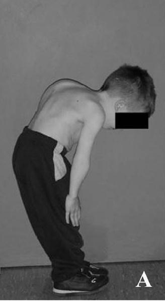
10 Clinical Example of Achondroplasia FGFR3
11 PTHrP Signaling In Growth Plate Resting chondrocytes PTHrP is produced in the resting zone. PTHrP signaling negatively regulate chondrocyte differentiation (#1 arrow). Morphogen gradient! Nature Vol May

12 PTHrP Knockout Mice Wild Type PTHrP-/- Genes Dev :


13 PTHrP Knockout Mice gp: Growth plate Wild Type PTHrP-/- Genes Dev :
.")
14 Indian Hedgehog Signaling In Growth Plate Resting chondrocytes Indian Hedgehog (Ihh) is produced in the prehypertrophic/early hypertrophic zone. Ihh acts on proliferating chondrocytes to keep dividing (#2 arrow). Ihh stimulates PTHrP production (#3 arrow), thus negatively regulate chondrocyte differentiation Nature Vol May
15 Ihh Knockout Mice Wild Type Ihh-/- Genes Dev :

16 Ihh Knockout Mice Wild Type Ihh-/- Genes Dev :

 Ihh-/- Fraction of chondrocytes that are hypertrophic increased Genes")
17 Ihh Knockout Mice Chondrocytes never displayed a clear stacked columnar organization Hypertrophic cells were observed in abnormal positions, close to the ends of the skeletal elements Wild Type (E18.5) Ihh-/- Fraction of chondrocytes that are hypertrophic increased Genes Dev :
18 Outline Introduction: bone and growth factors Growth Factors in Bone Development a) FGF b) PTHrP/Ihh Growth Factors in Bone Remodeling a) Coupling (bone formation and bone resorption) b) WNT c) BMP
19 Bone Development chondrocytes <--> osteoblasts FGF, PTHrP, Ihh Bone Remodeling osteoblasts <--> osteoclasts

20 Four phases Bone Remodeling Activation - initiating event that converts a quiescent bone surface into a remodeling one - recruitment, penetration, fusion Resorption - fully differentiated osteoclasts acidify the area bordered by the sealing zone Reversal - resorption lacunae is occupied by mononuclear cells, coupling signals are initiated Formation - osteoblasts synthesize the organic matrix and then mineralize it

21 Bone Remodeling Cycle

22 Bone Remodeling


.")

23 Bone remodeling coupling of formation and resorption 1. Osteoclasts chew up bone matrixes to release growth factors (TGF-b). (Yellow) 2. Osteoblasts produce factors to regulate osteoclasts (RANKL/OPG). (Orange)
24 How are bone formation and resorption coupled? Osteoblast (bone formation) OPG/RANKL Bone resorption RANKL OPG Osteoclast (bone resorption)
25 Physiological: Normal bone mass Bone Formation = Bone Resorption (osteoblasts) (osteoclasts)
26 Why is the coupling so important? Bone mass & Bone quality Bone Formation <----> Bone Resorption Bone mass normal, but Dr. Yuji Mishina
27 Post-menopausal Osteoporosis
28 Canonical Wnt signaling Wnt1 Wnt3 Nucleus *adapted from Montcouquiol M, et al , Annu. Rev. Neurosci. 29:363-86
29 _ + + Mesenchymal progenitors Pre-chondrocytes Proliferating chondrocytes + + Hypertrophic chondrocytes Runx2 + Osx + Ocn + Osteoblast progenitors Osteoblast progenitors Osteoblasts OPG/RANKL + _ HSC CFU-GM Pre-osteoclasts Osteoclasts


30 High Bone Mass diseases are caused by high Wnt signaling Mutations in an Wnt co-receptor, LRP5, increase Wnt signaling. (Gain-of-function type mutations)
31 Wnt Receptor Wnt Signaling In Osteoblasts Can Regulate Osteoclast Through OPG and RANKL Wnt1 Wnt3 Sclerostin OPG up RANKL down Osteoclast Osteoblast
32 Wnt Receptor Wnt Signaling In Osteoblasts Can Regulate Osteoclast Through OPG and RANKL Wnt1 Wnt3 Sclerostin OPG up RANKL down Osteoclast Osteoblast Dr. Yuji Mishina
33 Wnt Receptor Wnt Signaling In Osteoblasts Can Regulate Osteoclast Through OPG and RANKL Wnt1 Wnt3 Sclerostin OPG down RANKL up Osteoclast Osteoblast Dr. Yuji Mishina
34 N Engl J Med 2014;370:412-20


35 BMP was found by Marshall Urist as a potent bone inducer. Dr. M. Urist and Dr. Yuji Mishina? years ago
36

37 BMPs (BMP2 and 7) have been clinically used to accelerate fracture healing.
 Implants")
. J. Clin. Periodontol.")
38 BMP2 and Dentistry Supraalveolar critical-size peri-implant defects (Hound Labrador) Implants coated with BMP2 induced bone formation in alveolar bones (right). J. Clin. Periodontol., 2008, 35,
39 Key features of BMPs - found by their ectopic bone formation abilities - induce proliferation / differentiation of osteoblasts - are approved by FDA for assisting fracture healing - No / reverse effects of BMPs in some clinical cases - May have synergistic or antagonistic functions with other growth factors / signaling pathways


40 Hyper-calcification on spine, ribs, and sternum when BMP signaling was shut off in osteoblasts. 34 weeks, Male Ribs/Sternum Spine WT cko WT cko cko; conditional knockout. In this case, one BMP receptor (Bmpr1a) is knocked out only in osteoblasts. Dr. Yuji Mishina


41 More bone volume when BMP signaling is off in osteoblasts. 34 weeks old, H&E staining, spine (Lumbar) WT cko 200 mm A lot more bones are formed in trabecular bone area in cko. Dr. Yuji Mishina
42 Why is the coupling so important? Bone mass & Bone quality Bone Formation <----> Bone Resorption Bone mass normal Dr. Yuji Mishina
43 BMP Receptor Wnt Receptor BMP signaling in osteoblasts is important for both bone formation and resorption via the regulation of Wnt signaling BMP Sclerostin Wnt1 Wnt3 sclerostin expression OPG up RANKL down Osteoclast Osteoblast Dr. Yuji Mishina
44 BMP Receptor Wnt Receptor BMP signaling in osteoblasts is important for both bone formation and resorption via inhibition of Wnt signaling Wnt1 Wnt3 BMP Sclerostin sclerostin expression OPG up RANKL down Osteoclast Osteoblast Dr. Yuji Mishina


45 BMP Receptor Wnt Receptor BMP signaling in osteoblasts is important for both bone formation and resorption via inhibition of Wnt signaling Wnt1 Wnt3 BMP Sclerostin sclerostin expression OPG down RANKL up Osteoclast Osteoblast Dr. Yuji Mishina
46 Summary Basic concepts in bone development and bone remodeling Bone development (growth plate as example) is regulated by FGF, PTHrP, and Ihh through different mechanisms The coupling of bone formation and bone resorption is critical in bone remodeling The regulation of bone remodeling by Wnt and BMP signaling
47
48


49 Disorders of the skull vault in human Craniosynostosis caused by premature fusion of sutures Parietal foramina Wilkie AO & Morriss-Kay GM, Nature reviews, Genetics, 2001
50 Craniosynostosis presents clinical problems -- If untreated; chronic headache, vision loss, deafness, mental retardation. -- Reconstitution surgery is the only way. Sutures will be fused again. -- Very variable symptoms; positions, degrees various underneath molecular reasons Understanding the developmental mechanisms of craniosynostosis is necessary. Craniosynostosis affects one in 2,500 live births.


51 Clinical symptoms of Craniosynostosis are viable. Sagittal synostosis/ scaphocephaly Metopic synostosis/ trigonocephaly Coronal synostosis/ lambdoid synostosis (Plagiocephaly) Lambdoid suture Sagittal suture Coronal sutures Metopic suture
52 Causative genes for Craniosynostosis Gene mutation suture FGF receptors GOF Coronal suture MSX2 GOF Coronal suture TWIST1 Het Coronal suture TCF12 LOF Coronal suture BBS9 SNP (GOF?) Sagittal suture Axin1 and Fgfr1 Compound LOF Sagittal suture BMP2 enhancer SNP (GOF?) Sagittal suture ERF LOF All sutures Bmpr1a GOF Metopic Lambdoid suture Sagittal suture Coronal sutures Metopic suture
 and bone remodeling Facial growth requires freedom of movement of facial")
53 Facial Bone Sutures zygomatico-frontal suture zygomatico-maxillary suture midpalatal suture Midfacial growth occurs by bone deposition at bony sutures and by periosteal apposition (bone deposition) and bone remodeling Facial growth requires freedom of movement of facial sutures
54 Molecular Genetics of Apert Syndrome Molecular Genetics of Crouzon Syndrome Mutation: Activating mutation in FGFR2 gene Mutation is in area of FGF binding site Two mutations only Molecular phenotype: Enhanced FGFR to ligand binding FGFR2 binding to wrong ligands Mutations can arise spontaneously Risk of mutation increases with paternal age Mutation: Activating mutation in FGFR2 gene Mutation is in area of FGF binding site Two common mutations with numerous additional less common mutations. Molecular phenotype: Ligand independent FGFR signaling Mutations can arise spontaneously Risk of mutation increases with paternal age


55 FGFR2 Mutations extracellular intracellular Ig1 Ig2 Ig3 SP TM TK1 TK2 FGF binding Apert mutations Crouzon common mutations

")

56 Apert Syndrome: Full penetrance, variable expression Prenatal Coronal Synostosis Increased intracranial pressure NL to diminished IQ Brain abnormalities (defects in septum pellucidum) Proptosis, hypertelorism Midface hypoplasia (facial sutures) Class III malocclusion Progressive cervical spine fusions Tracheal abnormalities Digit abnormalities: syndactyly Higher risk with higher paternal age
 NL intelligence")

57 Crouzon Syndrome: Full penetrance, variable expression Coronal Synostosis, rare pansynostosis Increased intracranial pressure Brain abnormalities (defects in corpus callosum) NL intelligence Proptosis, hypertelorism Midface hypoplasia (facial sutures) Class III malocclusion Vertebral fusions Stylohyoid ligament calcification Tracheal abnormalities Digit abnormalities: None Higher risk with higher paternal age
58 Summary Basic concepts in bone development and bone remodeling Bone development (growth plate as example) is regulated by FGF, PTHrP, and Ihh through different mechanisms The coupling of bone formation and bone resorption is critical in bone remodeling The regulation of bone remodeling by Wnt and BMP signaling
59
Interactions of the endocrine system, bone and oral health
 Interactions of the endocrine system, bone and oral health All bones are not equal! Dense high proportion of cortical bone High proportion of trabecular bone Mandible Functions: mastication, respiration,
Lecture 2: Skeletogenesis
 Jilin University School of Stomatology Skeletogenesis Lecture 2: Skeletogenesis Aug. 18, 2015 Yuji Mishina, Ph.D. mishina@umich.edu Student will describe Development of Bone - the general anatomy of bone
Skeletal System. Prof. Dr. Malak A. Al-yawer Department of Anatomy/Embryology Section
 Skeletal System Prof. Dr. Malak A. Al-yawer Department of Anatomy/Embryology Section Learning objectives At the end of this lecture, the medical student will be able to: State the embryonic origin of skeletal
Bone. Development. Tim Arnett. University College London. Department of Anatomy and Developmental Biology
 Bone Development Tim Arnett Department of Anatomy and Developmental Biology University College London Bone development Outline Bone composition matrix + mineral Bone formation - intramembranous & endochondral
What is Craniosynostosis?
 What is Craniosynostosis? Craniosynostosis is defined as the premature closure of the cranial sutures (what some people refer to as soft spots). This results in restricted and abnormal growth of the head.
Skeletal Development Multiple Cellular Origins. Intramembranous Bone. Endochondrial Bone. Cartilage template of the limb in the Chick wing
 Skeletal Development Multiple Cellular Origins 1 - Paraxial Mesoderm Somite, Sclerotome Axial Skeleton (e.g. vertebra) 2 - Lateral Plate Mesoderm Appendicular Skeleton (e.g. limb) 3 - Neural Crest Head
SKELETAL SYSTEM I NOTE: LAB ASSIGNMENTS for this topic will run over 3 Weeks. A SEPARATE WORKSHEET WILL BE PROVIDED.
 BIO 211; Anatomy and Physiology I REFERENCE: CHAPTER 07 1 Dr. Lawrence Altman Naugatuck Valley Community College LECTURE TOPICS OUTLINE SKELETAL SYSTEM I NOTE: LAB ASSIGNMENTS for this topic will run over
Supplemental Data. Wnt/β-Catenin Signaling in Mesenchymal Progenitors. Controls Osteoblast and Chondrocyte
 Supplemental Data Wnt/β-Catenin Signaling in Mesenchymal Progenitors Controls Osteoblast and Chondrocyte Differentiation during Vertebrate Skeletogenesis Timothy F. Day, Xizhi Guo, Lisa Garrett-Beal, and
Rama Nada. - Mousa Al-Abbadi. 1 P a g e
 - 1 - Rama Nada - - Mousa Al-Abbadi 1 P a g e Bones, Joints and Soft tissue tumors Before we start: the first 8 minutes was recalling to Dr.Mousa s duties, go over them in the slides. Wherever you see
Week 14. Development of the Musculoskeletal System
 Week 14 Development of the Musculoskeletal System Skeletal System Derived from: paraxial mesoderm somites and somitomeres sclerotome sclerotome differentiation induced by SHH from notochord and floor plate
Functions of the Skeletal System
 SKELETAL SYSTEM Functions of the Skeletal System Support: Internal framework that supports and anchors all soft organs. Protection: Bones protect soft body organs Body movement skeletal muscle attached
The Skeletal System:Bone Tissue
 The Skeletal System:Bone Tissue Dynamic and ever-changing throughout life Skeleton composed of many different tissues cartilage, bone tissue, epithelium, nerve, blood forming tissue, adipose, and dense
Ossification and Bone Remodeling
 Ossification and Bone Remodeling Pre-natal Ossification Embryonic skeleton: fashioned from fibrous membranes or cartilage to accommodate mitosis. 2 types of pre-natal ossification (bone formation) 1.
Bones are made of OSSEOUS TISSUE
 SKELETAL SYSTEM Functions of the Skeletal System Bones are made of OSSEOUS TISSUE Support and Protection Body movement Blood cell formation (bone marrow) Storage of inorganic materials (salt, calcium,
Osteoporosis: current treatment and future prospects. Juliet Compston Professor Emeritus of Bone Medicine Cambridge Biomedical Campus
 Osteoporosis: current treatment and future prospects Juliet Compston Professor Emeritus of Bone Medicine Cambridge Biomedical Campus Disclosures Consultancy and speaking fees for Gilead, related to development
Skeletal Tissues. Skeletal tissues. Frame; muscles, organs and CT attach. Brain, spinal cord, thoracic organs; heart and lungs.
 Skeletal Tissues Functions 1) support 2) protection 3) movement Skeletal tissues Frame; muscles, organs and CT attach. Brain, spinal cord, thoracic organs; heart and lungs. Aids muscle contraction; generate
For more information about how to cite these materials visit
 Author(s): University of Michigan Medical School, Department of Cell and Developmental Biology License: Unless otherwise noted, the content of this course material is licensed under a Creative Commons
Bones are supremely local in their function:
 Developmental regulation of the growth plate Henry M. Kronenberg Endocrine Unit, Massachusetts General Hospital and Harvard Medical School, Boston, Massachusetts 02114-2696, USA (e-mail: kronenberg.henry@mgh.harvard.edu)
Functions of the Skeletal System
 SKELETAL SYSTEM Functions of the Skeletal System Bones are made of OSSEOUS TISSUE Support and Protection Body movement Blood cell formation (bone marrow) - hemopoiesis Storage of inorganic materials (salt,
International Journal of Current Research and Academic Review ISSN: Volume 3 Number 1 (January-2015) pp
 pp") International Journal of Current Research and Academic Review ISSN: 47 Volume Number (January) pp. 66 www.ijcrar.com Clinical Profile of Patients with Craniosynostosis: A Descriptive Study Nagaraj V. Gadwal*
International Journal of Current Research and Academic Review ISSN: 47 Volume Number (January) pp. 66 www.ijcrar.com Clinical Profile of Patients with Craniosynostosis: A Descriptive Study Nagaraj V. Gadwal*
Dr. Heba Kalbouneh. Saba Alfayoumi. Heba Kalbouneh
 11 Dr. Heba Kalbouneh Saba Alfayoumi Heba Kalbouneh 2- Bone Bone tissue is also classified into primary bone and secondary bone. In the beginning, the first bone that is deposited by the osteoblasts is
DISEASES WITH ABNORMAL MATRIX
 DISEASES WITH ABNORMAL MATRIX MSK-1 FOR 2 ND YEAR MEDICAL STUDENTS Dr. Nisreen Abu Shahin CONGENITAL DISEASES WITH ABNORMAL MATRIX OSTEOGENESIS IMPERFECTA (OI): also known as "brittle bone disease" a group
KEY CONCEPTS Unit 6 THE SKELETAL SYSTEM
 ANATOMY & PHYSIOLOGY 1 (101-805 - AB) PAUL ANDERSON 2011 KEY CONCEPTS Unit 6 THE SKELETAL SYSTEM A Overview of The Skeletal System 1. Definition: Anatomically the SKELETAL SYSTEM consists of bones, cartilages,
Functions of the Skeletal System. Chapter 6: Osseous Tissue and Bone Structure. Classification of Bones. Bone Shapes
 Chapter 6: Osseous Tissue and Bone Structure Functions of the Skeletal System 1. Support 2. Storage of minerals (calcium) 3. Storage of lipids (yellow marrow) 4. Blood cell production (red marrow) 5. Protection
SKELETAL SYSTEM CHAPTER 07. Bone Function BIO 211: ANATOMY & PHYSIOLOGY I. Body Movement interacts with muscles bones act as rigid bar of a lever
 Page 1 BIO 211: ANATOMY & PHYSIOLOGY I 1 CHAPTER 07 SKELETAL SYSTEM Dr. Lawrence G. G. Altman www.lawrencegaltman.com Some illustrations are courtesy of McGraw-Hill. Some illustrations are courtesy of
SKELETAL SYSTEM CHAPTER 07 BIO 211: ANATOMY & PHYSIOLOGY I
 BIO 211: ANATOMY & PHYSIOLOGY I 1 CHAPTER 07 SKELETAL SYSTEM Dr. Lawrence G. G. Altman www.lawrencegaltman.com Some illustrations are courtesy of McGraw-Hill. Some illustrations are courtesy of McGraw-Hill.
Figure ) The area that causes the lengthwise growth of a long bone is indicated by letter. Diff: 2 Page Ref:
 The area that causes the lengthwise growth of a long bone is indicated by letter. Diff: 2 Page Ref:") Essentials of Anatomy and Physiology, 9e (Marieb) Chapter 5 The Skeletal System Short Answer Figure 5.1 Using Figure 5.1, identify the following: 1) Spongy bone is indicated by letter. Diff: 1 Page Ref:
Essentials of Anatomy and Physiology, 9e (Marieb) Chapter 5 The Skeletal System Short Answer Figure 5.1 Using Figure 5.1, identify the following: 1) Spongy bone is indicated by letter. Diff: 1 Page Ref:
Supplementary Figure 1 (Related with Figure 4). Molecular consequences of Eed deletion. (a) ChIP analysis identifies 3925 genes that are associated
. Molecular consequences of Eed deletion. (a) ChIP analysis identifies 3925 genes that are associated") Supplementary Figure 1 (Related with Figure 4). Molecular consequences of Eed deletion. (a) ChIP analysis identifies 3925 genes that are associated with the H3K27me3 mark in chondrocytes (see Table S1,
Supplementary Figure 1 (Related with Figure 4). Molecular consequences of Eed deletion. (a) ChIP analysis identifies 3925 genes that are associated with the H3K27me3 mark in chondrocytes (see Table S1,
The Skeletal System:Bone Tissue
 The Skeletal System:Bone Tissue Dynamic and ever-changing throughout life Skeleton composed of many different tissues cartilage, bone tissue, epithelium, nerve, blood forming tissue, adipose, and dense
4.1 Classification of Craniosynostosis: Therapeutical implications.
 ISPN course 23 rd Nov, 2015 Cranial & Craniofacial disorders 4.1 Classification of Craniosynostosis: Therapeutical implications. Kazuaki Shimoji, Masakazu Miyajima and Hajime Arai Department of Neurosurgery,
Ossification = Osteogenesis
 Ossification = Osteogenesis Ossification = Osteogenesis Parts of the fetal skeleton form during the first few weeks after conception By the end of the 8 th week, the skeletal pattern is formed : cartilage
Role of FGF/FGFR signaling in skeletal development and homeostasis: learning from mouse models
 OPEN Citation: Bone Research (2014) 2, 14003; doi:10.1038/boneres.2014.3 ß 2014 Sichuan University. All rights reserved 2095-4700/14 www.boneresearch.org REVIEW ARTICLE Role of FGF/FGFR signaling in skeletal
Hole s Human Anatomy and Physiology Eleventh Edition. Mrs. Hummer. Chapter 7 Skeletal System
 Hole s Human Anatomy and Physiology Eleventh Edition Mrs. Hummer Chapter 7 Skeletal System 1 Chapter 7 Skeletal System Bone Classification Long Bones Short Bones Flat Bones Irregular Bones Sesamoid (Round)
Chapter 6: Skeletal System: Bones and Bone Tissue
 Chapter 6: Skeletal System: Bones and Bone Tissue I. Functions A. List and describe the five major functions of the skeletal system: 1. 2. 3.. 4. 5.. II. Cartilage A. What do chondroblasts do? B. When
Chapter 7: Head & Neck
 Chapter 7: Head & Neck Osteology I. Overview A. Skull The cranium is composed of irregularly shaped bones that are fused together at unique joints called sutures The skull provides durable protection from
The Skeletal System. Chapter 7a. Skeletal System Introduction Functions of the skeleton Framework of bones The skeleton through life
 The Skeletal System Skeletal System Introduction Functions of the skeleton Framework of bones The skeleton through life Chapter 7a Support Protection Movement Storage areas Minerals Lipids Hemopoiesis
Skeletal System: Skull.
 Skeletal System: Skull www.fisiokinesiterapia.biz Bones of the Skull SPLANCHNOCRANIUM Nasal (2) Maxilla (2) Lacrimal (2) Zygomatic (2) Palatine (2) Inferior concha (2) Vomer Mandible NEUROCRANIUM Frontal
Skeletal System worksheet
 Skeletal System worksheet Name Section A: Intro to Skeletal System The skeletal system performs vital functions that enable us to move through our daily lives. Support - The skeleton provides support and
ANATOMY AND ULTRASTRUCTURE OF BONE HISTOGENESIS, GROWTH AND REMODELING
 ANATOMY AND ULTRASTRUCTURE OF BONE HISTOGENESIS, GROWTH AND REMODELING Updated: May 13, 2008 Authors: Roland Baron, DDS, PhD. Summary 1. Bones have three major functions: to serve as mechanical support,
Subject Index. AXIN2, cleft defects 24, 26
 Subject Index ADAMTS, mouse mutants and palate development 37, 38 Africa, cleft lip and palate prevalence 6, 7 Alcohol dependence, pregnancy risks for cleft 25, 61 Altitude, pregnancy risks for cleft 25,
SUPPLEMENTARY INFORMATION
 SUPPLEMENTARY INFORMATION Supplementary Figure 1. Generation of a conditional allele of the Kindlin-2 gene. (A) A restriction map of the relevant genomic region of Kindlin-2 (top), the targeting construct
SKELETAL TISSUES CHAPTER 7 INTRODUCTION TO THE SKELETAL SYSTEM TYPES OF BONES
 SKELETAL TISSUES CHAPTER 7 By John McGill Supplement Outlines: Beth Wyatt Original PowerPoint: Jack Bagwell INTRODUCTION TO THE SKELETAL SYSTEM STRUCTURE Organs: Bones Related Tissues: Cartilage and Ligaments
A KL/R / AN A K/O / P O G G
 Outline and New Treatments on the Horizon Steven R. Cummings, MD CPMC and UCSF San Francisco Coordinating Center Support from Lilly and Amgen New treatments, new mechanisms of action Cathepsin K inhibition
Bone Formation, Growth, and Remodeling
 Bone Formation, Growth, and Remodeling Pre-natal Ossification Embryonic skeleton: fashioned from fibrous membranes or cartilage to accommodate mitosis. 2 types of pre-natal ossification (bone formation)
Sheets 16&17. Dr. Heba Kalbouneh. Dr. Heba Kalbouneh. Dr. Heba Kalbouneh
 Sheets 16&17 Dr. Heba Kalbouneh Dr. Heba Kalbouneh Dr. Heba Kalbouneh Ossification (formation of bone) - Osteoblasts are responsible for producing the extracellular matrix of the bone and these osteoblasts
Fig Articular cartilage. Epiphysis. Red bone marrow Epiphyseal line. Marrow cavity. Yellow bone marrow. Periosteum. Nutrient foramen Diaphysis
 Fig. 7.1 Articular cartilage Epiphysis Red bone marrow Epiphyseal line Marrow cavity Yellow bone marrow Nutrient foramen Diaphysis Site of endosteum Compact bone Spongy bone Epiphyseal line Epiphysis Articular
Pathophysiology of Postmenopausal & Glucocorticoid Induced Osteoporosis. March 15, 2016 Bone ECHO Kate T Queen, MD
 Pathophysiology of Postmenopausal & Glucocorticoid Induced Osteoporosis March 15, 2016 Bone ECHO Kate T Queen, MD Review: normal bone formation Bone Modeling Remodeling Peak Bone Mass Maximum bone mass
Chapter 5 The Skeletal System:Bone Tissue. Functions of Bone. Bones
 Chapter 5 The Skeletal System:Bone Tissue Dynamic and ever-changing throughout life Skeleton composed of many different tissues cartilage, bone tissue, epithelium, nerve, blood forming tissue, adipose,
Chapter 7. Skeletal System
 Chapter 7 Skeletal System 1 Skull A. The skull is made up of 22 bones: 8 cranial bones, 13 facial bones, and the mandible. B. The Cranium encloses and protects the brain, provides attachments for muscles,
Distinct Roles Of CCN1 And CCN2 In Limb Development
 Distinct Roles Of CCN1 And CCN2 In Limb Development Jie Jiang, PhD 1, Jessica Ong, BS 1, Faith Hall-Glenn, PhD 2, Teni Anbarchian, BS 1, Karen M. Lyons, PhD 1. 1 University of California, Los Angeles,
Bio 103 Skeletal System 45
 45 Lecture Outline: SKELETAL SYSTEM [Chapters 7, 8] Introduction A. Components B. Functions 1. 2. 3. 4. Classification and Parts A. Bone Shapes 1. Long: 2. Short: 3. Flat: 4. Irregular: 5. Sesamoid: B.
ANATOMY & PHYSIOLOGY - CLUTCH CH. 8 - BONE AND CARTILAGE.
 !! www.clutchprep.com CONCEPT: BONE CLASSIFICATIONS There are four classifications of bones based on their 1. Long bones are greater in length than in width - Found in the upper and lower limbs (ex: arm,
BIOH111. o Cell Module o Tissue Module o Integumentary system o Skeletal system o Muscle system o Nervous system o Endocrine system
 BIOH111 o Cell Module o Tissue Module o Integumentary system o Skeletal system o Muscle system o Nervous system o Endocrine system Endeavour College of Natural Health endeavour.edu.au 1 TEXTBOOK AND REQUIRED/RECOMMENDED
NEUROFIBROMIN REGULATED SIGNALING PATHWAYS IN ENDOCHONDRAL OSSIFCIATION. Matthew Ross Karolak. Dissertation. Submitted to the Faculty of the
 NEUROFIBROMIN REGULATED SIGNALING PATHWAYS IN ENDOCHONDRAL OSSIFCIATION by Matthew Ross Karolak Dissertation Submitted to the Faculty of the Graduate School of Vanderbilt University in partial fulfillment
Craniosynostosis - making the head fit the hat
 Craniosynostosis - making the head fit the hat Poster No.: R-0173 Congress: 2014 CSM Type: Scientific Exhibit Authors: S. Constantine, B. Clark; NORTH ADELAIDE/AU Keywords: Head and neck, Bones, Pediatric,
FORMATION OF BONE. Intramembranous Ossification. Bone-Lec-10-Prof.Dr.Adnan Albideri
 FORMATION OF BONE All bones are of mesodermal origin. The process of bone formation is called ossification. We have seen that formation of most bones is preceded by the formation of a cartilaginous model,
Chapter 7. Skeletal System
 Chapter 7 Skeletal System 1 Introduction: A. Bones are very active, living tissues B. Each bone is made up of several types of tissues and so is an organ. C. Bone functions include: muscle attachment,
Biology. Dr. Khalida Ibrahim
 Biology Dr. Khalida Ibrahim BONE TISSUE Bone tissue is a specialized form of connective tissue and is the main element of the skeletal tissues. It is composed of cells and an extracellular matrix in which
Unit 5 Skeletal System
 Unit 5 Skeletal System Nov 21 10:24 PM I. Functions A. Support: > internal framework, structure, anchors & supports soft tissue organs B. Protection: > protects vital organs C. Movement: > provides attach
ZUSAMMENSTELLUNG VON WISSENSCHAFTLICHEN PUBLIKATIONEN ZUR WIRKUNG DER PULSIERENDER MAGNETFELDTHERAPIE AUF KNOCHEN- UND KNORPELZELLEN
 ZUSAMMENSTELLUNG VON WISSENSCHAFTLICHEN PUBLIKATIONEN ZUR WIRKUNG DER PULSIERENDER MAGNETFELDTHERAPIE AUF KNOCHEN- UND KNORPELZELLEN 1982 Effects of a pulsed electromagnetic field on a mixed chondroplastic
NEUROCRANIUM VISCEROCRANIUM VISCEROCRANIUM VISCEROCRANIUM
 LECTURE 4 SKULL NEUROCRANIUM VISCEROCRANIUM VISCEROCRANIUM VISCEROCRANIUM CRANIUM NEUROCRANIUM (protective case around brain) VISCEROCRANIUM (skeleton of face) NASOMAXILLARY COMPLEX MANDIBLE (DESMOCRANIUM)
In Vitro Assessment of Osteoblast Behavior in. Craniosynostosis
 In Vitro Assessment of Osteoblast Behavior in Craniosynostosis by Tatiana Karine Simon Cypel A thesis submitted in conformity with the requirements for the degree of Master of Science The Institute of
Principles of Anatomy and Physiology
 Principles of Anatomy and Physiology 14 th Edition CHAPTER 6 The Skeletal System: Bone Tissue Introduction The skeletal system has 6 important functions: Provides support Protects the internal organs (brain,
Abdullah Alaraj. Mousa al abbadi. Mohammad Alfarra. 1 Page
 f #2 Abdullah Alaraj Mohammad Alfarra Mousa al abbadi 1 Page introduction to what we will cover: -dysostosis is caused by genetic abnormalities in homeobox genes, cytokines and its receptors. However,
Outline. Skeletal System. Functions of Bone. Bio 105: Skeletal System 3/17/2016. The material from this lecture packet will be on the lecture exam
 Bio 105: Skeletal System Lecture 8 Chapter 5 The material from this lecture packet will be on the lecture exam The identification that you do after this lecture will be on the lab exam Outline I. Overview
Coding For Craniosynostosis. Peggy Feeley RHIA, CCS, CCS-P, COC AHIMA Approved ICD-10-CM/PCS Trainer
 Coding For Craniosynostosis Peggy Feeley RHIA, CCS, CCS-P, COC AHIMA Approved ICD-10-CM/PCS Trainer Cranial sagittal Synostosis Cranium job is to protect the brain The top portion of the skull, which protects
Headlines Craniofacial Support
 Other leaflets available from Headlines Craniofacial Support Please contact Group Administrator Justine Tweedie - info@headlines.org.uk for details of how to obtain copies 1 What causes Craniosynostosis?
Unit 5 Skeletal System
 Unit 5 Skeletal System I. Functions A. Support: > Internal framework, structure, anchors & supports soft tissue organs B. Protection: > Protects vital organs C. Movement: > Provides attach point for muscles
NOTES: Skeletal System (Ch 5, part 1)
") NOTES: Skeletal System (Ch 5, part 1) Individual bones are the organs of the skeletal system. A bone contains very active tissues. BONE STRUCTURE: *Bone structure reflects its function. Parts of a long
NOTES: Skeletal System (Ch 5, part 1) Individual bones are the organs of the skeletal system. A bone contains very active tissues. BONE STRUCTURE: *Bone structure reflects its function. Parts of a long
Tetrapod Limb Development
 IBS 8102 Cell, Molecular and Developmental Biology Tetrapod Limb Development February 11, 2008 Tetrapod Limbs Merlin D. Tuttle Vicki Lockard and Paul Barry Father Alejandro Sanchez Anne Fischer Limb Patterning
2 PROCESSES OF BONE OSSIFICATION
 2 PROCESSES OF BONE OSSIFICATION ENDOCHONDRAL OSSIFICATION 6 STEPS 1. CARTILAGE ENLARGES, BY APPOSITIONAL GROWTH; CHONDROCYTES AT CENTER OF CARTILAGE GROW IN SIZE; MATRIX REDUCES IN SIZE & SPICULES CALCIFY;
The Skeletal System. BIOLOGY OF HUMANS Concepts, Applications, and Issues. Judith Goodenough Betty McGuire
 BIOLOGY OF HUMANS Concepts, Applications, and Issues Fifth Edition Judith Goodenough Betty McGuire 5 The Skeletal System Lecture Presentation Anne Gasc Hawaii Pacific University and University of Hawaii
Types of Bones. 5 basic types of bones: Sutural bones - in joint between skull bones
 The Skeletal System The Skeletal System Bone and their cartilage, ligaments & tendons. Dynamic and ever changing throughout life Skeleton contains all 4 tissue types; Epithelial, connective, muscle and
Chapter 6: SKELETAL SYSTEM
 Chapter 6: SKELETAL SYSTEM I. FUNCTIONS A. Support B. Protection C. Movement D. Mineral storage E. Lipid storage (Fig. 6.8b) F. Blood cell production (Fig. 6.4) II. COMPONENTS A. Cartilage 1. Hyaline 2.
Osteology. Dr. Carmen E. Rexach Anatomy 35 Mt San Antonio College
 Osteology Dr. Carmen E. Rexach Anatomy 35 Mt San Antonio College Functions of the Skeletal System: Support Movement Protection Hemopoiesis Electrolyte balance (Ca ++ /PO -3 4 ) Acid-base balance Storage
Skeletal System. The skeletal System... Components
 Skeletal System The skeletal System... What are the general components of the skeletal system? What does the skeletal system do for you & how does it achieve these functions? Components The skeletal system
Tanya Zappitelli. Copyright by Tanya Zappitelli 2015
 Characterization of the Gja1 Jrt /+ Skeletal Phenotype and the Cellular and Molecular Effects of the G60S Connexin 43 Mutation in the Long Bone Microenvironment by Tanya Zappitelli A thesis submitted in
Support and protection. Body movement. Blood cell formation = hemopoiesis (occurs in bone marrow)
") SKELETAL SYSTEM Functions of the Skeletal System Support and protection Body movement Blood cell formation = hemopoiesis (occurs in bone marrow) Storage of inorganic materials (salt, calcium, potassium.)
SKELETAL SYSTEM Functions of the Skeletal System Support and protection Body movement Blood cell formation = hemopoiesis (occurs in bone marrow) Storage of inorganic materials (salt, calcium, potassium.)
Skeletal System worksheet
 Skeletal System worksheet Name Section A: Intro to Skeletal System The skeletal system performs vital functions that enable us to move through our daily lives. Support - The skeleton provides support and
Skeletal System. Chapter 6.1 Human Anatomy & Physiology
 Skeletal System Chapter 6.1 Human Anatomy & Physiology Overview of Skeletal System Bones Joints Skeletal System Cartilage Tendons (bone to muscle) Ligaments (bone to bone) Function of the Skeletal System
Chapter 6 Part B Bones and Skeletal Tissue
 Chapter 6 Part B Bones and Skeletal Tissue 6.5 Bone Development Ossification (osteogenesis) is the process of bone tissue formation Formation of bony skeleton begins in month 2 of development Postnatal
1. Define Wolff s Law and give three examples of its application.
 Harvard-MIT Division of Health Sciences and Technology HST.021: Musculoskeletal Pathophysiology, IAP 2006 Course Director: Dr. Dwight R. Robinson Final Examination HST.021 February 2, 2001 Name 1. Define
The Skeletal System Vertebral column Sacrum. Osseous tissue For the body and soft organs. Magnesium, sodium, fluoride Levers for muscle action
 10/1/2016 Cranium Facial s Skull Clavicle Scapula Sternum Rib Humerus Vertebra Radius Ulna Carpals Thoracic cage (ribs and sternum) The Skeletal System Vertebral column Sacrum Phalanges Metacarpals Femur
GENETIC ALTERATIONS IN CRANIOSYNOSTOSIS, GENOTYPE- PHENOTYPE CORRELATIONS. Beáta Bessenyei. Supervisor: Prof. Dr. Éva Oláh
 SHORT THESIS FOR THE DEGREE OF DOCTOR OF PHILOSOPHY (PhD) GENETIC ALTERATIONS IN CRANIOSYNOSTOSIS, GENOTYPE- PHENOTYPE CORRELATIONS by Beáta Bessenyei Supervisor: Prof. Dr. Éva Oláh UNIVERSITY OF DEBRECEN
Craniosynostosis and Plagiocephaly
 Craniosynostosis and Plagiocephaly Andrew Jea MD MHA FAAP Professor and Chief Section of Pediatric Neurosurgery Riley Hospital for Children Department of Neurosurgery Indiana University School of Medicine
Bones. The division of bones anatomically is : long, short, irregular, flat and sesamoid.
 Bones Osteocytes : Are responsible for maintenance of bones Present in lacunae, and send processes. Unable to divide. The division of bones anatomically is : long, short, irregular, flat and sesamoid.
Bone Tissue- Chapter 5 5-1
 Bone Tissue- Chapter 5 5-1 Bone Functions Support Protection Assistance in movement Mineral storage and release Blood cell production Triglyceride storage 5-2 Bone Chemistry Water (25%) Organic Constituent
UNIT 5 THE SKELETAL SYSTEM
 UNIT 5 THE SKELETAL SYSTEM Nov 20 12:02 PM I. Functions A. Support: Internal framework, Structure, Anchors & Supports soft tissue/organs B. Protection: Protects vital organs C. Movement: Provide attach
Abdullah Alaraj. Mohammad Alfarra. Mousa al abbadi. Modified by : Saif Yamin. 1 P a g e
 #2 Abdullah Alaraj Mohammad Alfarra Mousa al abbadi Modified by : Saif Yamin 1 P a g e introduction to what we will cover: -dysostosis is caused by genetic abnormalities in homeobox genes, cytokines and
Cranium Facial bones. Sternum Rib
 Figure 7.1 The human skeleton. Skull Thoracic cage (ribs and sternum) Cranium Facial bones Sternum Rib Bones of pectoral girdle Vertebral column Sacrum Vertebra Bones of pelvic girdle (a) Anterior view
An Introduction to the Skeletal System Skeletal system includes Bones of the skeleton Cartilages, ligaments, and connective tissues
 An Introduction to the Skeletal System Skeletal system includes Bones of the skeleton Cartilages, ligaments, and connective tissues Functions of the Skeletal System Support Storage of minerals (calcium)
pressure 6-7 and cranial growth restriction.
 Josephine Jung graduated from Medical School in Berlin in 2014. After completing her M.D. Thesis in Berlin she moved to London and is working as a Clinical Research Fellow in Neurosurgery at King s College
The Skeletal System PART A
 5 The Skeletal System PART A PowerPoint Lecture Slide Presentation by Jerry L. Cook, Sam Houston University ESSENTIALS OF HUMAN ANATOMY & PHYSIOLOGY EIGHTH EDITION ELAINE N. MARIEB The Skeletal System
BONE LABORATORY DEMONSTRATIONS. These demonstrations are found on the bulletin boards outside the MCO Bookstore.
 BONE LABORATORY DEMONSTRATIONS These demonstrations are found on the bulletin boards outside the MCO Bookstore. COMPACT & TRABECULAR BONE - LM When viewed under the polarizing light microscope, the layering
Pathophysiology of fracture healing
 Pathophysiology of fracture healing Bone anatomy and biomechanics Fracture patterns Bone healing and blood supply Influence of implants 1 What is the structure of bone? 2 Bone structure Four levels: Chemical
BIOL 2457 CHAPTER 6 SI 1. irregular ectopic: sutural (Wormian) The is between the shaft and end. It contains cartilage that is
 The is between the shaft and end. It contains cartilage that is") BIOL 2457 CHAPTER 6 SI 1 1. List 5 functions of bones: 2. Classify bones according to shape: give descriptions and examples: long short flat irregular ectopic: sutural (Wormian) ectopic: sesamoid 3. The
BIOL 2457 CHAPTER 6 SI 1 1. List 5 functions of bones: 2. Classify bones according to shape: give descriptions and examples: long short flat irregular ectopic: sutural (Wormian) ectopic: sesamoid 3. The
Tetrapod Limb Development
 Biology 4361 Developmental Biology Tetrapod Limb Development July 29, 2009 Tetrapod Limbs Merlin D. Tuttle Vicki Lockard and Paul Barry Father Alejandro Sanchez Anne Fischer Limb Development - Overview
4/20/2009. From: Ham, AW & Cormack, DH Ham s Histology. Lippincott, Philadelphia.
 Bone: regulation of development and growth Bone Modeling From: Ham, AW & Cormack, DH. 1987. Ham s Histology. Lippincott, Philadelphia. 1 Bone Remodeling Removal of damaged/aged bone by osteoclasts t (microdamage)