BENIGN MESENCHYMAL TUMOR
|
|
|
- Reginald Davis
- 5 years ago
- Views:
Transcription

1 IN THE NAME OF GOD

2 BENIGN MESENCHYMAL TUMOR
3 o Giant Cell Fibroma o Fibrous Histiocytoma o Fibromatosis o Myofibroma o Lipoma o Neurilemoma o Neurofibroma o Neurofibromatosis Type I o Multiple Endocrine Neoplasia Type 2B
4 o Hemangioma and Vascular Malformations o Sturge-Weber Angiomatosis o Nasopharyngeal Angiofibroma o Lymphangioma o Leiomyoma o Rhabdomyoma o Osseous and Cartilaginous Choristomas


 Mandibular")
5 GIANT CELL FIBROMA Nodule Sessile or pedunculated Asymptomatic Papillary Papilloma Female Gingiva(50%) Mandibular gingiva(2) Tongue and palate

6 RETROCUSPID PAPILLA Gingiva lingual to the mandibular cuspid Bilateral Small, pink papule Common 25% to 99% Children and young adults Normal anatomic variation Disappears with age

7
8 HP Vascular firous connective tissue Hallmark numerous large, stellate firoblasts within the superfiial connective tissue. Epithelium thin and atrophic Rete ridges narrow and elongated
9 FIBROMATOSIS Intermediate tumor Fibrous proliferations Head and neck: juvenile aggressive fibromatosis Bone : desmoplastic fibromas Firm Painless Rapid growth Children or young adults Most common oral site : paramandibular soft tissue Destruction of adjacent bone

10
11 HISTOPATHOLOGIC FEATURES Cellular proliferation Spindle-shaped Streaming fascicles Poorly circumscribed Infiltrates the adjacent tissues
12 TREATMENT AND PROGNOSIS Locally aggressive Wide excision (margin of adjacent normal Tissues) Recurrence
 Oral and perioral region (rare) Painless Nodular mass Vary in size Deeper tumors")
13 FIBROUS HISTIOCYTOMA Firoblastic and histiocytic diffrentiation Dermatofiroma / sclerosing hemangioma / firoxanthoma / nodular subepidermal fibrosis True neoplasm Most common (skin of the extremities) Oral and perioral region (rare) Painless Nodular mass Vary in size Deeper tumors larger


14
15 HP o Spindle-shaped firoblastic cells o Short, intersecting fascicles o Storiform pattern o Histiocyte-like cells o Multinucleated giant cells can be seen Larger lesions of the deeper soft tissues have a greater potential to recur
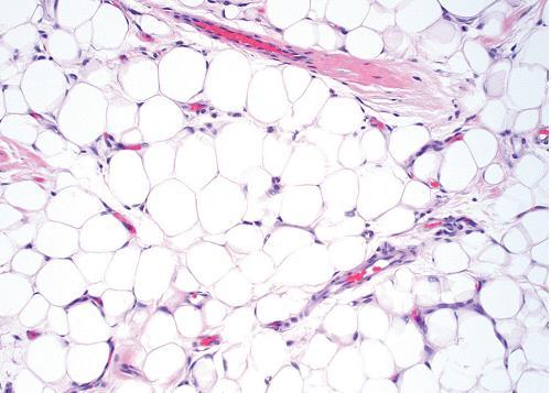
16 LIPOMA Benign tumor of fat Most common mesenchymal neoplasm Trunk and proximal of the extremities Oral and maxillofacial : less frequent More common in obese people Metabolism independent of the normal body fat
17 Nodular masses Sessile or pedunculated Soft Smooth-surfaced Asymptomatic Less than 3 cm Most common intraoral sites : buccal mucosa and buccal vestibule Tongue. floor of the mouth, and lips 40 y/o

18

19
20 HISTOPATHOLOGIC FEATURES Mature fat cells Well circumscribed Lobular arrangement Microscopic variants : o Fibrolipoma o Angiolipoma o Myxoid lipoma o Pleomorphic lipomas o Intramuscular (infiltrating) lipomas
21 TREATMENT AND PROGNOSIS Conservative local excision Recurrence : rare Intramuscular lipomas : higher recurrence
22 NEURILEMOMA (SCHWANNOMA) Benign neural neoplasm of Schwann cell Uncommon Head and neck : 25% to 48% Slow-growing Encapsulated

23
24 Pushes the nerve Asymptomatic Young and middle-aged adults Most common location : tongue Intraosseous : posterior mandible / unilocular or multilocular radiolucencies
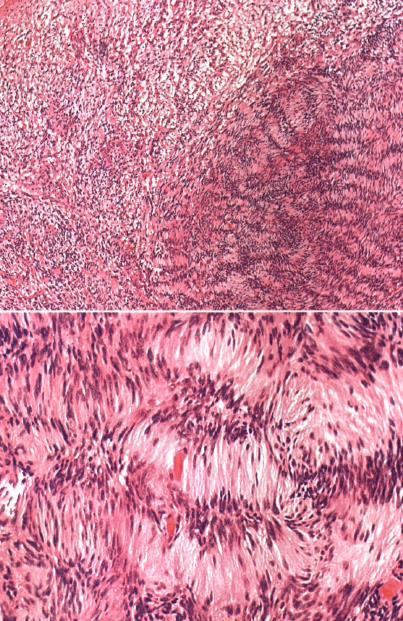
25 HISTOPATHOLOGIC FEATURE Encapsulated Two microscopic patterns : 1)Antoni A 2)Antoni B
26
27 Antoni A o Streaming fascicles of spindle-shaped Schwalm cells o Palisaded arrangement around central acellular, eosinophilic areas known as Verocay bodies Verocay bodies consist of reduplicated basement membrane and cytoplasmic processes Antoni B o Spindle cells o Less cellular and less organized o Randomly arranged

28
29 Ancient neurilemomas : Hemorrhage Hemosiderin deposits Inflammation Fibrosis Nuclear atypia Still benign
30 TREATMENT AND PROGNOSIS Surgical excision Malignant transformation
 Autosomal dominant trait Mutations : NF1")
31 NEUROFIBROMATOSIS TYPE I (VON RECKLINGHAUSEN'S DISEASE OF THE SKIN) Hereditary Most common : neurofibromatosis type I (NFl) Autosomal dominant trait Mutations : NF1 gene
32 Multiple neurofibromas (skin) : Small papules to larger soft nodules to massive baggy (elephantiasis neuromatosa) Plexiform neurofibroma : Bag of worms" Pathognomonic for NF 1 During puberty Wide variability in the expression of the disease : Only a few neurofibromas Hundreds or thousands of tumor

33

34
35 Cafe au lait (coffee with milk) pigmentation on the skin: Another highly characteristic feature Macules Vary in diameter At birth (during the first year) Axillary freckling (Crowe's sign) : Highly suggestive sign Lisch nodules: Translucent brown-pigmented spots on the iris All

36

37
38 The most common finding : enlargement of the fungiform papillae
39 Radiographic findings: 1. Enlargement of the mandibular foramen 2. Enlargement or branching of the mandibular canal 3. Increased bone density 4. Concavity of the medial surface of the ramus 5. Increase in dimension of the coronoid notch Unilateral enlargement that mimics hemifacial hyperplasia
40 TREATMENT AND PROGNOSIS No specific therapy Prevention or management of complications Cancer development : Malignant peripheral nerve sheath tumor Pheochromocytoma Leukemia Rhabdomyosarcoma Wilms tumor Average lifespan (15 years less than the general population)

41
42 MULTIPLE ENDOCRINE NEOPLASIA TYPE 2B (MULTIPLE ENDOCRINE NEOPLASIA TYPE 3; MULTIPLE MUCOSAL NEUROMA SYNDROME) MEN 3 / MEN 2B o MEN syndromes o Group of rare conditions o Tumors or hyperplasias of the neuroendocrine tissues MEN type 1 (benign tumors of the pancreatic islets. adrenal cortex. Parathyroid glands. pituitary gland) MEN type 2A (adrenal pheochromocytomas and medullary thyroid carcinoma) MEN type 2B (mucosal neuromas of oral mucousa)
43 Marfanoid body : thin / elongated limbs / muscle wasting / narrow face Lips : thick and protuberant Oral mucosal neuromas : first sign Neuromas : o Soft o Painless o Papules or nodules o Lips and anterior tongue o Bilateral neuromas of the commissural mucosa (highly characteristic) Pheochromocytomas Neuroendocrine tumors Medullary carcinoma (thyroid gland) : 90% / 18 and 25


44
45 LABORATORY VALUES Medullary carcinoma of the thyroid gland Calcitonin (serum or urinary) Pheochromocytomas Urinary vanillylmandelic acid (VMA) Epinephrine-to-norepinephrine ratios
46 HISTOPATHOLOGIC FEATURES Mucosal neuromas : hyperplasia of nerve bundles


47
48 TREATMENT AND PROGNOSIS Removal of the thyroid gland at an early age
49 HEMANGIOMA AND VASCULAR MALFORMATIONS Hemangiomas Benign tumors of infancy Rapid growth Endothelial cell proliferation Cannot be recognized at birth (during the first 8 weeks) Vascular malformations o Structural anomalies of blood vessels o Without endothelial proliferation o Present at birth and persist throughout life o Categorized : type of vessel / hemodynamic features

50
51 HEMANGIOMA OF INFANCY Most common tumors of infancy Females Whites Most common location: head and neck Single lesions Rapid development
52 Superficial tumors : Bosselated Bright-red color Strawberry hemangioma Firm and rubbery Blood cannot be evacuated by applying pressure Deeper tumors : Bluish hue
53 5 y/o 9 y/o Permanent changes : atrophy, scarring, wrinkling, or telangiectasias Complications : Most common problem : ulceration (secondary infection) Tumors in the neck and laryngeal region : airway obstruction
54 PHACE(S) syndrome Posterior fossa brain anomalies Hemangioma Arterial anomalies Cardiac defects Eye anomalies.sternal cleft Kasabach-Merritt phenomenon Serious coagulopathy (associated with hemangioma)
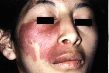
55 VASCULAR MALFORMATIONS 1. Port wine stains : o Common o Capillary malformation o Newborns o Trigeminal nerve o Sturge-Weber angiomatosis o Pink or purple macule
56
57 2. Venous malformations : o At birth o Grow with the patient o Secondary thrombosis 3. Arteriovenous malformations : o Persistent direct arterial and venous communication o Bruit o Overlying skin (warmer.pain. bleeding. Ulceration)
58 INTRABONY VASCULAR MALFORMATIONS Arteriovenous malformations First 3 decades Females Mandible Radiographic appearance : variable Multilocular radiolucent (honeycomb or soap bubble) Ill-defined radiolucent Well.defined(cystlike radiolucency ) o Sunburst" radiographic

59
60 HISTOPATHOLOGIC FEATURES Early hemangiomas of infancy: Numerous plump endothelial Indistinct vascular lumina Juvenile or cellular hemangiomas Mature lesions: Endothelial cells (flattened) Capillary-sized vascular spaces GLUTI


61
62 TREATMENT AND PROGNOSIS Hemangiomas Port wine stains Venous malformations Surgical excision Sclerotherapy
63 LYMPHANGIOMA Benign Hamartomatous tumors of lymphatic Three types of lymphangioma: I. Lymphangioma simplex (capillary lymphangioma) 2. Cavemous lymphangioma : mouth 3. Cystic lymphangioma (cystic hygroma) : neck and axilla

64
65 Head and neck : 50% to 75% At birth : 50% 2 years of age : 90% Oral lymphangiomas : Two thirds of the tongue (macroglossia) Pebbly surface (frog eggs or tapioca pudding) Small lymphangiomas on the alveolar ridge in black neonates
66
67 HISTOPATHOLOGIC FEATURES lymphatic vessels lining endothelium : thin Intraoral tumors : lymphatic vessels Beneath the epithelial surface Translucent, vesicle


68
69 TREATMENT AND PROGNOSIS Surgical excision Recurrence is common (cavernous lymphangiomas)
70 LEIOMYOMA Oral 1. Solid leiomyomas 2. Vascular leiomyomas (angiomyomas) 3. Epithelioid leiomyomas
71 Mucosal nodule Firm Slow-growing Asymptomatic Most common sites : lips, tongue,palate, and cheek Rare intraosseous (unilocular radiolucencies)

72

73

74
75 RHABDOMYOMA 1) Adult rhabdomyomas o Middle-aged and older (Men) o Pharynx, oral cavity. and larynx o Intraoral lesions(floor of the mouth, soft palate and base of tongue) o Rapid growth 2) Fetal rhabdomyomas Young children Face and periauricular region


76
77 OSSEOUS AND CARTILAGINOUS CHORISTOMAS Tumorlike growth Normal tissue in an abnormal location Most common choristomas : bone, cartilage, or both Soft tissue osteomas Soft tissue chondromas Not true neoplasms
78 Sessile or pedunculated nodule Firm Smooth-surfaced Females Tongue : 85% Most common location : posterior tongue near the foramen cecum


79
80
81
82
83
IN THE NAME OF GOD Dr. Kheirandish Oral and maxillofacial pathology
 IN THE NAME OF GOD Dr. Kheirandish Oral and maxillofacial pathology ORAL FOCAL MUCINOSIS Uncommon Tumorlike Cutaneous myxoid cyst Overproduction of hyaluronic acid by firoblasts Young adults Female Gingiva
Malignant Peripheral Nerve Sheath Tumor
 C H A P T E R 120 Malignant Peripheral Nerve Sheath Tumor Currently, malignant peripheral nerve sheath tumor (MPNST) is the most commonly used generic name for the neoplasms known in the past as neurosarcoma,
Pigmented lesions of the Oral cavity
 Oral medicine أ.م.د احسان عبد هللا كميل Pigmented lesions of the Oral cavity Pigmented oral lesions are a large group of disorders in which the dark or brown color is the essential clinical characteristic.
Neurocutaneous Syndromes. Phakomatoses
 Neurocutaneous Syndromes Phakomatoses Financial Disclosures I have NO SIGNIFICANT FINANCIAL, GENERAL, OR OBLIGATION INTERESTS TO REPORT Neurocutaneous Syndomes Definition Entities Diagnosis/ Presentation
Differential Diagnosis of Oral Masses. Gingival Lesions
 Differential Diagnosis of Oral Masses Gingival Lesions Gingival/Alveolar Ridge Masses Parulis Periodontal Abscess Tori and Exostoses Reactive Proliferations Peripheral Odontogenic Cysts Peripheral Odontogenic
4Ps LUMPS AND BUMPS B.L.&T. BUMPS, LUMPS, AND TATTOOS. Most Common BUMP in the oral cavity Fibroma INTERDENTAL PAPILLAE LESIONS
 B.L.&T. BUMPS, LUMPS, AND TATTOOS LUMPS AND BUMPS DIFFERENTIAL DIAGNOSIS FOR LUMPS AND BUMPS Traumatic Fibroma Papilloma Epulis Fissuratum Inflammatory Papillary Hyperplasia Lesions of Attached Gingiva
The Relevance of Cytologic Atypia in Cutaneous Neural Tumors
 The Relevance of Cytologic Atypia in Cutaneous Neural Tumors Recent Findings - New Developments New Problems Zsolt B. Argenyi, M.D. Professor of Pathology & Dermatology Director of Dermatopathology Department
Differential Diagnosis of Radiolucent Lesions of the Jaws
 Differential Diagnosis of Radiolucent Lesions of the Jaws Multilocular Multilocular Radiolucencies Odontogenic Keratocyst Botryoid Odontogenic Cyst Glandular odontogenic Cyst Invasive Ameloblastoma Central
Hemangioma. Hemangioma is an abnormal build up of blood vessels in the skin or internal organs.
 LESSON 5 Oncology. Tumours of maxillofacial area and neck. Hemangioma Hemangioma is an abnormal build up of blood vessels in the skin or internal organs. Causes The classically recognized hemangioma is
Index. oralmaxsurgery.theclinics.com. Note: Page numbers of article titles are in boldface type.
 Index Note: Page numbers of article titles are in boldface type. A Adenomatoid odontogenic tumor, pediatric, 50 51 Ameloblastic carcinoma, pediatric, 17, 49 Ameloblastic fibro-odontoma, pediatric, 54 Ameloblastic
أملس عضلي غرن = Leiomyosarcoma. Leiomyosarcoma 1 / 5
 Leiomyosarcoma 1 / 5 EPIDEMIOLOGY Exact incidence is unknown, but older studies suggest that leiomyosarcomas comprise approximately 3 percent of soft-tissue sarcomas. Superficial leiomyosarcoma occurs
DISORDERS OF THE SALIVARY GLANDS Neoplasms Dr.M.Baskaran Selvapathy S IV
 DISORDERS OF THE SALIVARY GLANDS Neoplasms Dr.M.Baskaran Selvapathy S IV NEOPLASMS A) Epithelial I. Benign Pleomorphic adenoma( Mixed tumour) Adenolymphoma (Warthin s tumour) Oxyphil adenoma (Oncocytoma)
Problem diagnoses. Current issues in Anatomic pathology. Problem Diagnoses in Tumors of the Oral Cavity 5/29/2009
 Current issues in Anatomic pathology Problem Diagnoses in Tumors of the Oral Cavity Richard Jordan DDS PhD FRCPath Professor of Oral Pathology & Pathology Director, UCSF Oral Pathology Diagnostic Laboratory
Differential Diagnosis of Oral Lesions. An Interactive Lecture Using Audience Response Polling. John L. Alonge, MS, DDS
 Differential Diagnosis of Oral Lesions An Interactive Lecture Using Audience Response Polling John L. Alonge, MS, DDS Goals 1. Review the diagnostic process needed to formulate a differential diagnosis
Neurilemmoma of the tongue: A case report
 www.edoriumjournals.com CASE REPORT OPEN ACCESS Neurilemmoma of the tongue: A case report Pallav Kumar Kinra, Jayakumar K, Manoj Joseph Michael ABSTRACT Introduction: Neurilemmomas, also referred to as
5/10. Pathology Soft tissue tumors. Farah Bhani. Mohammed Alorjani
 5/10 Pathology Soft tissue tumors Mohammed Alorjani Farah Bhani Slides are included in this sheet. Objectives: Soft tissue tumors 1. Describe soft tissue tumors. 2. Understand the classification of soft
Case Presentation 主治醫師 : 宋文鑫日期 :
 Case Presentation 主治醫師 : 宋文鑫日期 : 2015-2-28 General Data Name:OOO Chart Number:OOOOOOO Date of Admission:2014 年 08 月 04 日 Age: 33 y/o Sex:female Occupation : 會計 Chief Complaint Palpable soft tissue mass
University of Washington Radiology Review Course: Strange and Specific Diagnoses. Case #1
 University of Washington Radiology Review Course: Strange and Specific Diagnoses Katherine E. Dee, MD Seattle Breast Center Via Radiology 2014 Case #1 37 year old presents with bilateral palpable lumps.
Year 2003 Paper two: Questions supplied by Tricia
 question 43 A 42-year-old man presents with a two-year history of increasing right facial numbness. He has a history of intermittent unsteadiness, mild hearing loss and vertigo but has otherwise been well.
Neoplasia literally means "new growth.
 NEOPLASIA Neoplasia literally means "new growth. A neoplasm, defined as "an abnormal mass of tissue the growth of which exceeds and is uncoordinated with that of the normal tissues and persists in the
Selected Pseudomalignant Soft Tissue Tumors of the Skin and Subcutis
 Selected Pseudomalignant Soft Tissue Tumors of the Skin and Subcutis Andrew L. Folpe, M.D. Professor of Laboratory Medicine and Pathology Mayo Clinic, Rochester, MN folpe.andrew@mayo.edu 2016 MFMER slide-1
Diplomate of the American Board of Pathology in Anatomic and Clinical Pathology
 A 33-year-old male with a left lower leg mass. Contributed by Shaoxiong Chen, MD, PhD Assistant Professor Indiana University School of Medicine/ IU Health Partners Department of Pathology and Laboratory
Case Report A Giant Cell Fibroma and Focal Fibrous Hyperplasia in a Young Child: A Case Report
 Hindawi Publishing Corporation Case Reports in Dentistry Volume 2012, Article ID 370242, 5 pages doi:10.1155/2012/370242 Case Report A Giant Cell Fibroma and Focal Fibrous Hyperplasia in a Young Child:
Congenital Neck Masses C. Stefan Kénel-Pierre, MD
 Congenital Neck Masses C. Stefan Kénel-Pierre, MD SUNY-LICH Medical Center Department of Surgery Case Presentation xx year old male presents with sudden onset left lower neck swelling x 1 week Denies pain,
Course Instructions: Check your for your CE certification of completion (please check your junk/spam folder as well). About SMS CE courses:
. About SMS CE courses:") 2018 Course #4 Self-Study Course Contact Us: Phone 614-292-6737 Toll Free 1-888-476-7678 Fax 614-292-8752 E-mail smsosu@osu.edu Web dentistry.osu.edu/sms Course Instructions: Read and review the course
2018 Course #4 Self-Study Course Contact Us: Phone 614-292-6737 Toll Free 1-888-476-7678 Fax 614-292-8752 E-mail smsosu@osu.edu Web dentistry.osu.edu/sms Course Instructions: Read and review the course
Ancient schwannoma of the mouth floor A case report and review
 Oral Oncology EXTRA (2006) 42, 281 285 available at www.sciencedirect.com journal homepage: http://intl.elsevierhealth.com/journal/ooex CASE REPORT Ancient schwannoma of the mouth floor A case report and
Sonography of soft-tissue vascular lesions
 Sonography of soft-tissue vascular lesions Oscar M. Navarro Associate Professor, University of Toronto Dept. of Diagnostic Imaging, The Hospital for Sick Children Toronto, Canada Declaration of Disclosure
Vascular. Extravasated blood. Melanocytic. Tattoo. Epidermolysis bullosa. Lichen planus. Pemphigoid Pemphigus Lupus. Candidosis. Surface Epithelial
 Oral Soft Tissue Pathology Epithelial Thickening (white) Combination Erythema migrans Epithelial atrophy (red) Surface Lesions Clinical Impression Enlargements Surface Debris Pigmented Vesicular Ulcerated
Desmoplastic Melanoma R/O BCC. Clinical Information. 74 y.o. man with lesion on left side of neck r/o BCC
 R/O BCC Sabine Kohler, M.D. Professor of Pathology and Dermatology Dermatopathology Service Stanford University School of Medicine Clinical Information 74 y.o. man with lesion on left side of neck r/o
Premalignant lesions may expose to a promoting. factor & may be induced to undergo malignant. Carcinoma in situ displays the cytologic features of
 بسم رلاهللا Def. Premalignant lesions may expose to a promoting factor & may be induced to undergo malignant transformation. Carcinoma in situ displays the cytologic features of malignancy without invasion
A CASE OF A Huge Submandibular Pleomorphic Adenoma
 ISPUB.COM The Internet Journal of Head and Neck Surgery Volume 4 Number 2 S VERMA Citation S VERMA.. The Internet Journal of Head and Neck Surgery. 2009 Volume 4 Number 2. Abstract Pleomorphic adenoma
Case Report Basal Cell Ameloblastoma of Mandible: A Rare Case Report with Review
 Case Reports in Dentistry Volume 2013, Article ID 187820, 4 pages http://dx.doi.org/10.1155/2013/187820 Case Report Basal Cell Ameloblastoma of Mandible: A Rare Case Report with Review Hemant Shakya, 1
Vascular tumors & a brief discussion on varicose veins
 Vascular tumors & a brief discussion on varicose veins Classification Locally aggressive but metastasize infrequently Robbins basic pathology 9 th edition Overview Can arise from: endothelium (e.g., hemangioma,
Tumors of Adipose Tissue Tumors Epidemiology Clinical Features. Morphology. Mature Adipocytes Separated by delicate fibrous septa
 Tumors of Adipose Tissue Lipoma Liposarcoma Most commonly happens in female The most common soft tissue tumor o Originates from matured Adipocytes Most commonly happes at the 4 th and 5 th decade of life
SESSION 1: GENERAL (BASIC) PATHOLOGY CONCEPTS Thursday, October 16, :30am - 11:30am FACULTY COPY
 PATHOLOGY CONCEPTS Thursday, October 16, :30am - 11:30am FACULTY COPY") SESSION 1: GENERAL (BASIC) PATHOLOGY CONCEPTS Thursday, October 16, 2008 9:30am - 11:30am FACULTY COPY GOAL: Describe the basic morphologic (structural) changes which occur in various pathologic conditions.
SESSION 1: GENERAL (BASIC) PATHOLOGY CONCEPTS Thursday, October 16, 2008 9:30am - 11:30am FACULTY COPY GOAL: Describe the basic morphologic (structural) changes which occur in various pathologic conditions.
Case Series Oral lipomas: A report of two cases ABSTRACT
 International Journal of Medicine and Biomedical Research Volume 3 Issue 1 January April 2014 www.ijmbr.com Michael Joanna Publications Case Series Oral lipomas: A report of two cases Omisakin O.O 1*,
My Journey into the World of Salivary Gland Sebaceous Neoplasms
 My Journey into the World of Salivary Gland Sebaceous Neoplasms Douglas R. Gnepp Warren Alpert Medical School at Brown University Rhode Island Hospital Pathology Department Providence RI Asked to present
EXTRA-ORAL HEMANGIOMA IN A 10 YEAR OLD GIRL A CASE REPORT WITH LITERATURE REVIEW
 EXTRA-ORAL HEMANGIOMA IN A 10 YEAR OLD GIRL A CASE REPORT WITH LITERATURE REVIEW 1 2 2 2 R.Kannan B. Sekar S.Murali Dominic Augustine 1 2 Department of Oral and Maxillofacial Surgery, Department of Oral
Upper arch. 1Prosthodontics. Dr.Bassam Ali Al-Turaihi. Basic anatomy & & landmark of denture & mouth
 1Prosthodontics Lecture 2 Dr.Bassam Ali Al-Turaihi Basic anatomy & & landmark of denture & mouth Upper arch Palatine process of maxilla: it form the anterior three quarter of the hard palate. Horizontal
Essential Dermatopathology: Neoplastic American Academy of Dermatology Annual Meeting NEURAL AND SMOOTH MUSCLE NEOPLASMS
 Essential Dermatopathology: Neoplastic American Academy of Dermatology Annual Meeting NEURAL AND SMOOTH MUSCLE NEOPLASMS Kevin P. White M.D. Oregon Health and Science University Associate Professor of
Neoplasia part I. Dr. Mohsen Dashti. Clinical Medicine & Pathology nd Lecture
 Neoplasia part I By Dr. Mohsen Dashti Clinical Medicine & Pathology 316 2 nd Lecture Lecture outline Review of structure & function. Basic definitions. Classification of neoplasms. Morphologic features.
Epithelial tumors. Dr. F.F. Khuzin, PhD Dr. M.O. Mavlikeev
 Epithelial tumors Dr. F.F. Khuzin, PhD Dr. M.O. Mavlikeev Epithelial tumors Tumors from the epithelium are the most frequent among tumors. There are 2 group features of these tumors: The presence in most
Adrenal masses in infancy and childhood: A clinical and radiological overview M. Mearadji
 Adrenal masses in infancy and childhood: A clinical and radiological overview M. Mearadji International Foundation for Pediatric Imaging Aid Introduction Neoplastic adrenal masses usually originate from
NEOPLASIA-I CANCER. Nam Deuk Kim, Ph.D.
 NEOPLASIA-I CANCER Nam Deuk Kim, Ph.D. 1 2 Tumor in the hieroglyphics of the Edwin Smith papyrus (1,600 B.C., Breasted s translation 1930) 3 War on Cancer (National Cancer Act, 1971) 4 Cancer Acts in Korea
Congenital and Neonatal Lumps and Bumps. Diagnostico y manejo de las manchas y tumoraciones cutaneas congenitas en el neonato
 Congenital and Neonatal Lumps and Bumps Diagnostico y manejo de las manchas y tumoraciones cutaneas congenitas en el neonato Miriam Weinstein MD FRCPC Hospital for Sick Children, Toronto ALAPE Cartagena,
Kidney Case 1 SURGICAL PATHOLOGY REPORT
 Kidney Case 1 Surgical Pathology Report February 9, 2007 Clinical History: This 45 year old woman was found to have a left renal mass. CT urography with reconstruction revealed a 2 cm medial mass which
SOFT TISSUE TUMOR PATHOLOGY: AN UPDATE
 SOFT TISSUE TUMOR PATHOLOGY: AN UPDATE Jason L. Hornick, MD, PhD July 18, 2013 Department of Pathology Brigham and Women s Hospital Harvard Medical School Boston, MA, USA I have no disclosures. New Soft
Subdivided into Vestibule & Oral cavity proper
 Extends from the lips to the oropharyngeal isthmus The oropharyngeal isthmus: Is the junction of mouth and pharynx. Is bounded: Above by the soft palate and the palatoglossal folds Below by the dorsum
DUSTURBANCES OF GROWTH. MLS Basic histological diagnosis MLS HIST 422 Semester 8- batch 7 L8 Uz: Musa
 DUSTURBANCES OF GROWTH MLS Basic histological diagnosis MLS HIST 422 Semester 8- batch 7 L8 Uz: Musa Agnesia: means complete absence of an organ (Kidney). Aplasia: s defined in general as "defective development
Gross appearance of peritoneal cysts. They have a thin, translucent wall and contain a clear fluid.
 Gross appearance of peritoneal cysts. They have a thin, translucent wall and contain a clear fluid. So-called multicystic benign mesothelioma. A, Gross appearance. So-called multicystic benign mesothelioma.
An Overview of Genital Stromal Tumors
 An Overview of Genital Stromal Tumors By Konstantinos Linos MD, FCAP, FASDP Bone, Soft Tissue and Dermatopathology Assistant Professor of Pathology Dartmouth-Hitchcock Medical Center Geisel School of Medicine
the urinary system pathology Dr. Fairoz A Eltorgman
 the urinary system pathology Dr. Fairoz A Eltorgman Tumors of the renal pelvis & kidney Benign tumors of the renal pelvis: Hemangioma Leiomyoma Malignant tumors: Transitional cell carcinoma Squamous cell
الفتوي الاصفر الحبيبوم = Xanthogranuloma_Juvenile JUVENILE XANTHOGRANULOMA 1 / 9
 JUVENILE XANTHOGRANULOMA 1 / 9 Clinical Findings CUTANEOUS LESIONS JXG is a benign, self-healing disorder that is characterized by asymptomatic yellowish papulonodular lesions of the skin and other organs
Spindle Cell Lesions Of The Breast. Emad Rakha Professor of Breast Pathology and Consultant Pathologist
 Spindle Cell Lesions Of The Breast Emad Rakha Professor of Breast Pathology and Consultant Pathologist * SCLs comprise a wide spectrum of diseases, ranging from reactive processes to aggressive malignant
Vascular Malformations Of The Oral Cavity In Children And Young Adolescents Insights Into Their Pathogenesis.
 ISPUB.COM The Internet Journal of Pediatrics and Neonatology Volume 12 Number 2 Vascular Malformations Of The Oral Cavity In Children And Young Adolescents Insights Into Their D Shetty, H Rai, P Rastogi,
Diseases of the breast (1 of 2)
") Diseases of the breast (1 of 2) Introduction A histology introduction Normal ducts and lobules of the breast are lined by two layers of cells a layer of luminal cells overlying a second layer of myoepithelial
Diseases of the breast (1 of 2) Introduction A histology introduction Normal ducts and lobules of the breast are lined by two layers of cells a layer of luminal cells overlying a second layer of myoepithelial
Vascular Tumors in Children and Adults. Thuy Phung, MD, PhD Houston Methodist Hospital Texas Children s Hospital Baylor College of Medicine
 Vascular Tumors in Children and Adults Thuy Phung, MD, PhD Houston Methodist Hospital Texas Children s Hospital Baylor College of Medicine What are these lesions? (Marcelo Hochman, MD) What are these lesions?
HEAD AND NECK PATHOLOGY
 Bosnian-British School of Pathology November 2012 HEAD AND NECK PATHOLOGY Slide seminar: Oral Pathology Preferred Diagnoses Dr A Sandison, Slide seminar: Pathology of the Oral Cavity Page 1 of 5 1. Female
Case Report Fibrolipoma of the Buccal Mucosa: A Case Report and Review of the Literature
 Case Reports in Pathology Volume 2016, Article ID 5060964, 4 pages http://dx.doi.org/10.1155/2016/5060964 Case Report Fibrolipoma of the Buccal Mucosa: A Case Report and Review of the Literature Masayasu
Contents Part I Introduction 1 General Description 2 Natural History: Importance of Size, Site, Histopathology
 Contents Part I Introduction 1 General Description... 3 1.1 Introduction... 3 1.2 Incidence and Prevalence... 5 1.3 Predisposing and Genetic Factors... 8 References... 16 2 Natural History: Importance
INFECTION. HIV Infection DWI
 HIV Infection INFECTION DWI Fig Axial CT and MRI images show multiple enlarged lymph nodes in the neck as well as in the parotid gland bilaterally. These nodes were suppurative with positive diffusion.
Neckmasses in infancy and childhood: Clinical and radiological classification and imaging approaches M. Mearadji
 Neckmasses in infancy and childhood: Clinical and radiological classification and imaging approaches M. Mearadji International Foundation for Pediatric Imaging Aid Introduction Neck masses are a frequent
Index. J Juvenile hyaline fibromatosis, 27 Juvenile xanthogranuloma, 57 Juxta-articular myxoma, 152
 A Adenomatoid tumor, 76, 77 Adipose tissue tumors benign tumors angiolipoma, 6 chondroid lipoma, 9 fibrolipoma, 5 hibernoma, 8 lipoblastoma, 9 lipoma (see Lipoma) myelolipoma, 6 pleomorphic lipoma, 8 spindle
Clinical History. 29 yo woman with polyhydramnios Cardiac mass at fetal ultrasound At 35 weeks, newborn died 30 minutes after delivery
 CASE 1 a Clinical History 29 yo woman with polyhydramnios Cardiac mass at fetal ultrasound At 35 weeks, newborn died 30 minutes after delivery Interface between tumor and normal myocardium Smaller well-demarcated
MVST BOD & NST PART IB Thurs. 2 nd & Fri. 3 rd March 2017 Pathology Practical Class 23
 MVST BOD & NST PART IB Thurs. 2 nd & Fri. 3 rd March 2017 Pathology Practical Class 23 Neoplasia I Neoplasia I: Benign and malignant neoplasms in glandular epithelium and mesenchyme 1.0. Aims 1. To understand
PEDIATRICS WK 3 HEAD AND NECK ALISON WALLACE MD, PHD
 PEDIATRICS WK 3 HEAD AND NECK ALISON WALLACE MD, PHD Topics 1. Cervical lymphadenopathy 2. Lymphatic malformation 3. Thyroglossal duct cysts 4. Branchial cleft cysts 5. Thyroid masses CASE 1 Case 1 A 2
ORAL FOCAL MUCINOSIS: A RARE CASE REPORT OF TWO CASES
 Oral Focal Micinosis: A Rare Case Sharma E. et al 178 CASE REPORT ORAL FOCAL MUCINOSIS: A RARE CASE REPORT OF TWO CASES Sharma Ena 1, Nadella Manjari 1, Chatterjee Anirban 1, Alampalli Ramesh 1 ABSTRACT
Von Recklinghausen s Disease with a Giant Lipoma
 Von Recklinghausen s Disease with a Giant Lipoma Daiki Iwana¹( ) Kazutaka Izawa¹ Mitsuhiro Kawamura¹ Takaharu Nabeshima¹ Hideki Yoshikawa² ¹Department of Orthopaedic Surgery, Toneyama National Hospital,
PLEOMORPHIC ADENOMA ( BENIGN MIXED TUMOR )
") ( BENIGN MIXED TUMOR ) Grossly, the tumor is freely movable, solid, sometimes lobulated and occasionally cystic. If recurrent, multinodular masses are common. Histologically, within a fibrous capsule,
( BENIGN MIXED TUMOR ) Grossly, the tumor is freely movable, solid, sometimes lobulated and occasionally cystic. If recurrent, multinodular masses are common. Histologically, within a fibrous capsule,
Chief Complaint. Retroperitoneal cystic mass incidentally found at health examination center.
 Personal Information Age: 34 y/o Sex: female Past history: major systemic medical history(-) surgical history(-), family history(-) Denied food or drug allergy Chief Complaint Retroperitoneal cystic mass
Benign and malignant epithelial lesions: Seborrheic keratosis: A common benign pigmented epidermal tumor occur in middle-aged or older persons more
 Benign and malignant epithelial lesions: Seborrheic keratosis: A common benign pigmented epidermal tumor occur in middle-aged or older persons more common on the trunk; but extremities, head and neck are
A swelling of the lateral hard palate
 A swelling of the lateral hard palate Jeanne Leduc, DMD Private practice in Longueuil, QC Canada (this manuscript was written while she was a dental resident at the Faculty of Dental Medicine, Universite
Conflict of Interest: none. Neonatal Airway Masses. Neonatal Respiratory Papillomatosis. Paul J. Samuels, MD
 Paul J. Samuels, MD Professor of Anesthesiology and Pediatrics Director of Education Cincinnati Children s Hospital Cincinnati, Ohio Conflict of Interest: none Neonatal Respiratory Papillomatosis Caused
Case Report Hypertrichotic Giant Nevus Spilus Tardivus and Neurofibroma of the Tongue in Sporadic von Recklinghausen s Disease
 Case Reports in Dermatological Medicine, Article ID 141075, 6 pages http://dx.doi.org/10.1155/2014/141075 Case Report Hypertrichotic Giant Nevus Spilus Tardivus and Neurofibroma of the Tongue in Sporadic
Advances In Orbital Neuropathology
 Advances In Orbital Neuropathology Charles G. Eberhart, MD PhD Associate Professor of Pathology, Ophthalmology and Oncology Johns Hopkins University School of Medicine Overview Non-neoplastic lesions Microphthalmos/pseudoglioma
CYSTIC TUMORS OF THE KIDNEY JOHN N. EBLE, M.D. CYSTIC NEPHROMA
 Page 1 CYSTIC TUMORS OF THE KIDNEY JOHN N. EBLE, M.D. Department of Pathology & Laboratory Medicine Phone (317) 274-4806 Medical Science A-128 FAX: (317) 278-2018 635 Barnhill Drive jeble @iupui.edu Indianapolis,
An Overview of Cutaneous Vascular Neoplasms
 An Overview of Cutaneous Vascular Neoplasms By Konstantinos Linos MD, FCAP, FASDP Bone, Soft Tissue and Dermatopathology Assistant Professor of Pathology Dartmouth-Hitchcock Medical Center Geisel School
Juvenile Angiofibroma
 Juvenile Angiofibroma Disclaimer The pictures used in this presentation have been obtained from a number of sources. Their use is purely for academic and teaching purposes. The contents of this presentation
Pathology of Mediastinal Tumors
 SAMO Meeting Lucerne 2009 Pathology of Mediastinal Tumors Alex Soltermann Most common lesions (adults) Clinical presentation 50% of the patients are asymptomatic, lesion discovered incidentally Symptoms
CELL AND TISSUE INJURY COURSE-II PATHOLOGY LABORATORY. PATHOLOGY of MASS LESIONS and TISSUE DEFECTS -MACROSCOPY Assoc. Professor Rengin Ahıskalı
 CELL AND TISSUE INJURY COURSE-II PATHOLOGY LABORATORY PATHOLOGY of MASS LESIONS and TISSUE DEFECTS -MACROSCOPY Assoc. Professor Rengin Ahıskalı M1 - RENAL TUBERCULOSIS cavitary areas caseous necrosis fibrous
Dysplasia, Mimics and Other Controversies
 Dysplasia, Mimics and Other Controversies Mary S. Richardson, MD Dept. of Pathology Medical University of South Carolina Charleston, SC Notice of Faculty Disclosure In accordance with ACGME guidelines,
Chief complaint. A mass at right chest
 Chief complaint A mass at right chest Present illness This 1-year-5-month-old girl had a mass at right side chest since one month ago. flat and not tender at first In the recent 2 days, the mass enlarged
Case 9087 Retropharyngeal nodular fasciitis
 Case 9087 Retropharyngeal nodular fasciitis Santiago I 1; Cavalheiro F 2; Noruégas MJ 3; Sanches MC3 1 Hospital Infante D. Pedro, Aveiro, Portugal 2 Hospitais da Universidade de Coimbra, Portugal 3 Hospital
Hemangioma of Tongue with Phlebolith: A Rare presentation
 Journal of Government Dental College and Hospital, October 2017, Vol.-04, Issue- 01, P. 20-25 Original article: Hemangioma of Tongue with Phlebolith: A Rare presentation 1 Dr. Jigna S Shah (MDS) 1, 2 Dr.
Neurofibromatosis type 1 and RASopathies
 Neurofibromatosis type 1 and RASopathies Dawn Siegel, MD Medical College of Wisconsin American Academy of Dermatology San Diego, CA February 19 th, 2018 Neurofibromatosis Type 1 NF1- diagnostic criteria
Neck lumps in children
 Neck lumps in children Midline Lateral Midline neck lumps Thyroglossal cyst - 80% Dermoid cyst Submental lymph node Ectopic thyroid Some rare lesions Thyroglossal cyst Diagnosis: midline, usually overlying
Essentials in Head and Neck Embryology. Part 3 Development of the head, face, and oral cavity
 Essentials in Head and Neck Embryology Part 3 Development of the head, face, and oral cavity Outline General overview of prenatal development Embryonic period phase 1 Formation of bilaminar disk Formation
Atlas of Eyelid and Conjunctival Tumors
 Atlas of Eyelid and Conjunctival Tumors Jerry A. Shields, M.D. Director, Ocular Oncology Service Wills Eye Hospital Professor of Ophthalmology Thomas Jefferson University Philadelphia, Pennsylvania Carol
List the conditions known as neurophakomatosis and demonstrate their clinical findings:
 Neurophakomatosis: List the conditions known as neurophakomatosis and demonstrate their clinical findings: Phacos (Greek): mole or freckle. Neurologic abnormalities combined with skin or retinal pigmented
Evening Specialty Conference Bone and Soft Tissue Pathology. Diagnostic pitfalls in bone and soft tissue pathology
 Evening Specialty Conference Bone and Soft Tissue Pathology. Case 1 Elizabeth G Demicco, MD, PhD Mount Sinai Hospital, New York Disclosure of Relevant Financial Relationships USCAP requires that all planners
AMELOBLASTIC FIBROMA: A RARE CASE REPORT
 Case Report International Journal of Dental and Health Sciences Volume 04, Issue 03 AMELOBLASTIC FIBROMA: A RARE CASE REPORT Namratha Patil 1 1.Sr lecturer, dept of oral medicine and radiology, KAHES VK
Case year female. Routine Pap smear
 Case 1 57 year female Routine Pap smear Diagnosis? 1. Atypical glandular cells of unknown significance (AGUS) 2. Endocervical AIS 3. Endocervical adenocarcinoma 4. Endometrial adenocarcinoma 5. Adenocarcinoma
3/27/2017. Disclosure of Relevant Financial Relationships
 Ophthalmic Pathology Evening Specialty Conference USCAP 2017 5 th March, 2017 Mukul K. Divatia, MD Assistant Professor Department of Pathology & Genomic Medicine Weill Cornell Medical College Houston Methodist
Case year old Chinese female. Radiological echo-distortion in the right breast at o clock. Core biopsy of the o clock lesion.
 Case 3 64 year old Chinese female. Radiological echo-distortion in the right breast at 10-12 o clock. Core biopsy of the 11-12 o clock lesion. Division of Pathology Courtesty of Dr Lester Leong ill-defined,
Grading of Bone Tumors
 Grading of Bone Tumors Joon Hyuk Choi, M.D. Department of Pathology College of Medicine, Yeungnam University Introduction to grading system of bone tumor used at Mayo Clinic WHO Histologic Classification
Normal endometrium: A, proliferative. B, secretory.
 Normal endometrium: A, proliferative. B, secretory. Nội mạc tử cung Nội mạc tử cung Cyclic changes in endometrium.. Approximate relationship of useful microscopic changes. Arias-Stella reaction in endometrial
Differential Diagnosis of Oral Masses. Palatal Lesions
 Differential Diagnosis of Oral Masses Palatal Lesions Palatal Masses Periapical Abscess Torus Palatinus Mucocele Lymphoid Hyperplasia Adenomatous Hyperplasia Benign Salivary Neoplasms Malignant Salivary
Primary bone tumors > metastases from other sites Primary bone tumors widely range -from benign to malignant. Classified according to the normal cell
 Primary bone tumors > metastases from other sites Primary bone tumors widely range -from benign to malignant. Classified according to the normal cell counterpart and line of differentiation. Among the
Fun with Fat. General Rules. Case
 Fun with Fat General Rules Imaging: location (deep vs. superficial) Superficial lesions are seldom liposarcomas Deep lesions may be benign or malignant Myxoid stroma is common in benign and malignant lesions
JMSCR Vol 04 Issue 11 Page November 2016
 www.jmscr.igmpublication.org Impact Factor 5.244 Index Copernicus Value: 83.27 ISSN (e)-2347-176x ISSN (p) 2455-0450 DOI: https://dx.doi.org/10.18535/jmscr/v4i11.117 Original Article Demographic Profile