Imaging Techniques in Radiation Therapy
|
|
|
- Delphia Watkins
- 5 years ago
- Views:
Transcription
1 Imaging Techniques in Radiation Therapy Wuhan University Medical Physics Course June 5, 2015 Jianguo Qian, PhD, DABR Medical Physicist Department of Radiation Oncology & Molecular Radiation Sciences Johns Hopkins University
2 Statement All materials used in this lecture were used for teaching purpose only without commercial or financial interest. Some materials in this lecture may come from public internet and previously collected resources whose original sources could not be identified. If you notice any material missing appropriate citation, please let me know.
3 Outline X-ray Imaging (2D & 3D) Magnetic Resonance Imaging (MRI) Nuclear Medicine Imaging (Scintigraphy, SPECT & PET) Ultrasound Imaging (Ultrasonography)
4 X-ray 2D Imaging Radiography Mammography Fluoroscopy Angiography Linac-based MV & kv imaging

5 X-ray System Principle of an X-ray system with image intensifier. X rays impinging on the image intensifier are transformed into a distribution of electrons, which produces an amplified light image on a smaller fluorescent screen after acceleration. The image is observed by a television camera and a film camera and can be viewed on a computer screen and stored on a CD-ROM or a PACS.

6 X-Ray Imaging Principle The intensity of the transmitted beam as a function of the attenuation coefficient of the pixels traversed. Top part, the intensity after crossing one volume element; bottom part, after traversing n volume elements

7 First medical imaging technology Radiography Became possible after physicist Wilhelm Roentgen discovered X- rays on November 8, 1895 First radiograph ---- Roentgen s hand


8 Radiography Systems

9 Fluoroscopy Real-time radiography, i.e. continuous acquisition of a sequence of x-ray images over time, essentially a realtime x-ray movie of the patient.


10 Mammography A radiography system specifically designed for breast imaging using much lower x-ray energies (usually ~30kVp)


11 Angiography Angiography (or arteriography) is a medical imaging technique used to visualize the inside, or lumen, of blood vessels and organs of the body, with particular interest in the arteries, veins, and the heart chambers.

12 Angiography and Liver Cancer Johns Hopkins Medicine Department of Gastroenterology & Hepatology


13 Types of Angiography Conventional Angiography (CA) Digital Subtraction Angiography (DSA) CA and DSAs at three progressive time points during contrast material injection Hendee & Ritenour, Medical Imaging Physics, 4 th Edition, Pg.245

14 Linac-Based X-ray Imaging System Purpose 1. Verify treatment setup patient position and iso position 2. Verify aperture shape of treatment fields (MV)

15 Linac-Based MV 2D Imaging Du, et al., JACMP, 11(4):3297, 2010

16 Linac-Based kv 2D Imaging Department of Radiation Oncology, UCSD


17 SRS/SBRT imaging ExacTrac System Cyberknife
18 X-ray 3D Imaging Computed Tomography (CT) 4D CT CBCT 4D CBCT
19 CT Principle Abdullah et al., Optical Engineering, 52(3),

20 CT Reconstruction E. Kristensson, et. al, Opt. Express 20, (2012);

21 CT Reconstruction Algorithms Courtesy of Mark Bangert through Matlab Central


22 CT Evolution 1 st generation 2 nd generation 3 rd generation 4 th generation 5 th generation Hendee & Ritenour, Medical Imaging Physics, 4 th Edition, Pg.254, 257
.")

23 CT Scanning Mode Sequential scan (step-and-shoot) and Spiral scan (continuous) Single-slice scan and multi-slice scan Radaideh, M. M. et al. (2001). Using multi-slice spiral CT scanner: The principles you need. The University of Texas MD Anderson Cancer Center.
24 CT Application in Treatment Planning Delineation of target volume and the surrounding structures in relation to the external contour Providing quantitative data (in the form of CT numbers) for tissue heterogeneity corrections


25 CT scanner and CT simulator Special features of CT simulator Flat table top Laser system Software
 and embedded lasers on CT to mark patient AAPM")
26 CT simulation with ordinary CT scanner Create new CT number to density table Put on a flat table Using small X-ray opaque ball bearings (BBs) and embedded lasers on CT to mark patient AAPM TG-66
27 CT Calibration CT dose - a measure of dose delivered during a scan CT resolution ability to distinguish two very small objects in close proximity CT contrast - ability to resolve relatively large objects with small density difference from background CT uniformity evaluation of CT image noise and artifacts CT geometry ability to accurately reproduce patient dimensions and shape in CT images without distortion CT number ability to accurately represent density of objects


28 CT Phantoms CTDI Phantom ACR Phantom CatPhan 700 Philips Daily QA phantom


29 CatPhan CT AAPM TG-66


30 CT number to density table Purpose in Pinnacle planning system: Look up mass attenuation coefficients and Scale the dose deposition kernel to include the effects of inhomogeneities on scattered radiation CIRS RMI
31 RMI Phantom Data


32 CT Density Curve Saw, et al., Med Dosim. 2005, 30(3):145-8

33 CT Artifacts Definition: A distortion or error in an image that is unrelated to the subject being imaged.
34 Source of CT Artifacts Patient Imaging protocols Imaging processes Equipment
35 Typical CT Artifacts Metal artifact Beam hardening artifact Ring artifact Motion artifact

36 Reason: Presence of metallic objects Solution: Remove external metallic objects Using metal artifact reduction image reconstruction algorithm, such as Philips OMAR Metal Artifact
 Decrease slice thickness Adding filter (bowtie")
37 Beam Hardening Artifact Reason: Hardened beam is less attenuated by tissue Solution: Inrease beam energy (kv) Decrease slice thickness Adding filter (bowtie for CBCT)

38 Ring Artifact Reason: One or more detector having malfunction or failure Solution: Scanner recalibration Detector replacement Image: St-Amant M., et al. on

39 Motion Artifact Reason: Voluntary patient motion Involuntary patient motion Solution Voluntary: Use positioning aids Explain to patient Involuntary: Use fast scanning protocol Audio or video coaching, Use motion management procedure such as 4D CT
40 4D CT One more dimension was involved in CT imaging Patient breathing phase or amplitude (or, time) Series of CT data are collected with the patient free-breathing and correlated to patient s respiratory trace Multi-phase reconstruction of the same scanned area can easily show the displacement of a lesion over time, during the normal respiration cycle. 4DCT allows the clinician to evaluate the tumor and tumor motion, plan a more accurate radiotherapy treatment.

41 Tumor motion effect Traditional CT MIP of 4D CT


42 Courtesy of Peter Maxim, Stanford University 4DCT GE LightSpeed + RPM RPM system Patient setup 4D CT workstation Select phase(s) 4D planning GE cine mode Infrared reflective markers Infrared camera

43 Real-time Position Management RPM software records respiratory trace (i.e., vertical block position vs. time)

44 4D CT scanning on GE Cine mode: step-and-shoot; multiple gantry rotations at each bed position
45 4D CT scan phase 50%

46 4DCT Philips Brilliance + Bellows Courtesy of John Givens, Philips Healthcare Inc.


47 The Philips Bellows Device A pneumatic mechanism measuring changes in pressure caused by respiratory motion via a transducer linked to the Brilliance CT scanner.

48 Amplitude vs Linear Phase Binning Phase-based binning can produce badly misaligned slices within an image volume 0% 25% 50% 75%

49 Amplitude vs Linear Phase Binning Phase-Based Amplitude-Based

50 Cone Beam CT (CBCT) MacDonald-Jankowski DS, Asian Journal of Oral and Maxillofacial Surgery 2006;18:

51 Linac-based kv CBCT Zhen, et al., 2012 Phys. Med. Biol


52 Linac-based MV CBCT Department of Radiation Oncology, UCSF

53 CBCT-guided Linac D. Jaffray et al. Int J. Radiat. Oncol. Biol. Phys. 2002
, pp.")
54 CBCT Scanning Mode Son, et al., JACMP 15(2), pp.4556, 2014 Lehmann, et al., JACMP 8(3), pp.2354, 2007

55 CBCT-guided radiation therapy

56 CBCT-guided radiation therapy

57 Linac-based 4D CBCT 4D CBCT 8x84 projections 3D CT 670 projections Sonke, AAPM 2006 summer school
58 MRI Principle Typical MRI Images MRI Application in Radiation Therapy Image Fusion MRI-based planning MRI-guided radiation therapy system

59 MRI Basics Nuclear Magnetic Dipole Moment : Spinning Charge P = No Net Magnetization Hydrogen


60 Dipole Moment of Bulk Hydrogen B 0 Net Magnetization
61 Longitudinal Relaxation (T1) and Transverse Relaxation (T2) Hendee, et al., J. Med :
62 MRI Images T1WI T2WI PDWI DWI ADC GE Perfusion images fmri BOLD images MRA MRV Post-Gd images Volumetric images MR arthrograms FLAIR STIR Etc Presentation of Kulkarni, K., NHS UK
63 Typical MRI images T1-weighted: Fat bright, good for look at brain structure Usually for normal anatomical details T2-weighted Water and fluid brighter, ideal for tissue oedema Usually for pathology since tissues with disease tends to have higher water content FLAIR Fluid Attenuation Inversion Recovery Water suppression technique, good for cerebral oedema and periventricular or cortical lesions STIR Short T1 Inversion Recovery Fat suppression technique, good for adrenal glands, bone marrow and fatty tumors Weegenaar C, Magnetic Resonance Imaging e-tutorial
 appears black in T1 and white in T2.")
64 T1-weighted vs T2-weighted MRI - White matter appears a light grey in T1 and a dark grey in T2. - Grey matter appears grey in both. - Cerebrospinal fluid (CSF) appears black in T1 and white in T2. Weegenaar C, Magnetic Resonance Imaging e-tutorial


65 FLAIR
66 MRI application in radiation therapy Used to delineate anatomical structures or treatment target accurately Image fusion Target volume determination such as Y90 brachytherapy MRI-based treatment planning MRI-guided treatment device
 shows a hyperdense lesion (arrow) in the right frontoparietal region, with surrounding white matter edema and mass effect.")
67 MRI vs CT MRI advantage - Excellent and controllable soft tissue contrast Figure 1 (A, B): A 16-year-old boy developed altered sensorium followed by loss of consciousness. Plain CT scan (A) shows a hyperdense lesion (arrow) in the right frontoparietal region, with surrounding white matter edema and mass effect. Axial plain T1W MRI (B) at the corresponding level shows a hyperintense periphery (arrow) due to presence of intracellular methemoglobin and central hypointensity (arrowhead) due to deoxyhemoglobin Kembhavi, et al. Neuroradiology, 22(2), pp98-105, 2012

68 MRI-CT fusion Figure 1. Axial CT- 3T MRI scan fusion. The red line is the MRI-delineated prostate contour, the purple line is the CTdelineated prostate contour and the blue line is the contour of the rectum delineated on CT-MRI fusion. Ingrosso, et al. Open Journal of Radiology, 1(1), 2011
69 MRI-based Treatment Planning Current stage: MRI/CT based Process CT scan + MRI scan Register MR contours to CT (rigid/deformable) Transfer MR contours to CT data set Planning Future: MRI alone radiation treatment planning (RTP)
70 Challenges for MRI-based RTP No electron/physical density information MRI geometric distortion Magnetic field inhomogeneity Nonlinear gradients from RF excitation Displacement of signal from actual location

71 Density-Assigned MRI-based RTP Comparison of isodose distributions between (a) MRI W -based, (b) MRI W+B -based, (c) MRI W+B+A -based, and (d) CT-based inverse-optimized IMRT plans for Patient 2. Images were obtained at the identical axial level based on CT-MRI registration. Isodose distributions were calculated on CT with heterogeneity correction to standardize comparisons. Dark blue segment delineates PTV70. White and black arrows highlight areas of under- and over- coverage, respectively Chin, et al., JACMP, 15(5) 4851, 2014

72 MRI-guided Radiotherapy System Viewray MRIdian system

73 MRIdian Components Courtesy of Lasitha Senadheera, ViewRay Inc.


74 CBCT vs MRI
75 Nuclear Medicine Imaging (NMI) X-ray, CT, MRI or ultrasonography Basically transmission imaging Mainly provide structural (anatomical) information May consist true physical parameters NMI Basically emission imaging Mainly provide functional information: physiology, metabolism, biochemistry at molecular level

76 Isotopes in Clinical NMI Adapted from presentation of Frederic H. Fahey, Harvard Medical School
77 NMI Modalities Scintigraphy (2D) Single photon emission computed tomography (SPECT) Positron emission tomography (PET)
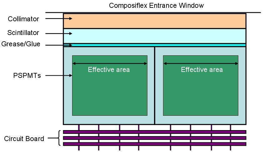
78 Scintigraphy and SPECT Gamma ray Parallel Collimator or Pinhole Collimator Pixellated Scintillator object Photons PSPMT Thansverse view

79 A Mouse-Sized Scintigraphy Detector


80 Scintigraphy Lactating Mouse Mice with tumors Each mouse was injected with about 14 µci Na 125 I.


81 Parallel-Hole SPECT Coronal images Sagittal images The mouse was injected with about 14 µci Na 125 I. Transaxial images

82 Pinhole SPECT 130 µci Na 125 I; Two-pinhole helical SPECT 200 µci Na 125 I; Two-pinhole helical SPECT
83 Scintigraphy Application in Radiation Therapy Lung shunt fraction determination for Y-90 Therasphere liver cancer brachytherapy Figure 4: During the mapping angiogram phase, 5 mci of technetium-99m labeled macroaggregated albumin was injected within the right hepatic artery to quantify the liver-lung shunt. The planar scintigraphic images demonstrate distribution of the radiotracer within the right lobe of the liver without any abnormal extrahepatic distribution. Regions of interest were drawn within the lungs and liver resulting in an estimated maximum lung shunting of 3%. Chamarthy, Case Rep Radiol. 2012:236732
84 SPECT Application in Radiation Therapy Assess cardiac toxicity of RT in breast cancer patients using myocardial perfusion SPECT Lung perfusion SPECT-guided IMRT planning to reduce dose delivery to highly functional lung McGuire, et al.,phys Med Biol. 2010;55(2):403-16
85 PET Li Z., et al. Advanced Drug Delivery Reviews 62 (2010)

86 PET Detector Siemens Healthcare Inc.
87 Clinical SPECT vs PET SPECT Resolution degrade with increasing object-detector distance Limited sensitivity, difficult for dynamic imaging Longer half-life radioisotope Less expensive PET Resolution has less dependence on object-detector distance High sensitivity, feasible do dynamic imaging Limited half-life radioisotope Expensive
 Adaptive radiation")
88 PET Application in Radiation Therapy Functional image-guided radiation therapy planning (PET/CT) Adaptive radiation therapy

89 PET-guided radiation therapy planning Department of Radiation Oncology, UCLA

90 PET-guided Adaptive Radiation Therapy Grootjans, et al., Nature Reviews Clinical Oncology, 2015
91 Ultrasound Imaging Advantage High sensitivity Technically easier to use Bedside test No radiation/contrast exposure Safe and well accepted Less expensive and widely available Disadvantage Operator-dependent Limited penetration (obesity) Small field of view False-negative in case of superficial and rare lesion

92 Ultrasound Imaging Basics Perry Sprawls, Ultrasound Production and Interaction
93 Two Major Effects Piezoelectric effect (Ultrasound probe, or transducer) Electric field causing certain crystal oscillate mechanically, thus generating acoustic waves ressure variation on the crystal causing electric potential Acoustic impedance A measure of how difficult the sound go through the material Defined as speed of sound x density of material Ultrasound reflections (echoes) are caused by variations in acoustic impedance of materials on opposite sides of the interfaces


94 A-mode and B-mode Scans A-mode Time/Amplitude B-mode Intensity Imaging Yale Fisher, Essential lectures in Ophthalmic Ultrasound


95 Ultrasound Image Courtesy of Wu Liu, Yale University

96 Ultrasound-based Prostate Seed Implant Brachytherapy Ultrasound image-based pre-implant planning


97 Ultrasound-based Prostate Seed Implant Brachytherapy Real-time ultrasound-guided implant procedure

98 Ultrasound-based Prostate Seed Implant Brachytherapy CT-based post-implant seed position verification


99 Ultrasound-guided Radiation Therapy Elekta Clarity
100 Thank you!
Imaging Rotation. University of Michigan Department of Radiation Oncology Division of Radiation Physics. Resident:
 University of Michigan Department of Radiation Oncology Division of Radiation Physics Imaging Rotation Resident: Rotation staff mentor/ advisor: James Balter, supplemental mentors: Dale Litzenberg, Don
Motion gating and tracking techniques: overview and recent developments
 Motion gating and tracking techniques: overview and recent developments Gig S Mageras, PhD, FAAPM Department of Medical Physics Memorial Sloan Kettering Cancer Center, New York MSK/gsm 15-Jun-2018 1 Disclosure
A. DeWerd. Michael Kissick. Larry. Editors. The Phantoms of Medical. and Health Physics. Devices for Research and Development.
 Larry Editors A. DeWerd Michael Kissick The Phantoms of Medical and Health Physics Devices for Research and Development ^ Springer Contents 1 Introduction to Phantoms of Medical and Health Physics 1 1.1
MR QA/QC for MRgRT. Rick Layman, PhD, DABR Department of Radiology July 13, 2015
 MR QA/QC for MRgRT Rick Layman, PhD, DABR Department of Radiology July 13, 2015 The Ohio State University Comprehensive Cancer Center Arthur G. James Cancer Hospital and Richard J. Solove Research Institute
Intensity-Modulated and Image- Guided Radiation Treatment. Outline. Conformal Radiation Treatment
 Intensity-Modulated and Image- Guided Radiation Treatment J. Daniel Bourland, PhD Professor Departments of Radiation Oncology, Physics, and Biomedical Engineering Wake Forest University School of Medicine
Image Guided Stereotactic Radiotherapy of the Lung
 Image Guided Stereotactic Radiotherapy of the Lung Jamie Marie Harris, MS DABR Avera McKennan Radiation Oncology September 25, 2015 Stereotactic Body Radiotherapy - Clinical Dose/Fractionation - Normal
ACR MRI Accreditation: Medical Physicist Role in the Application Process
 ACR MRI Accreditation: Medical Physicist Role in the Application Process Donna M. Reeve, MS, DABR, DABMP Department of Imaging Physics University of Texas M.D. Anderson Cancer Center Educational Objectives
X-Ray & CT Physics / Clinical CT
 Computed Tomography-Basic Principles and Good Practice X-Ray & CT Physics / Clinical CT INSTRUCTORS: Dane Franklin, MBA, RT (R) (CT) Office hours will be Tuesdays from 5pm to 6pm CLASSROOM: TIME: REQUIRED
Herlev radiation oncology team explains what MRI can bring
 Publication for the Philips MRI Community Issue 46 2012/2 Herlev radiation oncology team explains what MRI can bring The radiotherapy unit at Herlev University Hospital investigates use of MRI for radiotherapy
Image Guided in Radiation Therapy (IGRT) Chumpot Kakanaporn Med Phys Radiation Oncology Siriraj Hospital
 Chumpot Kakanaporn Med Phys Radiation Oncology Siriraj Hospital") Image Guided in Radiation Therapy (IGRT) Chumpot Kakanaporn Med Phys Radiation Oncology Siriraj Hospital EBT Process Diagnosis Simulation Tx Planning Tx Verification Tx Delivery X-ray CT MRI NM Patho Process
Image Guided in Radiation Therapy (IGRT) Chumpot Kakanaporn Med Phys Radiation Oncology Siriraj Hospital EBT Process Diagnosis Simulation Tx Planning Tx Verification Tx Delivery X-ray CT MRI NM Patho Process
5/28/2015. The need for MRI in radiotherapy. Multiparametric MRI reflects a more complete picture of the tumor biology
 Ke Sheng, Ph.D., DABR Professor of Radiation Oncology University of California, Los Angeles The need for MRI in radiotherapy T1 FSE CT Tumor and normal tissues in brain, breast, head and neck, liver, prostate,
MR Advance Techniques. Vascular Imaging. Class II
 MR Advance Techniques Vascular Imaging Class II 1 Vascular Imaging There are several methods that can be used to evaluate the cardiovascular systems with the use of MRI. MRI will aloud to evaluate morphology
ACR MRI Accreditation Program. ACR MRI Accreditation Program Update. Educational Objectives. ACR accreditation. History. New Modular Program
 ACR MRI Accreditation Program Update Donna M. Reeve, MS, DABR, DABMP Department of Imaging Physics University of Texas M.D. Anderson Cancer Center Educational Objectives Present requirements of the new
Nuclear Medicine and PET. D. J. McMahon rev cewood
 Nuclear Medicine and PET D. J. McMahon 150504 rev cewood 2018-02-15 Key Points Nuclear Medicine and PET: Imaging: Understand how Nuc Med & PET differ from Radiography & CT by the source of radiation. Be
UCLA UCLA UCLA 7/10/2015. The need for MRI in radiotherapy. Multiparametric MRI reflects a more complete picture of the tumor biology
 Ke Sheng, Ph.D., DABR Professor of Radiation Oncology University of California, Los Angeles The need for MRI in radiotherapy T1 FSE CT Tumor and normal tissues in brain, breast, head and neck, liver, prostate,
PHYS 383: Applications of physics in medicine (offered at the University of Waterloo from Jan 2015)
") PHYS 383: Applications of physics in medicine (offered at the University of Waterloo from Jan 2015) Course Description: This course is an introduction to physics in medicine and is intended to introduce
PHYS 383: Applications of physics in medicine (offered at the University of Waterloo from Jan 2015) Course Description: This course is an introduction to physics in medicine and is intended to introduce
Linac or Non-Linac Demystifying And Decoding The Physics Of SBRT/SABR
 Linac or Non-Linac Demystifying And Decoding The Physics Of SBRT/SABR PhD, FAAPM, FACR, FASTRO Department of Radiation Oncology Indiana University School of Medicine Indianapolis, IN, USA Indra J. Das,
RADIATION ONCOLOGY RESIDENCY PROGRAM Competency Evaluation of Resident
 Resident s Name: RADIATION ONCOLOGY RESIDENCY PROGRAM Competency Evaluation of Resident Rotation: PHYS 703: Clinical Rotation 2 Inclusive dates of rotation: Feb. 26, 2016 Aug. 25, 2016 Director or Associate
Typical PET Image. Elevated uptake of FDG (related to metabolism) Lung cancer example: But where exactly is it located?
 Lung cancer example: But where exactly is it located?") Typical PET Image Elevated uptake of FDG (related to metabolism) Lung cancer example: But where exactly is it located? PET/CT Oncology Imaging Anatometabolic fusion images are useful in the management
Typical PET Image Elevated uptake of FDG (related to metabolism) Lung cancer example: But where exactly is it located? PET/CT Oncology Imaging Anatometabolic fusion images are useful in the management
I. Equipments for external beam radiotherapy
 I. Equipments for external beam radiotherapy 5 linear accelerators (LINACs): Varian TrueBeam 6, 10 & 18 MV photons, 6-18 MeV electrons, image-guided (IGRT) and intensity modulated radiotherapy (IMRT),
Collapsed Cone Convolution 2D illustration
 Collapsed Cone Convolution 2D illustration 8 cones Energy desposition decreases very quickly with distance Energy is absorber in blue pixels only. IGRT1 technologies Paweł Kukołowicz Warsaw, Poland IGRT
Which Planning CT Should be Used for Lung SBRT? Ping Xia, Ph.D. Head of Medical Physics in Radiation Oncology Cleveland Clinic
 Which Planning CT Should be Used for Lung SBRT? Ping Xia, Ph.D. Head of Medical Physics in Radiation Oncology Cleveland Clinic Outline Image quality and image dose Free breathing CT, 4DCT, and synthetic
An audit of radiation dose of 4D CT in a radiotherapy department
 An audit of radiation dose of 4D CT in a radiotherapy department Poster No.: R-0097 Congress: Type: Authors: Keywords: DOI: 2014 CSM Scientific Exhibit T. Hubbard, J. Callahan, J. Cramb, R. Budd, T. Kron;
ADVANCES IN RADIATION TECHNOLOGIES IN THE TREATMENT OF CANCER
 ADVANCES IN RADIATION TECHNOLOGIES IN THE TREATMENT OF CANCER Bro. Dr. Collie Miller IARC/WHO Based on trends in the incidence of cancer, the International Agency for Research on Cancer (IARC) and WHO
Pitfalls and Remedies in PET/CT imaging for RT planning
 Pitfalls and Remedies in PET/CT imaging for RT planning Tinsu Pan, Ph.D. M.D. Anderson Cancer Center The University of Texas Outlines Background Average CT (< 1 msv) to reduce mis-alignment of PET and
4D PET: promises and limitations
 4D PET: promises and limitations Tinsu Pan, Ph.D. M.D. Anderson Cancer Center The University of Texas Background Outlines Gating techniques: Deep inspiration breath hold 4D PET/CT Non-gating techniques
Learning objective. Outline. Acknowledgements. KV CBCT Imaging Part I. R Hammoud AAPM 2008 CE-Therapy (SAM) 1
 1") 1 2 KV CBCT Imaging Part I Rabih Hammoud, MS, DABR Henry Ford Health System Detroit, Michigan Acknowledgements Indrin Chetty, PhD Teamour Nurushev, PhD Harrison Guan, PhD Jinkoo Kim, PhD JianYue Jin, PhD
1 2 KV CBCT Imaging Part I Rabih Hammoud, MS, DABR Henry Ford Health System Detroit, Michigan Acknowledgements Indrin Chetty, PhD Teamour Nurushev, PhD Harrison Guan, PhD Jinkoo Kim, PhD JianYue Jin, PhD
Varian Edge Experience. Jinkoo Kim, Ph.D Henry Ford Health System
 Varian Edge Experience Jinkoo Kim, Ph.D Henry Ford Health System Disclosures I participate in research funded by Varian Medical Systems. Outline of Presentation Review advanced imaging in Varian Edge Linear
IGRT1 technologies. Paweł Kukołowicz Warsaw, Poland
 IGRT1 technologies Paweł Kukołowicz Warsaw, Poland Minimal prerequisite for good, efficient radiotherapy ICTP 2015 Paweł Kukołowicz 2/29 Minimal prerequisite for good, efficient radiotherapy Well trained
Guideline & Reports 医学物理学会教育委員会資料
 Guideline & Reports 医学物理学会教育委員会資料 2017/11 更新 report title year keyword 1 keyword 2 AAPM TG211 Classification and evaluation strategies of auto-segmentation approaches for PET 2017 PET autosegmentation
PINPOINTING RADIATION THERAPY WITH THE PRECISION OF MR.
 GE Healthcare PINPOINTING RADIATION THERAPY WITH THE PRECISION OF MR. MR Radiation Oncology Suite MAXIMIZE YOUR PRECISION. HELP MINIMIZE PATIENT COMPLICATIONS. Our goal in MR radiation oncology is to
Managing the imaging dose during image-guided radiation therapy
 Managing the imaging dose during image-guided radiation therapy Martin J Murphy PhD Department of Radiation Oncology Virginia Commonwealth University Richmond VA Imaging during radiotherapy Radiographic
SRS Uncertainty: Linac and CyberKnife Uncertainties
 SRS Uncertainty: Linac and CyberKnife Uncertainties Sonja Dieterich, PhD Linac/CyberKnife Technological Uncertainties 1 Linac Mechanical/Radiation Isocenters Depuydt, Tom, et al. "Computer aided analysis
TG-128: Quality Assurance for Prostate Brachytherapy Ultrasound
 TG-128: Quality Assurance for Prostate Brachytherapy Ultrasound STEVEN SUTLIEF DOUG PFEIFFER (HEATHER PIERCE, WENGZHENG FENG, JIM KOFLER) AAPM ANNUAL MEETING 2010 Educational Objectives To describe the
Learning Objectives. New Developments in Radiation Therapy Targeting. Respiration-Induced Motion. Targeting Uncertainty in RT
 New Developments in Radiation Therapy Targeting D.A. Jaffray, Ph.D. Radiation Therapy Physics Princess Margaret Hospital/Ontario Cancer Institute Associate Professor Departments of Radiation Oncology and
EORTC Member Facility Questionnaire
 Page 1 of 9 EORTC Member Facility Questionnaire I. Administrative Data Name of person submitting this questionnaire Email address Function Phone Institution Address City Post code Country EORTC No Enter
Computed tomography Acceptance testing and dose measurements
 Computed tomography Acceptance testing and dose measurements Jonas Andersson Medical Physicist, Ph.D. Department of Radiation Sciences University Hospital of Norrland, Umeå Sweden Contents The Computed
SBRT fundamentals. Outline 8/2/2012. Stereotactic Body Radiation Therapy Quality Assurance Educational Session
 Stereotactic Body Radiation Therapy Quality Assurance Educational Session J Perks PhD, UC Davis Medical Center, Sacramento CA SBRT fundamentals Extra-cranial treatments Single or small number (2-5) of
In-Room Radiographic Imaging for Localization
 In-Room Radiographic Imaging for Localization Fang-Fang Yin, Zhiheng Wang, Sua Yoo, Devon Godfrey, Q.-R. Jackie Wu Department of Radiation Oncology Duke University Medical Center Durham, North Carolina
Managing the imaging dose during Image-guided Radiotherapy. Martin J Murphy PhD Department of Radiation Oncology Virginia Commonwealth University
 Managing the imaging dose during Image-guided Radiotherapy Martin J Murphy PhD Department of Radiation Oncology Virginia Commonwealth University Radiographic image guidance has emerged as the new paradigm
Combined Anatomical and Functional Imaging with Revolution * CT
 GE Healthcare Case studies Combined Anatomical and Functional Imaging with Revolution * CT Jean-Louis Sablayrolles, M.D. Centre Cardiologique du Nord, Saint-Denis, France Case 1 Whole Brain Perfusion and
Quality Assurance of Ultrasound Imaging in Radiation Therapy. Zuofeng Li, D.Sc. Murty S. Goddu, Ph.D. Washington University St.
 Quality Assurance of Ultrasound Imaging in Radiation Therapy Zuofeng Li, D.Sc. Murty S. Goddu, Ph.D. Washington University St. Louis, Missouri Typical Applications of Ultrasound Imaging in Radiation Therapy
45 Hr PET Registry Review Course
 45 HR PET/CT REGISTRY REVIEW COURSE Course Control Document Timothy K. Marshel, MBA, R.T. (R), (N)(CT)(MR)(NCT)(PET)(CNMT) The PET/CT Training Institute, Inc. SNMMI-TS 028600-028632 45hr CEH s Voice Credits
8/2/2018. Disclosure. Online MR-IG-ART Dosimetry and Dose Accumulation
 Online MR-IG-ART Dosimetry and Dose Accumulation Deshan Yang, PhD, Associate Professor Department of Radiation Oncology, School of Medicine Washington University in Saint Louis 1 Disclosure Received research
8/3/2016. The EPID Strikes Back! - EPID In-Vivo Dosimetry. EPID Research Number of Publications. Why EPID in-vivo? Detectable errors: patient
 Number of Publications Number of publications 8/3/2016 The Strikes Back! - In-Vivo Dosimetry AAPM, Washington D.C, USA, 2016 Peter Greer 1,2 (1) University of Newcastle, Australia, (2) Calvary Mater Newcastle,
HSC Physics. Module 9.6. Medical Physics
 HSC Physics Module 9.6 Medical Physics Contextual Outline 9.6 Medical Physics (28 indicative hours) The use of other advances in technology, developed from our understanding of the electromagnetic spectrum,
A Comparison of IMRT and VMAT Technique for the Treatment of Rectal Cancer
 A Comparison of IMRT and VMAT Technique for the Treatment of Rectal Cancer Tony Kin Ming Lam Radiation Planner Dr Patricia Lindsay, Radiation Physicist Dr John Kim, Radiation Oncologist Dr Kim Ann Ung,
Magnetic Resonance Imaging on Soft Tissue. Jiten K. Mistry Calvin Gan
 Magnetic Resonance Imaging on Soft Tissue 1 Jiten K. Mistry Calvin Gan Outline Background of Medical Imaging Introduction to MRI How MRI works MRI of Soft Tissue Benefits & Risks Recent Advances 2 The
In-Room Radiographic Imaging for Localization
 In-Room Radiographic Imaging for Localization Fang-Fang Yin, Zhiheng Wang, Sua Yoo, Devon Godfrey, Q.-R. Jackie Wu Department of Radiation Oncology Duke University Medical Center Durham, North Carolina
A TREATMENT PLANNING STUDY COMPARING VMAT WITH 3D CONFORMAL RADIOTHERAPY FOR PROSTATE CANCER USING PINNACLE PLANNING SYSTEM *
 Romanian Reports in Physics, Vol. 66, No. 2, P. 394 400, 2014 A TREATMENT PLANNING STUDY COMPARING VMAT WITH 3D CONFORMAL RADIOTHERAPY FOR PROSTATE CANCER USING PINNACLE PLANNING SYSTEM * D. ADAM 1,2,
Compact Gamma Camera for Detection of Prostate Cancer
 Compact Gamma Camera for Detection of Prostate Cancer Presented at: Human Interest Panel Federal Laboratory Consortium Annual Conference Nashville, Tennessee Brookhaven National Laboratory and Hybridyne
Efficient SIB-IMRT planning of head & neck patients with Pinnacle 3 -DMPO
 Investigations and research Efficient SIB-IMRT planning of head & neck patients with Pinnacle 3 -DMPO M. Kunze-Busch P. van Kollenburg Department of Radiation Oncology, Radboud University Nijmegen Medical
Image Fusion, Contouring, and Margins in SRS
 Image Fusion, Contouring, and Margins in SRS Sarah Geneser, Ph.D. Department of Radiation Oncology University of California, San Francisco Overview Review SRS uncertainties due to: image registration contouring
Simulations of Preclinical andclinical Scans in Emission Tomography, Transmission Tomography and Radiation Therapy. Using GATE
 GATE Simulations of Preclinical andclinical Scans in Emission Tomography, Transmission Tomography and Radiation Therapy Using GATE Quick tour & Highlights! GATE Training, INSTN-Saclay, October 2015 Albertine
M. J. Maryanski, Three Dimensional BANG Polymer Gel Dosimeters AAPM'99, CE Course
 Three Dimensional BANG Polymer Gel Dosimeters Marek J. Maryanski MGS Research, Inc. Guilford, CT Educational objectives: Describe the need for high-resolution 3D dosimetry in 3D CRT. Explain the physics
CT Imaging at the Point-of-Care
 ENGLISH True Dedication The new Planmed Verity Extremity CT Scanner revolutionizes extremity CT imaging. The compact unit brings 3D imaging at emergency departments, orthopedic clinics or trauma centers
Implementation of the 2012 ACR CT QC Manual in a Community Hospital Setting BRUCE E. HASSELQUIST, PH.D., DABR, DABSNM ASPIRUS WAUSAU HOSPITAL
 Implementation of the 2012 ACR CT QC Manual in a Community Hospital Setting BRUCE E. HASSELQUIST, PH.D., DABR, DABSNM ASPIRUS WAUSAU HOSPITAL Conflict of Interest Disclaimer Employee of Aspirus Wausau
Imaging of Scattered Radiation for Real Time Tracking of Tumor Motion During Lung SBRT
 Imaging of Scattered Radiation for Real Time Tracking of Tumor Motion During Lung SBRT April 25 nd, 2015 Lung Cancer Lung cancer is the most lethal cancer: Over 224,000 new diagnoses in the U.S. predicted
PGY-1. Resident Review Session Schedule
 1. August Simulation & Treatment 1.1. Sim Setup 1.2. Sim Techniques 1.3. 4DCT 1.4. Breath Hold / Gating 1.5. Treatment Setup 1.6. Treatment Delivery 1.7. Filming 1.7.1. Port film 1.7.2. kv 1.7.3. CBCT
A Snapshot on Nuclear Cardiac Imaging
 Editorial A Snapshot on Nuclear Cardiac Imaging Khalil, M. Department of Physics, Faculty of Science, Helwan University. There is no doubt that nuclear medicine scanning devices are essential tool in the
Bone PET/MRI : Diagnostic yield in bone metastases and malignant primitive bone tumors
 Bone PET/MRI : Diagnostic yield in bone metastases and malignant primitive bone tumors Lars Stegger, Benjamin Noto Department of Nuclear Medicine University Hospital Münster, Germany Content From PET to
Normal tissue doses from MV image-guided radiation therapy (IGRT) using orthogonal MV and MV-CBCT
 using orthogonal MV and MV-CBCT") Received: 28 September 2017 Revised: 17 November 2017 Accepted: 28 December 2017 DOI: 10.1002/acm2.12276 RADIATION ONCOLOGY PHYSICS Normal tissue doses from MV image-guided radiation therapy (IGRT) using
Received: 28 September 2017 Revised: 17 November 2017 Accepted: 28 December 2017 DOI: 10.1002/acm2.12276 RADIATION ONCOLOGY PHYSICS Normal tissue doses from MV image-guided radiation therapy (IGRT) using
Basics of nuclear medicine
 Basics of nuclear medicine Prof. dr. Davor Eterović Prof. dr. Vinko Marković Radioisotopes are used both in diagnostics and in therapy Diagnostics gamma emitters are used since gamma rays can penetrate
Toshiba Aquillion 64 CT Scanner. Phantom Center Periphery Center Periphery Center Periphery
 Comparison of radiation dose and imaging performance for the standard Varian x-ray tube and the Richardson Healthcare ALTA750 replacement tube for the Toshiba Aquillion CT scanners. by Robert L. Dixon,
Medical imaging X-ray, CT, MRI, scintigraphy, SPECT, PET Györgyi Műzes
 Medical imaging X-ray, CT, MRI, scintigraphy, SPECT, PET Györgyi Műzes Semmelweis University, 2nd Dept. of Medicine Medical imaging: definition technical process of creating visual representations about
Mammography. Background and Perspective. Mammography Evolution. Background and Perspective. T.R. Nelson, Ph.D. x41433
 - 2015 Background and Perspective 2005 (in US) Women Men Mammography Invasive Breast Cancer Diagnosed 211,240 1,690 Noninvasive Breast Cancer Diagnosed 58,940 Deaths from Breast Cancer 40,410 460 T.R.
Measurement of Respiratory and Cardiac Motion Using a Multi Antenna Continuous Wave Radar Operating in the Near Field
 Measurement of Respiratory and Cardiac Motion Using a Multi Antenna Continuous Wave Radar Operating in the Near Field Florian Pfanner 1,2, Thomas Allmendinger 2, Thomas Flohr 2, and Marc Kachelrieß 1,3
Magnetic Resonance Angiography
 Magnetic Resonance Angiography 1 Magnetic Resonance Angiography exploits flow enhancement of GR sequences saturation of venous flow allows arterial visualization saturation of arterial flow allows venous
Impaired Regional Myocardial Function Detection Using the Standard Inter-Segmental Integration SINE Wave Curve On Magnetic Resonance Imaging
 Original Article Impaired Regional Myocardial Function Detection Using the Standard Inter-Segmental Integration Ngam-Maung B, RT email : chaothawee@yahoo.com Busakol Ngam-Maung, RT 1 Lertlak Chaothawee,
Introduction. Cardiac Imaging Modalities MRI. Overview. MRI (Continued) MRI (Continued) Arnaud Bistoquet 12/19/03
 MRI (Continued) Arnaud Bistoquet 12/19/03") Introduction Cardiac Imaging Modalities Arnaud Bistoquet 12/19/03 Coronary heart disease: the vessels that supply oxygen-carrying blood to the heart, become narrowed and unable to carry a normal amount
Introduction Cardiac Imaging Modalities Arnaud Bistoquet 12/19/03 Coronary heart disease: the vessels that supply oxygen-carrying blood to the heart, become narrowed and unable to carry a normal amount
CLINICAL RADIATION SCIENCES (CLRS)
") Clinical Radiation Sciences (CLRS) 1 CLINICAL RADIATION SCIENCES (CLRS) CLRS 101. Introduction to Clinical Radiologic Sciences. 1 Hour. Semester course; 1 lecture hour. 1 credit. Presentation and discussion
Clinical Radiation Sciences (CLRS) 1 CLINICAL RADIATION SCIENCES (CLRS) CLRS 101. Introduction to Clinical Radiologic Sciences. 1 Hour. Semester course; 1 lecture hour. 1 credit. Presentation and discussion
PET-CT for radiotherapy planning in lung cancer: current recommendations and future directions
 PET-CT for radiotherapy planning in lung cancer: current recommendations and future directions Gerry Hanna Centre for Cancer Research and Cell Biology Queen s University of Belfast @gerryhanna Talk Outline
Implementing New Technologies for Stereotactic Radiosurgery and Stereotactic Body Radiation Therapy
 Implementing New Technologies for Stereotactic Radiosurgery and Stereotactic Body Radiation Therapy Implementation of radiosurgery and SBRT requires a fundamentally sound approach Errors don t blur out
FROM ICARO1 TO ICARO2: THE MEDICAL PHYSICS PERSPECTIVE. Geoffrey S. Ibbott, Ph.D. June 20, 2017
 FROM ICARO1 TO ICARO2: THE MEDICAL PHYSICS PERSPECTIVE Geoffrey S. Ibbott, Ph.D. June 20, 2017 1 DISCLOSURES My institution holds Strategic Partnership Research Agreements with Varian, Elekta, and Philips
Precision of pre-sirt predictive dosimetry
 International Course on THERANOSTICS AND MOLECULAR RADIOTHERAPY Precision of pre-sirt predictive dosimetry Hugo Levillain Department of Nuclear Medicine Medical Physics Jules Bordet Institute, Université
Magnetic Resonance Imaging. Basics of MRI in practice. Generation of MR signal. Generation of MR signal. Spin echo imaging. Generation of MR signal
 Magnetic Resonance Imaging Protons aligned with B0 magnetic filed Longitudinal magnetization - T1 relaxation Transverse magnetization - T2 relaxation Signal measured in the transverse plane Basics of MRI
Varian Acuity BrachyTherapy Suite One Room Integrated Image-Guided Brachytherapy
 Varian Acuity BrachyTherapy Suite One Room Integrated Image-Guided Brachytherapy The Acuity BrachyTherapy Suite Integrating Imaging, Planning, and Treatment in a Single Room Each component draws on the
Credentialing for the Use of IGRT in Clinical Trials
 Credentialing for the Use of IGRT in Clinical Trials James M. Galvin, DSc Thomas Jefferson University Hospital Jefferson Medical College Philadelphia, PA and The Radiation Therapy Oncology Group RADIATION
Acknowledgments. A Specific Diagnostic Task: Lung Nodule Detection. A Specific Diagnostic Task: Chest CT Protocols. Chest CT Protocols
 Personalization of Pediatric Imaging in Terms of Needed Indication-Based Quality Per Dose Acknowledgments Duke University Medical Center Ehsan Samei, PhD Donald Frush, MD Xiang Li PhD DABR Cleveland Clinic
Laura Tormoehlen, M.D. Neurology and EM-Toxicology Indiana University
 Laura Tormoehlen, M.D. Neurology and EM-Toxicology Indiana University Disclosures! No conflicts of interest to disclose Neuroimaging 101! Plain films! Computed tomography " Angiography " Perfusion! Magnetic
Cardiac CT - Coronary Calcium Basics Workshop II (Basic)
") Cardiac CT - Coronary Calcium Basics Workshop II (Basic) J. Jeffrey Carr, MD, MSCE Dept. of Radiology & Public Health Sciences Wake Forest University School of Medicine Winston-Salem, NC USA No significant
Cardiac CT - Coronary Calcium Basics Workshop II (Basic) J. Jeffrey Carr, MD, MSCE Dept. of Radiology & Public Health Sciences Wake Forest University School of Medicine Winston-Salem, NC USA No significant
AAPM Task Group 180 Image Guidance Doses Delivered During Radiotherapy: Quantification, Management, and Reduction
 AAPM Task Group 180 Image Guidance Doses Delivered During Radiotherapy: Quantification, Management, and Reduction Parham Alaei, Ph.D. Department of Radiation Oncology University of Minnesota NCCAAPM Fall
Radiosurgery. Most Important! 8/2/2012. Stereotactic Radiosurgery: State of the Art Technology and Implementation Linear Accelerator Radiosurgery
 Therapy SAM Symposium: WE-A-BRCD-1 Stereotactic Radiosurgery: State of the Art Technology and Implementation Linear Accelerator Radiosurgery Kamil M. Yenice, PhD Associate Professor Chief of Clinical Physics
Introduction. Modalities used in imaging guidance. Flat panel detector. X-ray Imaging Dose to Patients in the Era of Image-Guided Radiation Therapy
 X-ray Imaging Dose to Patients in the Era of Image-Guided Radiation Therapy George Ding, Ron Price, Charles Coffey Vanderbilt-Ingram Cancer Center Vanderbilt University Medical Center, Nashville, TN Introduction
UNIVERSITY OF WISCONSIN-LA CROSSE Graduate Studies
 UNIVERSITY OF WISCONSIN-LA CROSSE Graduate Studies A SINGLE INSTITUTION S EXPERIENCE IN DEVELOPING A PURPOSEFUL AND EFFICIENT OFF-LINE TECHNIQUE FOR ADAPTIVE RADIOTHERAPY IN A CLINICAL ENVIRONMENT A Research
Principles of Ultrasound. Cara C. Prideaux, M.D. University of Utah PM&R Sports Medicine Fellow March 14, 2012
 Principles of Ultrasound Cara C. Prideaux, M.D. University of Utah PM&R Sports Medicine Fellow March 14, 2012 None Disclosures Outline Introduction Benefits and Limitations of US Ultrasound (US) Physics
COMPUTED TOMOGRAPHY COURSE
 CT Radiography RAD 421 4 th year semester 2 Course Lecture Tutorial Practical Credit hours CT Radiography 2-1 2 Course Description The course explores the basic physical and technical principles of CT
BioMedical quantitative X-Ray Imaging. Emmanuel Brun Researcher Inserm Université Grenoble Alpes
 BioMedical quantitative X-Ray Imaging Emmanuel Brun Researcher Inserm Université Grenoble Alpes 1 Outline Introduction K-Edge Imaging Patient imaging at the European synchrotron Medical Phase Contrast
Overview of Advanced Techniques in Radiation Therapy
 Overview of Advanced Techniques in Radiation Therapy Jacob (Jake) Van Dyk Manager, Physics & Engineering, LRCP Professor, UWO University of Western Ontario Acknowledgements Glenn Bauman Jerry Battista
Radiologic Imaging Magnetic Resonance Imaging (MRI)
") Radiologic Imaging X-ray has always been the golden rule in diagnosing and treating podiatric patients. Unfortunately, for some patients the diagnosis is not as evident. That is when we need to utilize
Radiologic Imaging X-ray has always been the golden rule in diagnosing and treating podiatric patients. Unfortunately, for some patients the diagnosis is not as evident. That is when we need to utilize
Chapters from Clinical Oncology
 Chapters from Clinical Oncology Lecture notes University of Szeged Faculty of Medicine Department of Oncotherapy 2012. 1 RADIOTHERAPY Technical aspects Dr. Elemér Szil Introduction There are three possibilities
MRI to fit your planning. Philips Panorama HFO Oncology Configuration
 MRI to fit your planning Philips Panorama HFO Oncology Configuration MR Imaging that fits Philips Panorama HFO Oncology Configuration allows radiation oncologists to take full advantage of MRI s excellent
3D Dosimetry with Polymer Gel
 3D Dosimetry with Polymer Gel Yoichi Watanabe, Ph.D. University of Minnesota Minneapolis, MN NCCAAPM Meeting, Minneapolis, MN NCCAAPM Meeting, Minneapolis, MN April 28, 2006 Outline I. Basics of polymer
Intensity modulated radiotherapy (IMRT) for treatment of post-operative high grade glioma in the right parietal region of brain
 for treatment of post-operative high grade glioma in the right parietal region of brain") 1 Carol Boyd March Case Study March 11, 2013 Intensity modulated radiotherapy (IMRT) for treatment of post-operative high grade glioma in the right parietal region of brain History of Present Illness:
1 Carol Boyd March Case Study March 11, 2013 Intensity modulated radiotherapy (IMRT) for treatment of post-operative high grade glioma in the right parietal region of brain History of Present Illness:
Applicable Neuroradiology
 For the Clinical Neurology Clerkship LSU Medical School New Orleans Amy W Voigt, MD Clerkship Director Introduction The field of Radiology first developed following the discovery of X-Rays by Wilhelm Roentgen
Additional Questions for Review 2D & 3D
 Additional Questions for Review 2D & 3D 1. For a 4-field box technique, which of the following will deliver the lowest dose to the femoral heads? a. 100 SSD, equal dmax dose to all fields b. 100 SSD, equal
"The Good Side of Radiation: Medical Applications"
 "The Good Side of Radiation: Medical Applications" J. Battista, Ph.D. Medical Physicist London Regional Cancer Program LHSC http://www.macmillan.org.uk/images/cancerinfo Role of Medical Physicists Diagnostic
Can we deliver the dose distribution we plan in HDR-Brachytherapy of Prostate Cancer?
 Can we deliver the dose distribution we plan in HDR-Brachytherapy of Prostate Cancer? Dimos Baltas Dept. of Medical Physics & Engineering, Strahlenklinik, Klinikum Offenbach GmbH 63069 Offenbach, Germany
Sasa Mutic a) Department of Radiation Oncology, Washington University School of Medicine, St. Louis, Missouri 63110
 Department of Radiation Oncology, Washington University School of Medicine, St. Louis, Missouri 63110") Quality assurance for computed-tomography simulators and the computedtomography-simulation process: Report of the AAPM Radiation Therapy Committee Task Group No. 66 Sasa Mutic a) Department of Radiation
Quality assurance for computed-tomography simulators and the computedtomography-simulation process: Report of the AAPM Radiation Therapy Committee Task Group No. 66 Sasa Mutic a) Department of Radiation
Multiple Gated Acquisition (MUGA) Scanning
 Scanning") Multiple Gated Acquisition (MUGA) Scanning Dmitry Beyder MPA, CNMT Nuclear Medicine, Radiology Barnes-Jewish Hospital / Washington University St. Louis, MO Disclaimers/Relationships Standard of care research
Multiple Gated Acquisition (MUGA) Scanning Dmitry Beyder MPA, CNMT Nuclear Medicine, Radiology Barnes-Jewish Hospital / Washington University St. Louis, MO Disclaimers/Relationships Standard of care research