TG-128: Quality Assurance for Prostate Brachytherapy Ultrasound
|
|
|
- Lynne Bryan
- 5 years ago
- Views:
Transcription
1 TG-128: Quality Assurance for Prostate Brachytherapy Ultrasound STEVEN SUTLIEF DOUG PFEIFFER (HEATHER PIERCE, WENGZHENG FENG, JIM KOFLER) AAPM ANNUAL MEETING 2010
2 Educational Objectives To describe the methods of TG-128: Quality assurance tests for prostate brachytherapy ultrasound systems. To highlight special techniques which may be used to accomplish the tests described in TG-128. To review the materials and time needed to complete the TG-128 tests.
3 Outline Ultrasound physics Equipment TG128 tests Tolerances Time estimates Materials
4 Report AAPM Task Group 128: Quality assurance tests for prostate brachytherapy ultrasound systems. Medical Physics, Vol. 35, No. 12, December 2008, pages The report is available on the Publications page of the AAPM web site.

5 Beam geometry Ultrasound physics
6 Ultrasound Physics Wavelength (axial resolution) λ = c f where f = ultrasound frequency c = speed of sound in the tissue Material Velocity ( m/s) Air 330 Water 1497 Bone 3500 Fat 1440 Blood 1570 Soft tissue 1540
7 Ultrasound Physics Sound Power Reflection Coefficient [ ] 2 [ Z 2 + Z 1 ] 2 α r = Z 2 Z 1 Z is the acoustic impedance Z 1 = 3500 (bone) Z 2 = 1540 (soft tissue) α r = 0.15 Stronger boundary signal Z 1 = 1450 (fat) Z 2 = 1540 (soft tissue) α r = Weaker boundary signal


8 Ultrasound physics The attenuation coefficient for a typical clinical tissue density and ultrasound frequency is 0.5 db/cm/mhz. P P o P P o = 0.5 db/cm/mhz 3 MHz 5 cm 2 = = = 0.5 db/cm/mhz 6 MHz 5 cm 2 = 15/10 15 db = (deeper resolution) 30/10 30 db = 10 = (less deep resolution)
9 Ultrasound Physics So why would you ever use 6 MHz? λ = c f You can t measure smaller than your wavelength m/s λ = = 0.5 mm 3 MHz (finer resolution) 1540 m/s λ = = 0.3 mm 6 MHz (coarser resolution)

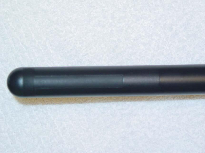
10 US equipment Probe

11 System US equipment
12 Prostate Brachytherapy US QA Complete QA will include: Ultrasound unit Needle template Treatment planning system Fluoroscope CT for post implant Template Needle Prostate Ultrasound Probe Ultrasound System

13 Setup for Phantom Measurements Clinical perspective, but coupling gel can leak out

14 Setup for Phantom Measurements Coupling gel stays in place, but image can be confusing
15 QC Testing Limit inter-observer variability Ideally performed by a single individual If multiple people, train for consistency
16 The Phantom CIRS Model 45 phantom. Used here for illustrative purposes only; no endorsement is implied. Wires spaced at known intervals Volumetric objects. TG128 report recommends a phantom design, but no manufacturer has implemented it yet
17 Test 1: Grayscale visibility Monitor must be adjusted appropriately for optimal visibility Calibration and brightness can drift over time Phosphors can fade
18 Test 1: Grayscale visibility Locate the gray scale strip on the side of the ultrasound screen. Depending on the type of strip, count the number of gray levels or measure the length of the gradation.
19 Test 2: Depth of penetration Reduced depth of penetration can reduce visibility of anterior capsule border Any increase in system noise will reduce depth of penetration Dead transducer elements (reducing signal) will also reduce depth of penetration
20 Test 2: Depth of penetration Find a relatively homogeneous region in the phantom. Using the digital calipers, determine the maximum depth that the static ultrasound speckle pattern of the phantom can be clearly distinguished from the dynamic electronic noise.

21

22 Test 3: Spatial resolution Negatively impacted by Poor probe condition Problems with pulse formation circuit boards Problems with pulse send/receive circuit boards 1 mm 2 mm 3 mm 4 mm 5 mm


23 Test 3: Spatial resolution Find a region of the phantom having single filament targets at various depths. Measure the dimensions of the filament image in both the axial and lateral directions. These dimensions are effectively the axial and lateral resolution limits. Switch the probe to the orthogonal direction and repeat
24 Test 4: Distance measurement accuracy Distance determined by pixel size and pixel calibration Pixel depth determined by range equation Differences from 1540 m/s sound speed will lead to errors in distance measurement Lateral distance calibration determined by FOV and pixel calibration. Errors can stem from image processing board and other circuitry

25 Test 4: Distance measurement accuracy Axial measurement: Align a column of fiber targets near to the center of the image, if possible. Freeze the image. Using the electronic calipers, measure the distance between the most proximal and the most distal targets. Lateral measurement: Repeat using a row of targets, measuring most lateral targets.
26 Test 5: Area measurement accuracy Area measurement is central to the implant procedure Since area measurement and distance measurement are so closely related, they have similar fault causes

27 Test 5: Area measurement accuracy Scan an object of known dimension such that the ultrasound beam intercepts it normally Using the appropriate tool on the ultrasound system, carefully trace the boundary of the object and record the calculated area of the object Measured area = 3.1 Nominal area = 3.05

28 Test 6: Volume measurement accuracy Complementary to distance and area measurements Errors are compounded multiplicatively

29 Test 6: Volume measurement accuracy Measured volume: 20.8 cc Certified volume: 20.6 cc Locate the base and apex of the phantom target; zero the stepper at the base Using the typical clinical procedure, perform a volume study After contouring the entire target, record the calculated volume


30 Test 7: Needle template alignment Depends on Accuracy of electronic template Distance measurement accuracy Physical needle template location
31 Test 7: Needle template alignment Place the probe with the needle template attached vertically in the water bath. Place needles at each corner of the needle template and one at the center. On the US system, verify that needle flashes in the image correspond to locations of needles on electronic grid overlay.
 US Measured volume: 20.8 cc Certified volume: 20.")
32 Test 8: TPS volume accuracy Perform a volume study of 3D target in the US phantom Import ultrasound images into treatment planning computer Retrace contours in treatment planning software Compare TPS volume to volume calculated by US system Variseed volume: 21.4 cc (3.9%) US Measured volume: 20.8 cc Certified volume: 20.6 cc
33 Tolerances Test Test name Tolerances 1 Grayscale visibility Δ > 2 steps or 10% from baseline 2 Depth of penetration Δ > 1 cm from baseline 3 Axial and lateral resolution Δ > 1 cm from baseline 4 Axial and lateral distance accuracy Error > 2 mm or 2% 5 Area measurement accuracy Error > 3 mm or 3% 6 Volume measurement accuracy Error > 5% 7 Needle template alignment Error > 3 mm 8 Treatment planning computer volume accuracy Error > 5%
34 Frequency # Test name Frequency 1 Grayscale visibility Annual 2 Depth of penetration Annual* 3 Axial and lateral resolution Annual* 4 Axial and lateral distance measurement accuracy Annual 5 Area measurement accuracy Annual 6 Volume measurement accuracy Annual 7 Needle template alignment Annual** 8 Treatment planning computer volume accuracy At Acceptance *Or following transport to another facility. **Or when a new template is used.
35 Time Estimates Test # Test name Typical duration 0 Gather and filling in preliminary information 10 minutes 1 Grayscale visibility 2 minutes 2 Depth of penetration 2 minutes 3 Axial and lateral resolution 1-5 minutes 4 Axial and lateral distance measurement accuracy 5 minutes 5 Area measurement accuracy 5 minutes 6 Volume measurement accuracy 10 minutes 7 Needle template alignment 15 minutes 8 Treatment planning computer volume accuracy 15 minutes Total: 70 minutes
36 Materials # Test name Equipment 1 Grayscale visibility Phantom 2 Depth of penetration Phantom 3 Axial and lateral resolution Phantom 4 Axial and lateral distance measurement accuracy Phantom 5 Area measurement accuracy Phantom 6 Volume measurement accuracy Phantom 7 Needle template alignment Water bath 8 Treatment planning computer volume accuracy TPS, Phantom

37 Ring-down artifact The signal is reflected multiple times within the needle.
38 Moisture in probe connection Electronic artifact

39 Poor contact between probe and object Contact artifact
40 Concluding Remarks Each of the tests rely on establishing a set of baseline measurements against which future measurements can be compared. The overall time commitment is manageable: the full set of measurements should be performed annually and will take about 90 minutes.
Introduction. The goal of TRUS QA is to ensure your system can do all of this accurately.
 TRUS QA Workshop Introduction The goals of using TRUS in prostate brachytherapy Visualize the prostate Need the US to penetrate deeply enough Need sufficient grey scale resolution to be able to visualize
Quality Assurance of Ultrasound Imaging in Radiation Therapy. Zuofeng Li, D.Sc. Murty S. Goddu, Ph.D. Washington University St.
 Quality Assurance of Ultrasound Imaging in Radiation Therapy Zuofeng Li, D.Sc. Murty S. Goddu, Ph.D. Washington University St. Louis, Missouri Typical Applications of Ultrasound Imaging in Radiation Therapy
Routine Quality Assurance Cookbook
 This Cookbook is a companion guide to the AIUM Routine Quality Assurance (QA) for Diagnostic Ultrasound Equipment document, which outlines the basic QA requirements for AIUM-accredited practices. The Guide
ULTRASOUND QA SOLUTIONS. Ensure Accurate Screening, Diagnosis & Monitoring DOPPLER FLOW PHANTOMS MULTI-PURPOSE PHANTOMS TRANSDUCER TEST PHANTOMS
 ULTRASOUND QA SOLUTIONS Ensure Accurate Screening, Diagnosis & Monitoring DOPPLER FLOW PHANTOMS MULTI-PURPOSE PHANTOMS TRANSDUCER TEST PHANTOMS INNOVATORS IN ADVANCED ULTRASOUND TECHNIQUES Gammex is the
ULTRASOUND QA SOLUTIONS. Ensure Accurate Screening, Diagnosis and Monitoring DOPPLER FLOW PHANTOMS MULTI-PURPOSE PHANTOMS TRAINING PHANTOMS
 ULTRASOUND QA SOLUTIONS Ensure Accurate Screening, Diagnosis and Monitoring DOPPLER FLOW PHANTOMS MULTI-PURPOSE PHANTOMS TRAINING PHANTOMS INNOVATORS IN ADVANCED ULTRASOUND TECHNIQUES Gammex is the only
The table below shows the density and velocity of waves in two different substances. Density / kg m 3 Velocity / m s 1
 Q1.(a) When ultrasound is incident at an interface between two different media some energy is transmitted and some is reflected. The ratio of the reflected energy intensity I r to the incident energy intensity
Outline. QA/QC of Ultrasound Imagers: Basic Physics, Procedures and Experiences. Frame Rate Limitation. US Imaging Range Equation.
 QA/QC of Ultrasound Imagers: Basic Physics, Procedures and Experiences Zheng F. Lu, PhD Radiology Department Columbia University Email: zfl1@columbia.edu Outline General overview of basic ultrasound physics
Ultrasound Physics and Knobology Alan Macfarlane. Consultant Anaesthetist Glasgow Royal Infirmary
 Ultrasound Physics and Knobology Alan Macfarlane Consultant Anaesthetist Glasgow Royal Infirmary RAPM 2009; 34: 40-46 Ultrasound Proficiency Understanding US image generation and device operation Image
Diploma of Medical Ultrasonography (DMU) Physical Principles of Ultrasound and Instrumentation Syllabus
 Physical Principles of Ultrasound and Instrumentation Syllabus") Diploma of Medical Ultrasonography (DMU) Physical Principles of Ultrasound and Instrumentation Syllabus Page 1 of 7 11/18 Candidates are expected to cover all of the content of this syllabus when preparing
Diploma of Medical Ultrasonography (DMU) Physical Principles of Ultrasound and Instrumentation Syllabus Page 1 of 7 11/18 Candidates are expected to cover all of the content of this syllabus when preparing
Lesson 03: Sound Wave Propagation and Reflection. This lesson contains 15 slides plus 14 multiple-choice questions.
 Lesson 03: Sound Wave Propagation and Reflection This lesson contains 15 slides plus 14 multiple-choice questions. Accompanying text for the slides in this lesson can be found on pages 8 through 14 in
8/3/2016. Diagnostic Ultrasound Imaging Quality Assurance. Purpose. Information on US QA. James A. Zagzebski 1, Ph.D. Zheng Feng Lu 2, Ph.D.
 Diagnostic Ultrasound Imaging Quality Assurance James A. Zagzebski 1, Ph.D. Zheng Feng Lu 2, Ph.D. 1 Dept. of Medical Physics University of Wisconsin, Madison 2 Dept. Of Radiology, University of Chicago,
Ultrasound Physics & Terminology
 Ultrasound Physics & Terminology This module includes the following: Basic physics terms Basic principles of ultrasound Ultrasound terminology and terms Common artifacts seen Doppler principles Terms for
Current Ultrasound Quality Control Recommendations and Techniques
 Current Ultrasound Quality Control Recommendations and Techniques Evan J. Boote, Ph.D. University of Missouri-Columbia Thank you for your interest - today I hope to provide some basic recommendations for
Hands-On Ultrasound Physics and Quality Control Workshop. Outline
 Slide 1 Hands-On Ultrasound Physics and Quality Control Workshop Z. F. Lu, PhD Radiology Department Columbia University New York, NY R. L. Kruger, PhD Radiology Department Marshfield Clinic Marshfield,
Preamble (disclaimer)
") Preamble (disclaimer) PHYSICS AND PRINCIPLES OF HEAD/NECK ULTRASOUND Joseph C. Sniezek, MD FACS LTC, MC, USA Otolaryngology/H&N Surgery Tripler Army Medical Center 1. I am not a physicist 2. ACS has recommended
Preamble (disclaimer) PHYSICS AND PRINCIPLES OF HEAD/NECK ULTRASOUND Joseph C. Sniezek, MD FACS LTC, MC, USA Otolaryngology/H&N Surgery Tripler Army Medical Center 1. I am not a physicist 2. ACS has recommended
BICOE Breast Imaging Center of Excellence. What is it? - Requirements. National Mammography Database. What do you get? ACR Accreditation in:
 BICOE Breast Imaging Center of Excellence What is it? - Requirements William Geiser, MS DABR Senior Medical Physicist MD Anderson Cancer Center Houston, Texas wgeiser@mdanderson.org ACR Accreditation in:
1. Fig. 1 shows data for the intensity of a parallel beam of X-rays after penetration through varying thicknesses of a material
 1. Fig. 1 shows data for the intensity of a parallel beam of X-rays after penetration through varying thicknesses of a material. intensity / MW m 2 thickness / mm 0.91 0.40 0.69 0.80 0.52 1.20 0.40 1.60
Principles of Ultrasound. Cara C. Prideaux, M.D. University of Utah PM&R Sports Medicine Fellow March 14, 2012
 Principles of Ultrasound Cara C. Prideaux, M.D. University of Utah PM&R Sports Medicine Fellow March 14, 2012 None Disclosures Outline Introduction Benefits and Limitations of US Ultrasound (US) Physics
Underwater Acoustic Measurements in Megahertz Frequency Range.
 Underwater Acoustic Measurements in Megahertz Frequency Range. Current State and Prospects of Development in Russia Alexander M. Enyakov,, Many medical applications of underwater acoustic measurements
ULTRASOUND. OB/Gyn (Core) Ultrasound PIEZOELECTRIC EFFECT. Principles of Ultrasound Physics and Instrumentation. Nathan Pinkney, BS, CDOS
 Ultrasound PIEZOELECTRIC EFFECT. Principles of Ultrasound Physics and Instrumentation. Nathan Pinkney, BS, CDOS") 1 OB/Gyn (Core) Ultrasound Principles of Ultrasound Physics and Instrumentation Nathan Pinkney, BS, CDOS Philadelphia College of Osteopathic Medicine 2016 ULTRASOUND CATEGORIES OF SOUND INFRASOUND = below
1 OB/Gyn (Core) Ultrasound Principles of Ultrasound Physics and Instrumentation Nathan Pinkney, BS, CDOS Philadelphia College of Osteopathic Medicine 2016 ULTRASOUND CATEGORIES OF SOUND INFRASOUND = below
Physical Principles of Ultrasound
 Physical Principles of Ultrasound Grateful appreciation to Richard A. Lopchinsky, MD, FACS and Nancy H. Van Name, RDMS, RTR, and MarleneKattaron, RDMS 2000 UIC All Rights Reserved. Course Objectives Identify
A. DeWerd. Michael Kissick. Larry. Editors. The Phantoms of Medical. and Health Physics. Devices for Research and Development.
 Larry Editors A. DeWerd Michael Kissick The Phantoms of Medical and Health Physics Devices for Research and Development ^ Springer Contents 1 Introduction to Phantoms of Medical and Health Physics 1 1.1
BICOE Stereotactic Breast Biopsy and Breast Ultrasound Accreditation. Introduction. Educational Objectives
 BICOE Stereotactic Breast Biopsy and Breast Ultrasound Accreditation William Geiser, MS DABR Senior Medical Physicist MD Anderson Cancer Center Houston, Texas wgeiser@mdanderson.org 1 Introduction Objectives
Ultrasound Applied Physics
 Ultrasound Applied Physics University of Toronto Department of Medical Imaging Applied Physics Mini-Course #3 2016 Ultrasound Laboratory Manual and Examination Booklet 1/21/2016 Ultrasound Applied Physics
Descriptions of NDT Projects Fall 2004 October 31, 2004
 Descriptions of NDT Projects Fall 2004 October 31, 2004 Introduction There are two separate NDT labs in Magister: ULTRA for ultrasound and EDDY for eddy current. Both labs are equipped with mechanical
Basic Physics of Ultrasound and Knobology
 WELCOME TO UTMB Basic Physics of Ultrasound and Knobology By Daneshvari Solanki, FRCA Laura B. McDaniel Distinguished Professor Anesthesiology and Pain Medicine University of Texas Medical Branch Galveston,
Dr Emma Chung. Safety first - Physical principles for excellent imaging
 Safety first - Physical principles for excellent imaging Dr Emma Chung Lecturer in Medical Physics, University of Leicester Clinical Scientist, University Hospitals of Leicester NHS Trust Thanks to Caroline
BICOE Stereotactic Breast Biopsy and Breast Ultrasound Accreditation. Introduction. Educational Objectives
 BICOE Stereotactic Breast Biopsy and Breast Ultrasound Accreditation William Geiser, MS DABR Senior Medical Physicist MD Anderson Cancer Center Houston, Texas wgeiser@mdanderson.org 1 Introduction Objectives
Ultrasound Principles cycle Frequency Wavelength Period Velocity
 ! Teresa S. Wu, MD, FACEP Director, EM Ultrasound Program & Fellowship Co-Director, Simulation Based Training Program & Fellowship Associate Program Director, EM Residency Program Maricopa Medical Center
Quality Control Program Of Real Time Medical Ultrasound Machines In Sudan
 Quality Control Program Of Real Time Medical Ultrasound Machines In Sudan Mamdouh Y Osman a*, Fathi A Taha b a sudan Atomic Energy commission, P.O.Box 3001, Khartoum, Sudan. b Department of Applied Physics,
Ultrasound. Principles of Medical Imaging. Contents. Prof. Dr. Philippe Cattin. MIAC, University of Basel. Oct 17th, 2016
 Ultrasound Principles of Medical Imaging Prof. Dr. Philippe Cattin MIAC, University of Basel Contents Abstract 1 Image Generation Echography A-Mode B-Mode M-Mode 2.5D Ultrasound 3D Ultrasound 4D Ultrasound
Diagnostic Ultrasound. Sutiporn Khampunnip, M.D.
 Diagnostic Ultrasound Sutiporn Khampunnip, M.D. Definition of Ultrasound Ultrasound is simply sound waves, like audible sound. High-frequency sound and refers to mechanical vibrations above 20 khz. Human
Introduction to Ultrasound Guided Region Anesthesia
 Introduction to Ultrasound Guided Region Anesthesia Brian D. Sites, MD Dept of Anesthesiology Dartmouth-Hitchcock Medical Center INTRODUCTION Welcome to Introduction to Ultrasound Guided Regional Anesthesia.
Terminology Tissue Appearance
 By Marc Nielsen, MD Advantages/Disadvantages Generation of Image Ultrasound Machine/Transducer selection Modes of Ultrasound Terminology Tissue Appearance Scanning Technique Real-time Portable No ionizing
APPLICATION AND DEPLOYMENT OF ADVANCED NDE TECHNIQUES IN HIGH PRESSURE VESSELS
 APPLICATION AND DEPLOYMENT OF ADVANCED NDE TECHNIQUES IN HIGH PRESSURE VESSELS Jeffrey P. Milligan, Daniel T. Peters, Structural Integrity Associates, Inc., USA Many advances in Non-Destructive Examination
Performance Evaluation of Ultrasound Systems: Advanced Methods and Applications
 Performance Evaluation of Ultrasound Systems: Advanced Methods and Applications NJ Hangiandreou PhD, Z Long PhD, DJ Tradup RDMS, SF Stekel BS, A Ferrero PhD, AT Tao PhD, W Zhou PhD, JE Browne PhD*, N Strissel
Ultrasonic Testing Level I:
 Ultrasonic Testing Level I: 1- Sound Wave - Introduction - ASNT Level I - Sound Wave Propagation - Velocity / Frequency / Wave Length - Acoustic Impedance - Energy / Intensity 2- Ultrasound Wave Modes
Debbie Childs RDMS, RVT Sonographer Murphy Medical Center Murphy, NC
 Debbie Childs RDMS, RVT Sonographer Murphy Medical Center Murphy, NC Worked at Murphy Medical Center as a sonographer for 18 years Registered in Abdomen, OB/GYN, Breast, & Vascular Ultrasound ACR Accredited
Development of Ultrasound Based Techniques for Measuring Skeletal Muscle Motion
 Development of Ultrasound Based Techniques for Measuring Skeletal Muscle Motion Jason Silver August 26, 2009 Presentation Outline Introduction Thesis Objectives Mathematical Model and Principles Methods
DIGITAL IMAGE PROCESSING IN ULTRASOUND IMAGES
 DIGITAL IMAGE PROCESSING IN ULTRASOUND IMAGES Kamaljeet Kaur Computer Science & Engineering Department Guru Nanak Dev Engg. College, Ludhiana. Punjab-India meetk.89@gmail.com ABSTRACT-- Image processing
Basic of Ultrasound Physics E FAST & Renal Examination. Dr Muhammad Umer Ihsan MBBS,MD, DCH CCPU,DDU1,FACEM
 Basic of Ultrasound Physics E FAST & Renal Examination Dr Muhammad Umer Ihsan MBBS,MD, DCH CCPU,DDU1,FACEM What is Sound? Sound is Mechanical pressure waves What is Ultrasound? Ultrasounds are sound waves
Ultrasound Measurements and Non-destructive Testing Educational Laboratory
 Session 3548 Ultrasound Measurements and Non-destructive Testing Educational Laboratory Vladimir Genis, Horacio Sosa Goodwin College of Professional Studies, Drexel University, Philadelphia, 19104 Emil
RADIATION PROTECTION IN DIAGNOSTIC AND INTERVENTIONAL RADIOLOGY. L19: Optimization of Protection in Mammography
 IAEA Training Material on Radiation Protection in Diagnostic and Interventional Radiology RADIATION PROTECTION IN DIAGNOSTIC AND INTERVENTIONAL RADIOLOGY L19: Optimization of Protection in Mammography
4.17. RESEARCHING MODELS WITH AN ULTRASONIC ECHOSCOPE
 4.17. RESEARCHING MODELS WITH AN ULTRASONIC ECHOSCOPE Purpose of experiment Determine the main characteristics of ultrasound waves, and the distances and positions of models using an ultrasonic echoscope.
ULTRASOUND IMAGING EE 472 F2018. Prof. Yasser Mostafa Kadah
 ULTRASOUND IMAGING EE 472 F2018 Prof. Yasser Mostafa Kadah www.k-space.org Recommended Textbook Diagnostic Ultrasound: Physics and Equipment, 2nd ed., by Peter R. Hoskins (Editor), Kevin Martin (Editor),
Tissue Strain Analytics Virtual Touch Tissue Imaging and Quantification
 Whitepaper Tissue Strain Analytics Virtual Touch Tissue Imaging and Quantification ACUSON S2000 Ultrasound System Answers for life. Page 1 Tissue Strain Analytics: Virtual Touch Tissue Imaging and Quantification
Point-of-Care Ultrasound: An Introduction
 Point-of-Care Ultrasound: An Introduction Delegation Teaching Package for Registered Respiratory Therapists and Anesthesia Assistants Developed by: Rob Bryan RRT, AA Edited by: Kelly Hassall RRT, FCSRT,
ADVANCED TECHNOLOGY CONSORTIUM (ATC) CREDENTIALING PROCEDURES FOR LUNG BRACHYTHERAPY IMPLANT PROTOCOLS
 CREDENTIALING PROCEDURES FOR LUNG BRACHYTHERAPY IMPLANT PROTOCOLS") ACOSOG-RTOG Lung Brachytherapy QA Page 1 of 8 ADVANCED TECHNOLOGY CONSORTIUM (ATC) CREDENTIALING PROCEDURES FOR LUNG BRACHYTHERAPY IMPLANT PROTOCOLS FACILITY QUESTIONNAIRE Institutions wishing to enter
ACOSOG-RTOG Lung Brachytherapy QA Page 1 of 8 ADVANCED TECHNOLOGY CONSORTIUM (ATC) CREDENTIALING PROCEDURES FOR LUNG BRACHYTHERAPY IMPLANT PROTOCOLS FACILITY QUESTIONNAIRE Institutions wishing to enter
A new method of sonograph lateral resolution measurement using PSF analysis of received signal
 A new method of sonograph lateral resolution measurement using PSF analysis of received signal L. Doležal, J. Hálek Faculty of Medicine Palacký University in Olomouc, Czech Republic E-mail: ladol@tunw.upol.cz
Implementation of the 2012 ACR CT QC Manual in a Community Hospital Setting BRUCE E. HASSELQUIST, PH.D., DABR, DABSNM ASPIRUS WAUSAU HOSPITAL
 Implementation of the 2012 ACR CT QC Manual in a Community Hospital Setting BRUCE E. HASSELQUIST, PH.D., DABR, DABSNM ASPIRUS WAUSAU HOSPITAL Conflict of Interest Disclaimer Employee of Aspirus Wausau
Ultrasound Accreditation and Regulatory Compliance from A to Z
 Jim Rickner presents: Ultrasound Accreditation and Regulatory Compliance from A to Z Sponsored by: Conquest Imaging Wednesday, September 28, 2016, 2:00pm ET PRESENTER: JIM RICKNER, CONQUEST IMAGING Jim
Linear Ultrasonic Wave Propagation in Biological Tissues
 Indian Journal of Biomechanics: Special Issue (NCBM 7-8 March 29) Linear Ultrasonic Wave Propagation in Biological Tissues Narendra D Londhe R. S. Anand 2, 2 Electrical Engineering Department, IIT Roorkee,
4.17. RESEARCHING MODELS WITH AN ULTRASONIC ECHOSCOPE
 4.17. RESEARCHING MODELS WITH AN ULTRASONIC ECHOSCOPE Purpose of experiment Determine the main characteristics of ultrasound waves, and the distances and positions of models using an ultrasonic echoscope.
Patient Safety Focused QA. LDR Brachytherapy Vrinda Narayana
 Patient Safety Focused QA LDR Brachytherapy Vrinda Narayana D < 2 Gy/h Old LDR Brachytherapy? Ra-226; Cs-137; Ir-192 New Gynecological; interstitial Pd-103; I-125; Cs-131 Prostate implants Eye plaques
The Physics of Ultrasound. The Physics of Ultrasound. Claus G. Roehrborn. Professor and Chairman. Ultrasound Physics
 The Physics of Ultrasound Pipe Organ 10-8000 Emission Dog 452-1080 Man 85-1100 Spectrum Bat 10,000-120,000 Porpoise 7000-120,000 Claus G. Roehrborn Professor and Chairman 10 20 Cycles per second Reception
Ultrasound: Past and Present. Lecturer: Dr. John M Hudson, PhD
 Ultrasound: Past and Present Lecturer: Dr. John M Hudson, PhD Disclosures 2 No conflicts of interest to declare Course Outline 3 1. Survey of ultrasound physics & applications 2. (Sep 21) 3. (Sep 28) 4.
Augmentation of the In Vivo Elastic Properties Measurement System to include Bulk Properties
 DISTRIBUTION STATEMENT A. Approved for public release; distribution is unlimited. Augmentation of the In Vivo Elastic Properties Measurement System to include Bulk Properties Peter H. Rogers and Michael
Trina Lynd, M.S. Medical Physicist Lifefirst Imaging & Oncology Cullman, AL Tri-State Alabama, Louisiana and Mississippi Spring 2016 Meeting April
 Trina Lynd, M.S. Medical Physicist Lifefirst Imaging & Oncology Cullman, AL Tri-State Alabama, Louisiana and Mississippi Spring 2016 Meeting April 17, 2016 Discuss permanent prostate brachytherapy and
Basic Ultrasound Physics Board Review Questions
 Basic Ultrasound Physics Board Review Questions Sidney K. Edelman, PhD ESP Ultrasound The Woodlands, TX Question 1 What is the wavelength of 2 MHz sound in soft tissue? 1. 1.54 mm 2. 0.75 mm 3. 0.75 cm
INTRODUCTION. Getting the best scan. Choosing a probe. Choosing the frequency
 Getting the best scan Choosing a probe Select the most appropriate probe for the particular scan required. s vary in their: operating frequency range higher ultrasound frequencies provide better discrimination
Pulse-Echo Ultrasound Imaging. Resolution in Ultrasound Imaging. Doppler Ultrasound. Resolution vs Penetration. Medical Imaging (EL582/BE620/GA4426)
") Medical Imaging (EL582/BE620/GA4426) Pulse-Echo Ultrasound Imaging Ultrasound Imaging Lecture 2 Daniel (Dan) Turnbull, Ph.D. Skirball Institute and Dept of Radiology NYU School of Medicine (daniel.turnbull@med.nyu.edu)
Medical Imaging (EL582/BE620/GA4426) Pulse-Echo Ultrasound Imaging Ultrasound Imaging Lecture 2 Daniel (Dan) Turnbull, Ph.D. Skirball Institute and Dept of Radiology NYU School of Medicine (daniel.turnbull@med.nyu.edu)
CHARACTERIZATION OF ANNULAR ARRAY TRANSDUCER
 Analele Universităţii de Vest din Timişoara Vol. LV, 2011 Seria Fizică CHARACTERIZATION OF ANNULAR ARRAY TRANSDUCER Luminita Moraru 1, Laura Onose 1, 2, Ana-Maria Chiselev 1 1 Dunărea de Jos University
Annular Array Transducer and Matched Amplifier for Therapeutic Ultrasound
 ARCHIVES OF ACOUSTICS 35, 4, 653 660 (2010) DOI: 10.2478/v10168-010-0049-6 Annular Array Transducer and Matched Amplifier for Therapeutic Ultrasound Wojciech SECOMSKI, Andrzej NOWICKI, Janusz WÓJCIK, Marcin
ACR Accreditation Update in MRI
 ACR Accreditation Update in MRI Whole Body Systems Extremity (MSK) Ron Price Vanderbilt University Medical Center Nashville, TN Dedicated Breast MRI Accreditation Update 1. ACR MRI Accreditation: Overview,
1 Fundamentals. Basic Definitions and Physics Principles. Fundamentals
 1 To become versed in the language of ultrasonography, it is necessary to review some of the basic principles of physics. The wave physics principles of ordinary (i.e., audible) sound apply to ultrasound
Automatic quality control processing for detection of element/channel dropout in ultrasound transducers
 Automatic quality control processing for detection of element/channel dropout in ultrasound transducers Paul L. Carson, Ph.D. University of Michigan, Department of Radiology, Ann Arbor, MI, USA Acknowledgements
Hands-on. Physics Workshop. 2-day Workshop June 1-2, 2018 Ann Arbor, MI. Program Director: Brian Fowlkes, PhD
 Hands-on Ultrasound Physics Workshop 2-day Workshop June 1-2, 2018 Ann Arbor, MI Program Director: Brian Fowlkes, PhD Faculty: Paul Carson, PhD Mitch Goodsitt, PhD Oliver Kripfgans, PhD Sandra Larson,
Ultrasound in Medicine
 Ultrasound in Medicine Experimental Equipment for Medical Education Universities Colleges Medical Schools Medical and Med-Technical Training Education can befun! WELCOME TO GAMPT Devices and accessories
Features and Benefits
 Features and Benefits DC7 Significant Features HIPAA Compliant Frequency Compound Imaging Standard i-clear Standard i-beam Standard SCI Digital Processing Channels 2048 Dynamic Range > 160 Db 10 Bit A/D
Ultrasonic Thickness Procedure Pulse-Echo Thickness Gage TABLE OF CONTENTS. 1.0 Introduction Reference Documents Summary of Practice 2
 Page: 1 of 6 TABLE OF CONTENTS SECTION PAGE NO. 1.0 Introduction 2 2.0 Reference Documents 2 3.0 Summary of Practice 2 4.0 Significance & Use 3 5.0 Apparatus 3 6.0 Calibration & Adjustment of Apparatus
Available online at ScienceDirect. Physics Procedia 70 (2015 )
") Available online at www.sciencedirect.com ScienceDirect Physics Procedia 70 (2015 ) 1139 1143 2015 International Congress on Ultrasonics, 2015 ICU Metz Attenuation Coefficient Estimation of the Healthy
Available online at www.sciencedirect.com ScienceDirect Physics Procedia 70 (2015 ) 1139 1143 2015 International Congress on Ultrasonics, 2015 ICU Metz Attenuation Coefficient Estimation of the Healthy
Evaluation of the Quality of Thick Fibre Composites Using Immersion and Air- Coupled Ultrasonic Techniques
 ECNDT 2006 - We.1.6.4 Evaluation of the Quality of Thick Fibre Composites Using Immersion and Air- Coupled Ultrasonic Techniques Kaj K. BORUM, Risø National Laboratory, Materials Research Department, Roskilde,
Review of TG-186 recommendations
 Review of TG-186 recommendations Implementation of advanced brachytherapy dose calculation algorithms beyond TG-43 Rowan M. Thomson Carleton Laboratory for Radiotherapy Physics Carleton University Ottawa
High resolution ultrasound scanner for skin imaging
 High resolution ultrasound scanner for skin imaging Christine Turlat Sales Director Atys medical 17 Parc d Arbora 69510 SOUCIEU EN JARREST Atys company Principle of ultrasound imaging DERMCUP Normal image
10 Years Experience in Industrial Phased Array Testing of Rolled Bars
 18th World Conference on Nondestructive Testing, 16-20 April 2012, Durban, South Africa 10 Years Experience in Industrial Phased Array Testing of Rolled Bars Josef MAIER 1 and Gerhard Ferstl 1, 1 Böhler
ULTRASONIC ARRAY APPROACH FOR THE EVALUATION OF ELECTROFUSION JOINTS OF POLYETHYLENE GAS PIPING
 ULTRASONIC ARRAY APPROACH FOR THE EVALUATION OF ELECTROFUSION JOINTS OF POLYETHYLENE GAS PIPING H. J. Shin 1, Y. H. Jang 1, J. R. Kwan 2, H. D. Lee 3 1 INDE System Co., Ltd., Suwon, Kyunggi-do, 440-746,
Dental ConeBeam Computed Tomography (CBCT) X-ray Systems
 X-ray Systems") Dental ConeBeam Computed Tomography (CBCT) X-ray Systems PROPOSED REVISIONS TO 4732.XXXX, 1.0 4732.#### DENTAL CONEBEAM COMPUTED TOMOGRAPHY (CBCT) X-RAY SYSTEMS; STATIONARY AND MOBILE. Subpart 1. Applicability.
Dental ConeBeam Computed Tomography (CBCT) X-ray Systems PROPOSED REVISIONS TO 4732.XXXX, 1.0 4732.#### DENTAL CONEBEAM COMPUTED TOMOGRAPHY (CBCT) X-RAY SYSTEMS; STATIONARY AND MOBILE. Subpart 1. Applicability.
Example 1: How would you evaluate this transducer? What is the most common problem found in ultrasound QC testing? Uniformity is Subjective
 AAPM Working Group on Quantitative B-mode Ultrasound Quality Control: Software for assessment of transducer artifacts Sandra Larson, PhD University of Michigan Medical Center, Ann Arbor, MI, What is the
Compact Gamma Camera for Detection of Prostate Cancer
 Compact Gamma Camera for Detection of Prostate Cancer Presented at: Human Interest Panel Federal Laboratory Consortium Annual Conference Nashville, Tennessee Brookhaven National Laboratory and Hybridyne
Ultrasound Physics & Doppler
 Ultrasound Physics & Doppler Endocrine University 2018 Mark Lupo, MD, FACE, ECNU Objectives Review the essential components of ultrasound physics in neck sonography Demonstrate the importance of ultrasound
Sound in medicine. CH.12. Dr.Rajaa أ.م.د. رجاء سهيل جنم جامعة تكريت كلية طب االسنان. General Properties of Sound
 CH.12. Dr.Rajaa Sound in medicine أ.م.د. رجاء سهيل جنم جامعة تكريت كلية Sound : It is the audible waves of frequency between 20 Hz and 20 khz. Infrasound : refers to the sound of frequency below the normal
Interim Report. Prostate Cancer Fight Foundation Motorcycle Ride for Dad. February 7, Supporting Kingston s university hospitals:
 Interim Report Prostate Cancer Fight Foundation Motorcycle Ride for Dad February 7, 2014 Supporting Kingston s university hospitals: Thank you for your continued support of prostate cancer research in
Feng Xiujuan National Institute of Metrology (NIM),China
,China") The acoustic calibration service in transportation at NIM Feng Xiujuan National Institute of Metrology (NIM),China 1. Calibration requirements 2. Calibration service at NIM 2.1 Microphone 2.2 Ultrasonic
The acoustic calibration service in transportation at NIM Feng Xiujuan National Institute of Metrology (NIM),China 1. Calibration requirements 2. Calibration service at NIM 2.1 Microphone 2.2 Ultrasonic
Characterization of Ultrasound Elevation Beamwidth Artefacts for Brachytherapy Needle Insertion
 Characterization of Ultrasound Elevation Beamwidth Artefacts for Brachytherapy Needle Insertion by Mohammad Peikari A thesis submitted to the School of Computing in conformity with the requirements for
AAPM Annual Meeting. ACR Accreditation Update in CT
 AAPM Annual Meeting ACR Accreditation Updates in CT, Ultrasound, Mammography and MRI: ACR Accreditation Update in CT Michael McNitt-Gray, PhD, DABR, FAAPM Professor, Department of Radiological Sciences
Performance of phased array and conventional ultrasonic probes on the new ISO reference block
 Performance of phased array and conventional ultrasonic probes on the new ISO 19675 reference block C. Udell, D. Chai 1 and F. Gattiker Proceq S.A., Ringstrasse 2, Schwerzenbach, Switzerland. More info
Musculoskeletal Ultrasound: Basics, Utility, and Clinical Applications
 Musculoskeletal Ultrasound: Basics, Utility, and Clinical Applications Andrew Lavigne, MD, FRCPC Physical Medicine and Rehabilitation CSCN Diplomat (EMG) Dip Sport Medicine Eugene Maida, MD, PGY-4 Resident
Quality Assurance of Ultrasound Imagers: Procedures, Expectations, and Philosophies
 Quality Assurance of Ultrasound Imagers: Procedures, Expectations, and Philosophies James M. Kofler, Jr. INTRODUCTION This handout discusses several crucial issues regarding quality assurance (QA) in diagnostic
Confident and Conclusive Diagnosis with Flexible Solutions
 Internal Medicine Confident and Conclusive Diagnosis with Flexible Solutions ALPINION MEDICAL SYSTEMS We are Ultrasound Professionals Ultrasound is widely used as the gold-standard for internal medicine
API. Defined Procedure. for. Ultrasonic Thickness Measurement API-UT-21
 API Defined Procedure for Ultrasonic Thickness Measurement API-UT-21 This Procedure Defines the Recommended Ultrasonic Methods and Techniques for Thickness Measurements Page 1 1.0 PURPOSE 1.1 This procedure
Joon-Il Choi, MD, Pyo Nyun Kim, MD, Woo Kyoung Jeong, MD, Hyun Cheol Kim, MD, Dal Mo Yang, MD, Sang Hoon Cha, MD, Jae-Joon Chung, MD
 ORIGINAL RESEARCH Establishing Cutoff Values for a Quality Assurance Test Using an Ultrasound Phantom in Screening Ultrasound Examinations for Hepatocellular Carcinoma An Initial Report of a Nationwide
Skin Characterization with High-Frequency Ultrasound
 Skin Characterization with High-Frequency Ultrasound Stephanie L. Shubert sls6626@rit.edu Advisor: Dr. María Helguera Ultrasound Imaging Lab Chester F. Carlson Center for Imaging Science Rochester Institute
Varian Acuity BrachyTherapy Suite One Room Integrated Image-Guided Brachytherapy
 Varian Acuity BrachyTherapy Suite One Room Integrated Image-Guided Brachytherapy The Acuity BrachyTherapy Suite Integrating Imaging, Planning, and Treatment in a Single Room Each component draws on the
On the feasibility of speckle reduction in echocardiography using strain compounding
 Title On the feasibility of speckle reduction in echocardiography using strain compounding Author(s) Guo, Y; Lee, W Citation The 2014 IEEE International Ultrasonics Symposium (IUS 2014), Chicago, IL.,
Pipeline Technology Conference 2007
 The Complete Solution: Combined Crack and Metal Loss Detection Tool using Phased Array Technology By A. Hugger, J. Franz, H. Charbon, R. Bauernschmitt, M. Tschuch, K.-H. Käshammer, I. Lachtchouk, J. Ehrhardt.
Imaging Rotation. University of Michigan Department of Radiation Oncology Division of Radiation Physics. Resident:
 University of Michigan Department of Radiation Oncology Division of Radiation Physics Imaging Rotation Resident: Rotation staff mentor/ advisor: James Balter, supplemental mentors: Dale Litzenberg, Don
What is Ultrasound? What is Ultrasound? B A. Basic Principles of Ultrasound. Basic Principles of Ultrasound. Basic Principles of Ultrasound
 Introduction to Ultrasound Principles Mani Montazemi, RDMS Baylor College of Medicine Division of Maternal-Fetal Medicine Department of Obstetrics and Gynecology Manager, Maternal Fetal Center Imaging
Quality management for Breast Brachytherapy.
 Quality management for Breast Brachytherapy. DORIN TODOR, Ph.D. Medical College of Virginia Campus Department of Radiation Oncology New England AAPM Chapter 2012 Summer Meeting, Providence, RI Quality
OPTION I TEST REVIEW
 IB PHYSICS 3 Name: Period: Date: DEVIL PHYSICS BADDEST CLASS ON CAMPUS OPTION I TEST REVIEW s2. This question is about defects of hearing. The graph below shows an audiogram for a person who has not been