Suyash Mohan MD, PDCC. Assistant Professor of Radiology Neuroradiology Division, Department of Radiology University of Pennsylvania
|
|
|
- Vivian Shields
- 5 years ago
- Views:
Transcription


1 Suyash Mohan MD, PDCC Assistant Professor of Radiology Neuroradiology Division, Department of Radiology University of Pennsylvania
2 Consultant, ACR Imaging Network (ACRIN) & ACR Image Metrix Gliobastoma multi-institutional trial ABTC 0901 RANO Reader (ACR Image Metrix) Eisai TM Study Phase III Trial RTOG 0825(4508)/ACRIN 6686 PI: High Resolution MRI/ MRS to Evaluate Therapeutic Response to Optune in Recurrent Glioblastomas NovoCure Advisory board Co-I: Guerbet Dose Finding Study in Central Nervous System MRI PI: RadDx (Galileo CDS Inc) Clinical Diagnostic Decision Support in Radiology Co-I: RSNA Education Scholar Grant: Development of a Novel Radiology Teaching Interface Using Bayesian Networks: Application to Neuroradiology as Proof of Concept Adaptive Radiology Interpretation Education System (ARIES)
3 1. MR Spectroscopy 1. 2D Localized Correlated Spectroscopy (L-COSY) at 7.0 T 2. Chemical Shift Imaging (CSI) at 3.0 T IDH1 mutation & 2HG 2. Whole brain Spectroscopic Imaging 1. CSI at 3.0 T for disease monitoring in IDH-mutant gliomas 2. 3-D Echo-Planar Spectroscopic Imaging (EPSI) at 3.0 T TP versus PsP 3. MR perfusion 1. TP versus PsP 2. Response Assessment(CAR-T, TTFields), Survival Pre therapy setting Post therapy setting 4. Multi-modal Approach (DTI/EPSI/Perfusion)


 Reported in ~")
4 Enzyme catalyzes the oxidative decarboxylation of isocitrate to α-kg Mutant IDH1/2 causes accumulation of 2-HG (5-35 mm) Reported in ~ 70% LGG/ sec GBM
5 1. Virtually no normal 2-HG background 2. 99% of tumors with IDH mutations have increased levels of 2-HG 3. Only other known cause of elevated 2HG is hydroxyglutaric aciduria, 1. Rare inborn error of metabolism with a different clinical phenotype Hence, tumors with levels of 2-HG are unlikely to represent false +ve cases for IDH mutations
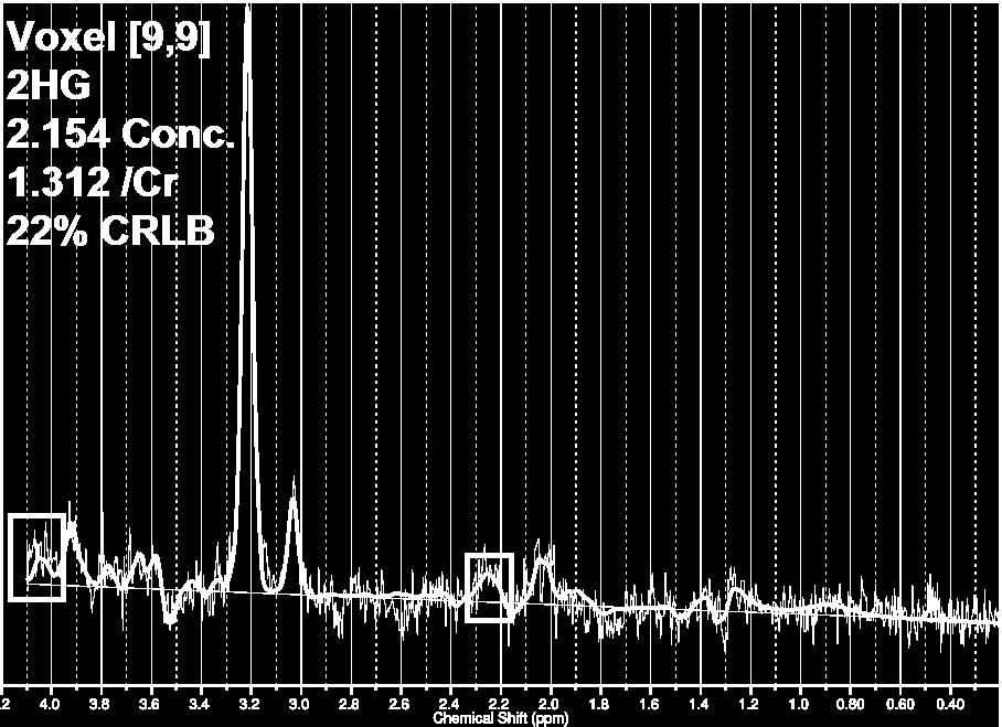
6 Unambiguous detection of 2-HG with MRS, in vivo, is challenging because its coupled fivespin system yields a complex spectral pattern Five resonance peaks 4.02, 2.27, 2.22, 1.98, 1.83 ppm 2-HG signals from Hβ (1.91 ppm) and Hγ (2.24 ppm) protons are superimposed by glutamate, glutamine, & GABA, while Hα (4.02 ppm) signals are obscured by mi, phosphocreatine, & lactate


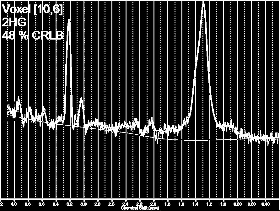
7 2HG
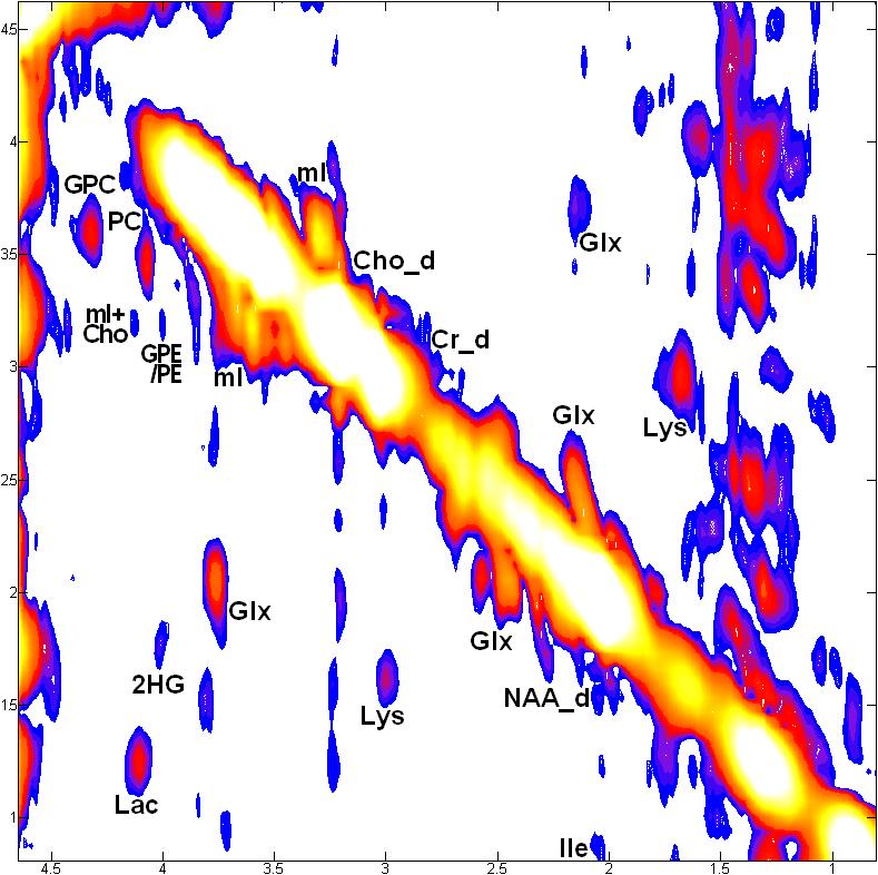
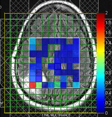
8 Uniquely resolve 2HG 1D MRS N-acetylasparate Creatine Choline Myo-Inositol Overlap Lactate/Lipids Overlap Glx 2D L-COSY MRS NAA Myo-Inositol Creatine Choline PC, GPC, PE, GPE Resolve Lac/Lipids Resolve Glu/Gln/GSH Amino Acids Ile, Leu, Lys


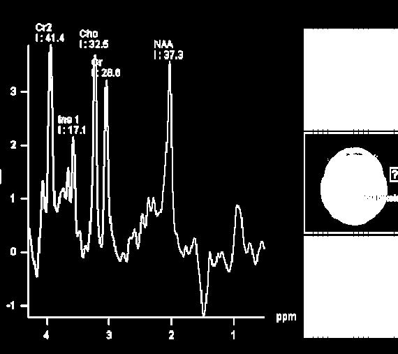
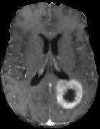
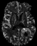
9 18 Y/M after a grand mal seizure

10 7T 2D L-COSY Spectrum


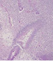
11 Mutant IDH 1 positive Slides courtesy: McLean Nasrallah MD, PhD


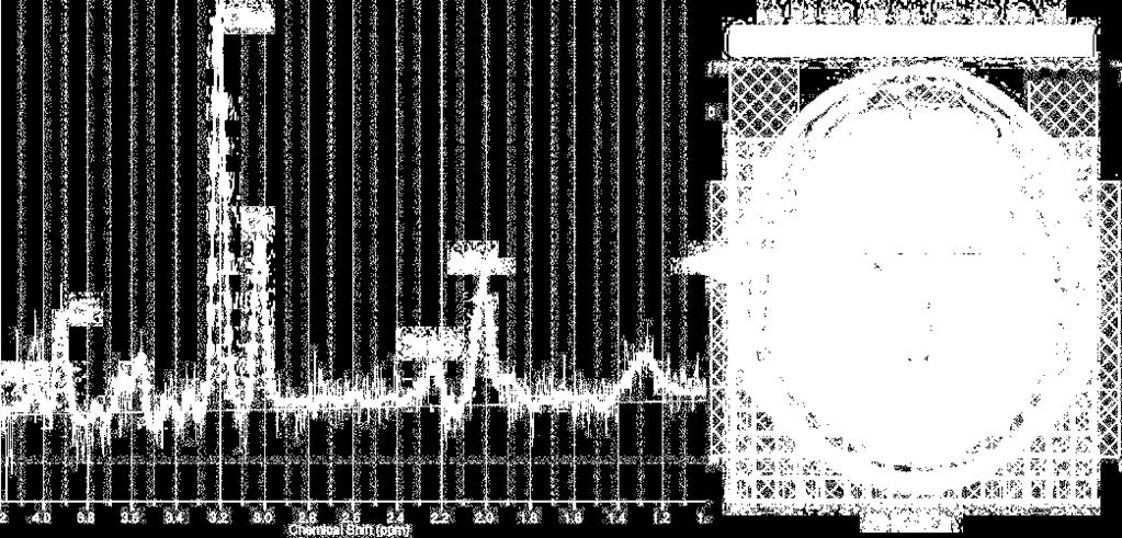
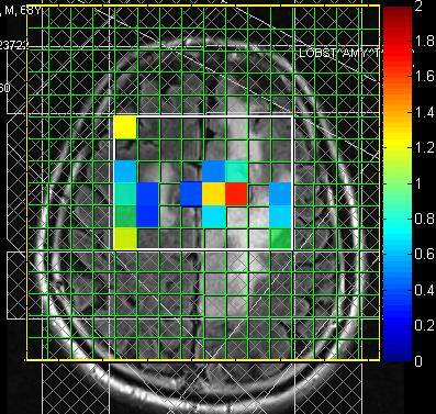
12 36 Y/M vision changes & seizure

13 7T 2D L-COSY Spectrum


14 Mutant IDH 1 positive Slides courtesy: McLean Nasrallah MD, PhD


15 No detectable 2HG WHO Grade I Ganglioglioma 8.8 ml VOI
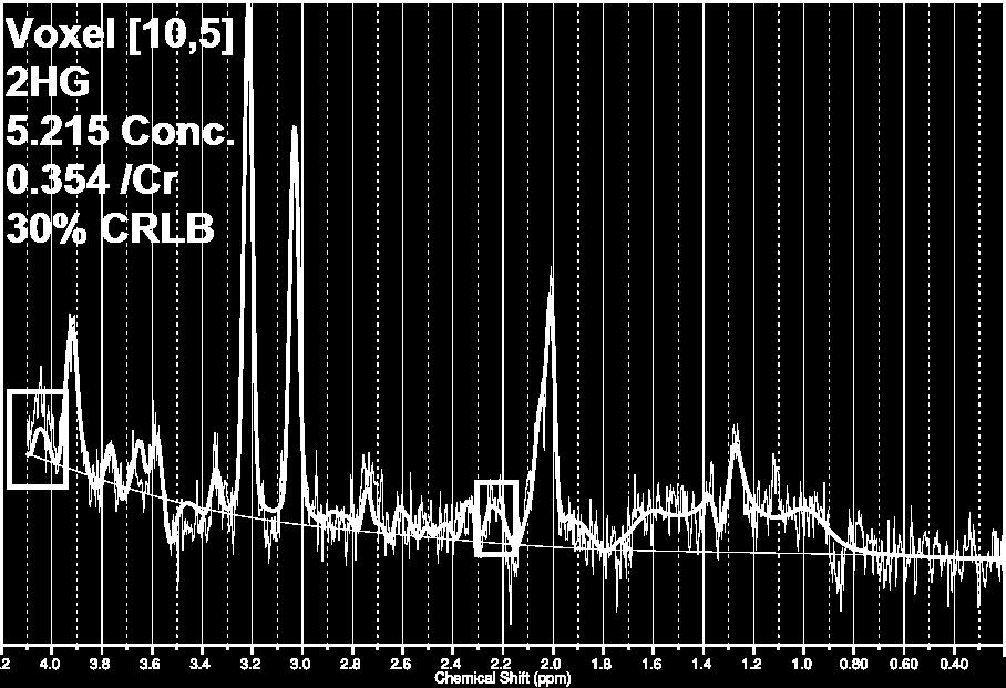
16 2D CSI (3T Tim-Trio clinical MRI scanner) TE/TR = 97/1700 ms, 3 averages, 16x16 Array 1.5 ml voxels, 6:53 min scan time Optimized for detection of 2HG 1 LCModel Fitting 40% CRLB, neighboring voxels Nine patients assessed 4 female, 5 male Ages 31 to 83, mean = 48 years 4 IDH1 mutants Compare to immunohistochemical staining


17 Grade III AA

18 2HG 2HG Grade III AOA

19 2HG 2HG Grade II Oligodendroglioma with some Grade III features
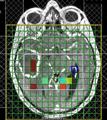

20 No detectable 2HG Path Result: Wild-Type Glioblastoma
21 1. MR Spectroscopy 1. 2D Localized Correlated Spectroscopy (L-COSY) at 7.0 T 2. Chemical Shift Imaging (CSI) at 3.0 T IDH1 mutation & 2HG 2. Whole brain Spectroscopic Imaging 1. CSI at 3.0 T for disease monitoring in IDH-mutant gliomas 2. 3-D Echo-Planar Spectroscopic Imaging (EPSI) at 3.0 T TP versus PsP 3. MR perfusion 1. TP versus PsP 2. Response Assessment(CAR-T, TTFields), Survival Pre therapy setting Post therapy setting 4. Multi-modal Approach (DTI/EPSI/Perfusion)
:")
22 Neuro Oncol Feb; 18(2):
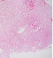
 to 91% for larger tumors (>8 ml) 2HG-MRS prior to surgery, corresponded with tumor cellularity Cytoreduction results in gradual decrease in 2HG levels CONCLUSIONS: 2HG-MRS can be linked with")
23 IDH mutant AA RESULTS: Detection of 2HG in IDH-mutant gliomas was closely linked to tumor volume Sensitivity, 8% for small tumors (<3.4 ml) to 91% for larger tumors (>8 ml) 2HG-MRS prior to surgery, corresponded with tumor cellularity Cytoreduction results in gradual decrease in 2HG levels CONCLUSIONS: 2HG-MRS can be linked with routine MRI to provide quantitative measurements of 2HG Useful imaging biomarker to non-invasively monitor the abundance of IDH-mutant tumor cells during therapy & disease monitoring Neuro Oncol Feb; 18(2):
24 Potential to detect IDH1 mutation in vivo To date, in vivo MRS is the only imaging method that is specific to IDH mutations Existing PET or SPECT radiotracers are not! Significant translational implications HGG better prognosis LGG early intervention before it transforms to a higher grade Potential use as an early biomarker of malignant progression Disease monitoring Endpoint for targeted therapy (AGIOS 121 drug)
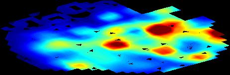


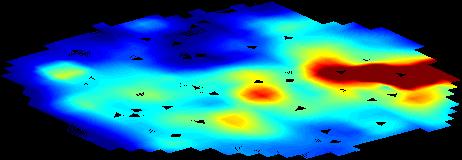

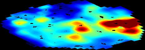

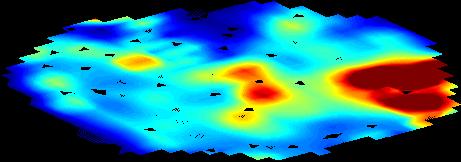


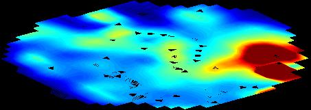

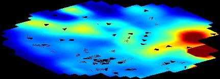

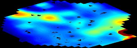

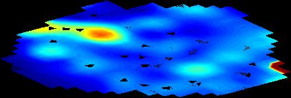

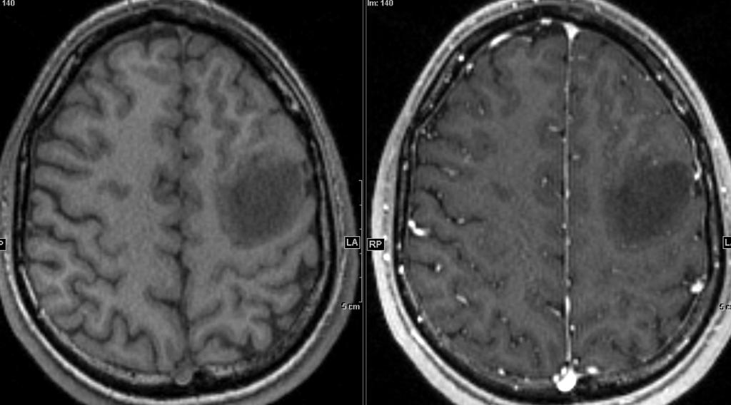
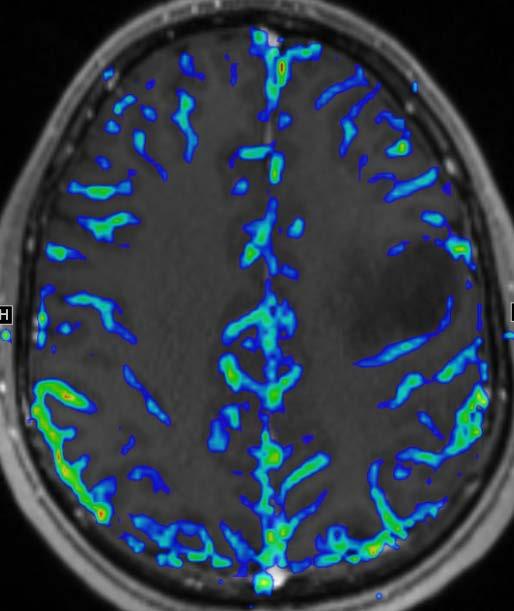
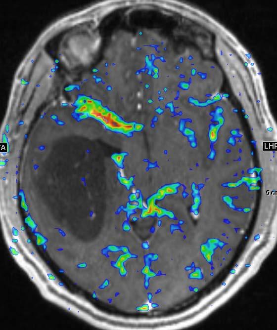
25 Whole-Brain High Resolution Single Scan Cho/Cr Map True Progression


26 Enhancing n = 20 Immediate Peritumoral n = 27 SEGMENTATION T 1 MPRAGE T 2 FLAIR EPSI Distant Peritumoral n = 132


27 Cho/NAA Cho/Cr NAA Cho Cr


28 P =.008 P =.045 P =.040 Enhancing Immediate Distal True Progression (N=7) Pseudoprogression (N=7) Verma G. et al. ISMRM 2016
29 Baseline Followup True Progression Pseudoprogression Verma G. et al. ISMRM 2016
30 EPSI Detects Tumor Heterogeneity Cho/Cr in True Progression Enhancing Lesion Immediate Peritumoral Histogram analysis of the enhancing lesions better separates PsP from TP
31 1. MR Spectroscopy 1. 2D Localized Correlated Spectroscopy (L-COSY) at 7.0 T 2. Chemical Shift Imaging (CSI) at 3.0 T IDH1 mutation & 2HG 2. Whole brain Spectroscopic Imaging 1. CSI at 3.0 T for disease monitoring in IDH-mutant gliomas 2. 3-D Echo-Planar Spectroscopic Imaging (EPSI) at 3.0 T TP versus PsP 3. MR perfusion 1. TP versus PsP 2. Response Assessment(CAR-T, TTFields), Survival Pre therapy setting Post therapy setting 4. Multi-modal Approach (DTI/EPSI/Perfusion)

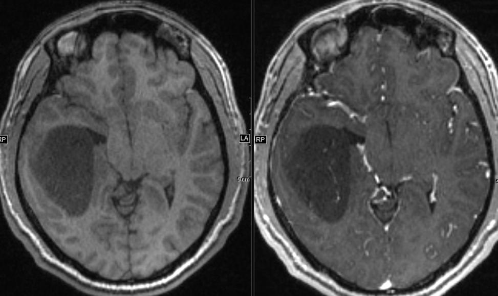
32 Pseudoprogression rcbvmax= 2.71 True Progression T1 gad CBV rcbvmax= 4.26 H&E

33 Quantitative Perfusion analysis provides a better estimation of treatment response Baseline rcbvmax month rcbvmax month rcbvmax month rcbvmax 2.00

34 Wang S. et al. ISMRM 2016

35 Baseline (before TTFields) 3 month follow up Post contrast T1 of 51 Y/F with left thalamic GBM


36 PC-T1 MD FA CBV Cho/Cr Baseline 2 month follow-up PC-T1 MD FA CBV Cho/Cr Mohan S, et al. Assessment of early response to tumor-treating fields in newly diagnosed glioblastoma using physiologic and metabolic MRI: initial experience. CNS Oncol Apr 14. Early response to TTFields showed trends toward Increasing MD Decreasing tumor volume, FA, rcbv and Cho/Cr Potential for physiologic and metabolic MRI to assess early treatment response to TTFields
:1247-52.")
37 Long Survivor (n=28) Short survivor (n=30) rcbv cutoff: 5.79 P<0.01 AJNR Jul;36(7):

38 Advanced techniques are establishing new paradigms in brain tumor research & therapy Immense translational potential Can be used for more sensitive assessment of tumor metabolism and response to therapy
39 Neuroradiology: Harish Poptani PhD, UK Gaurav Verma PhD Sumei Wang MD Sanjeev Chawla PhD Ronald Wolf MD PhD Neurosurgery: Steven Brem MD Donald O Rourke MD Neurooncology: Arati Desai MD Michelle A Basanta MD Robert Lustig MD Neuropathology: MacLean Nasrallah MD PhD Maria Martinez-Lage MD 7T COSY Prodev; IRB# R21 Grant: 1R21CA Novocure: IST Novartis COSY & EPSI Development Group Dr. Andrew Maudsley Dr. Albert Thomas Sulaiman Sheriff Dr. Mohammad Sabati MRI Technicians Jacqui, Tanya, Pat, Doris, Alicia, Jeffrey Research Personnel Lisa Desiderio Krista Huff Lauren karpf
MRS and Perfusion of Brain Tumors
 Department of Radiology University of California San Diego MRS and Perfusion of Brain Tumors John R. Hesselink, M.D. MRS & Perfusion of Brain Tumors Tumor histology Degree of malignancy Delineate tumor
Clinical Trials for Adult Brain Tumors - the Imaging Perspective
 Clinical Trials for Adult Brain Tumors - the Imaging Perspective Whitney B. Pope, M.D., Ph.D. Department of Radiology David Geffen School of Medicine at UCLA August 22, 2015 1 Disclosure of Financial Relationships
PENN BRAIN TUMOR ACADEMY:
 F R I D AY, MAY 13, 2016 7:30 AM 4:30 PM Smilow Center for Translational Research 3400 Civic Center Blvd Philadelphia, PA 19104 Penn Medicine s Abramson Cancer Center and Brain Tumor Center P R E S E N
General Identification. Name: 江 X X Age: 29 y/o Gender: Male Height:172cm, Weight: 65kg Date of admission:95/09/27
 General Identification Name: 江 X X Age: 29 y/o Gender: Male Height:172cm, Weight: 65kg Date of admission:95/09/27 Chief Complaint Sudden onset of seizure for several minutes Present illness This 29-year
Correlation of quantitative proton MR spectroscopy with local histology from stereotactic brain biopsy to evaluate heterogeneity of brain tumors
 Correlation of quantitative proton MR spectroscopy with local histology from stereotactic brain biopsy to evaluate heterogeneity of brain tumors Steve H. Fung, MD 1, Edward F. Jackson, PhD 2, Samuel J.
Stereotactic Radiosurgery for Intracranial Metastases
 Stereotactic Radiosurgery for Intracranial Metastases Michelle Alonso-Basanta, MD PhD Helene Blum Assistant Professor Chief, Central Nervous System Section Associate Chief of Clinical Operations Department
Translating MRS into clinical benefit for children with brain tumours
 Translating MRS into clinical benefit for children with brain tumours Andrew Peet NIHR Research Professor Childhood Cancer The Facts Cancer is the most common cause of death from disease in childhood Brain
Brain Tumors: Radiologic Perspective
 Brain Tumors: Radiologic Perspective Alberto Bizzi, M.D. Neuroradiology Humanitas Research Hospital Milan, Italy The job of the neuroradiologist in the work-up of brain tumors has quite changed in the
Joana Ramalho, MD C. Ryan Miller, MD, PhD
 Joana Ramalho, MD C. Ryan Miller, MD, PhD Case 1 56 year old female Presented with: 3-4 weeks of visual symptoms (asymmetric vision loss, blurry & dark vision, photosensitivity & decreased peripheral vision)
Proton MRS Signal - Outline. Why? Proton MR Signal Spectral content of brain MR signal. Spectral content of brain MR signal
 Proton MR Signal Spectral content of brain MR signal water Why N ow April 5 th 2018 Eva Maria Ratai, Ph.D. Department of Radiology, Neuroradiology Division A.A. Martinos Center for Biomedical Imaging,
Correlation of Myo-inositol Levels and Grading of Cerebral Astrocytomas
 AJNR Am J Neuroradiol 21:1645 1649, October 2000 Correlation of Myo-inositol Levels and Grading of Cerebral Astrocytomas Mauricio Castillo, J. Keith Smith, and Lester Kwock BACKGROUND AND PURPOSE: In a
Analysis of human biopsy specimens in a hospital by HR-MAS NMR Martial Piotto Bruker BioSpin France
 Analysis of human biopsy specimens in a hospital by HR-MAS NMR Martial Piotto Bruker BioSpin France Benutzertagung, Karlsruhe, November 8 rd -9 th, 2016 OUTLINE General principles of HR-MAS NMR Practical
Handzettel 1. Multiparametric Functional Imaging in Radiation Therapy. Functional and Quantitative Imaging with MR
 Multiparametric Functional Imaging in Radiation Therapy Himanshu Bhat, Ph.D. Siemens Healthcare MR in RT Adding valuable information on tissue properties CT provides: Geometric accuracy Delineation of
Role of proton magnetic resonance spectroscopy in diagnosis of pilocytic astrocytoma in children
 Alexandria Journal of Medicine (2012) 48, 131 137 Alexandria University Faculty of Medicine Alexandria Journal of Medicine www.sciencedirect.com ORIGINAL ARTICLE Role of proton magnetic resonance spectroscopy
In Vivo Proton MR Spectroscopy of Untreated and Treated Brain Abscesses
 AJNR Am J Neuroradiol 20:1049 1053, June/July 1999 Case Report In Vivo Proton MR Spectroscopy of Untreated and Treated Brain Abscesses Isabella M. Burtscher and Stig Holtås Summary: MR spectroscopy was
Lara A. Brandão, MD a,b, *, Mark S. Shiroishi, MD c, Meng Law, MD c. mri.theclinics.com KEYWORDS KEY POINTS
 Brain Tumors A Multimodality Approach with Diffusion- Weighted Imaging, Diffusion Tensor Imaging, Magnetic Resonance Spectroscopy, Dynamic Susceptibility Contrast and Dynamic Contrast-Enhanced Magnetic
Effects of Contrast Material on Single-volume Proton MR Spectroscopy
 AJNR Am J Neuroradiol 21:1084 1089, June/July 2000 Effects of Contrast Material on Single-volume Proton MR Spectroscopy J. Keith Smith, Lester Kwock, and Mauricio Castillo BACKGROUND AND PURPOSE: Administration
1) Diffusion weighted imaging DWI is a term used to describe moving molecules due to random thermal motion. This motion is restricted by boundaries
 Diffusion weighted imaging DWI is a term used to describe moving molecules due to random thermal motion. This motion is restricted by boundaries") 1) Diffusion weighted imaging DWI is a term used to describe moving molecules due to random thermal motion. This motion is restricted by boundaries such as ligaments, membranes and macro molecules. Diffusion
1) Diffusion weighted imaging DWI is a term used to describe moving molecules due to random thermal motion. This motion is restricted by boundaries such as ligaments, membranes and macro molecules. Diffusion
Comparison of 1.5T and 3T 1 H MR Spectroscopy for Human Brain Tumors
 Comparison of 1.5T and 3T 1 H MR Spectroscopy for Human Brain Tumors Ji-hoon Kim, MD 1 Kee-Hyun Chang, MD 2-4 Dong Gyu Na, MD 2 In Chan Song, PhD 2,3 Seung Ja Kim, MD 2 Bae Ju Kwon, MD 2 Moon Hee Han,
Proton therapy. Low Grade and Maligant Gliomas
 Proton therapy Low Grade and Maligant Gliomas Michelle Alonso-Basanta, MD PhD Helene Blum Assistant Professor Associate Chief of Clinical Operations Director of Quality Assurance Chief, Central Nervous
Role of functional MRI in evaluating intraaxial brain tumors Advances and pitfalls.
 Role of functional MRI in evaluating intraaxial brain tumors Advances and pitfalls. Poster No.: C-1685 Congress: ECR 2014 Type: Educational Exhibit Authors: A. R. Udare, A. Mahajan, S. Juvekar, P. Shetty,
PRINCESS MARGARET CANCER CENTRE CLINICAL PRACTICE GUIDELINES
 PRINCESS MARGARET CANCER CENTRE CLINICAL PRACTICE GUIDELINES CENTRAL NERVOUS SYSTEM ANAPLASTIC GLIOMAS CNS Site Group Anaplastic Gliomas Author: Dr. Norm Laperriere Date: February 20, 2018 1. INTRODUCTION
MR spectroscopy in post-treatment follow up of brain tumors
 The Egyptian Journal of Radiology and Nuclear Medicine (2011) 42, 413 424 Egyptian Society of Radiology and Nuclear Medicine The Egyptian Journal of Radiology and Nuclear Medicine www.elsevier.com/locate/ejrnm
MR spectroscopy in diagnosing intracranial lesions: comparison of diagnostic accuracy at different TE
 MR spectroscopy in diagnosing intracranial lesions: comparison of diagnostic accuracy at different TE Poster No.: C-1359 Congress: ECR 2013 Type: Authors: Keywords: DOI: Scientific Exhibit A. S. DUNGDUNG;
FUNCTIONAL MAGNETIC RESONANCE IMAGING IN FOLLOW-UP OF CEREBRAL GLIAL TUMORS
 Anvita Bieza FUNCTIONAL MAGNETIC RESONANCE IMAGING IN FOLLOW-UP OF CEREBRAL GLIAL TUMORS Summary of Doctoral Thesis to obtain PhD degree in medicine Specialty Diagnostic Radiology Riga, 2013 Doctoral thesis
fmri and Tractography in Preoperative Neurosurgical Planning Ronald L. Wolf, M.D., Ph.D. University of Pennsylvania Medical Center
 fmri and Tractography in Preoperative Neurosurgical Planning Ronald L. Wolf, M.D., Ph.D. University of Pennsylvania Medical Center Acknowledgements/Disclosures Radiology Ragini Verma Birkan Tunc Sumei
The Significance of Lactate and Lipid Peaks for Predicting Primary Neuroepithelial Tumor Grade with Proton MR Spectroscopy
 Magn Reson Med Sci 207; XX; XXX XXX doi:0.2463/mrms.mp.207-0042 Published Online: August 6, 207 MAJOR PAPER The Significance of Lactate and Lipid Peaks for Predicting Primary Neuroepithelial Tumor Grade
MRI/MRS Biomarkers. Robert E. Lenkinski, Ph.D.
 MRI/MRS Biomarkers Robert E. Lenkinski, Ph.D. Disclosure GE Healthcare-Research Grant Aspect MR-Scientific Advisor Aposense-Scientific Advisor Brainwatch-Scientific Advisor I will be discussing off-label
Removal of Nuisance Signal from Sparsely Sampled 1 H-MRSI Data Using Physics-based Spectral Bases
 Removal of Nuisance Signal from Sparsely Sampled 1 H-MRSI Data Using Physics-based Spectral Bases Qiang Ning, Chao Ma, Fan Lam, Bryan Clifford, Zhi-Pei Liang November 11, 2015 1 Synopsis A novel nuisance
Role of Perfusion MRI and 1H Spectroscopy in the Evaluation of Brain Tumors- a Study in the Asian Subpopulation
 ISSN (Online): 2393-915X; (Print): 2454-7379 IJCMR 1159 ORIGINAL RESEARCH Role of Perfusion MRI and 1H Spectroscopy in the Evaluation of Brain Tumors- a Study in the Asian Subpopulation Shaafiya Ashraf
Goals for this Lecture. Case 1. Key Points MRI TECHNIQUES FOR DIFFERENTIAL DIAGNOSIS OF RECURRENT BRAIN LESIONS
 MRI TECHNIQUES FOR DIFFERENTIAL DIAGNOSIS OF RECURRENT BRAIN LESIONS Goals for this Lecture 1. Review common appearances for recurrent tumor and treatment effects on conventional MRI 2. Discuss current
MAGNETIC RESONANCE SPECTROSCOPY (MRS)
") UnitedHealthcare Commercial Medical Policy MAGNETIC RESONANCE SPECTROSCOPY (MRS) Policy Number: 2018T0063O Effective Date: July 1, 2018 Table of Contents Page INSTRUCTIONS FOR USE... 1 BENEFIT CONSIDERATIONS...
UnitedHealthcare Commercial Medical Policy MAGNETIC RESONANCE SPECTROSCOPY (MRS) Policy Number: 2018T0063O Effective Date: July 1, 2018 Table of Contents Page INSTRUCTIONS FOR USE... 1 BENEFIT CONSIDERATIONS...
IEHP UM Subcommittee Approved Authorization Guidelines Magnetic Resonance Spectroscopy
 Policy: Based on the information reviewed, IEHP s UM Subcommittee consider Magnetic Resonance Spectroscopy (MRS) to be investigational and not medically necessary. Although MRS can accurately delineate
University of Groningen. Biomarkers in premanifest Huntington's disease van Oostrom, Joost Cornelis Hendricus
 University of Groningen Biomarkers in premanifest Huntington's disease van Oostrom, Joost Cornelis Hendricus IMPORTANT NOTE: You are advised to consult the publisher's version (publisher's PDF) if you
Diffusion Tensor Imaging in brain tumours
 Diffusion Tensor Imaging in brain tumours @MarionSmits, MD PhD Associate Professor of Neuroradiology Dept. of Radiology, Erasmus MC, Rotterdam (NL) Honorary Consultant and Reader UCLH National Hospital
Fig. 1. Localized single voxel proton MR spectroscopy was performed along the long axis of right hippocampus after extension of patient s head to
 125 A B C Fig. 1. Localized single voxel proton MR spectroscopy was performed along the long axis of right hippocampus after extension of patient s head to obtain entire dimension of the hippocampal body.
Related Symposia in AAPM 2007
 Related Symposia in AAPM 7 Functional and Physiological MR Imaging for Therapy Assessment Yue Cao,, Ph.D. Departments of Radiation Oncology and Radiology, University of Michigan President s s symposium:
New Imaging Concepts in Central Nervous System Neoplasms
 New Imaging Concepts in Central Nervous System Neoplasms Maarten Lequin Department of Pediatric Radiology Wilhelmina Children s Hospital/University Medical Center Utrecht New Imaging Concepts in Central
PREPARED FOR: U.S. Army Medical Research and Materiel Command Fort Detrick, Maryland
 AWARD NUMBER: W81XWH-16-1-0524 TITLE: Non-Uniformly Sampled MR Correlated Spectroscopic Imaging in Breast Cancer and Nonlinear Reconstruction PRINCIPAL INVESTIGATOR: Michael Albert Thomas CONTRACTING ORGANIZATION:
Clinical ph, Hypoxia, and Vascular Architecture MR Imaging in Human Brain Tumors
 Clinical ph, Hypoxia, and Vascular Architecture MR Imaging in Human Brain Tumors Benjamin M. Ellingson, Ph.D. Director, UCLA Brain Tumor Imaging Laboratory Co-Director, UCLA Center for Computer Vision
Amide Proton Transfer Imaging: A Novel MR Method for High-grade Brain Tumors.
 Amide Proton Transfer Imaging: A Novel MR Method for High-grade Brain Tumors. Poster No.: C-1732 Congress: ECR 2013 Type: Scientific Exhibit Authors: M. Ida, M. Ishizuka, T. Suzuki, Y. Kubo, K. Hino, S.
NIH Public Access Author Manuscript Magn Reson Insights. Author manuscript; available in PMC 2011 May 3.
 NIH Public Access Author Manuscript Published in final edited form as: Magn Reson Insights. 2010 June 15; 2010(4): 1 9. doi:10.4137/mri.s4739. Detection of Amide and Aromatic Proton Resonances of Human
/13/$ IEEE
 Multivariate Discriminant Analysis of Multiparametric Brain MRI to Differentiate High Grade and Low Grade Gliomas - A Computer- Aided Diagnosis Development Study *, Zeynep Firat, Ilhami Kovanlikaya, Ugur
RADIOLOGY NEURORADIOLOGY. Iranian Journal of
 NEURORADIOLOGY Iranian Journal of RADIOLOGY RADIOLOGYwww.iranjradiol.com Value of Proton-MR-Spectroscopy in the Diagnosis of Temporal Lobe Epilepsy; Correlation of Metabolite Alterations With Electroencephalography
Laura Tormoehlen, M.D. Neurology and EM-Toxicology Indiana University
 Laura Tormoehlen, M.D. Neurology and EM-Toxicology Indiana University Disclosures! No conflicts of interest to disclose Neuroimaging 101! Plain films! Computed tomography " Angiography " Perfusion! Magnetic
MOLECULAR DIAGNOSTICS OF GLIOMAS
 MOLECULAR DIAGNOSTICS OF GLIOMAS Arie Perry, M.D. Director, Neuropathology Division DIFFUSE GLIOMAS Cell types Astrocytomas (A) Oligodendrogliomas (O) Mixed oligoastrocytoma (MOA) Three WHO grades: II,
Disclosures. Diffusion and Perfusion Imaging in the Head and Neck. Learning objectives ???
 Disclosures No relevant financial disclosures Diffusion and Perfusion Imaging in the Head and Neck Ashok Srinivasan, MD Associate Professor Director of Neuroradiology University of Michigan Health System
BrainProtonMagnetic Resonance Spectroscopy Introduction and Overview
 BrainProtonMagnetic Resonance Spectroscopy Introduction and Overview Débora Bertholdo, MD, Arvemas Watcharakorn, MD, Mauricio Castillo, MD* KEYWORDS 1 H Magnetic resonance spectroscopy Stimulated echo
MAGNETIC RESONANCE SPECTROSCOPY (MRS)
") UnitedHealthcare Commercial Medical Policy MAGNETIC RESONANCE SPECTROSCOPY (MRS) Policy Number: 2017T0063N Effective Date: August 1, 2017 Table of Contents Page INSTRUCTIONS FOR USE... 1 BENEFIT CONSIDERATIONS...
UnitedHealthcare Commercial Medical Policy MAGNETIC RESONANCE SPECTROSCOPY (MRS) Policy Number: 2017T0063N Effective Date: August 1, 2017 Table of Contents Page INSTRUCTIONS FOR USE... 1 BENEFIT CONSIDERATIONS...
Clinical significance of genetic analysis in glioblastoma treatment
 Clinical significance of genetic analysis in glioblastoma treatment Department of Neurosurgery, Graduate School of Medical Sciences, Kyushu University, Fukuoka, Japan Koji Yoshimoto Can we get prognostic
Original Research Article
 Original Research Article Comparison of Single Voxel and Multi Voxel Magnetic Resonance Spectroscopy in Evaluation of Brain Tumors Anith Alfred J 1, Abubacker Sulaiman F 2, Divya Shree 3, Ashraf Ahmed
International Journal of Research and Review E-ISSN: ; P-ISSN:
 International Journal of Research and Review www.ijrrjournal.com E-ISSN: 2349-9788; P-ISSN: 2454-2237 Original Research Article Grading of Intracranial Neoplasms with MR Perfusion and MR Spectroscopy Niharika
Benjamin M. Ellingson, Ph.D.
 Simultaneous ph- and Oxygen-Weighted Metabolic Imaging of Brain Tumors using Multi-Echo Amine Chemical Exchange Saturation Transfer (CEST) Echo Planar Imaging Benjamin M. Ellingson, Ph.D. Associate Professor
University of Groningen. Neuro-imaging of visual field defects Boucard, Christine
 University of Groningen Neuro-imaging of visual field defects Boucard, Christine IMPORTANT NOTE: You are advised to consult the publisher's version (publisher's PDF) if you wish to cite from it. Please
Dynamic 1H-MRS assessment of brain tumors: A novel approach for differential diagnosis of glioma
 Dynamic 1H-MRS assessment of brain tumors: A novel approach for differential diagnosis of glioma The Harvard community has made this article openly available. Please share how this access benefits you.
Diffusion Restriction Precedes Contrast Enhancement in Glioblastoma Multiforme
 Diffusion Restriction Precedes Contrast Enhancement in Glioblastoma Multiforme Adil Bata 1, Jai Shankar 2 1 Faculty of Medicine, Class of 2017 2 Department of Diagnostic Radiology, Division of Neuroradiology,
Structural and functional imaging for the characterization of CNS lymphomas
 Structural and functional imaging for the characterization of CNS lymphomas Cristina Besada Introduction A few decades ago, Primary Central Nervous System Lymphoma (PCNSL) was considered as an extremely
Dr. Timothy Shepherd T2 mapping in the optic nerves and spinal cord of MS patients
 List for Website Dr. Timothy Shepherd timothy.shepherd@nyumc.org T2 mapping in the optic nerves and spinal cord of MS patients Dr. Oded Gonen (CBI) Oded.Gonen@nyumc.org Spectroscopic Imaging of the human
Detection of oncogenic IDH1 mutations using magnetic resonance spectroscopy of 2-hydroxyglutarate
 Review series Detection of oncogenic IDH1 mutations using magnetic resonance spectroscopy of 2-hydroxyglutarate Ovidiu C. Andronesi, 1 Otto Rapalino, 2 Elizabeth Gerstner, 3,4 Andrew Chi, 3,4 Tracy T.
Proton Magnetic Resonance Spectroscopy
 1432/Cap.10/2b 12-11-2001 16:55 Pagina 3 Chapter 10 Proton Magnetic Resonance Spectroscopy Z. CARAMANOS, A.C. SANTOS, S.J. FRANCIS, S. NARAYANAN, D. PELLETIER, D.L. ARNOLD Introduction Primary Progressive
Lecture #16 Clinical 1 H Spectroscopy
 Lecture #16 Clinical 1 H Spectroscopy Neurospectroscopy in clinical practice and research Body applications References P., Clinical MR Spectroscopy Techniques and Applications, Cambridge University Press,
Medical Policy An independent licensee of the Blue Cross Blue Shield Association
 Magnetic Resonance Spectroscopy Page 1 of 20 Medical Policy An independent licensee of the Blue Cross Blue Shield Association Title: Magnetic Resonance Spectroscopy Professional Institutional Original
Glioblastoma: Adjuvant Treatment Abdulrazag Ajlan, MD, MSc, FRCSC, UCNS(D)
") Glioblastoma: Adjuvant Treatment Abdulrazag Ajlan, MD, MSc, FRCSC, UCNS(D) *Neurosurgery Consultant, King Saud University, Riyadh, KSA *Adjunct Teaching Faculty, Neurosurgery, Stanford School Of Medicine,
Glioblastoma: Adjuvant Treatment Abdulrazag Ajlan, MD, MSc, FRCSC, UCNS(D) *Neurosurgery Consultant, King Saud University, Riyadh, KSA *Adjunct Teaching Faculty, Neurosurgery, Stanford School Of Medicine,
Brain Pain Infections of the CNS
 FRIDAY, OCTOBER 28, 2016 Brain Pain Infections of the CNS Suyash Mohan MD, PDCC Assistant Professor of Radiology & Neurosurgery Division of Neuroradiology, Department of Radiology Perelman School of Medicine
Brain tumors are very often associated with perilesional
 ORIGINAL RESEARCH R. Ricci A. Bacci V. Tugnoli S. Battaglia M. Maffei R. Agati M. Leonardi Metabolic Findings on 3T 1 H-MR Spectroscopy in Peritumoral Brain Edema BACKGROUND AND PURPOSE: Little is known
The current treatment regimen for patients with
 Neuro-Oncology 15(5):515 534, 2013. doi:10.1093/neuonc/nos307 Advance Access publication January 16, 2013 NEURO-ONCOLOGY Differentiating tumor recurrence from treatment necrosis: a review of neurooncologic
CME. In Vivo MRS Study of Intraventricular Tumors. Original Research
 CME JOURNAL OF MAGNETIC RESONANCE IMAGING 34:1053 1059 (2011) Original Research In Vivo MRS Study of Intraventricular Tumors Tariq Shah, PhD, 1 * Rama Jayasundar, PhD, 1 Virendera Paul Singh, MD, 2 and
Grading of Brain Tumors by Mining MRS Spectrums Using LabVIEW
 Open Journal of Medical Imaging, 2017, 7, 17-27 http://www.scirp.org/journal/ojmi ISSN Online: 2164-2796 ISSN Print: 2164-2788 Grading of Brain Tumors by Mining MRS Spectrums Using LabVIEW Metabolite Peak
Hypothalamic hamartomas are non-neoplastic congenital
 ORIGINAL RESEARCH D.R. Amstutz S.W. Coons J.F. Kerrigan H.L. Rekate J.E. Heiserman Hypothalamic Hamartomas: Correlation of MR Imaging and Spectroscopic Findings with Tumor Glial Content BACKGROUND AND
Clinical Policy: Electric Tumor Treating Fields (Optune) Reference Number: PA.CP.MP.145
 Reference Number: PA.CP.MP.145") Clinical Policy: Electric Tumor Treating Fields (Optune) Reference Number: PA.CP.MP.145 Effective Date: 01/18 Last Review Date: 04/18 Coding Implications Revision Log Description Electric tumor treating
Clinical Policy: Electric Tumor Treating Fields (Optune) Reference Number: PA.CP.MP.145 Effective Date: 01/18 Last Review Date: 04/18 Coding Implications Revision Log Description Electric tumor treating
Proton MR spectroscopy of brain tumors: The cause of wrong diagnosis and grading
 Proton MR spectroscopy of brain tumors: The cause of wrong diagnosis and grading Poster No.: C-2644 Congress: ECR 2010 Type: Topic: Educational Exhibit Neuro Authors: S. J. Lee, K. J. Ahn, S. L. Jung,
Gliomatosis Cerebri: Imaging Findings on Traditional and Advanced Techniques
 Gliomatosis Cerebri: Imaging Findings on Traditional and Advanced Techniques Poster No.: C-1464 Congress: ECR 2015 Type: Educational Exhibit Authors: L. M. Cruz Hernandez, I. Herrera, A. L. Reyes Ortiz,
The New WHO Classification and the Role of Integrated Molecular Profiling in the Diagnosis of Malignant Gliomas
 The New WHO Classification and the Role of Integrated Molecular Profiling in the Diagnosis of Malignant Gliomas Stefan Prokop, MD Neuropathology Fellow Hospital of the University of Pennsylvania Background
UPDATES ON CHEMOTHERAPY FOR LOW GRADE GLIOMAS
 UPDATES ON CHEMOTHERAPY FOR LOW GRADE GLIOMAS Antonio M. Omuro Department of Neurology Memorial Sloan-Kettering Cancer Center II International Neuro-Oncology Congress Sao Paulo, 08/17/12 CHALLENGES IN
Magnetic resonance spectroscopic imaging of brain injury after nasopharyngeal cancer radiation in early delayed reaction
 Magnetic resonance spectroscopic imaging of brain injury after nasopharyngeal cancer radiation in early delayed reaction W.-S. Chen 1, J.-J. Li 1, J.-H. Zhang 2, L. Hong 1, Z.-B. Xing 1, F. Wang 1 and
Correlation between Choline Level Measured by Proton MR Spectroscopy and Ki-67 Labeling Index in Gliomas
 AJNR Am J Neuroradiol 1:659 665, April 000 Correlation between Choline Level Measured by Proton MR Spectroscopy and Ki-67 Labeling Index in Gliomas Hiroaki Shimizu, Toshihiro Kumabe, Reizo Shirane, and
MR spectroscopy in stroke
 Dawn E Saunders Department of Radiology, King's College Hospital, London, UK Magnetic resonance spectroscopy (MRS) is a non-invasive in vivo method that allows the investigation of biochemical changes
Phase II Pediatric Study With Dabrafenib in Combination With Trametinib in Patients With HGG and LGG
 Find Studies About Studies Submit Studies Resources About Site Phase II Pediatric Study With Dabrafenib in Combination With Trametinib in Patients With HGG and LGG The safety and scientific validity of
MAGNETIC RESONANCE SPECTROSCOPY (MRS)
") UnitedHealthcare Community Plan Medical Policy MAGNETIC RESONANCE SPECTROSCOPY (MRS) Policy Number: CS073.E Effective Date: August 1, 2017 Table of Contents Page INSTRUCTIONS FOR USE... 1 BENEFIT CONSIDERATIONS...
UnitedHealthcare Community Plan Medical Policy MAGNETIC RESONANCE SPECTROSCOPY (MRS) Policy Number: CS073.E Effective Date: August 1, 2017 Table of Contents Page INSTRUCTIONS FOR USE... 1 BENEFIT CONSIDERATIONS...
Functional aspects of anatomical imaging techniques
 Functional aspects of anatomical imaging techniques Nilendu Purandare Associate Professor & Consultant Radiologist Tata Memorial Centre Functional/metabolic/molecular imaging (radioisotope scanning) PET
Edited 1 H Magnetic Resonance Spectroscopy In Vivo: Methods and Metabolites
 SPECTROSCOPIC METHODOLOGY - Review Magnetic Resonance in Medicine 77:1377 1389 (2017) Edited 1 H Magnetic Resonance Spectroscopy In Vivo: Methods and Metabolites Ashley D. Harris, 1,2,3 * Muhammad G. Saleh,
Review Article MRI in Glioma Immunotherapy: Evidence, Pitfalls, and Perspectives
 Hindawi Journal of Immunology Research Volume 2017, Article ID 5813951, 16 pages https://doi.org/10.1155/2017/5813951 Review Article MRI in Glioma Immunotherapy: Evidence, Pitfalls, and Perspectives Domenico
PRINCESS MARGARET CANCER CENTRE CLINICAL PRACTICE GUIDELINES
 PRINCESS MARGARET CANCER CENTRE CLINICAL PRACTICE GUIDELINES CENTRAL NERVOUS SYSTEM LOW GRADE GLIOMAS CNS Site Group Low Grade Gliomas Author: Dr. Norm Laperriere 1. INTRODUCTION 3 2. PREVENTION 3 3. SCREENING
Key Words: Parkinson's disease, Magnetic resonance spectroscopy (MRS) 대한신경과학회지 21 권 6 호
 대한신경과학회지 21 권 6 호") Jong-Ki Kim,.D., Byeong-Chae Kim,.D., Kee-Ra Lee,.D., in-kyung Song,.D., an-seok Park,.D., yeong-kyu Kim,.D., Ki-Hyun Cho,.D., Jeong-Jin Seo,.D. Background: Parkinson's disease is a progressive, common
Jong-Ki Kim,.D., Byeong-Chae Kim,.D., Kee-Ra Lee,.D., in-kyung Song,.D., an-seok Park,.D., yeong-kyu Kim,.D., Ki-Hyun Cho,.D., Jeong-Jin Seo,.D. Background: Parkinson's disease is a progressive, common
Challenges for multivariate and multimodality analyses in "real life" projects: Epilepsy
 Challenges for multivariate and multimodality analyses in "real life" projects: Epilepsy Susanne Mueller M.D. Center for Imaging of Neurodegenerative Diseases Background: Epilepsy What is epilepsy? Recurrent
Patterns of Brain Tumor Recurrence Predicted From DTI Tractography
 Patterns of Brain Tumor Recurrence Predicted From DTI Tractography Anitha Priya Krishnan 1, Isaac Asher 2, Dave Fuller 2, Delphine Davis 3, Paul Okunieff 2, Walter O Dell 1,2 Department of Biomedical Engineering
Asma Bashir, MD, 1 Jannick Brennum, MD, DMSc, 2 Helle Broholm, MD, 3 and Ian Law, MD, PhD, DMSc 1
 CLINICAL ARTICLE The diagnostic accuracy of detecting malignant transformation of low-grade glioma using O-(2-[ 18 F]fluoroethyl)-l-tyrosine positron emission tomography: a retrospective study Asma Bashir,
Publication for the Philips MRI Community Issue 39 December 2009
 FieldStrength Publication for the Philips MRI Community Issue 39 December 2009 32-channel coil boosts 3.0T neuro imaging at Kennedy Krieger Kennedy Krieger Institute sees significantly better fmri, DTI,
Metabolites in Proton Spectroscopy of the Brain: Neurochemistry and Physiology
 Metabolites in Proton Spectroscopy of the Brain: Neurochemistry and Physiology Josef Pfeuffer Max-Planck Institute for Biological Cybernetics, Department of Neurophysiology, Tübingen, Germany josef.pfeuffer@tuebingen.mpg.de
Abstract. Background. Objective
 Molecular epidemiology of clinical tissues with multi-parameter IHC Poster 237 J Ruan 1, T Hope 1, J Rheinhardt 2, D Wang 2, R Levenson 1, T Nielsen 3, H Gardner 2, C Hoyt 1 1 CRi, Woburn, Massachusetts,
SCIENTIFIC PROGRAMME SNOLA UPDATE ON NEURO- ONCOLOGY th March
 SCIENTIFIC PROGRAMME SNOLA UPDATE ON NEURO- ONCOLOGY 2016 24th March 13h 13h45 pathology case case parasellar meningeoma case : posterior fossa pediatric tumor 13h45 16h Imaging for CNS lymphomas Parasellar
Systemic Treatment. Third International Neuro-Oncology Course. 23 May 2014
 Low-Grade Astrocytoma of the CNS: Systemic Treatment Third International Neuro-Oncology Course São Paulo, Brazil 23 May 2014 John de Groot, MD Associate Professor, Neuro-Oncology UT MD Anderson Cancer
Supporting Information
 Supporting Information Santagata et al. 10.1073/pnas.1404724111 Extended Description of Fig. 4A Intraoperative MS allows for significant advances in the frequency of intraoperative tissue sampling as well
DISTINCTION BETWEEN RECURRENT GLIOMA AND RADIATION INJURY USING MAGNETIC RESONANCE SPECTROSCOPY IN COMBINATION WITH DIFFUSION-WEIGHTED IMAGING
 doi:10.1016/j.ijrobp.2006.12.001 Int. J. Radiation Oncology Biol. Phys., Vol. 68, No. 1, pp. 151 158, 2007 Copyright 2007 Elsevier Inc. Printed in the USA. All rights reserved 0360-3016/07/$ see front
Role of Diffusion-Weighted Imaging and Proton MR Spectroscopy in Distinguishing between Pyogenic Brain Abscess and Necrotic Brain Tumor
 107 Role of Diffusion-Weighted Imaging and Proton MR Spectroscopy in Distinguishing between Pyogenic Brain Abscess and Necrotic Brain Tumor Ping-Hong Lai 1,4, Shu-Shong Hsu 2,4, Yuk-Keung Lo 3,4, and Shang-Wu
CT & MRI Evaluation of Brain Tumour & Tumour like Conditions
 CT & MRI Evaluation of Brain Tumour & Tumour like Conditions Dr. Anjana Trivedi 1, Dr. Jay Thakkar 2, Dr. Maulik Jethva 3, Dr. Ishita Virda 4 1 M.D. Radiology, Professor and Head, P.D.U. Medical College
The effect of diets on neurodegenerative diseases
 The effect of diets on neurodegenerative diseases Results from the FP7 project LIPIDIDIET A Kiliaan Donders Institute for Brain, Cognition and Behaviour, Centre for Neuroscience Dept Anatomy and dept Cognitive
PETER PAZMANY CATHOLIC UNIVERSITY Consortium members SEMMELWEIS UNIVERSITY, DIALOG CAMPUS PUBLISHER
 PETER PAZMANY CATHOLIC UNIVERSITY SEMMELWEIS UNIVERSITY Development of Complex Curricula for Molecular Bionics and Infobionics Programs within a consortial* framework** Consortium leader PETER PAZMANY
Precision medicine for gliomas
 Precision medicine for YAZMIN ODIA, MD MS LEAD PHYSICIAN OF MEDICAL NEURO-ONCOLOGY DISCLOSURES Novocure: Advisory Board for Optune in No other financial conflicts of interest Glioma OVERVIEW INFILTRATIVE,
AJNR Am J Neuroradiol 24: , November/December 2003
 AJNR Am J Neuroradiol 24:1989 1998, November/December 2003 Glioma Grading: Sensitivity, Specificity, and Predictive Values of Perfusion MR Imaging and Proton MR Spectroscopic Imaging Compared with Conventional