Hiding in Plain Sight / Site: Archictectural Distortions and Breast Asymmetries
|
|
|
- Samuel Ward
- 5 years ago
- Views:
Transcription


1 Hiding in Plain Sight / Site: Archictectural Distortions and Breast Asymmetries Dianne Georgian-Smith MD Associate Professor Harvard Med School Brigham and Women s Hospital

2 Financial Disclosures Book Publication Available through Amazon Otherwise, nothing relevant
3 Challenges Architectural Distortion Asymmetry Architectural Distortion- The perception What to look for Summation tissues vs ill-defined mass Asymmetry Which asymmetry?: Asymmetry < previously density Global asymmetry < previously asymmetric breast tissue Focal asymmetry < previously focal asymmetric density Developing asymmetry < Neodensity Summation tissues vs. ill-defined mass
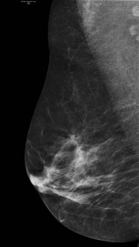
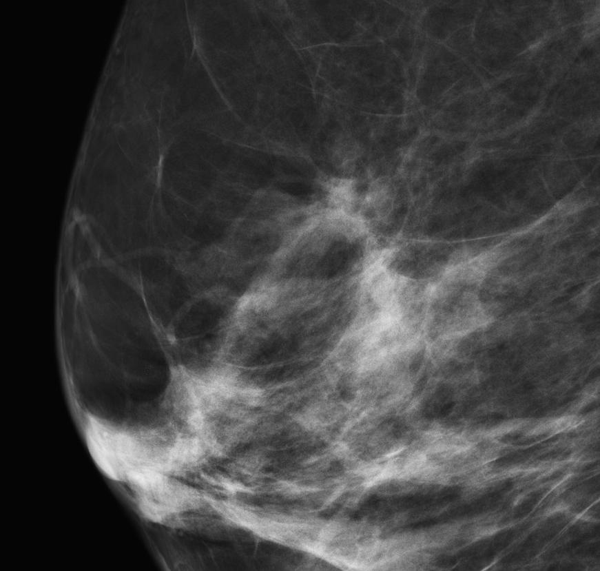
4 Architectural Distortion Definition BIRADS (4 th ed.) The normal architecture (of the breast) is distorted with no definite mass visible. This includes spiculations radiating from a point and focal retraction or distortion at the edge of parenchyma. Can be difficult to visualize finding based on written description
5 Architectural Distortion Perception: Most commonly missed mammographic finding Burrell HD et al. Rad 1996; 199: Know what to look for To be able to answer Is it real?
6 Architectural Distortion Look for Straight lines Radiating/ Spoke wheel appearance Fine, hair-like lines in Fat away from the center Optimize image Fatty tissue- gray color medium to light


7 Calls of Architectural Distortion: Real and Summation Shadows
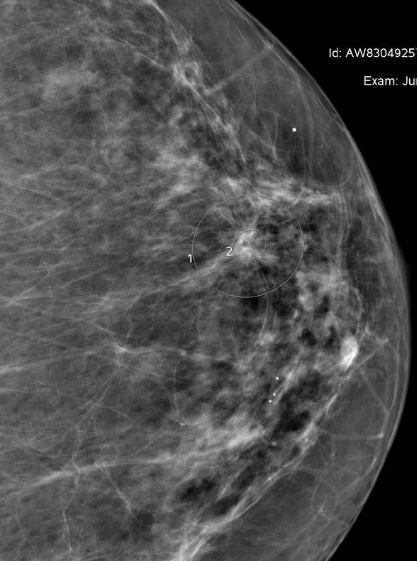
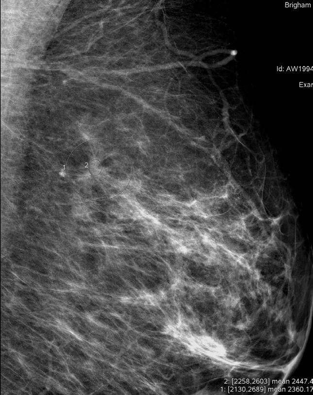
8 Calls of Architectural Distortion

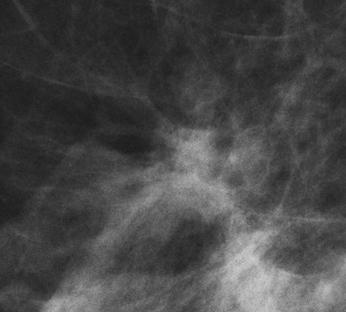
9 Architectural Distortion Straight lines Sort out vessels and ducts Can be a fine, brushhair appearance In fat Spoke wheel Angular fat-glandular interfaces
10 Arch Distortion vs Spiculated Mass Perceptual features are the same: Straigthened lines, radiation pattern Architectural distortion No central density; hence the center is radiolucent/ fat density Spiculated Mass Features of a mass Spiculations describe the margins of the mass

11 Courtesy Dr Sughra Raza

12 Courtesy Dr Sughra SBI/ACR 2016, Austin, Raza TX
13 Arch Distortion vs Spiculated Mass Perceptual features are the same: Straigthened lines, radiation pattern Like the peacock, these features may vary with projections
14 Architectural Distortion vs Summation tissues Question at Diagnostic Work-up? IS IT REAL??
15

16 First question- Is it real? Spot Magnification CC If not present on the additional view, is it because the technologist spotted the wrong area, off the page, or is it just a summation shadow?
17 For Location Correlation between spots and full views, Verify with Continents of Density- both white and grey, as well as skin, nipple, chest wall. Spot Magnification CC
18 For Location Correlation between spots and full views, Verify with Continents of Density- both white and grey, as well as skin, nipple, chest wall. Spot Magnification CC SUMMATION TISSUES
19 Is it Real Spiculated Mass?
20 MLO: electronic zoom

21 Where on CC? Yes Real!


22 Spot MLO LM MLO What do you do If the 90 degree Does not help?
23 ROLLED LAT ROLLED MED


24 ROLLED LAT ROLLED MED YES- Real!!
25 VALUE OF ROLLED VIEWS Confirm/ determine 3D location Determine 3D location BEFORE Ultrasound Can roll the breast in any position Place BB on breast to help determine location Guard against rolling tissue off the page.
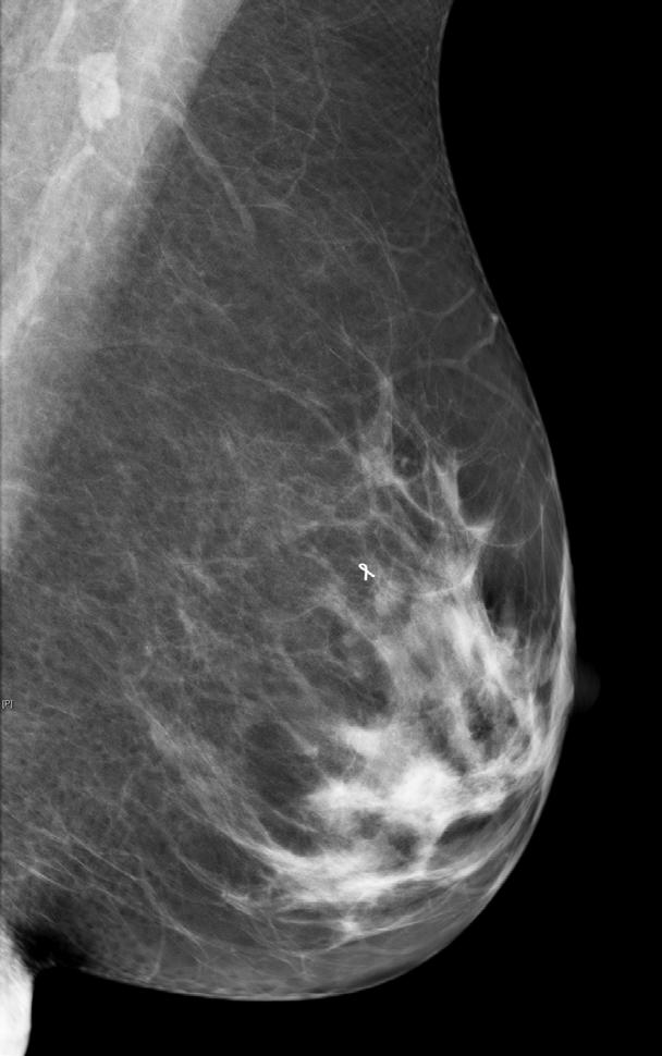
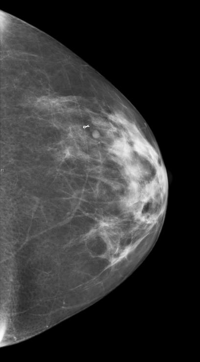
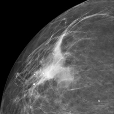
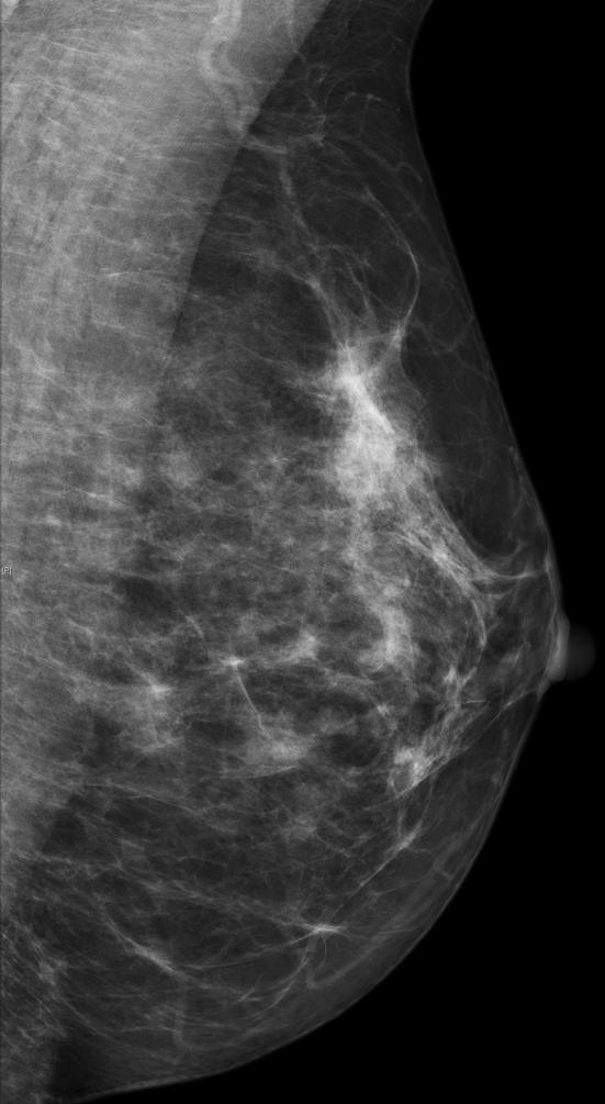
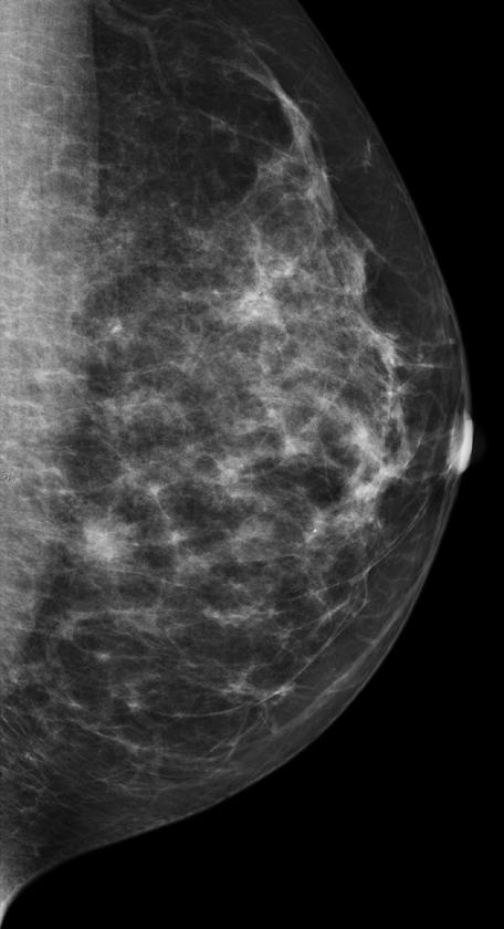
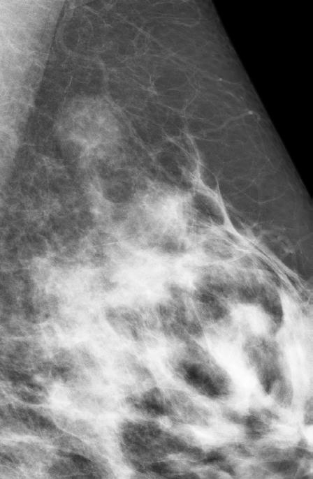
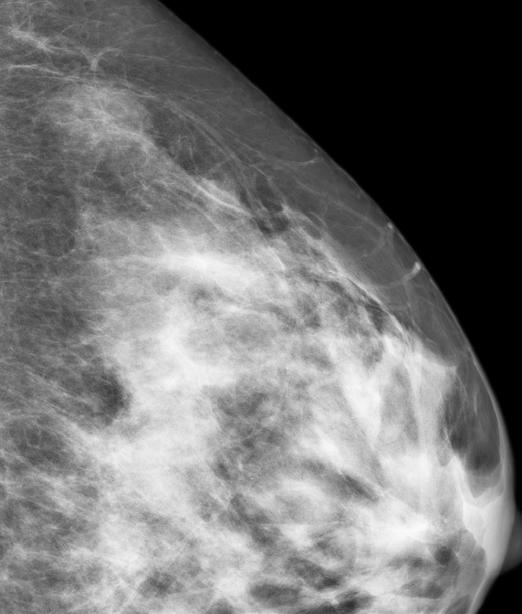
26 AD with Tomo MLO

27 AD with Tomo MLO

28 AD with Tomo MLO
29 AD with Tomo MLO
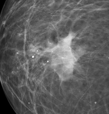
30 AD with Tomo MLO Note Central Lucency- BIOPSY!!


31 AD with Tomo MLO Note Central Lucency- BIOPSY!! Invasive Ductal Ca
32 The Asymmetries The Spectrum of Breast Asymmetries: Imaging Features, Work-up, Management Sickles EA. Radiological Clinics of No Am. 2007: Asymmetry- one view only Global Asymmetry >,= 1 quadrant Focal Asymmetry < 1 quadrant Developing asymmetry (neodensity)
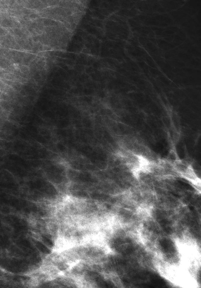
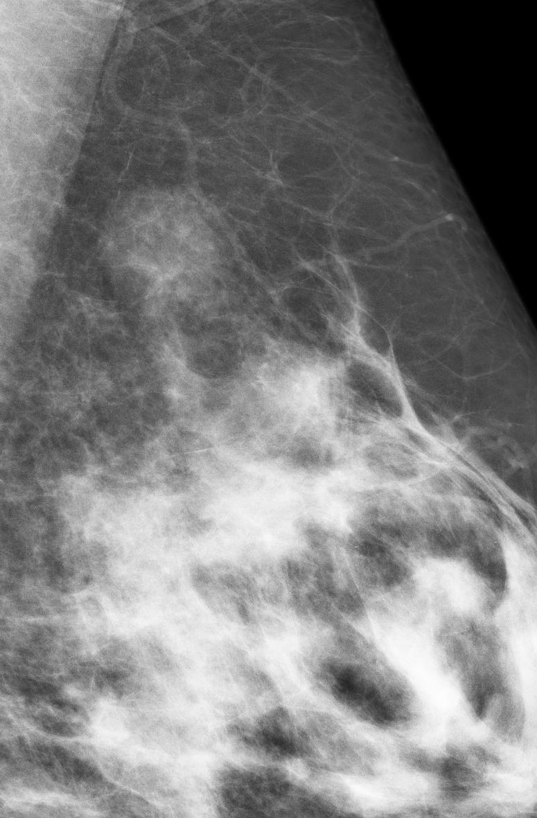
33 Focal Asymmetry Is it real? But in What Way?
34 Focal Asymmetry Is it real? But in What Way? The Differential: - Is it an ill defined mass, hence likely to be Ca, OR Is it summation tissues, hence negative for Ca? An island of breast tissue
35 Focal Asymmetry Is it a Mass? 3 Objective Criteria to Apply Feig SA. Radiol Clin No Am (30): Margins- Convexed vs Concaved from center Density- Homogeneous vs Heterogeneous Shape - Rounded (mass) vs Planar / Amorphous

36 Breast Imaging: Basic Principles Solid Ball at Xray- A Mass - Round Shape - Margins convexed outward - Homogeneous soft-tissue pattern - Centrally more dense because thicker in the center
37 Breast Imaging: Basic Principles Shape Determination of a Mass What is the radiographic density pattern of a fibrocystic Cyst? Same color (soft-tissue density) as a solid mass, but the pattern is different. Why?

38 Breast Imaging: Basic Principles Shape Determination of a Mass What is the radiographic density pattern of a Cystic mass? Because the SHAPE of a cyst is different than a solid mass in a Mammogram!!


39 Screening Call-back Focal Asymmetry

40 Spot CC Spot MLO

41 Spot CC Spot MLO Spiculated Mass


42 Focal Asymmetry MLO CC LM RL CC

43 1 year prior Developing Asymmetry Is it a New Mass?


44 Spot MLO Are margins white LINES?
45 ML Rolled ML views


46 ML Rolled ML views Summation tissues
47 Rolled Views One can roll the breast in ANY projection. Very helpful with BB s for laterality determination Use them if you need additional data AND can be used with Tomo!!
48 Developing Focal Asymmetries MLO Two years prior MLO CC
49 Developing Focal Asymmetries LM BIOPSY! represents focal change
50 Developing Focal Asymmetries LM PASH

51 Developing Focal Asymmetries 2 years later
52 Developing Focal Asymmetries 2 years later
53 Developing Focal Asymmetries If Real = Ill Defined Mass BIOPSY!


54 Developing Focal Asymmetries If Real = Ill Defined Mass BIOPSY! Invasive Ductal Ca
55 The A s: - Summary Differentiate findings between A and summation shadows Architectural Distortion Look in the fatty tissues for straight lines, spoke wheel Focal Asymmetry Analyze shape, density pattern and margins on all views Use rolled views, in any projection, including Tomo Developing Asymmetry If real = Ill-defined Mass. BIOPSY!!
56
Imaging in breast cancer. Mammography and Ultrasound Donya Farrokh.MD Radiologist Mashhad University of Medical Since
 Imaging in breast cancer Mammography and Ultrasound Donya Farrokh.MD Radiologist Mashhad University of Medical Since A mammogram report is a key component of the breast cancer diagnostic process. A mammogram
S. Murgo, MD. Chr St-Joseph, Mons Erasme Hospital, Brussels
 S. Murgo, MD Chr St-Joseph, Mons Erasme Hospital, Brussels? Introduction Mammography reports are sometimes ambiguous and indecisive. ACR has developped the BIRADS. BIRADS consists of a lexicon in order
BI-RADS Update. Martha B. Mainiero, MD, FACR, FSBI Brown University Rhode Island Hospital
 BI-RADS Update Martha B. Mainiero, MD, FACR, FSBI Brown University Rhode Island Hospital No Disclosures BI-RADS History 1980s Quality Issues ACR Accreditation BI-RADS 1994 2003 4 th Edition MRI, US January
Leonard M. Glassman MD
 BI-RADS The New BI-RADS Leonard M. Glassman MD FACR Former Chief of Breast Imaging American Institute for Radiologic Pathology Washington Radiology Associates, PC Breast Imaging Reporting and Data System
Breast asymmetries in mammography: Management
 Breast asymmetries in mammography: Management Poster No.: C-1026 Congress: ECR 2015 Type: Educational Exhibit Authors: V. de Lara Bendahan 1, F. J. Hidalgo Ramos 2, J. L. Ortega Garcia 3, Keywords: DOI:
Breast Imaging Lexicon
 9//201 200 BI RADS th Edition 201 BI RADS th Edition Breast Imaging Lexicon Mammographic Pathology and Assessment Categories Deborah Thames, R.T.(R)(M)(QM) The Advanced Health Education Center Nonmember:
Amammography report is a key component of the breast
 Review Article Writing a Mammography Report Amammography report is a key component of the breast cancer diagnostic process. Although mammographic findings were not clearly differentiated between benign
8/31/2016 HIDING IN PLAIN SITE, ARCHITECTURAL DISTORTIONS AND BREAST ASYMMETRIES ARCHITECTURAL DISTORTIONS ARCHITECTURAL DISTORTIONS
 HIDING IN PLAIN SITE, ARCHITECTURAL DISTORTIONS AND BREAST ASYMMETRIES DEBORAH THAMES R.T. (R)(M)(QM) ARCHITECTURAL DISTORTIONS Definition is disruption of the natural flow of breast pattern towards the
Diagnostic Dilemmas of Breast Imaging
 Diagnostic Dilemmas of Breast Imaging Common Causes of Error in Breast Cancer Detection By: Jason Cord, M.D. Mammography: Initial Imaging The standard for detection of breast cancer Screening mammography
ACRIN 6666 IM Additional Evaluation: Additional Views/Targeted US
 Additional Evaluation: Additional Views/Targeted US For revised or corrected form check box and fax to 215-717-0936. Instructions: The form is completed based on recommendations (from ID form) for additional
Intracystic papillary carcinoma of the breast
 Intracystic papillary carcinoma of the breast Poster No.: C-1932 Congress: ECR 2011 Type: Educational Exhibit Authors: V. Dimarelos, F. TZIKOS, N. Kotziamani, G. Rodokalakis, 1 2 3 1 1 1 2 T. MALKOTSI
THE DIAGNOSTIC WORKUP: THE TEAM APPROACH
 X-Ray Associates of New Mexico, P.C. THE DIAGNOSTIC WORKUP: THE TEAM APPROACH MICHAEL N. LINVER, MD, FACR DAWN DERENBURGER, RTRM Disclosure There are no conflicts of interest or relevant financial interests
UW Radiology Review Course Breast Calcifications. BI-RADS 5 th Edition
 UW Radiology Review Course Breast Calcifications Grace Kalish, MD Vantage Radiology BI-RADS 5 th Edition Benign Skin Vascular Large rod like Coarse popcorn Suspicious Amorphous Coarse heterogenous Fine
Ultrasound of the Breast BASICS FOR THE ORDERING CLINICIAN
 Ultrasound of the Breast BASICS FOR THE ORDERING CLINICIAN Breast Ultrasound Anatomy Skin Breast Parenchyma Pectoralis Fascia Pectoralis Breast Ultrasound Anatomy Indications for Breast Ultrasound Palpable
Ana Sofia Preto 19/06/2013
 Ana Sofia Preto 19/06/2013 Understanding the underlying pathophysiologic processes leading to the various types of calcifications Description and illustration of the several types of calcifications, according
Since its introduction in 2000, digital mammography has become
 Review Article Smith A, PhD email : Andrew.smith@hologic.com Since its introduction in 2000, digital mammography has become an accepted standard of care in breast cancer screening and has paved the way
Mammographic imaging of nonpalpable breast lesions. Malai Muttarak, MD Department of Radiology Chiang Mai University Chiang Mai, Thailand
 Mammographic imaging of nonpalpable breast lesions Malai Muttarak, MD Department of Radiology Chiang Mai University Chiang Mai, Thailand Introduction Contents Mammographic signs of nonpalpable breast cancer
University of Washington Radiology Review Course: Strange and Specific Diagnoses. Case #1
 University of Washington Radiology Review Course: Strange and Specific Diagnoses Katherine E. Dee, MD Seattle Breast Center Via Radiology 2014 Case #1 37 year old presents with bilateral palpable lumps.
Outline. Digital Breast Tomosynthesis: Update and Pearls for Implementation. Tomosynthesis Dataset: 2D/3D (Hologic Combo Acquisition)
") Outline Digital Breast Tomosynthesis (DBT) the new standard of care Digital Breast Tomosynthesis: Update and Pearls for Implementation Emily F. Conant, M.D. Professor, Chief of Breast Imaging Department
Outline Digital Breast Tomosynthesis (DBT) the new standard of care Digital Breast Tomosynthesis: Update and Pearls for Implementation Emily F. Conant, M.D. Professor, Chief of Breast Imaging Department
Armed Forces Institute of Pathology.
 Armed Forces Institute of Pathology www.radpath.com Armed Forces Institute of Pathology Breast Disease www.radpath.org Armed Forces Institute of Pathology Interpretation of Breast MRI Leonard M. Glassman
Optimizing Breast Sonography
 Optimizing Breast Sonography Cindy Rapp BS, RDMS, FSDMS, FAIUM Denver, Colorado Breast Sonography general goal to make a more specific diagnosis than can be made with clinical and mammographic findings
Contrast Enhanced Spectral Mammography (CESM) Updates
 Updates") Contrast Enhanced Spectral Mammography (CESM) Updates Georgeta Mihai, PhD, DABR Medical Physicist, BIDMC, Boston Assistant Professor, Harvard Medical School, Boston Disclosures None Acknowledgments: Da
Contrast Enhanced Spectral Mammography (CESM) Updates Georgeta Mihai, PhD, DABR Medical Physicist, BIDMC, Boston Assistant Professor, Harvard Medical School, Boston Disclosures None Acknowledgments: Da
FACT: FACT: Breast Cancer Staging. For cancer to occur, something must damage nucleus of the cell. Stage I. Stage II 10/9/2018
 Digital Breast Tomosynthesis (DBT) Pathology Findings: Case Studies and Beyond For cancer to occur, something must damage nucleus of the cell. Advanced Health Education Center www.aheconline.com Copyright
Non-mass Enhancement on Breast MRI. Aditi A. Desai, MD Margaret Ann Mays, MD
 Non-mass Enhancement on Breast MRI Aditi A. Desai, MD Margaret Ann Mays, MD Breast MRI Important screening and diagnostic tool, given its high sensitivity for breast cancer detection Breast MRI - Indications
What s New in Breast Imaging. Jennifer A. Harvey, M.D., FACR Professor of Radiology University of Virginia
 What s New in Breast Imaging Jennifer A. Harvey, M.D., FACR Professor of Radiology University of Virginia Disclosure Hologic, Inc. Shareholder and research agreement. Volpara Solutions, Ltd. Shareholder
Imaging Guidelines for Breast Cancer Screening
 Imaging Guidelines for Breast Cancer Screening Sarah Colwick, MD Dr. Sarah Colwick was born and raised in Sikeston, MO. She attended college and medical school at the University of Missouri-Kansas City
AMSER Case of the Month: September 2018
 AMSER Case of the Month: September 2018 60-year-old woman with a left breast mass noted on screening mammography. Catherine McNulty, MS4 Tulane University School of Medicine Dr. Robin Sobolewski Breast
Ductal carcinoma in situ: ultrasound, mammography and MRI features with pathologic correlation
 Ductal carcinoma in situ: ultrasound, mammography and MRI features with pathologic correlation Poster No.: C-2252 Congress: ECR 2013 Type: Educational Exhibit Authors: L. Fernandes, H. A. M. R. Tinto,
BI-RADS and Breast MRI. Kathy Borovicka, M.D. Thursday February 15, 2018
 BI-RADS and Breast MRI Kathy Borovicka, M.D. Thursday February 15, 2018 Learning Objectives Be familiar with the Breast Imaging Reporting and Data System (BI-RADS) Understand the components of a breast
ORIGINAL ARTICLE EVALUATION OF BREAST LESIONS USING X-RAY MAMMOGRAM WITH HISTOPATHOLOGICAL CORRELATION
 Available online at www.journalijmrr.com INTERNATIONAL JOURNAL OF MODERN RESEARCH AND REVIEWS IJMRR ISSN: 2347-8314 Int. J. Modn. Res. Revs. Volume 3, Issue 10, pp 807-814, October, 2015 ORIGINAL ARTICLE
AMSER Case of the Month: November 2018
 AMSER Case of the Month: November 2018 42 year old with right breast mass Rina Kiyota Petek Lake Erie College of Osteopathic Medicine, OMS-III Kossivi Dantey, MD Bibianna Klepchick, MD Matthew Hartman,
PLACE LABEL HERE. ACRIN 6657 MRI Form: Pre-Treatment (MRI-1)
") M3 ACRIN 6657 MRI Form: Pre-Treatment (MRI-1) If this is a revised or corrected form,indicate by checking box. ACRIN Study 6657 Case # Instructions: In accordance with the protocol, four MRI exams are
M3 ACRIN 6657 MRI Form: Pre-Treatment (MRI-1) If this is a revised or corrected form,indicate by checking box. ACRIN Study 6657 Case # Instructions: In accordance with the protocol, four MRI exams are
Evaluation of Abnormal Screening Mammograms
 342 Evaluation of Abnormal Screening Mammograms Ellen Shaw de Paredes, M.D. The purpose of routine screening mammography is to detect unsuspected cancer that has the potential to be cured. Abnormalities
TOMOSYNTHESIS: WORTH ALL THE HYPE?
 X-Ray Associates of New Mexico, P.C. TOMOSYNTHESIS: WORTH ALL THE HYPE? MICHAEL N. LINVER, MD, FACR MAMMOGRAPHY: THE GOOD, THE PRETTY GOOD, & THE NOT SO GOOD MAMMOGRAPHY: THE GOOD, THE PRETTY GOOD, & THE
Fat Necrosis: A Grand Imposter
 Fat Necrosis: A Grand Imposter Poster No.: C-0751 Congress: ECR 2015 Type: Educational Exhibit Authors: L. C. Flores Salinas, Y. A. Ramirez Galvan, A. Garza Báez, C. M. Ferrara Chapa; Monterrey/MX Keywords:
Digital breast tomosynthesis (DBT) occult breast cancers: clinical, radiological and histopathological features.
 occult breast cancers: clinical, radiological and histopathological features.") Digital breast tomosynthesis (DBT) occult breast cancers: clinical, radiological and histopathological features. Poster No.: C-1707 Congress: ECR 2015 Type: Scientific Exhibit Authors: V. Vinci 1, A. Iqbal
Digital breast tomosynthesis (DBT) occult breast cancers: clinical, radiological and histopathological features. Poster No.: C-1707 Congress: ECR 2015 Type: Scientific Exhibit Authors: V. Vinci 1, A. Iqbal
Leonard M. Glassman MD Analysis of Breast Calcifications
 Importance of Calcification Leonard M. Glassman MD FACR American Institute for Radiologic Pathology Washington Radiology Associates, PC Washington DC 45% of all breast cancers present as calcification
Developing Asymmetry Identified on Mammography: Correlation with Imaging Outcome and Pathologic Findings
 Asymmetry on Mammography Women s Imaging Original Research WOMEN S IMAGING Jessica W. T. Leung 1 Edward A. Sickles Leung JWT, Sickles EA Keywords: breast, breast cancer, mammography, screening, sonography
Invasive lobular carcinoma of the breast; spectrum of imaging findings.
 Invasive lobular carcinoma of the breast; spectrum of imaging findings. Poster No.: C-0847 Congress: ECR 2014 Type: Educational Exhibit Authors: D. Mandich, T. Diaz de Bustamante, L. Koren, M. Arroyo,
A Breast Surgeon s Use of Three Dimensional Specimen Tomosynthesis
 A Breast Surgeon s Use of Three Dimensional Specimen Tomosynthesis Cary S. Kaufman MD, FACS Associate Clinical Professor of Surgery A Breast Surgeon s Use of Three Dimensional Specimen Tomosynthesis Cary
Imaging the Symptomatic Patient. Avice M.O Connell MD,FACR,FSBI Professor of Imaging Sciences Director, Women s Imaging University of Rochester
 Imaging the Symptomatic Patient Avice M.O Connell MD,FACR,FSBI Professor of Imaging Sciences Director, Women s Imaging University of Rochester The four most common symptoms Mass Pain Discharge Infection
PLACE LABEL HERE BASELINE / PRE-TREATMENT. ACRIN 6657 Extension MRI Form: Baseline / Pre-Treatment MRI 1. o Unknown
 T1 ACRIN 6657 Extension MRI Form: Baseline / Pre-Treatment MRI 1 If this is a revised or corrected form, please box. ACRIN Study 6657 No. Instructions: In accordance with the protocol, four MRI exams are
Standard Breast Imaging Modalities. Lilian Wang, M.D. Breast Imaging Section Department of Radiology Northwestern Medicine
 Standard Breast Imaging Modalities Lilian Wang, M.D. Breast Imaging Section Department of Radiology Northwestern Medicine Overview Standard breast imaging modalities Mammography Ultrasound MRI Imaging
RADIOLOGIC EVALUATION OF BREAST CANCER
 RADIOLOGIC EVALUATION OF BREAST CANCER Orsolya Farkas, Gabriella Bodrogi and Gábor Szalai Department of Radiology, Pécs University Orsifarkas@yahoo.com Complex evaluation of the breast Patient history
Pitfalls and Limitations of Breast MRI. Susan Orel Roth, MD Professor of Radiology University of Pennsylvania
 Pitfalls and Limitations of Breast MRI Susan Orel Roth, MD Professor of Radiology University of Pennsylvania Objectives Review the etiologies of false negative breast MRI examinations Discuss the limitations
Radiology Review Course Hotel del Coronado Coronado, California
 37 th Annual Radiology Review Course Hotel del Coronado Coronado, California Friday, April 21, 2017 - PM TABLE OF CONTENTS Friday, April 21, 2017 - PM SAM Session - Breast Imaging Update 12:45 PM 1:30
Current issues and controversies in breast imaging. Kate Brown, South GP CME 2015
 Current issues and controversies in breast imaging Kate Brown, South GP CME 2015 JUDICIOUS USE OF RESOURCES IN REFERRALS FOR BREAST IMAGING THE DILEMMA How do target referrals for breast imaging? Want
MEDICAL IMAGING AND BREAST DISEASE HOW CAN WE HELP YOU?
 MEDICAL IMAGING AND BREAST DISEASE HOW CAN WE HELP YOU? Barbara M. Preston, M.D. SCREENING MAMMOGRAPHY AVERAGE RISK PATIENTS KAISER RECOMMENDATION: ALL WOMEN (INCLUDING TRANSGENDER FEMALES) Every 1-21
BIRADS 3 and 4 lesions viewed by ultrasound and not seen in digital mammograms and tomosynthesis.
 Original article Anales de Radiología México 2016 Jul;15(3):205-213. BIRADS 3 and 4 lesions viewed by ultrasound and not seen in digital mammograms and tomosynthesis. García-Quintanilla JF 1, González-Coronado
The radiologic workup of a palpable breast mass
 Imaging in Practice CME CREDIT EDUCTIONL OJECTIVE: The reader will consider which breast masses require further workup and which imaging study is most appropriate Lauren Stein, MD Imaging Institute, Cleveland
STEREOTACTIC BREAST BIOPSY: CORRELATION WITH HISTOLOGY
 3-rd Baltic Congress of Radiology, October 8-9, 2010 Riga Rūta Briedienė, Rūta Grigienė, Raimundas Meškauskas Institute of Oncology Vilnius University, National Centre of Pathology STEREOTACTIC BREAST
Breast imaging in general practice
 Breast series CLINICAL PRACTICE Breast imaging in general practice Nehmat Houssami, MBBS, FAFPHM, FASBP, PhD, is Associate Clinical Director, NSW Breast Cancer Institute, Westmead Hospital, Honorary Senior
Breast Cancer Screening with Mammography
 Progress in Public Health Breast Cancer Screening with Mammography JMAJ 44(7): 318 324, 2001 Tokiko ENDO Director, Department of Radiology, National Nagoya Hospital Abstract: Breast cancer has been increasing
POSITIONING ACR REQUIREMENTS IMAGE REVIEW CATEGORIES DEFICIENCIES IN POSITIONING ACR REQUIREMENTS 3/28/2016 NUMBER 1 REASON FOR ACR FAILURE
 CERTIFYING AGENCIES MAMMOGRAPHY CLINICAL IMAGE EVALUATION Pam Fulmer, BA RT (R)(M)(QM) FDA approved certifying states States can only certify facilities within their state borders Illinois Iowa South Carolina
Avoiding Pitfalls in Mammographic Interpretation
 Canadian Association of Radiologists Journal 62 (2011) 50e59 www.carjonline.org Thoracic and Cardiac Imaging / Imagerie cardiaque et imagerie thoracique Avoiding Pitfalls in Mammographic Interpretation
CDIS: what's beyond microcalcifications? - Pictorial essay
 CDIS: what's beyond microcalcifications? - Pictorial essay Poster No.: C-1096 Congress: ECR 2014 Type: Educational Exhibit Authors: R. N. Lucas, C. A. S. Ruano, I. Oliveira, J. M. G. Lourenco, Z. 1 1 1
Supplemental Screening for Dense Breasts. Reagan Leverett, MD, MS
 Supplemental Screening for Dense Breasts Reagan Leverett, MD, MS Outline Anatomy and Density Risk of dense breasts Theory of Supplemental Screening Options for supplemental screening Tomosynthesis Ultrasound
DCIS of the Breast--MRI findings with mammographic correlation.
 DCIS of the Breast--MRI findings with mammographic correlation. Poster No.: C-1560 Congress: ECR 2013 Type: Educational Exhibit Authors: N. B. Ibrahim, P. Morris, S. ANANDAN; Burlington, MA/US Keywords:
Digital Breast Tomosynthesis in the Diagnostic Environment: A Subjective Side-by-Side Review
 Women s Imaging Original Research Hakim et al. Digital Breast Tomosynthesis Women s Imaging Original Research Christiane M. Hakim 1 Denise M. Chough 1 Marie A. Ganott 1 Jules H. Sumkin 1 Margarita L. Zuley
Can Digital Breast Tomosynthesis(DBT) Perform Better than Standard Digital Mammography Workup in a Breast Cancer Assessment Clinic?
 Perform Better than Standard Digital Mammography Workup in a Breast Cancer Assessment Clinic?") Can Digital Breast Tomosynthesis(DBT) Perform Better than Standard Digital Mammography Workup in a Breast Cancer Assessment Clinic? Accepted for publication in European Radiology Authors: S Mall, J Noakes,
Can Digital Breast Tomosynthesis(DBT) Perform Better than Standard Digital Mammography Workup in a Breast Cancer Assessment Clinic? Accepted for publication in European Radiology Authors: S Mall, J Noakes,
Benign, Reactive and Inflammatory Lesions of the Breast
 Benign, Reactive and Inflammatory Lesions of the Breast Marilin Rosa, MD Associate Member Section Head of Breast Pathology Department of Anatomic Pathology Program Director, Breast Pathology Fellowship
AB MR Interpretation Overview
 AB MR Interpretation Overview Goal of AB MR interpretation is to maintain high sensitivity and specificity In order to minimize false positives and short term follow ups, it is fundamental to focus only
Journal of Medical Imaging and Radiation Oncology
 Journal of Medical Imaging and Radiation Oncology 60 (2016) 506 513 MEDICAL IMAGING PICTORIAL ESSAY Malignant hyperechoic breast lesions at ultrasound: A pictorial essay Stephen Tiang, 1 Cecily Metcalf,
Evaluation of surgical margins by specimen in impalpable breast carcinoma: a radiopathological correlation
 Evaluation of surgical margins by specimen in impalpable breast carcinoma: a radiopathological correlation Poster No.: C-1146 Congress: ECR 2014 Type: Scientific Exhibit Authors: D. Mandich, L. Koren,
SIGNIFICANT OTHERS. Miscellaneous Benign Breast Conditions
 SIGNIFICANT OTHERS Miscellaneous Benign Breast Conditions Epworth HealthCare 1 FAT NECROSIS TRAUMATIC Cell rupture Seat-Belt injury Blunt trauma Iatrogenic injury Surgery, Flaps, Radiotherapy Pathology
Tomosynthesis and breast imaging update. Dr Michael J Michell Consultant Radiologist King's College Hospital NHS Foundation Trust
 Tomosynthesis and breast imaging update Dr Michael J Michell Consultant Radiologist King's College Hospital NHS Foundation Trust Breast imaging new technology BREAST CANCER FLT PET shows different grades
BI-RADS 3 category, a pain in the neck for the radiologist which technique detects more cases?
 BI-RADS 3 category, a pain in the neck for the radiologist which technique detects more cases? Poster No.: B-0966 Congress: ECR 2013 Type: Scientific Paper Authors: J. Etxano Cantera, I. Simon-Yarza, G.
ISSN X (Print) Research Article. *Corresponding author Dr. Amlendu Nagar
 Research Article. *Corresponding author Dr. Amlendu Nagar") Scholars Journal of Applied Medical Sciences (SJAMS) Sch. J. App. Med. Sci., 2015; 3(3A):1069-1073 Scholars Academic and Scientific Publisher (An International Publisher for Academic and Scientific Resources)
Scholars Journal of Applied Medical Sciences (SJAMS) Sch. J. App. Med. Sci., 2015; 3(3A):1069-1073 Scholars Academic and Scientific Publisher (An International Publisher for Academic and Scientific Resources)
BI-RADS classification in breast tomosynthesis. Our experience in breast cancer cases categorized as BI-RADS 0 in digital mammography
 BI-RADS classification in breast tomosynthesis. Our experience in breast cancer cases categorized as BI-RADS 0 in digital mammography Poster No.: C-0562 Congress: ECR 2017 Type: Scientific Exhibit Authors:
Lesion Imaging Characteristics Mass, Favoring Benign Circumscribed Margins Intramammary Lymph Node
 Lesion Imaging Characteristics Mass, Favoring Benign Circumscribed Margins Intramammary Lymph Node Oil Cyst Mass, Intermediate Concern Microlobulated Margins Obscured Margins Mass, Favoring Malignant Indistinct
Mammography is a most effective imaging modality in early breast cancer detection. The radiographs are searched for signs of abnormality by expert
 Abstract Methodologies for early detection of breast cancer still remain an open problem in the Research community. Breast cancer continues to be a significant problem in the contemporary world. Nearly
Breast Ultrasound: Improving Your Skills & Patient Care
 Breast Ultrasound: Improving Your Skills & Patient Care Objectives Discuss US techniques available for image optimization. Review & compare the US appearances of benign & malignant masses. Cherie M. Kuzmiak,
Breast Imaging! Ravi Adhikary, MD!
 Breast Imaging! Ravi Adhikary, MD! ACS Estimated Cancers Statistics 2014! Breast! New Cases in Women! 232,670 (+67,570 in situ)! Deaths in Women! 40,000! Colon! 48,380! 24,040! Cervical! 12,360! 4,020!
Mammographic evaluation of palpable breast masses with pathological correlation: a tertiary care centre study in Nepal
 Original article 21 Mammographic evaluation of palpable breast masses with pathological correlation: a tertiary care centre study in Nepal G. Gurung, R. K. Ghimire, B. Lohani Department of Radiology and
Scottsdale, AZ Plaza Hotel, 7200 N. Scottsdale Rd
 Mammography Education, Inc. 2014 3D image of the breast tissue LÁSZLÓ TABÁR, M.D.,F.A.C.R (Hon) and STAMATIA DESTOUNIS, M.D., F.A.C.R. The normal TDLUs have bud-like acini Multimodality Approach to Detection
Updates in Mammography. Dr. Yang Faridah A. Aziz Department of Biomedical Imaging University Malaya Medical Centre
 Updates in Mammography Dr. Yang Faridah A. Aziz Department of Biomedical Imaging University Malaya Medical Centre Updates in Mammography Breast Imaging Dr. Yang Faridah A. Aziz Department of Biomedical
Contrast-Enhanced Spectral Mammography
 Contrast-Enhanced Spectral Mammography Illuminating Breast Cancer Detection SenoBright HD TM gehealthcare.com/senobright Mammography is the most reliable imaging technique for breasts, but limitations
CURRENTLY IN THE United States, 431,284
 Increased Breast Calcifications in Women With ESRD on Dialysis: Implications for Breast Cancer Screening Mario Castellanos, MD, Seema Varma, MD, Kathleen Ahern, PhD, RN, Sue-Jane Grosso, MD, Shalom Buchbinder,
Pitfalls of Dynamic Contrast Enhanced MR Mammography (DCE-MRM) in Evaluation of Post-Biopsy Suspicious Breast Lesions
 in Evaluation of Post-Biopsy Suspicious Breast Lesions") Med. J. Cairo Univ., Vol. 86, No. 3, June: 1513-1522, 2018 www.medicaljournalofcairouniversity.net Pitfalls of Dynamic Contrast Enhanced MR Mammography (DCE-MRM) in Evaluation of Post-Biopsy Suspicious
Med. J. Cairo Univ., Vol. 86, No. 3, June: 1513-1522, 2018 www.medicaljournalofcairouniversity.net Pitfalls of Dynamic Contrast Enhanced MR Mammography (DCE-MRM) in Evaluation of Post-Biopsy Suspicious
One or Two Clusters of Crushed Stone like Calcifications on the Mammogram Produced by Malignancy
 66 One or Two Clusters of Crushed Stone like Calcifications on the Mammogram Produced by Malignancy Example 2.13 A 36-year-old woman who recentlyfelt a small hard lump in the upper-outer quadrant of her
Correlation between lesion type and the additional value of digital breast tomosynthesis
 Correlation between lesion type and the additional value of digital breast tomosynthesis Poster No.: C-1604 Congress: ECR 2011 Type: Scientific Exhibit Authors: C. Van Ongeval, L. Cockmartin, A. Van Steen,
Breast Health. Learning Objectives. Breast Anatomy. Poll Question. Breast Anatomy
 Learning Objectives Describe breast anatomy to a patient Breast Health Answer questions about causes of breast pain and masses Explain breast cancer screening/diagnostic modalities Appropriately triage
OPTO-ACOUSTIC BREAST IMAGING
 OPTO-ACOUSTIC BREAST IMAGING A Novel Fusion of Functional and Morphologic Imaging Reni S. Butler, MD A. Thomas Stavros, MD F. Lee Tucker, MD Michael J. Ulissey, MD PURPOSE 1. Explain opto-acoustic (OA)
BREAST IMAGING and NEW IMAGING MODALITIES- A Surgeons view
 BREAST IMAGING and NEW IMAGING MODALITIES- A Surgeons view DR CHANTEL THORNTON SPECIALIST BREAST CANCER SURGEON BMSc (hons) MBBS (hons) FRACS Epworth Hospital, Richmond- Agora Centre for Women s Health
Here are examples of bilateral analog mammograms from the same patient including CC and MLO projections.
 Good afternoon. It s my pleasure to be discussing Diagnostic Breast Imaging over the next half hour. I m Wei Yang, Professor of Diagnostic Radiology and Chief, the Section of Breast Imaging as well as
DR AISHA A UMAR CHIEF CONSULTANT RADIOLOGIST NATIONAL HOSPITAL ABUJA.
 DR AISHA A UMAR CHIEF CONSULTANT RADIOLOGIST NATIONAL HOSPITAL ABUJA. OUTLINE WHY DO WE IMAGE WHOM TO IMAGE WHEN TO IMAGE HOW TO IMAGE WHAT TO IMAGE WITH PERSONAL EXPERIENCE CONCLUSION/RECOMMENDATIONS
Imaging of radial scars on Digital Breast Tomosynthesis: a pictorial review.
 Imaging of radial scars on Digital Breast Tomosynthesis: a pictorial review. Poster No.: C-2114 Congress: ECR 2017 Type: Educational Exhibit Authors: E. Zanelli, I. Bednarova, A. Dallorto, A. Linda, C.
Fundamentals of Breast Tomosynthesis
 Fundamentals of Breast Tomosynthesis Improving the Performance of Mammography Andrew Smith, Ph.D. This white paper is one in a series of research overviws on advanced technologies in women s healthcare.
The role of MRI in assessment of asymmetrical breast densities
 The Egyptian Journal of Radiology and Nuclear Medicine (2010) 41, 501 508 Egyptian Society of Radiology and Nuclear Medicine The Egyptian Journal of Radiology and Nuclear Medicine www.elsevier.com/locate/ejrnm
Breast Imaging Essentials
 Breast Imaging Essentials Module 8 Transcript 2017 ASRT. All rights reserved. Breast Imaging Essentials Module 8 Digital Procedures and Techniques 1. ASRT Animation 2. Welcome Welcome to Module 8 of Breast
Building Blocks for Effective Report Communication
 2016 SBI/ACR Breast Imaging Symposium Building Blocks for Effective Report Communication Richard L. Ellis, M.D. Breast Imaging Section Mayo Clinic Franciscan Health Care La Crosse, WI JW Marriott Austin
Case Scenario 1 History and Physical 3/15/13 Imaging Pathology
 Case Scenario 1 History and Physical 3/15/13 The patient is an 84 year old white female who presented with an abnormal mammogram. The patient has a five year history of refractory anemia with ringed sideroblasts
Contrast-Enhanced Digital Mammography
 2015 ARRS Breast Symposium Contrast-Enhanced Digital Mammography John Lewin, M.D. Diversified Radiology of Colorado CEDM - Outline History Technique Literature Review / Cases Clinical Status Inexpensive,
Alena Levit MD Avice O Connell MD University of Rochester, Rochester, NY
 Alena Levit MD Avice O Connell MD University of Rochester, Rochester, NY Purpose Review imaging spectrum of both common benign and malignant breast lesions Describe and demonstrate CT features with mammogram,
EARLY DETECTION: MAMMOGRAPHY AND SONOGRAPHY
 EARLY DETECTION: MAMMOGRAPHY AND SONOGRAPHY Elizabeth A. Rafferty, M.D. Avon Comprehensive Breast Center Massachusetts General Hospital Harvard Medical School Breast Cancer Screening Early detection of
Tissue Breast Density
 Tissue Breast Density Reporting breast density within the letter to the patient is now mandated by VA law. Therefore, this website has been established by Peninsula Radiological Associates (PRA), the radiologists
Financial Disclosures
 Financial Disclosures 3D Mammography: The Latest Developments in the Breast Imaging Arena I have no financial disclosures Dr. Katharine Lampen-Sachar Breast and Body Radiologist Radiology Associates of
Mammographically non-calcified ductal carcinoma in situ: sonographic features with pathological correlation in 35 patients
 Clinical Radiology (2009) 64, 628e636 ORIGINAL PAPER Mammographically non-calcified ductal carcinoma in situ: sonographic features with pathological correlation in 35 patients B. Mesurolle a, *, M. El-Khoury