Columnar Cell Lesions and Flat Epithelial Atypia
|
|
|
- Gordon Grant
- 5 years ago
- Views:
Transcription
1 Columnar Cell Lesions and Flat Epithelial Atypia Laura C. Collins, M.D. Department of Pathology Beth Israel Deaconess Medical Center and Harvard Medical School, Boston, MA
2 Terminology for Columnar Cell Lesions Columnar cell change Columnar cell hyperplasia Flat epithelial atypia (Columnar cell change with atypia) (Columnar cell hyperplasia with atypia)
3 Columnar Cell Lesions Increasingly seen in an era of mammographic screening due to the identification of microcalcifications Further increase noted with introduction of digital mammography (Anoek, Mod Pathol, 2011)
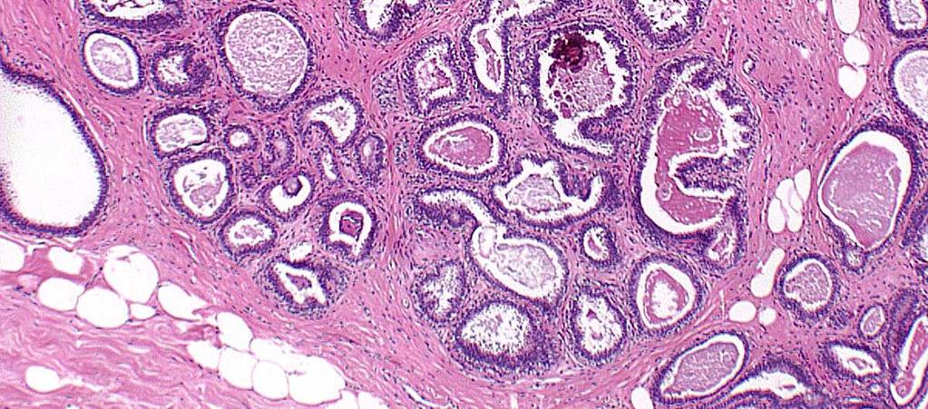
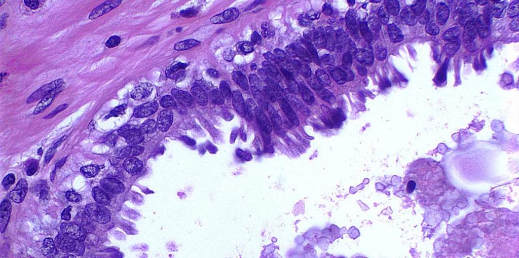
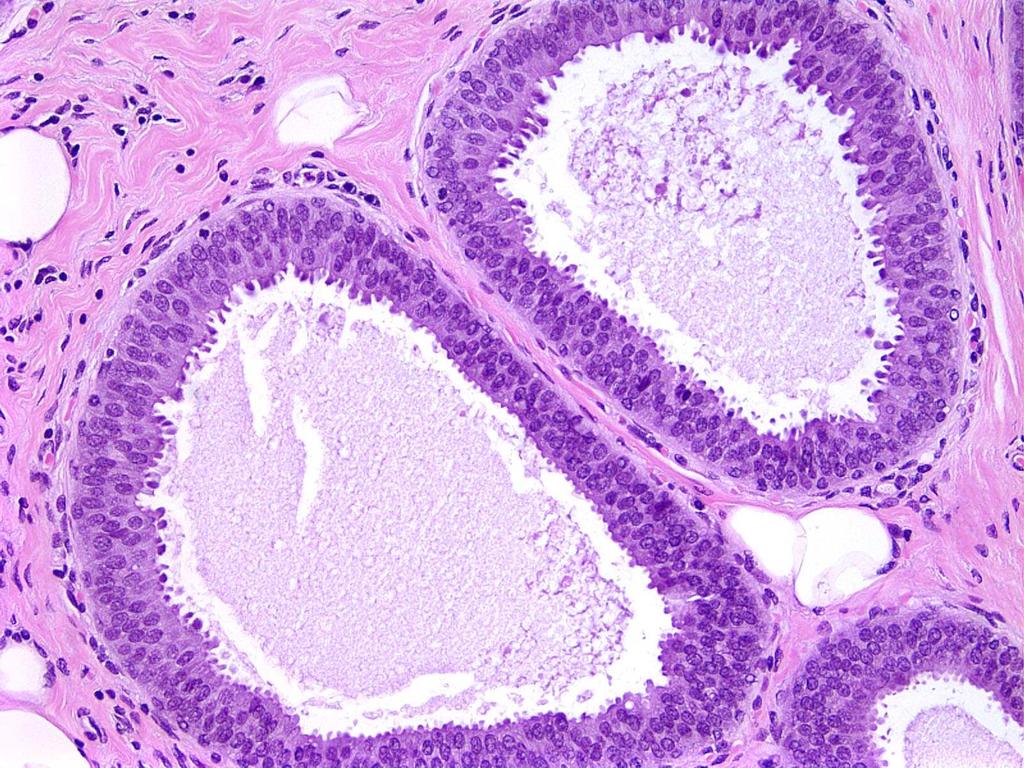
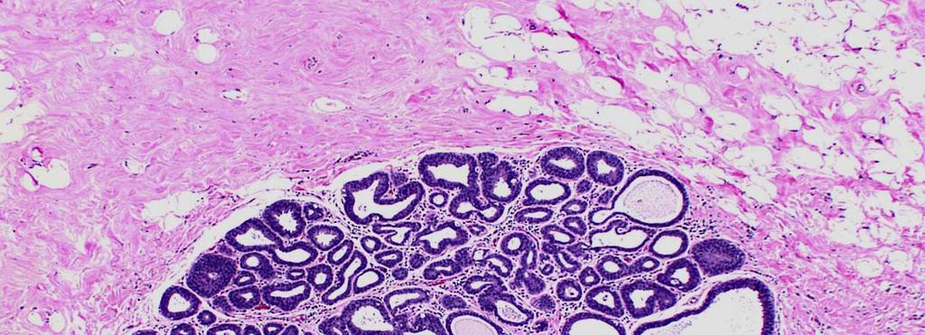
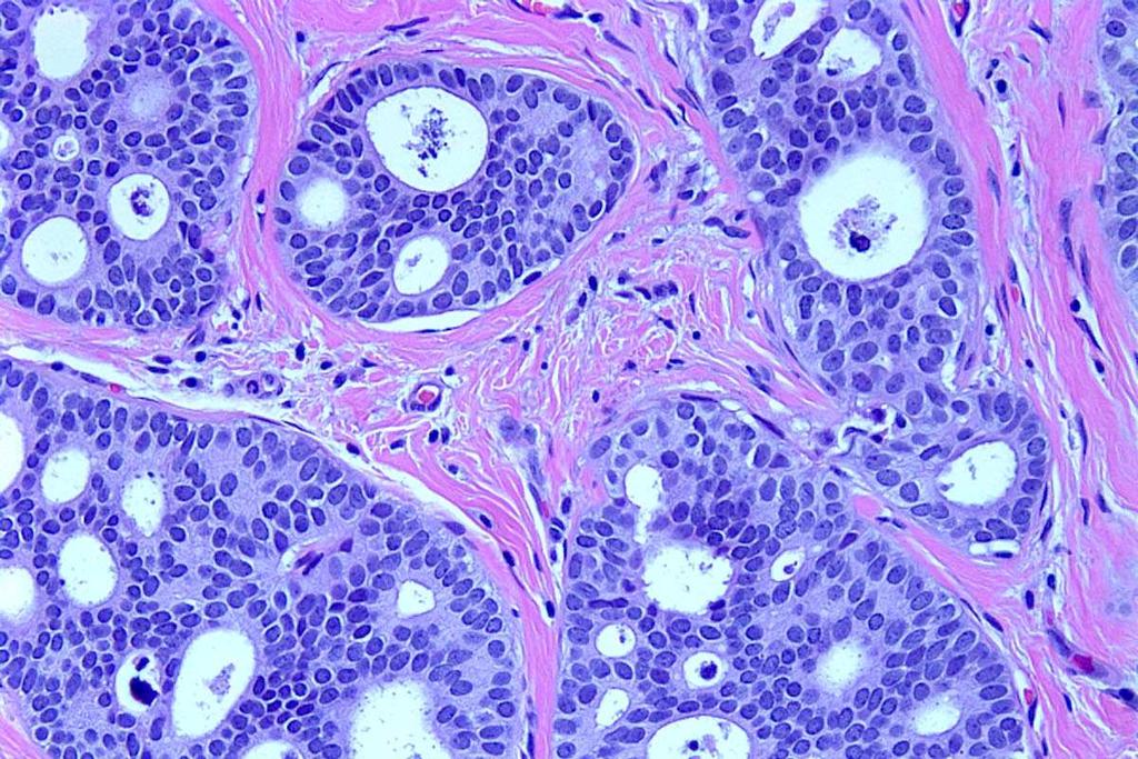
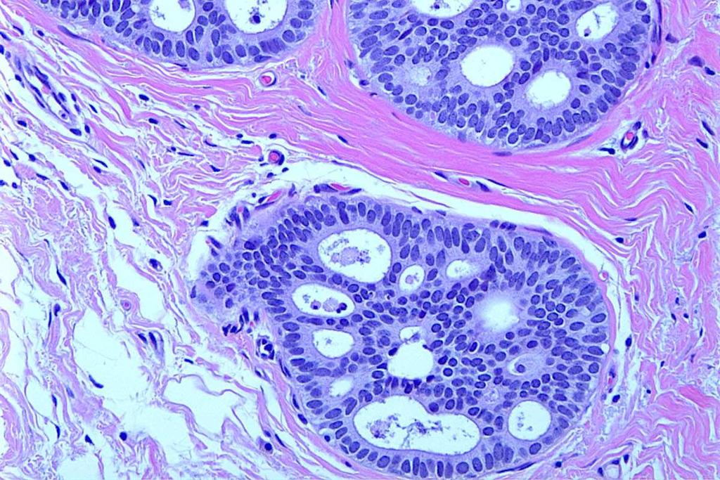
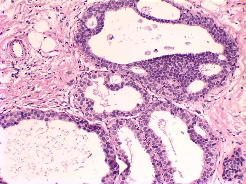
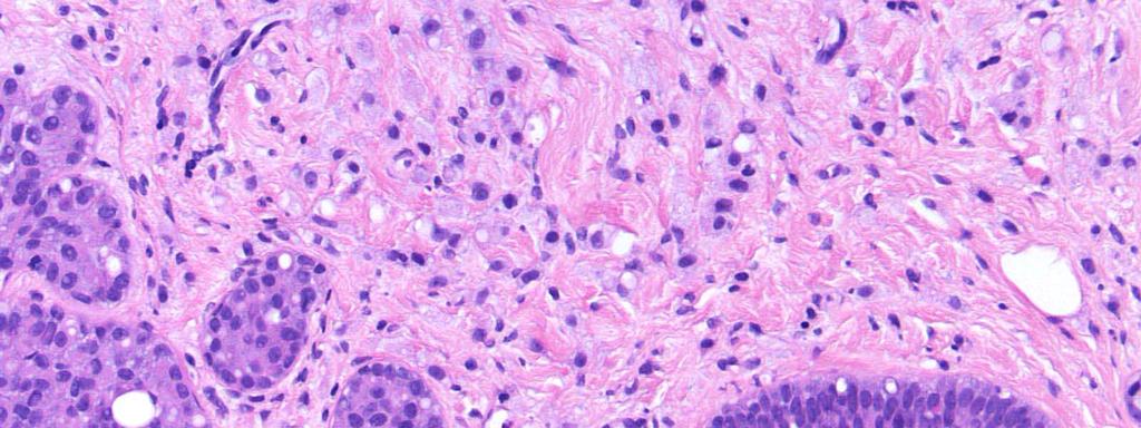
4 Columnar Cell Change WHO, 2003 Variably distended acini of TDLUs lined by columnar cells arranged perpendicular to basement membrane Apical snouts Secretions and microcalcifications in lumens
5
6
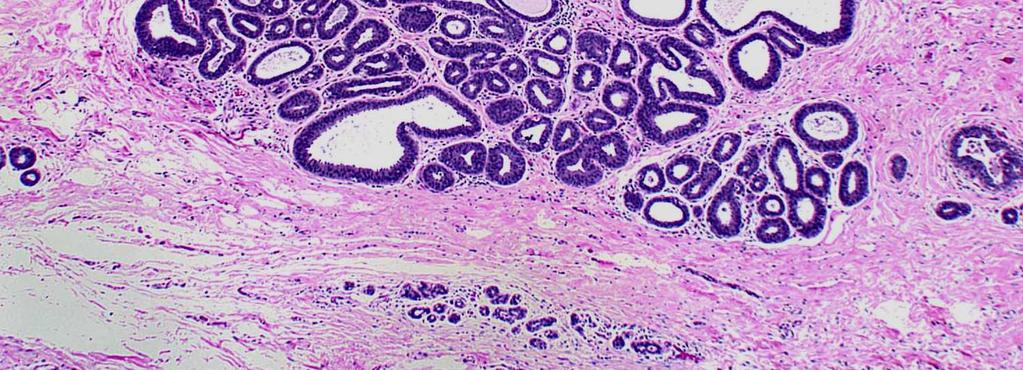
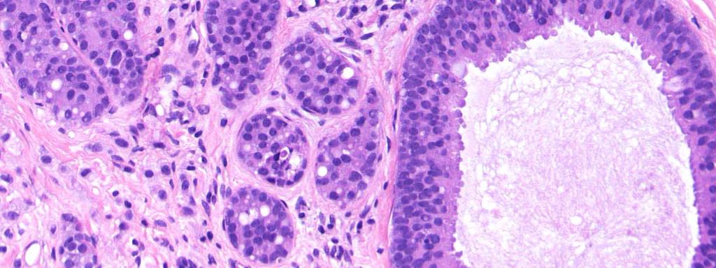
7 Columnar Cell Hyperplasia WHO, 2003 Variably distended acini of TDLUs lined by columnar cells arranged perpendicular to basement membrane, some multilayer and piling up of nuclei, but no true micropapillations Apical snouts Secretions and microcalcifications in lumens, may be more pronounced

8
9
10
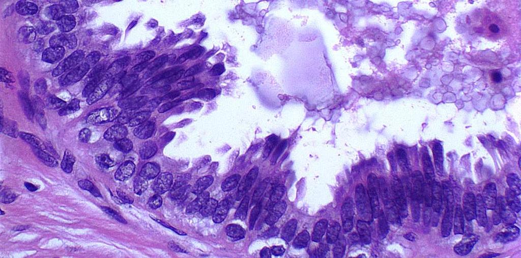
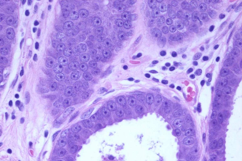
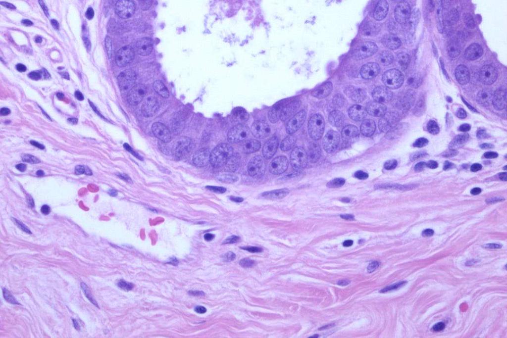
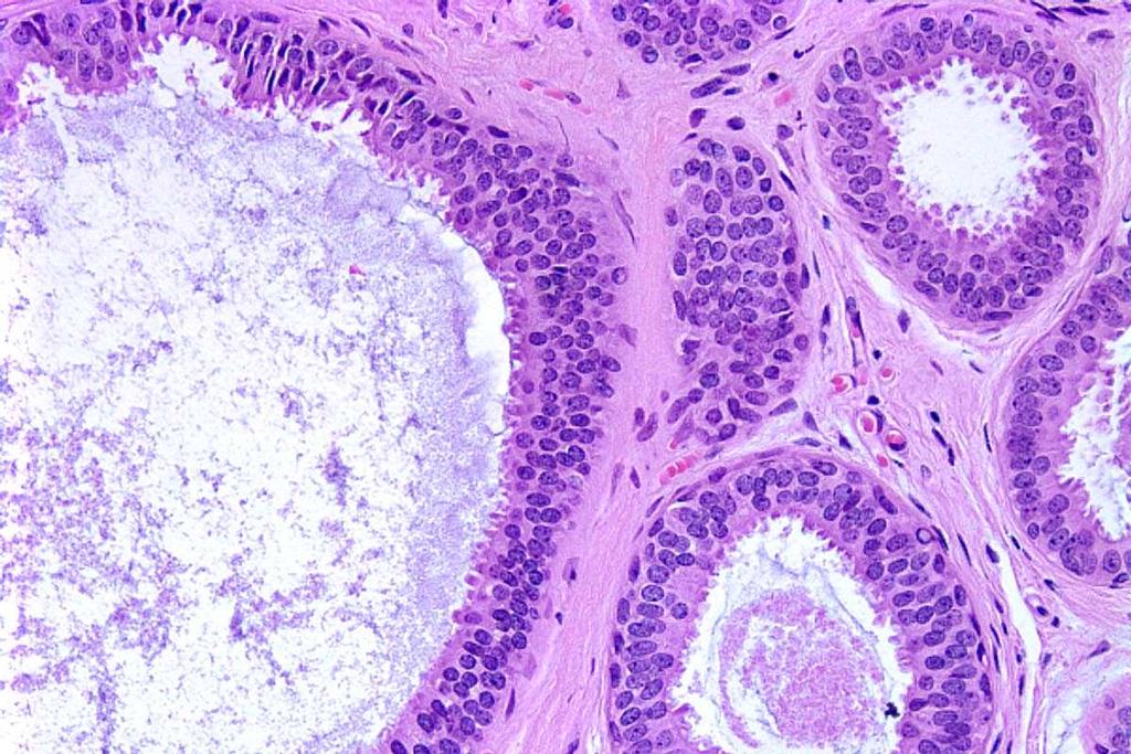
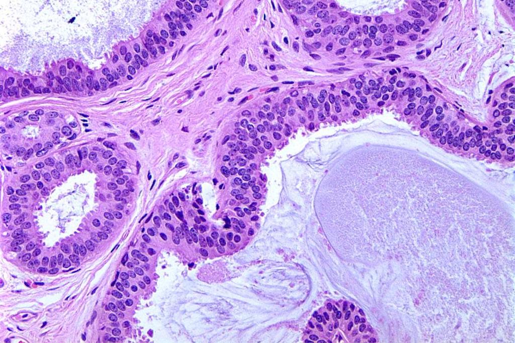
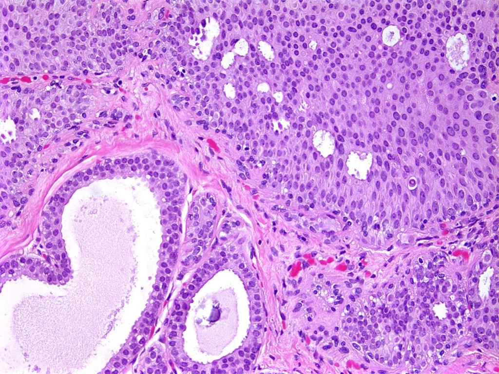
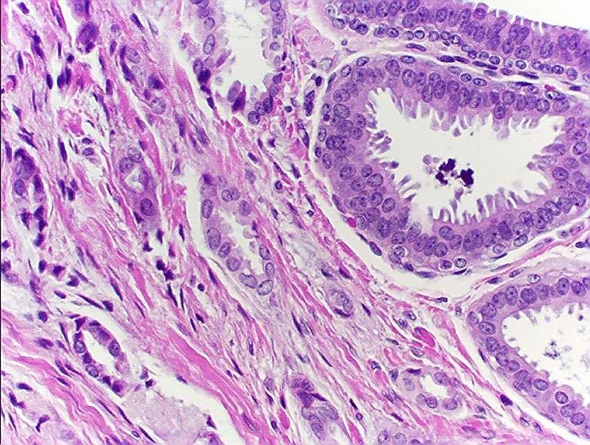
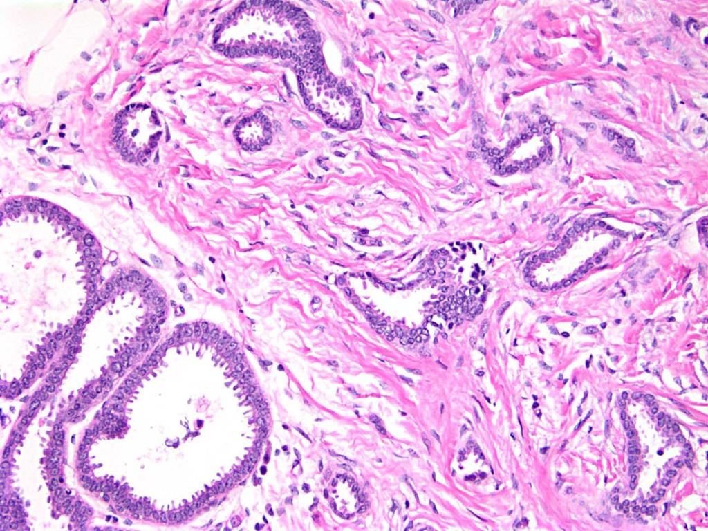
11 Flat Epithelial Atypia WHO, 2003 Variably distended acini of TDLUs Monotonous proliferation of cuboidal to columnar epithelial cells Apical snouts Secretions and microcalcifications in lumens
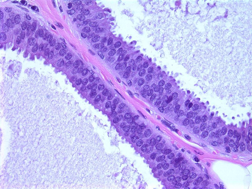
12 Flat Epithelial Atypia Low-grade cytologic atypia Nuclei usually round rather than elongated Loss of polarization Flat growth pattern
13 Flat Epithelial Atypia Features do not fulfill combined architectural and cytologic criteria for diagnosis of ADH or DCIS
14 Flat Epithelial Atypia Given a number of different names Clinging carcinoma (monomorphic type) Atypical cystic lobules type A Columnar cell change/hyperplasia with atypia CAPSS with atypia DIN of the flat type
15 Problems in Breast Pathology Azzopardi, 1979 Underdiagnosis of Malignancy There is a more common form of clinging carcinoma..the involved structures are lined by a single or a few layers of neoplastic epithelial cells.the lesion can be missed entirely since the alteration is cytological rather than anatomical (architectural).
16 Flat Epithelial Atypia The missing link in breast cancer progression?* *Simpson, Am J Surg Pathol, 2005

17 No consistent genetic alterations +1q, -16q, -17p ILC UDH LCIS ALH -CDH1 (e-cad) Normal Epithelium FEA ADH LG-DCIS IG-DCIS HG-DCIS +1q, -16q, -17p LG-Invasive Cancer? +1q, -16q, -17p Low Grade Pathway ER+, HER2-, low prolif rate? -16q??? IG-Invasive? Cancer?? Apocrine Metaplasia??? HG-Invasive Cancer
18 Normal Low grade intraepithelial neoplasia Lobular Neoplasia Columnar cell lesions ADH/DCIS ILC TLC Tubular Ca Low grade invasive carcinomas Low grade breast neoplasia family Adapted from Ellis, Mod Pathol 2010
19
20
21
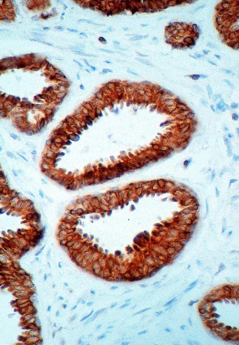
22
23
24
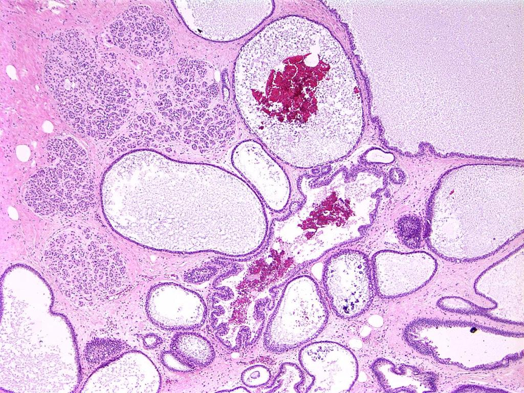
25 Differential Diagnosis
26 Differential Diagnosis of Flat Epithelial Atypia Apocrine metaplasia Microcysts ADH Cystic Hypersecretory Hyperplasia FEA LG-DCIS Columnar Cell Change/Hyperplasia


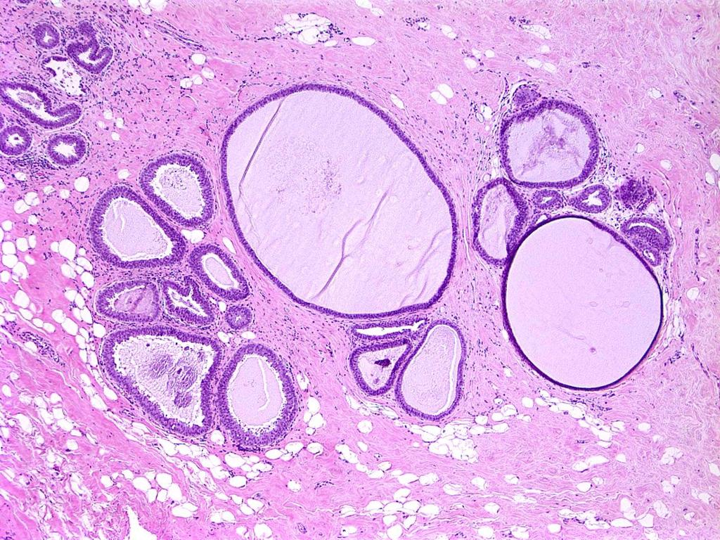
27 Microcysts Single layer of low cuboidal/attenuated epithelial cells Small nuclei May have secretions


28 Apocrine cysts Single layer of low cuboidal cells Abundant pink cytoplasm Round nuclei with prominent nucleoli Calcifications often calcium oxalate

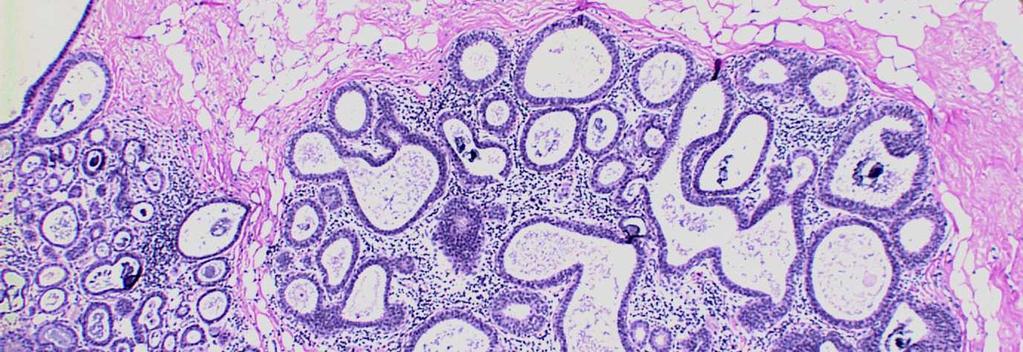
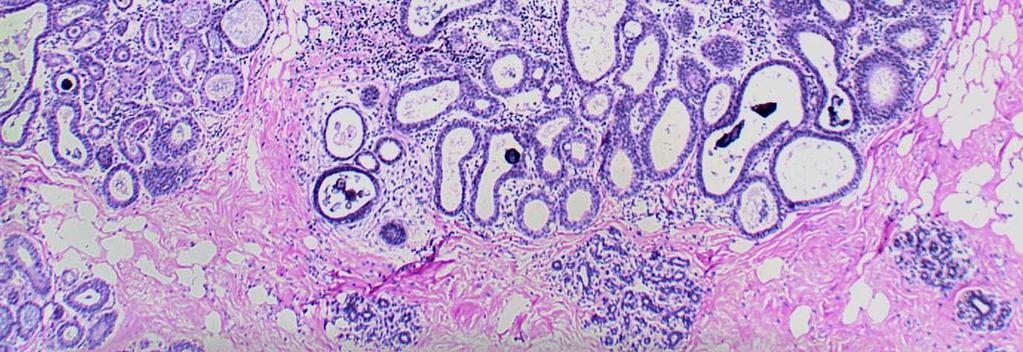
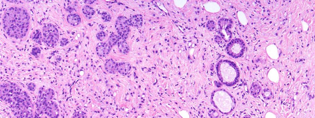
29 Flat Epithelial Atypia Apocrine Metaplasia
30 Flat Epithelial Atypia vs. Apocrine Epithelium FEA Apocrine Epithelium Apical snouts yes yes Granular eosinophilic cytoplasm no yes Hobnail cells yes no Psammomatous calcifications yes no ER expression yes no bcl-2 expression yes no

31 ER and bcl2 Expression FEA Apocrine met ER bcl2
32 Columnar cell lesions without atypia Columnar cells Nuclei elongated Polarization maintained May stratify

33 Columnar Cell Change Flat Epithelial Atypia Columnar Cell Hyperplasia


34 Columnar Cell Change Flat Epithelial Atypia Columnar Cell Hyperplasia Irregular contours Smooth contours Irregular contours

35 FEA CCC

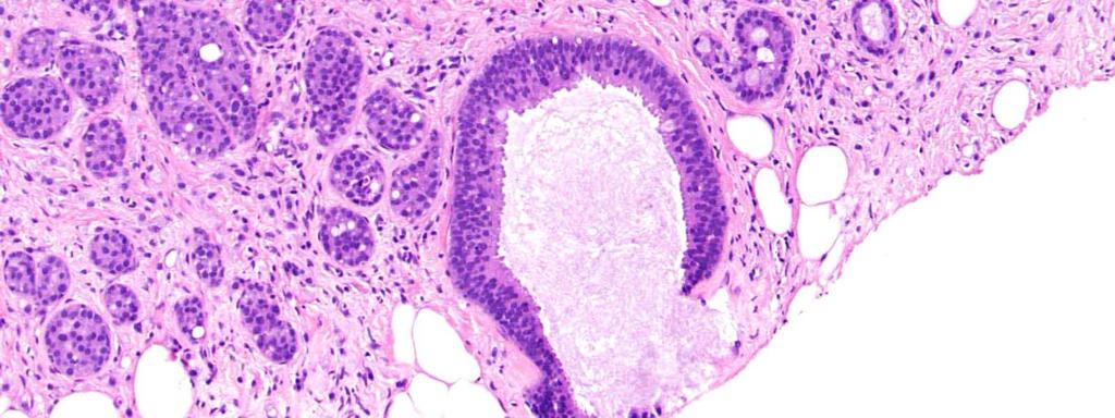
36 Cystic Hypersecretory Hyperplasia

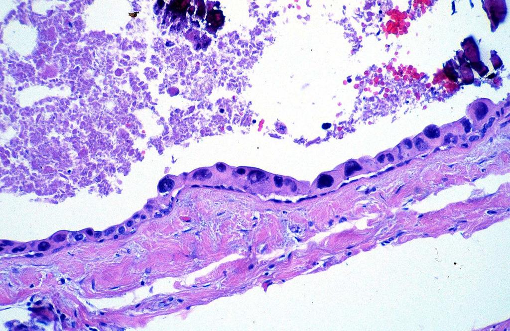
37 Cystic Hypersecretory Hyperplasia


38 Atypical Ductal Hyperplasia Cells with low grade cytologic atypia Architectural atypia also present


39 Low Grade DCIS Cells with low grade cytologic atypia Architectural atypia also present, completely filling a ductal unit
40
41
42
43 Flat Epithelial Atypia vs. ADH or DCIS Low-grade cytologic atypia Complex architectural patterns FEA Yes No ADH/DCIS Yes Yes


44 High Grade DCIS Clinging carcinoma may present as dilated spaces with single layer of atypical epithelial cells
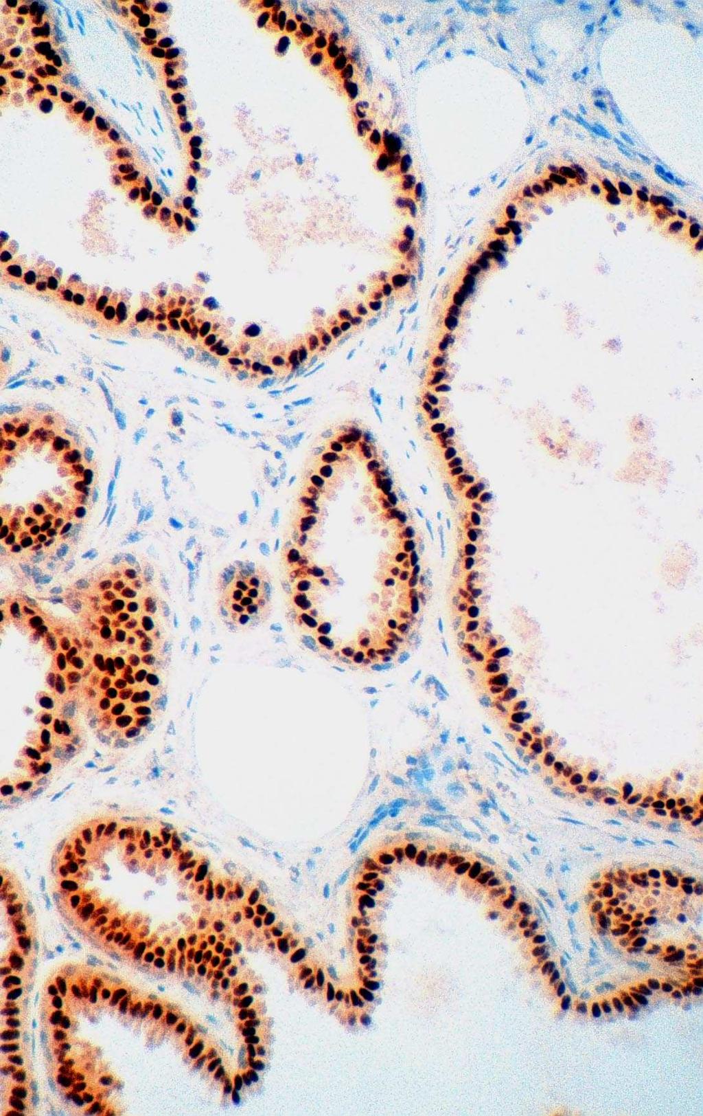
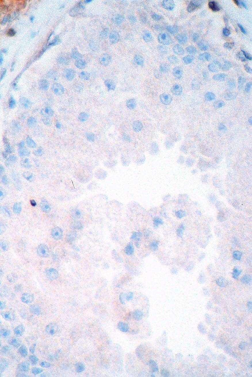
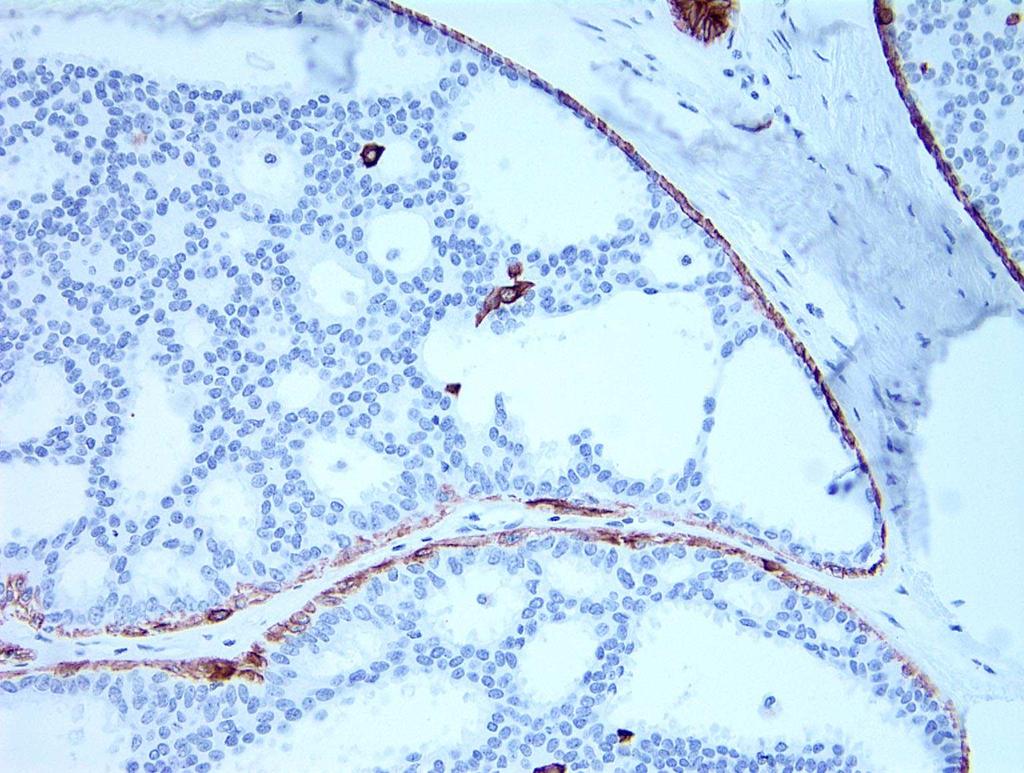
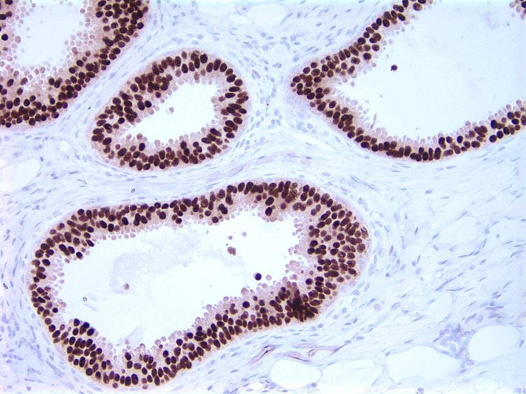
45
46 Differential Diagnosis of Flat Epithelial Atypia Apocrine metaplasia Microcysts ADH Cystic CYTOLOGY Hypersecretory Hyperplasia FEA ARCHITECTURE LG-DCIS Columnar Cell Change/Hyperplasia
47 Interobserver Reproducibility
48 Can Pathologists Reproducibly Distinguish Columnar Cell Lesions That Are Not Atypical From FEA? (O Malley, Mod Pathol 2006) 8 pathologists (including 1 reference pathologist) Training tutorial Written criteria Representative images Test set of 30 cases Each scored as either FEA or not atypical by each of the 8 observers
49 Interobserver Agreement (FEA vs. Not Atypical) Overall agreement among 8 pathologists: 91.8% Multi-rater kappa value: 0.83 ( excellent agreement )
50 Immunophenotype
51 Immunophenotype Immunoperoxidase studies not helpful for separating FEA and CCC/CCH Express CK 19 Lack expression of HMW-CK (CK 5/6) Strong expression of ER and PR Overall higher expression of proliferation and anti-apoptotic markers than normal TDLUs

52 UDH ADH CCC FEA

53 ER CCC FEA
54 Molecular Alterations
55 Molecular Alterations FEA is a clonal proliferation 16q loss Genetic changes relatively few in number Recurrent loss of 16q reported Abdel-Fatah et al, AJSP, 2007 Aulmann, AJSP, 2012 Verschuur-Maes, BCRT, 2012 Go, Human Pathol, 2012
56 Molecular Alterations Promoter methylation of tumor suppressor genes seen in CCL (CCND2 and ID4) Similar alterations identified in associated LGDCIS or invasive carcinoma Direct transitions from FEA to LG DCIS seen on studies of mdna Abdel-Fatah et al, AJSP, 2007 Aulmann, AJSP, 2012 Verschuur-Maes, BCRT, 2012 Go, Human Pathol, 2012
57 Lesions often associated with FEA Atypical ductal hyperplasia Low grade ductal carcinoma in situ Tubular carcinoma Lobular neoplasia (ALH/LCIS) Fraser, 1998; Rosen, 1999; Brogi, 2001; Collins, 2007; Abdel-Fatah, 2007
58
59
60
61
62
63
64 Clinical Significance
65 Clinical Significance of FEA Evidence to support relationship to ADH, LG-DCIS, tubular carcinoma and lobular neoplasia Cytologic similarities Coexistence and geographic proximity Immunophenotypic similarities Molecular similarities Brogi, Int J Surg Pathol, 2001 Collins, Am J Surg Pathol, 2007 Abdel-Fatah, Am J Surg Pathol, 2007 Brandt, Adv Anat Pathol, 2008 Aulmann, Am J Surg Pathol, 2012
66 Clinical Significance of FEA Based on observations of association with other lesions and genetic data, FEA appears to be a precursor to, or the earliest manifestation of LG DCIS
67 Relationship between DCIS and FEA Collins, Mod Pathol, 2007 FEA is preferentially associated with DCIS with particular features Low nuclear grade Micropapillary pattern Absence of comedo necrosis Absence of stromal inflammation/desmoplasia
68 Implications Our observations provide further evidence for a precursor-product relationship between FEA and DCIS that exhibit such features
69 Rosen Triad Brandt et al., Adv Anat Pathol, cases of tubular carcinoma 100% of cases with associated CCLs 53% of cases with associated LCIS
70 Rosen Triad Brandt et al., Adv Anat Pathol, 2008 Tubular carcinoma Columnar cell lesions LCIS (Rosen, Am J Surg Pathol, 1999)


71 Tubular ca LN FEA Tubular ca FEA

72 Proposed Evolutionary Pathway for LG Neoplasia Abdel-Fatah et al, AJSP, 2007
73 Clinical Follow-up Studies
74 Eusebi et al 1994 EORTC de Mascarel, et al 2006 # of patients Type of study Retrospective review, biopsies originally considered benign Prospective, randomized clinical trial Retrospective review, clinging carcinoma of the monomorphic type Treatment Diagnostic biopsy; no attempt at excision Excision alone or Excision and radiation Biopsy alone (n=70) Biopsy and radiation (n=45) Follow-up 19.2 years 5.4 years 13.3 years (mean) (median) (median) # (%) with LR 1 (4%) 0 3 (2.6%) # with subsequent IBC (2.6%)
75 Columnar Cell Lesions and Subsequent Breast Cancer Risk Evaluated overall cancer risk in >1,000 CCLs Mild increase in overall cancer risk RR=1.47 with 17 yrs of follow-up (Nashville) OR=1.44 with 9 yrs median follow-up (NHS) RR=2.75 with 13.5 yrs mean follow-up (Mayo) No difference among 3 categories of CCL with regard to future breast cancer risk 2-3 fold increase in AH in presence of CCLs May indicate the presence of a concomitant worse lesion Boulos, Cancer, 2008 Aroner, Breast Cancer Res Treat, 2010 Said, Mod Pathol, A
76 Management Issues

77 Columnar Cell Change/Hyperplasia Found on excision: No further treatment Found on CNB: No excision necessary No additional levels obtained


78 Flat Epithelial Atypia FEA on core biopsy Upgraded in 0-30% of cases But need for excision remains uncertain Rad-path correlation required Khoumais Ann Surg Oncol, 2013 WHO, 2012 Bianchi, Virchow s Arch, 2012 Peres, BCRT, 2012 Lavoue, BCRT, 2011 Lee, Breast Journal, 2010 Ingegnoli, Breast Journal, 2010 Tomasino, J Cell Physiol, 2009 Chivukula, Am J Clin Pathol, 2009 Senetta, Mod Pathol, 2009 Piubello, Am J Surg Pathol, 2009
79 Important to separate implications of FEA on CNB For current management (upgrade rate) And ongoing management (subsequent breast cancer risk)
80 FEA on excisional biopsy Obtain multiple levels looking for areas diagnostic of ADH or LG DCIS Submit all tissue


81 Management Issues FEA in association with ADH Manage as ADH FEA in association with LG DCIS Manage as DCIS Question as to whether FEA included in extent/size of DCIS Question of how to manage FEA at margins remains unanswered

82 FEA is a red flag for the presence of other worse lesions
83 Conclusions CCL and FEA increasingly prevalent in an era of mammographic screening (especially digital mammography) May represent a precursor to LG DCIS Risk of progression to invasive carcinoma appears to be very low (with limited data)
84 Conclusions Presence of FEA should alert pathologist to possible presence of other lesions with which it is frequently associated (e.g., ADH, DCIS, tubular ca, LN) On CNB look very carefully for subtle examples of tubular carcinoma or microinvasive foci of ILC/TC
Columnar Cell Lesions
 Columnar Cell Lesions Laura C. Collins, M.D. Department of Pathology Beth Israel Deaconess Medical Center and Harvard Medical School Boston, MA Question? Columnar cell lesions are: a) Annoying lesions
Columnar Cell Lesions. Columnar Cell Lesions and Flat Epithelial Atypia
 Columnar Cell Lesions and Stuart J. Schnitt, M.D. Beth Israel Deaconess Medical Center and Harvard Medical School Boston, MA, USA Columnar Cell Lesions Lesions characterized by columnar epithelial cells
Epithelial Columnar Breast Lesions: Histopathology and Molecular Markers
 29th Annual International Conference Advances in the Application of Monoclonal Antibodies in Clinical Oncology and Symposium on Cancer Stem Cells 25 th -27t h June, 2012, Mykonos, Greece Epithelial Columnar
Incidence of ductal lesions
 Ductal Proliferative Lesions of the Breast: From FEA to ADH to DCIS Incidence of ductal lesions Pre-mammography: DCIS < 3% of breast cancers, large palpable masses, with invasion Mammography: DCIS 25%
Flat Epithelial Atypia
 Flat Epithelial Atypia Richard Owings, M.D. University of Arkansas for Medical Sciences Department of Pathology Flat epithelial atypia can be a difficult lesion May be a subtle diagnosis Lots of changes
Proliferative Epithelial lesions of the Breast. Sami Shousha, MD, FRCPath Charing Cross Hospital & Imperial College, London
 Proliferative Epithelial lesions of the Breast Sami Shousha, MD, FRCPath Charing Cross Hospital & Imperial College, London Amman, November2013 Proliferative Epithelial Lesions of the Breast Usual type
04/10/2018 HIGH RISK BREAST LESIONS. Pathology Perspectives of High Risk Breast Lesions ELEVATED RISK OF BREAST CANCER HISTORICAL PERSPECTIVES
 Pathology Perspectives of High Risk Breast Lesions Savitri Krishnamurthy MD Professor of Pathology Deputy Division Head Director of Clinical Trials, Research and Development The University of Texas MD
Flat Epithelial Atypia: A Review of Current Concepts
 90 The Open Breast Cancer Journal, 2010, 2, 90-94 Flat Epithelial Atypia: A Review of Current Concepts Amy L. Adams * Open Access Department of Pathology and Laboratory Medicine, Emory University School
Ductal Carcinoma in Situ. Laura C. Collins, M.D. Department of Pathology Beth Israel Deaconess Medical Center and Harvard Medical School Boston, MA
 Ductal Carcinoma in Situ Laura C. Collins, M.D. Department of Pathology Beth Israel Deaconess Medical Center and Harvard Medical School Boston, MA Definition of DCIS WHO 2012 A neoplastic proliferation
Proliferative Breast Disease: implications of core biopsy diagnosis. Proliferative Breast Disease
 Proliferative Breast Disease: implications of core biopsy diagnosis Jean F. Simpson, M.D. Breast Pathology Consultants, Inc. Nashville, TN Proliferative Breast Disease Must be interpreted in clinical and
The Diagnosis and Management of Pre-invasive Breast Disease: Flat Epithelial Atypia Classification, Pathologic Features and Clinical Significance
 The Diagnosis and Management of Pre-invasive Breast Disease: Flat Epithelial Atypia Classification, Pathologic Features and Clinical Significance The Harvard community has made this article openly available.
CLINICAL SIGNIFICANCE OF BENIGN EPITHELIAL CHANGES
 Papillomas. Papillomas are composed of multiple branching fibrovascular cores, each having a connective tissue axis lined by luminal and myoepithelial cells ( Fig. 23-11 ). Growth occurs within a dilated
Controversies and Problematic Issues in Core Needle Biopsies (To excise or not to excise)
") Controversies and Problematic Issues in Core Needle Biopsies (To excise or not to excise) Laura C. Collins, M.D. Beth Israel Deaconess Medical Center and Harvard Medical School Boston, MA Schematic Representation
Controversies and Problematic Issues in Core Needle Biopsies (To excise or not to excise) Laura C. Collins, M.D. Beth Israel Deaconess Medical Center and Harvard Medical School Boston, MA Schematic Representation
Pathology of Lobular & Ductal Preneoplasia. Syed A Hoda, MD Weill-Cornell, New York, NY
 Pathology of Lobular & Ductal Preneoplasia Syed A Hoda, MD Weill-Cornell, New York, NY Proliferative Epithelial Changes in Breast A wide range of proliferative epithelial changes occur in the breast There
Ductal Proliferations of the Breast: The Good, the Bad, and the Ugly
 Ductal Proliferations of the Breast: The Good, the Bad, and the Ugly Melinda F. Lerwill, MD CRITERIA FOR DISTINGUISHING LOW-GRADE DUCTAL CARCINOMA IN SITU FROM USUAL DUCTAL HYPERPLASIA CYTOLOGY Low-grade
1 NORMAL HISTOLOGY AND METAPLASIAS
 1 NORMAL HISTOLOGY AND METAPLASIAS, MD Anatomy and Histology 1 Metaplasias 2 ANATOMY AND HISTOLOGY The female breast is composed of a branching duct system, which begins at the nipple with the major lactiferous
6/3/2010. Outline of Talk. Lobular Breast Cancer: Definition of lobular differentiation. Common Problems in Diagnosing LCIS in Core Biopsies
 Outline of Talk Lobular Breast Cancer: Common Problems in Diagnosing LCIS in Core Biopsies Definition of lobular differentiation Variants of LCIS that: carry risk for unsampled invasive cancer mimic DCIS
Significance of flat epithelial atypia on mammotome core needle biopsy: should it be excised? B
 Human Pathology (2007) 38, 35 41 www.elsevier.com/locate/humpath Original contribution Significance of flat epithelial atypia on mammotome core needle biopsy: should it be excised? B Lakshmi P. Kunju MD,
Disclosures 5/27/2012. Outline of Talk. Outline of Talk. When Is LCIS Clinically Significant? Classic LCIS. Classic LCIS
 When Is LCIS Clinically Significant? Disclosures I have nothing to disclose Yunn-Yi Chen, MD, PhD Professor Outline of Talk Outline of Talk Classic LCIS Classic LCIS Definition of lobular differentiation
Papillary Lesions of the Breast
 Papillary Lesions of the Breast Laura C. Collins, M.D. Associate Professor of Pathology Associate Director, Division of Anatomic Pathology Beth Israel Deaconess Medical Center and Harvard Medical School
Papillary Lesions of the Breast: WHO Update
 Papillary Lesions of the Breast: WHO Update Stuart J. Schnitt, M.D. Department of Pathology Beth Israel Deaconess Medical Center and Harvard Medical School Boston, MA, USA Papillary Lesions of the Breast
Cytyc Corporation - Case Presentation Archive - March 2002
 FirstCyte Ductal Lavage History: 68 Year Old Female Gail Index: Unknown Clinical History: Negative Mammogram in 1995 6 yrs. later presents with bloody nipple discharge Subsequent suspicious mammogram Suspicious
Treatment options for the precancerous Atypical Breast lesions. Prof. YOUNG-JIN SUH The Catholic University of Korea
 Treatment options for the precancerous Atypical Breast lesions Prof. YOUNG-JIN SUH The Catholic University of Korea Not so benign lesions? Imaging abnormalities(10% recall) lead to diagnostic evaluation,
Case study 1. Rie Horii, M.D., Ph.D. Division of Pathology Cancer Institute Hospital, Japanese Foundation for Cancer Research
 NCCN/JCCNB Seminar in Japan April 15, 2012 Case study 1 Rie Horii, M.D., Ph.D. Division of Pathology Cancer Institute Hospital, Japanese Foundation for Cancer Research Present illness: A 50y.o.premenopausal
The Hot Topic for today is a biopsy from a 58-year-old woman who had worrisome mammographic calcifications on screening.
 The Hot Topic for today is a biopsy from a 58-year-old woman who had worrisome mammographic calcifications on screening. 1 My name is Dan Visscher; I am a consultant in the Division of Anatomic Pathology
Benign Breast Disease and Breast Cancer Risk
 Benign Breast Disease and Breast Cancer Risk Jean F. Simpson, M.D. Vanderbilt University Nashville, Tennessee December 1, 2011 Nashville Nashville Lebanon 1 Cedars of Lebanon State Park The American University
Papillary Lesions of the breast
 Papillary Lesions of the breast Emad Rakha Professor of Breast Pathology The University of Nottingham Papillary lesions of the breast are a heterogeneous group of disease, which are characterised by neoplastic
Interpretation of Breast Pathology in the Era of Minimally Invasive Procedures
 Shahla Masood, M.D. Professor and Chair Department of Pathology and Laboratory Medicine University of Florida College of Medicine Jacksonville Medical Director, UF Health Breast Center Chief of Pathology
Promise of a beautiful day
 Promise of a beautiful day Ductal carcinoma in Situ Lobular Carcinoma in Situ Natural History Manosmed Tartous Oct 2009 Gérard ABADJIAN MD Pathology Department Hôtel-Dieu de France. Associate Professor
Good afternoon everyone. First of all many thanks to Dr. Bonaventura and Dr. Arn for inviting
 PATHOLOGY IN-SITU CARCINOMA, ROHIT BHARGAVA, MD 1 Good afternoon everyone. First of all many thanks to Dr. Bonaventura and Dr. Arn for inviting me here, it s great to be here and I m going to talk about
Benign Mimics of Malignancy in Breast Pathology
 Arthur Purdy Stout Society of Surgical Pathologists Companion Meeting Benign Mimics of Malignancy in Breast Pathology Stuart J. Schnitt, M.D. Beth Israel Deaconess Medical Center and Harvard Medical School,
Papillary Lesions of the Breast
 Papillary Lesions of the Breast Texas Society of Pathologists 2013 Laura C. Collins, M.D. Associate Professor of Pathology Associate Director, Division of Anatomic Pathology Beth Israel Deaconess Medical
Breast: Difficulties in Core Biopsies
 Breast: Difficulties in Core Biopsies Anna Marie Mulligan, MB, MSc, FRCPath University Health Network and University of Toronto E-mail: annamarie.mulligan@uhn.ca No conflicts of interest Role of Core Needle
The management of B3 lesions with emphasis on lobular neoplasia
 The management of B3 lesions with emphasis on lobular neoplasia Abeer Shaaban Queen Elizabeth Hospital Birmingham NHSBSP core biopsy categories B1 - Normal B2 - Benign B3 Uncertain malignant potential
Disclosures. Premalignant Lesions of the Breast: What Clinicians Want and Why. NY Times: Prone to Error: Earliest Steps to Find Cancer.
 Disclosures Premalignant Lesions of the Breast: What Clinicians Want and Why I have nothing to disclose Rick Baehner, MD Assistant Professor, UCSF Pathology NY Times: Prone to Error: Earliest Steps to
04/10/2018. Intraductal Papillary Neoplasms Of Breast INTRADUCTAL PAPILLOMA
 Intraductal Papillary Neoplasms Of Breast Savitri Krishnamurthy MD Professor of Pathology Deputy Division Head The University of Texas MD Anderson Cancer Center 25 th Annual Seminar in Pathology Pittsburgh,
In Situ Breast Carcinoma. James L. Connolly, M.D Beth Israel Deaconess Medical Center Professor of Pathology Harvard Medical School Boston, MA
 In Situ Breast Carcinoma James L. Connolly, M.D Beth Israel Deaconess Medical Center Professor of Pathology Harvard Medical School Boston, MA Content In Situ Ductal Carcinoma In Situ Lobular Carcinoma
Papillary Lesions of the Breast A Practical Approach to Diagnosis. (Arch Pathol Lab Med. 2016;140: ; doi: /arpa.
 Papillary Lesions of the Breast A Practical Approach to Diagnosis (Arch Pathol Lab Med. 2016;140:1052 1059; doi: 10.5858/arpa.2016-0219-RA) Papillary lesions of the breast Span the spectrum of benign,
Breast Pathology. Breast Development
 Breast Pathology Lecturer: Hanina Hibshoosh, M.D. Reading: Kumar, Cotran, Robbins, Basic Pathology, 6th Edition, pages 623-635 Breast Development 5th week - thickening of the epidermis - milk line 5th
High risk lesions of the breast : Review of the current diagnostic and management strategies
 High risk lesions of the breast : Review of the current diagnostic and management strategies Poster No.: C-1204 Congress: ECR 2016 Type: Educational Exhibit Authors: P. Jagmohan, F. J. Pool, P. G. Pillay,
Atypical proliferative lesions diagnosed on core biopsy - 6 year review
 Atypical proliferative lesions diagnosed on core biopsy - 6 year review Dr Angela Harris, Dr Julie Weigner & Dr Ricardo Vilain NSW Health Pathology Pathology North, Hunter Anatomical Pathology & Cytology
Enterprise Interest None
 Enterprise Interest None B3 lesions of the breast What are they at surgery? Case 4 Edi Brogi MD PhD Attending Pathologist - Director of Breast Pathology Memorial Sloan Kettering Cancer Center New York
3/27/2017. Disclosure of Relevant Financial Relationships. Papilloma???
 Management of Papillary Lesions Diagnosed at Rad Path Concordant Core Biopsy (CNB) Disclosure of Relevant Financial Relationships USCAP requires that all planners (Education Committee) in a position to
Diseases of the breast (1 of 2)
") Diseases of the breast (1 of 2) Introduction A histology introduction Normal ducts and lobules of the breast are lined by two layers of cells a layer of luminal cells overlying a second layer of myoepithelial
Diseases of the breast (1 of 2) Introduction A histology introduction Normal ducts and lobules of the breast are lined by two layers of cells a layer of luminal cells overlying a second layer of myoepithelial
Current issues in diagnostic breast pathology
 1 Cancer Studies and Molecular Medicine, University of Leicester, Leicester, UK 2 Academic Unit of Pathology, Leeds University, Leeds, UK 3 Academic Oncology/Breast Pathology, King s College London, London,
In situ lobular neoplasia of the breast with marked myoepithelial proliferation
 In situ lobular neoplasia of the breast with marked myoepithelial proliferation Sami Shousha To cite this version: Sami Shousha. In situ lobular neoplasia of the breast with marked myoepithelial proliferation.
Controversies on the Management of High Risk Breast Lesions on Core Biopsy: An Update on the Literature
 Controversies on the Management of High Risk Breast Lesions on Core Biopsy: An Update on the Literature Dianne Georgian- Smith MD Brigham and Women s Hospital Associate Professor of Radiology, Harvard
Surgical Pathology Issues of Practical Importance
 Surgical Pathology Issues of Practical Importance Anne Moore, MD Medical Oncology Syed Hoda, MD Surgical Pathology The pathologist is central to the team approach needed to manage the patient with breast
BREAST PATHOLOGY. Fibrocystic Changes
 BREAST PATHOLOGY Lesions of the breast are very common, and they present as palpable, sometimes painful, nodules or masses. Most of these lesions are benign. Breast cancer is the 2 nd most common cause
Histological Type. Morphological and Molecular Typing of breast Cancer. Nottingham Tenovus Primary Breast Cancer Study. Survival (%) Ian Ellis
 Ian Ellis") Morphological and Molecular Typing of breast Cancer Ian Ellis Molecular Medical Sciences, University of Nottingham Department of Histopathology, Nottingham University Hospitals NHS Trust Histological Type
Morphological and Molecular Typing of breast Cancer Ian Ellis Molecular Medical Sciences, University of Nottingham Department of Histopathology, Nottingham University Hospitals NHS Trust Histological Type
Notice of Faculty Disclosure
 California Society of Pathology Diagnostic Problems in Surgical Pathology December 2015 Case 2 Laura C. Collins, M.D. Associate Professor of Pathology Associate Director of Anatomic Pathology Beth Israel
Breast pathology. 2nd Department of Pathology Semmelweis University
 Breast pathology 2nd Department of Pathology Semmelweis University Breast pathology - Summary - Benign lesions - Acute mastitis - Plasma cell mastitis / duct ectasia - Fat necrosis - Fibrocystic change/
Short Term Cancer Risk
 in a Cohort of 2312 Women with High Risk Breast Lesions Kubat E 1, Puligandla B 2, Collins L 3, Jiang SF 4, Callahan M 5, Kutner S 6, Habel L 4, Shim V 1 1 Surgery, Kaiser Permanente, Oakland, CA; 2 Pathology,
Management of B3 lesions
 Management of B3 lesions Pathological view Abeer Shaaban Queen Elizabeth Hospital Birmingham FEA AIDP B3 lesions In situ Lobular neoplasia Papilloma Radial scar Fibroaepithelial lesion Mucocoele like lesion
Ana Sofia Preto 19/06/2013
 Ana Sofia Preto 19/06/2013 Understanding the underlying pathophysiologic processes leading to the various types of calcifications Description and illustration of the several types of calcifications, according
ACCME/Disclosures. Cribriform Lesions of the Prostate. Case
 Cribriform Lesions of the Prostate Ming Zhou, MD, PhD Departments of Pathology and Urology New York University Langone Medical Center New York, NY Ming.Zhou@NYUMC.ORG ACCME/Disclosures The USCAP requires
ARTHUR PURDY STOUT SOCIETY COMPANION MEETING: DIFFICULT NEW DIFFERENTIAL DIAGNOSES IN PROSTATE PATHOLOGY. Jonathan I. Epstein.
 1 ARTHUR PURDY STOUT SOCIETY COMPANION MEETING: DIFFICULT NEW DIFFERENTIAL DIAGNOSES IN PROSTATE PATHOLOGY Jonathan I. Epstein Professor Pathology, Urology, Oncology The Reinhard Professor of Urological
Title malignancy. Issue Date Right 209, 12, (2013)
") NAOSITE: Nagasaki University's Ac Title Author(s) A case of intracystic apocrine papi malignancy Hayashi, Hiroko; Ohtani, Hiroshi; Y Citation Pathology - Research and Practice, Issue Date 2013-12 URL Right
NAOSITE: Nagasaki University's Ac Title Author(s) A case of intracystic apocrine papi malignancy Hayashi, Hiroko; Ohtani, Hiroshi; Y Citation Pathology - Research and Practice, Issue Date 2013-12 URL Right
CNB vs Surgical Excision
 Update on Core Needle Biopsy of Non-palpable Breast Lesions Nour Sneige, M.D. UT MD Anderson Cancer Center Houston, Tx Image-Guided CNB of Breast Lesions An alternative to surgical biospy CNB vs Surgical
IBCM 2, April 2009, Sarajevo, Bosnia and Herzegovina
 Preoperative diagnosis and treatment planning in breast cancer The pathologist s perspective L. Mazzucchelli Istituto Cantonale di Patologia Locarno, Switzerland IBCM 2, 23-25 April 2009, Sarajevo, Bosnia
LOBULAR CARCINOMA IN SITU: WHAT DOES IT MEAN? THE SURGEON'S PERSPECTIVE
 : WHAT DOES IT MEAN? THE SURGEON'S PERSPECTIVE Benjamin O. Anderson, M.D. Director, Breast Health Clinic Professor of Surgery and Global Health, University of Washington Joint Member, Fred Hutchinson Cancer
Protocol for the Examination of Biopsy Specimens From Patients With Invasive Carcinoma of the Breast
 Protocol for the Examination of Specimens From Patients With Invasive Carcinoma of the Breast Version: BreastInvasive 1.0.0.0 Protocol Posting Date: February 2019 Accreditation Requirements The use of
Image guided core biopsies:
 Recommendations on the Surgical, Radiologic and Pathologic Approaches to Breast Disease: Using best practices based on multidisciplinary methodologies developed through the Allina Breast Committee. Image
Papillary Lesions in Breast Pathology Practice: Diagnostic Challenges and Practical Approach. A Six- Year Experience from a Tertiary Care Hospital
 Open Access Journal Research Article DOI: 10.23958/ijirms/vol02-i05/12 Papillary Lesions in Breast Pathology Practice: Diagnostic Challenges and Practical Approach. A Six- Year Experience from a Tertiary
Excisional biopsy or long term follow-up results in breast high-risk lesions diagnosed at core needle biopsy
 Excisional biopsy or long term follow-up results in breast high-risk lesions diagnosed at core needle biopsy Poster No.: C-2515 Congress: ECR 2015 Type: Authors: Scientific Exhibit Ö. S. Okcu 1, A. Oktay
Mousa. Israa Ayed. Abdullah AlZibdeh. 0 P a g e
 1 Mousa Israa Ayed Abdullah AlZibdeh 0 P a g e Breast pathology The basic histological units of the breast are called lobules, which are composed of glandular epithelial cells (luminal cells) resting on
Benign and preinvasive breast lesions
 58 Benign and preinvasive breast lesions Anke Kleine-Tebbe 1, Aurelia Noske 2 1 DRK Clinics Berlin Köpenick, Berlin, Germany 2 Institute for pathology, Charité Campus Mitte, Charité University Medical
CASE REPORT Malignant transformation of breast ductal adenoma: a diagnostic pitfall
 Malaysian J Pathol 2015; 37(3) : 281 285 CASE REPORT Malignant transformation of breast ductal adenoma: a diagnostic pitfall Hiroko HAYASHI, Hiroshi OHTANI,* Junzo YAMAGUCHI,** and Isao SHIMOKAWA Department
Lobular Carcinoma In Situ Variants in Breast Cores
 Lobular Carcinoma In Situ Variants in Breast Cores Potential for Misdiagnosis, Upgrade Rates at Surgical Excision, and Practical Implications Megan E. Sullivan, MD; Seema A. Khan, MD; Yurdanur Sullu, MD;
Benign, Reactive and Inflammatory Lesions of the Breast
 Benign, Reactive and Inflammatory Lesions of the Breast Marilin Rosa, MD Associate Member Section Head of Breast Pathology Department of Anatomic Pathology Program Director, Breast Pathology Fellowship
LYMPHATIC DRAINAGE AXILLARY (MOSTLY) INTERNAL MAMMARY SUPRACLAVICULAR
 INTERNAL MAMMARY SUPRACLAVICULAR") BREAST LYMPHATIC DRAINAGE AXILLARY (MOSTLY) INTERNAL MAMMARY SUPRACLAVICULAR HISTOLOGY LOBE: (10 in whole breast) LOBULE: (many per lobe) ACINUS/I, aka ALVEOLUS/I: (many per lobule) DUCT(S): INTRA- or
BREAST LYMPHATIC DRAINAGE AXILLARY (MOSTLY) INTERNAL MAMMARY SUPRACLAVICULAR HISTOLOGY LOBE: (10 in whole breast) LOBULE: (many per lobe) ACINUS/I, aka ALVEOLUS/I: (many per lobule) DUCT(S): INTRA- or
Surgical Management of High- Risk Breast Lesions
 Surgical Management of High- Risk Breast Lesions Amy C. Degnim, MD a, Tari A. King, MD b, * KEYWORDS High-risk lesion Atypical hyperplasia Lobular carcinoma in situ Percutaneous breast biopsy Breast cancer
CPC 4 Breast Cancer. Rochelle Harwood, a 35 year old sales assistant, presents to her GP because she has noticed a painless lump in her left breast.
 CPC 4 Breast Cancer Rochelle Harwood, a 35 year old sales assistant, presents to her GP because she has noticed a painless lump in her left breast. 1. What are the most likely diagnoses of this lump? Fibroadenoma
Guidance on the management of B3 lesions
 Guidance on the management of B3 lesions Lesion diagnosed on 14g or vacuumassisted biopsy (VAB) Risk of upgrade Recommended investigation Suggested approach for follow-up if no malignancy on VAE awaiting
AMSER Case of the Month: November 2018
 AMSER Case of the Month: November 2018 52 year old female with an abnormal screening mammogram Areeg Rehman, MS 4 Nova Southeastern University Rebecca T. Sivarajah, MD Penn State University College of
COMMON BENIGN DISORDERS AND DISEASES OF THE BREAST
 COMMON BENIGN DISORDERS AND DISEASES OF THE BREAST Aberrations of Normal Development and Involution (ANDI). The basic principles underlying the aberrations of normal development and involution (ANDI) classification
Non-mass Enhancement on Breast MRI. Aditi A. Desai, MD Margaret Ann Mays, MD
 Non-mass Enhancement on Breast MRI Aditi A. Desai, MD Margaret Ann Mays, MD Breast MRI Important screening and diagnostic tool, given its high sensitivity for breast cancer detection Breast MRI - Indications
Mody. AIS vs. Invasive Adenocarcinoma of the Cervix
 Common Problems in Gynecologic Pathology Michael T. Deavers, M.D. Houston Methodist Hospital, Houston, Texas Common Problems in Gynecologic Pathology Adenocarcinoma in-situ (AIS) of the Cervix vs. Invasive
Review Article. ISSN: (Print) ISSN: (Online)
 ISSN: (Online)") Scholars International Journal of Obstetrics and Gynecology Abbreviated key title: Sch. Int. J. Obstet. Gynec. A Publication by Scholars Middle East Publishers Dubai, United Arab Emirates ISSN: 2616-8235
Scholars International Journal of Obstetrics and Gynecology Abbreviated key title: Sch. Int. J. Obstet. Gynec. A Publication by Scholars Middle East Publishers Dubai, United Arab Emirates ISSN: 2616-8235
Columnar cell lesions associated with breast calcifications on vacuum-assisted core biopsies: clinical, radiographic, and histological correlations
 & 2009 USCAP, Inc All rights reserved 0893-3952/09 $32.00 www.modernpathology.org Columnar cell lesions associated with breast calcifications on vacuum-assisted core biopsies: clinical, radiographic, and
Emad A Rakha, Bernard Chi-Shern Ho, Veena K Naik, Soumadri Sen, Lisa Hamilton, Zsolt Hodi, Ian Ellis, Andrew Hs Lee
 Outcome of breast lesions diagnosed as lesion of uncertain malignant potential (B3) or suspicious of malignancy (B4) on needle core biopsy including detailed review of epithelial atypia Emad A Rakha, Bernard
INTRADUCTAL LESIONS OF THE PROSTATE. Jonathan I. Epstein
 INTRADUCTAL LESIONS OF THE PROSTATE Jonathan I. Epstein Topics Prostatic intraepithelial neoplasia (PIN) Intraductal adenocarcinoma (IDC-P) Intraductal urothelial carcinoma Ductal adenocarcinoma High Prostatic
Anatomic Pathology / Mucocele-like Lesions on Breast Core Biopsy. Mucocele-like Lesions Diagnosed on Breast Core Biopsy
 Anatomic Pathology / Mucocele-like Lesions on Breast Core Biopsy Mucocele-like Lesions Diagnosed on Breast Core Biopsy Assessment of Upgrade Rate and Need for Surgical Excision Brian Sutton, MD, 1 Simone
Diagnosis of Fibroepithelial and Mesenchymal Lesions on Core Needle Biopsy
 Diagnosis of Fibroepithelial and Mesenchymal Lesions on Core Needle Biopsy Emmanuel Agosto-Arroyo, MD Assistant Member Department of Anatomic Pathology 3/3/2018 Disclosure There are no conflicts of interest.
Biliary tract tumors
 Short Course 2010 Annual Fall Meeting of the Korean Society for Pathologists Biliary tract tumors Joon Hyuk Choi, M.D., Ph.D. Professor, Department of Pathology, Yeungnam Univ. College of Medicine, Daegu,
Question 1 A. ER-, PR-, HER+ B. ER+, PR+, HER2- C. ER-, PR+, HER2- D. ER-, PR-, HER2- E. ER-, PR+, HER2+
 Triple Negative Breast Cancer Laura C. Collins, M.D. Department of Pathology Beth Israel Deaconess Medical Center and Harvard Medical School, Boston, MA Question 1 The tumor depicted on the next slide
Basement membrane in lobule.
 Bahram Memar, MD Basement membrane in lobule. Normal lobule-luteal phase Normal lobule-follicular phase Lactating breast Greater than 95% are adenocarcinomas in situ carcinomas and invasive carcinomas.
Endometrial hyperplasia vs. Intraepithelial neoplasia. Martin Chang, MD PhD FRCPC Pathology Update Friday November 9, 2012
 Endometrial hyperplasia vs. Intraepithelial neoplasia Martin Chang, MD PhD FRCPC Pathology Update Friday November 9, 2012 Disclosure No relevant financial conflicts to declare. Case 1 Gland crowding Gland
Atypical Hyperplasia/EIN
 EIN Atypical Hyperplasia/EIN Based on scientific and diagnostic advances, in 2014 the WHO moved that the precursor lesion for endometrioid carcinoma be atypical hyperplasia/ein, rather than what was previously
Original Articles. Clinical Implications of Margin Involvement by Pleomorphic Lobular Carcinoma In Situ
 Original Articles Clinical Implications of Margin Involvement by Pleomorphic Lobular Carcinoma In Situ Erinn Downs-Kelly, DO; Diana Bell, MD; George H. Perkins, MD; Nour Sneige, MD; Lavinia P. Middleton,
Advocating Nonsurgical Management of Patients With Small, Incidental Radial Scars at the Time of Needle Core Biopsy. A Study of 77 Cases
 Advocating Nonsurgical Management of Patients With Small, Incidental Radial Scars at the Time of Needle Core Biopsy A Study of 77 Cases Cathleen Matrai, MD; Timothy M. D Alfonso, MD; Lindsay Pharmer, MD;
Evaluation of Breast Specimens Removed by Needle Localization Technique
 Evaluation of Breast Specimens Removed by Needle Localization Technique Specimen Handling: The breast specimen when received should be measured and grossly inspected for any orientation designated by the
Atypical papillary lesions after core needle biopsy and subsequent breast carcinoma
 Asian Biomedicine Vol. 5 No. 2 April 2011; 243-248 DOI: 10.5372/1905-7415.0502.031 Original article Atypical papillary lesions after core needle biopsy and subsequent breast carcinoma Tuenchit Khamapirad
FIBROEPITHELIAL LESIONS
 DEFINITIONS FIBROEPITHELIAL LESIONS Suzanne Moore FIBROADENOMA- A discrete benign tumour showing evidence of connective tissue and epithelial proliferation- WHO Fibrous stromal element of these tumours
Atypical Ductal Hyperplasia and Papillomas: A Comparison of Ultrasound Guided Breast Biopsy and Stereotactic Guided Breast Biopsy
 Atypical Ductal Hyperplasia and Papillomas: A Comparison of Ultrasound Guided Breast Biopsy and Stereotactic Guided Breast Biopsy Breast Cancer is the most common cancer diagnosed in women in the United
A712(19)- Test slide, Breast cancer tissues with corresponding normal tissues
- Test slide, Breast cancer tissues with corresponding normal tissues") A712(19)- Test slide, Breast cancer tissues with corresponding normal tissues (formalin fixed) For research use only Specifications: No. of cases: 12 Tissue type: Breast cancer tissues with corresponding
A712(19)- Test slide, Breast cancer tissues with corresponding normal tissues (formalin fixed) For research use only Specifications: No. of cases: 12 Tissue type: Breast cancer tissues with corresponding
Papillary lesions of the breast: selected diagnostic and management issues
 Histopathology 2008, 52, 20 29. DOI: 10.1111/j.1365-2559.2007.02898.x REVIEW Papillary lesions of the breast: selected diagnostic and management issues L C Collins & S J Schnitt Department of Pathology,
Classic lobular neoplasia on core biopsy: a clinical and radio-pathologic correlation study with follow-up excision biopsy
 762 & 2013 USCAP, Inc All rights reserved 0893-3952/13 $32.00 Classic lobular neoplasia on core biopsy: a clinical and radio-pathologic correlation study with follow-up excision biopsy Shweta Chaudhary
Diagnostically Challenging Cases in Gynecologic Pathology
 Diagnostically Challenging Cases in Gynecologic Pathology Eric C. Huang, M.D., Ph.D. Department of Pathology and Laboratory Medicine University of California, Davis Medical Center Case 1 Presentation 38