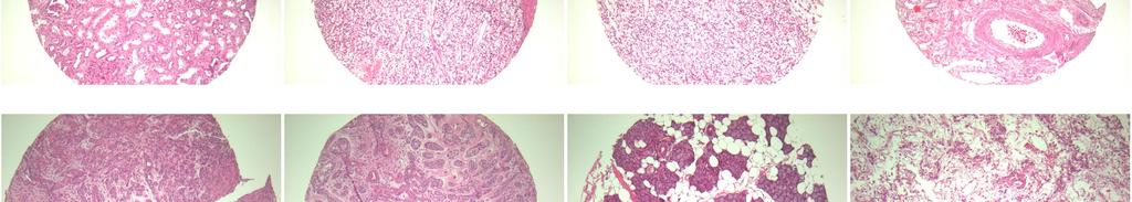
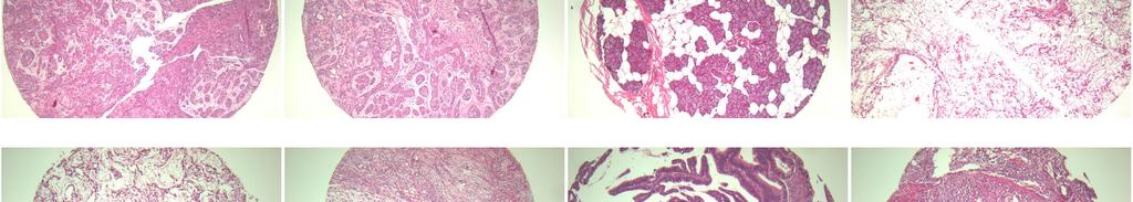
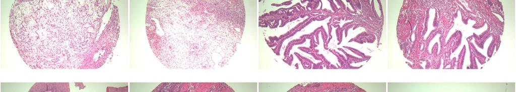
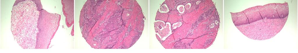
A301 VI- Various cancer tissues with corresponding normal tissues
|
|
|
- Emmeline O’Neal’
- 5 years ago
- Views:
Transcription
 28 non-neoplastic spots (28 spots) Total spots: 84 Corresponding normal tissues with cancers: Yes Diameter: 1.")
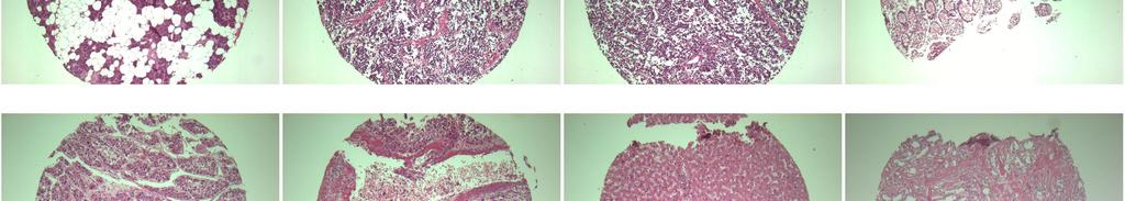
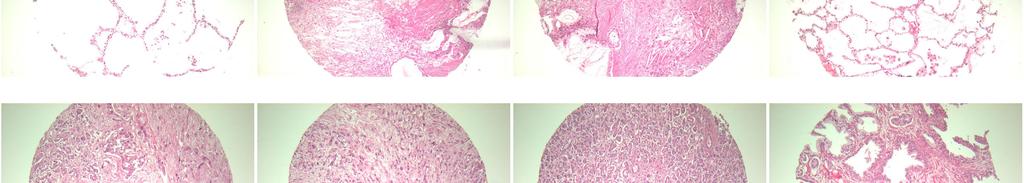
1 A301 VI- Various cancer tissues with (formalin fixed) For research use only Specifications: No. of cases: 28 Tissue type: Various cancer tissues with No. of spots: 2 spots from each cancer case (56 spots) 28 non-neoplastic spots (28 spots) Total spots: 84 Corresponding normal tissues with cancers: Yes Diameter: 1. 0 mm Documents: Product specification: layout, summary of tissue spots H&E stained images Detailed pathological information Layout: P T
 For research use only")
2 A301 VI- Various cancer tissues with Summary of tissue spots (formalin fixed) For research use only P T
 For research use only")
3 A301 VI- Various cancer tissues with Summary of tissue spots (formalin fixed) For research use only P T


4 A301 VI- Various cancer tissues with (formalin fixed) For research use only QC sheet_lot# Hematoxylin and Eosin staining A1 A2 A3 A4 A5 A6 A7 A8 A9 A10 A11 A12 B1 B2 B3 B4 B5 B6 B7 B8 B9 B10 B11 B12 P T


5 A301 VI- Various cancer tissues with (formalin fixed) For research use only QC sheet_lot# Hematoxylin and Eosin staining C1 C2 C3 C4 C5 C6 C7 C8 C9 C10 C11 C12 D1 D2 D3 D4 D5 D6 D7 D8 D9 D10 D11 D12 P T


6 A301 VI- Various cancer tissues with (formalin fixed) For research use only QC sheet_lot# Hematoxylin and Eosin staining E1 E2 E3 E4 E5 E6 E7 E8 E9 E10 E11 E12 F1 F2 F3 F4 F5 F6 F7 F8 F9 F10 F11 F12 P T
")


7 A301 VI- Various cancer tissues with (formalin fixed) For research use only QC sheet_lot# Hematoxylin and Eosin staining G1 G2 G3 G4 G5 G6 G7 G8 G9 G10 G11 G12 P T
8 A301 VI: Various cancer tissues with 1 A 1,2 f 52 adrenal gland: pheochromocytoma 2 A 3 f 52 non-neoplastic non-neoplastic from A1,2 1. Adrenal gland, right, adrenalectomy: pheochromocytoma 1) high nuclear grade 2) clear cells: less than 25% 3) presence of capsular invasion, 4) consistent with adrenal cortical carcinoma, by modified Weiss histopathologic criteria. 5) immunohistochemical stain: chromogranin A, NSE, Synaptophysin: (+) 3 A 4,5 m 62 esophagus: squamous cell carcinoma Esophagus and cardia of stomach, esophagectomy and node dissection: 1.Ulcerative squamous cell carcinoma, poorly differentiated, 1) size: 3.3x1cm 2) extension to the outer muscle layer 3) lymphovascular permeation: Positive 2.Resection margins, proximal and distal: Free of tumor. 3.Regional lymph nodes: Free of tumor in all nodes(0/24), in detail, subcarinal(0/10), hilar(0/2), pretracheal(0/6),lower paratracheal(0/3), left paracardial(0/2), 8L(0/1),omental mass(free of tumor). 4 A 6 m 62 non-neoplastic non-neoplastic from A4,5 5 A 7,8 m 35 salivary gland: pleomorphic adenoma 6 A 9 m 35 non-neoplastic non-neoplastic from A7,8 Salivary gland, parotid, right, parotidectomy: Pleomorphic adenoma. 7 A 10,1 1 m 20 terminal ileum: diffuse large cell lymphoma Terminal ileum, segmental resection: diffuse large B cell lymphoma 1) resection margin: lymphoid hyperplasia 2) lymph node: reactive hyperplasia 3) appendix: no pathological diagnosis 4) immunohistochemical staining results: CD20, CD10: diffuse (+), Bcl-6: focal (+), CD3: (-) Ki-67 proliferation index: about 70%. 8 A 12 f 60 non-neoplastic non-neoplastic from A10,11
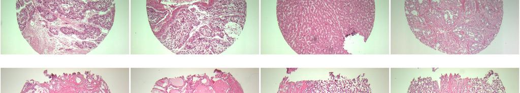
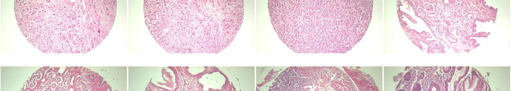
9 A301 VI: Various cancer tissues with 9 B 1,2 m 59 liver: hepatocellular carcinoma Liver, right lobe, lobectomy: hepatocellular carcinoma 1) size: 8x8x6.8cm 2) edmondson-steiner grade III 3) clear cell(majority) and trabecular histologic type 4) single nodular type with perinodal spreading growth 5) necrosis: less than 10% of total volume 6) microvascular invasion 7) intact intrahepatic resection margin 8) nonneoplastic liver showing: chronic hepatitis, B viral, with mild inflammatory activity(grade II) and early bcirrhosis (stage III). 10 B 3 m 59 non-neoplastic non-neoplastic from B1,2 11 B 4,5 m 51 thyroid gland: papillary carcinoma 12 B 6 m 51 non-neoplastic non-neoplastic from B4,5 13 B 7,8 m 70 soft tissue: diffuse large cell lymphoma 14 B 9 m 70 non-neoplastic non-neoplastic from B7,8 15 B 10,1 1 m 36 skin: invasive squamous cell carcinoma 16 B 12 m 36 non-neoplastic non-neoplastic from B10,11 Thyroid gland, total thyroidectomy with neck node dissection: 1.Papillary carcinoma 1) involving bilateral whole lobe(size: 6.5x3.7cm) 2) extension to perithyroidal fat tissue 2.Regional lymph node: tumor metastasis to 4 out of 20(4/20), in detail, level II(1/2), level III(3/13), level IV(0/5). Soft tissue and lymph node, neck, left, radical neck dissection: 1) soft tissue: diffuse large B cell lymphoma 2) lymph nodes, separately submitted as level ia(0/7), level ib(0/11), level iv(0/16) and level V(0/17): (0/51): reactive hyperplasia 3) salivary gland in level IB: free of tumor 4) tonsil, left, tonsillectomy: chronic follicular tonsillitis 5) immunohistochemical staining results CD20: positive to tumor cells CD3, CD4, CD8, Granzyme, CD30, CD56, ALK: negative to tumor cells 6) EBER ISH (-). Skin, scalp, excision: Invasive squamous cell carcinoma, 1) well-differentiated with extension to the subcutaneous fat tissue (invasion depth: about 3cm) 2) resection margins, all circumference and base: free of tumor 3) lymph nodes, level 2a(0/36), level 2b(0/11), level III(0/25), level IV(0/13) and level V(0/11):(0/96): free of tumor.
10 A301 VI: Various cancer tissues with 17 C 1,2 m 52 urinary bladder: Papillary urothelial carcinoma Urinary bladder including prostate, seminal vesicle and ureter, radical cystectomy: 1. Urinary bladder: Papillary urothelial carcinoma, high grade,with squamous differentiation, with various pathologic state including low grade papillary urothelial carcinoma, urothelial tumor of low malignant potential, carcinoma in situ and papilloma, with extension to the perivesical soft tissue and prostate, and with extensive lymphatic, venous and perineural invasion, incompletely excised. 2. Seminal vesicle: Free of tumor. 3. Ureters, bilateral, resection margins: Free of tumor. 18 C 3 m 52 non-neoplastic non-neoplastic from C1,2 19 C 4,5 m 79 tonsil: squamous cell carcinoma Tonsil, left, wide excision with right neck dissection: squamous cell carcinoma, poorly differentiated 1) size: 3.6x1.5x1.2cm 2) lymphatic permeation: uncertain 3) resection margin, base: free of tumor, margin is very close (less than 1mm) 4) resection margin, lateral: free of tumor 5) lymph node, right, level Ia(0/2), II(0/11), III(0/5), IV(0/4): (0/22): free of tumor 6) lymph node, left, level Ib(0/7), IIa(3/3), IIb(1/2), III(5/7), IV(1/1), V(0/16):(10/36): tumor metastasis, see note. 7) separately submitted lymph node, Ib(0/1), IIb(0/1): free of tumor Note: The largest lymph node involved by tumor measures 1.8x1.3cm in level III of the left side. 20 C 6 m 79 non-neoplastic non-neoplastic from C4,5 21 C 7,8 f 70 lung: squamous cell carcinoma 22 C 9 f 70 non-neoplastic non-neoplastic from C7,8 Lung, right lower lobe, lobectomy: squamous cell carcinoma, moderately differentiated 1) size: 7x5cm 2) extension to the visceral pleura 3) associated obstructive pneumonitis 4) bronchial resection margin: free of tumor.
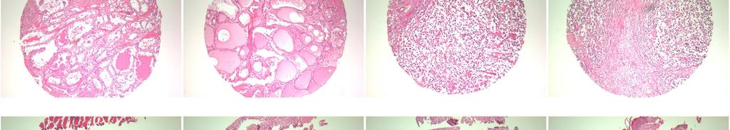
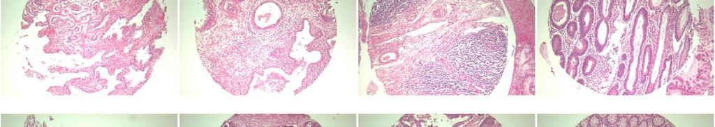
11 A301 VI: Various cancer tissues with 23 C 10,1 1 m 55 lung: adenocarcinoma 24 C 12 m 55 non-neoplastic non-neoplastic from C10,11 Lung, left lower lobe, lobectomy: adenocarcinoma, acinic and papillary with 1) marked mucin production 2) size: 4.7x4.6cm 3) extension to visceral pleura 4) bronchial resection margin: free of tumor 5) regional lymph node, peribronchial: tumor metastasis(1/1). 25 D 1,2 m 52 Pancreas: Adenocarcinoma Pancreas, distal pancreatectomy: 1.Adenocarcinoma, moderately differentiated 1) Size: 3.5x2.5cm 2) Tumor involvement: Extension to peripancreatic soft tissue and encircling the common hepatic artery without invasion. 3) Vascular invasion: Present 4) Neural and perineural invasion: Frequently present 2.Resection margin, pancreas: Free from tumor 3.Resection margin, circumferential: Free from tumor (margin of clearance: 20um) [Non-neoplastic pancreas] Chronic pancreatitis with parenchymal atrophy at tail portion of pancreas. Lymph nodes, peripancreatic : (4/13) Metastatic carcinoma in 4 out of 13 lymph nodes. 26 D 3 m 52 non-neoplastic non-neoplastic from D1,2 27 D 4,5 m 62 Prostate: adenocarcinoma Prostate, radical prostatectomy: 1.Adenocarcinoma, Gleason score 7(4+3) involving both sides of lobes, multifocally, confined to prostatic parenchyme without capsular involvement. 2.Seminal vesicles, bilateral: Free of tumor. 3.Lymph node, iliac, dissection: Free of tumor in all 14 lymph nodes (0/14), in detail, right(0/7) and left (0/7). 28 D 6 m 62 non-neoplastic non-neoplastic from D4,5
12 A301 VI: Various cancer tissues with 29 D 7,8 m 66 colon: adenocarcinoma 30 D 9 m 66 non-neoplastic non-neoplastic from D7,8 31 D 10,1 1 m 45 colon: adenocarcinoma 32 D 12 m 45 non-neoplastic non-neoplastic from D10,11 33 E 1,2 f 70 stomach: adenocarcinoma 34 E 3 f 70 non-neoplastic non-neoplastic from E1,2 Colon, rectum, lower anterior resection: adenocarcinoma, well differentiated, polypoid 1) size: 4.2x2.5cm 2) confined to the mucosa 3) associated low and high grade tubulo-villous adenoma 4) resection margins, proximal and distal: free of tumor 5) regional lymph nodes: free of tumor in all nodes(0/21), regional(0/21), principal(0/0). Colon, rectum, lower anterior resection: 1.colon: ulcerofungating adenocarcinoma, moderately differentiated, 1) size: 5.5x4.5cm 2) extension to the pericolic fat tissue(pt3c) 3) lateral margin: free of tumor 4) lymphovascular permeation(+) 2.urinary bladder: free of tumor 3.resection margins, proximal and distal: free of tumor 4.regional lymph nodes: tumor metastasis to 4 out of 11(4/11), in detail, regional(4/8), principal(0/3). Stomach, pancreas and spleen, radical total gastrectomy, splenectomy and pancreatectomy: 1.Stomach: ulceroinfiltrative adenocarcinoma, moderately differentiated, 1) size: 10.7x9cm 2) extension to the serosa and pancreas parenchyme 3) lymphovascular permeation: positive 4) Intestinal type by Lauren's classification 2.Resection margins, proximal and distal: free of tumor 3.Resection margin, pancreas: free of tumor 4.Regional lymph nodes: tumor metastasis to 2 out of 47(2/47), in detail, N1(0/5), N3(0/5), N4(0/8), N5(0/2), N6(1/16), N7(0/2), N8(1/4), N9(0/2), N10(0/3) 5.Pancreas: tumor extension 6.Spleen: congestion.
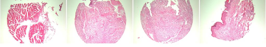
13 A301 VI: Various cancer tissues with 35 E 4,5 m 67 stomach: signet ring cell carcinoma 36 E 6 m 67 non-neoplastic non-neoplastic from E4,5 37 E 7,8 f 65 kidney: renal cell carcinoma 38 E 9 f 65 non-neoplastic non-neoplastic from E7,8 39 E 10,1 1 m 52 kidney: renal cell carcinoma Stomach, subtotal gastrectomy: 1.ulceroinfiltrative signet ring cell carcinoma 1) size: 4.5x4cm 2) extension to the serosa(serosa attached) 3) lymphovascular permeation: positive 4) perineural invasion: positive 5) diffuse type by lauren's classification 2. resection margins, proximal and distal: free of tumor 3. regional lymph nodes: tumor metastasis to 3 out of 31(3/31), in detail, N3(0/6), N4(0/7), N5(1/2), N6(2/5), N7(0/10), N12(0/1). Kidney, left, radical nephrectomy: 1.Renal cell carcinoma, conventional(clear cell) type 1) Fuhrman's nuclear grade: IV 2) Foci of sarcomatoid differentiation 3) Invasion to the perinephric fat tissue but not to Gerota's fascia 4) Renal vein involvement(pt3b) 2.Resection margin, ureter: Free of tumor. 3.Resection margin, renal vein: Presence of tumor. 4.Lymph nodes, perihilar(0/11), paraaortic(0/2) and left common iliac(0/3): Free of tumor metastasis in all 16 nodes. 5.Adrenal gland: Free of tumor. 1. Kidney, left, nephrectomy: renal cell carcinoma, clear cell type 1) size: 4x4cm 2) confined within the renal capsule 3) nuclear grade II 4) renal artery and vein: free of tumor 2. Ureter resection margin: free of tumor 3. Adrenal gland: free of tumor. 40 E 12 m 52 non-neoplastic non-neoplastic from E10,11 41 F 1,2 f 21 parotid gland: pleomorphid adenoma Parotid gland, right, parotidectomy: 1.Pleomorphic adenoma. 2.Lymph node: Reactive hyperplasia. 42 F 3 f 21 non-neoplastic non-neoplastic from F1,2
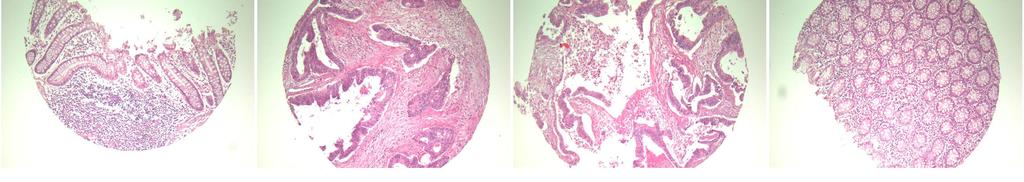
14 A301 VI: Various cancer tissues with 43 F 4,5 f 15 ovary: yolk sac tumor 44 F 6 f 15 non-neoplastic non-neoplastic from F4,5 45 F 7,8 f 47 uterus: adenocarcinoma 46 F 9 f 47 non-neoplastic non-neoplastic from F7,8 47 F 10,1 1 f 40 uterus: squamous cell carcinoma 48 F 12 f 40 non-neoplastic non-neoplastic from F10,11 49 G 1,2 f 52 breast: Invasive lobular carcinoma. 50 G 3 f 52 non-neoplastic non-neoplastic from G1,2 Ovary and fallopian tube, right, right salpingooophorectomy: 1) ovary: malignant germ cell tumor, probably yolk sac tumor (endodermal sinus tumor) 2) fallopian tube: paraovarian cyst 3) result of special stain: AFP(+), HCG(-), PAS(+), D-PAS(-). Uterus with right adnexa, total abdominal hysterectomy with right salpingooophorectomy: 1.Cervix: Invasive squamous cell carcinoma, moderately differentiated, mixed with adenocarcinoma, moderately differentiated 1) size: 4x3cm 2) extension to lower 1/3 of the wall (invasion depth: 1.9cm) 3) lymphatic permeation: positive 4) lateral margin: Free of tumor. 2.Endometrium: Secretory phase. 3.Myometrium : 1. Leiomyomas, intramural. Uterus with lymph nodes, pelvic, radical abdominal hysterectomy with dissection: 1.Cervix: Invasive squamous cell carcinoma, large cell, keratinizing (invasion depth: 1.1cm) with focal lymphovascular permeation. 2.Endometrium: Proliferative phase. 3.Myometrium : Adenomyosis. 4.Parametria, bilateral: Free of tumor. 5.Vaginal cuff: Free of tumor. 6.Lymph nodes, right pelvic(0/4), left pelvic(0/6), regional(0/2):(0/12): Free of tumor. Breast, left, lumpectomy: Invasive lobular carcinoma.

15 A301 VI: Various cancer tissues with 51 G 4,5 m 52 cecum: adenocarcinoma 52 G 6 m 52 non-neoplastic non-neoplastic from G4,5 53 G 7,8 m 36 Testis:Seminoma 54 G 9 m 36 non-neoplastic non-neoplastic from G7,8 55 G 10,1 1 m 31 Thymus: thymoma 56 G 12 m 31 non-neoplastic non-neoplastic from G10,11 Cecum, ascending colon, terminal ileum and appendix, right hemicolectomy: 1.Ulcerofungating adenocarcinoma, moderately differentiated, inascending colon 1) size: 5x4cm 2) expanding growth 3) invasion to pericolic soft tissue. 4) vascular invasion. 5) intact proximal (terminal ileum) and distal (colon) resection margins. 6) metastasis to 2 out of 32 pericolic lymph nodes. 7) a tubular adenoma with low grade dysplasia in the surrounding mucosa. 8) Terminal ileum: Prominent peyer's patch. 9) Appendix: Free from tumor extension. Testis, left, orchiectomy: 1.Seminoma with invasion beyond tunica albuginea(pt2) and associated with intratubular germ cell neoplasia. 2.Spermatic cord: Free of tumor Thymus, thymectomy: 1.Type B2 thymoma (cortical thymoma by Muller-Hermelink scheme) 1) size: 8x5.5cm 2) confined to the thymic capsule(pt1) (Masaoka's stage I). 3) multilocular thymic cyst 4) resection margin: Free of tumor. 2. Adenomyosis. 3.Ovary: No pathological diagnosis. 4.Fallopian tube: Paratubal cyst. 5.Parametrium, bilateral: Free of tumor. 6.Vaginal cuff: Free of tumor. 7.Regional lymph nodes: Free of tumor in all nodes(0/4), in detail, paraaortic(0/0), left pelvic(0/2), right pelvic(0/2).
A103(9)- Normal tissues, more than single spots
- Normal tissues, more than single spots") A103(9)- Normal tissues, more than single spots (formalin fixed) For research use only Specifications: No. of cases: 45 Tissue type: Normal tissues, more than single spots No. of spots: 2 spots from each
A103(9)- Normal tissues, more than single spots (formalin fixed) For research use only Specifications: No. of cases: 45 Tissue type: Normal tissues, more than single spots No. of spots: 2 spots from each
A201(VI)- Various cancer tissues
- Various cancer tissues") A201(VI)- Various cancer tissues (formalin fixed) For research use only Specifications: No. of cases: 44 Tissue type: Various cancer tissues No. of spots: 2 spots from each cancer case (88 spots) Total
A201(VI)- Various cancer tissues (formalin fixed) For research use only Specifications: No. of cases: 44 Tissue type: Various cancer tissues No. of spots: 2 spots from each cancer case (88 spots) Total
(formalin fixed) 6 non-neoplastic spots (6 spots) Corresponding normal tissues with cancers: Yes Diameter: 1. 0 mm
 6 non-neoplastic spots (6 spots) Corresponding normal tissues with cancers: Yes Diameter: 1. 0 mm") CBA729-Test slide, Head and neck cancer tissues (formalin fixed) For research use only Specifications: No. of cases: 6 Tissue type: Test slide, Head and neck cancer tissues No. of spots: 6 spots from each
CBA729-Test slide, Head and neck cancer tissues (formalin fixed) For research use only Specifications: No. of cases: 6 Tissue type: Test slide, Head and neck cancer tissues No. of spots: 6 spots from each
A215- Urinary bladder cancer tissues
 A215- Urinary bladder cancer tissues (formalin fixed) For research use only Specifications: No. of cases: 45 Tissue type: Urinary bladder cancer tissues No. of spots: 2 spots from each cancer case (90
A218 : Esophagus cancer tissues. (formalin fixed)
") (formalin fixed) For research use only Specifications: No. of cases: 40 Tissue type: Esophagus cancer tissues No. of spots: 2 spots from each cancer case (80 spots) 4 non-neoplastic spots (4 spots) Total
(formalin fixed) For research use only Specifications: No. of cases: 40 Tissue type: Esophagus cancer tissues No. of spots: 2 spots from each cancer case (80 spots) 4 non-neoplastic spots (4 spots) Total
Definition of Synoptic Reporting
 Definition of Synoptic Reporting The CAP has developed this list of specific features that define synoptic reporting formatting: 1. All required cancer data from an applicable cancer protocol that are
A916: rectum: adenocarcinoma
 General facts of colorectal cancer The colon has cecum, ascending, transverse, descending and sigmoid colon sections. Cancer can start in any of the r sections or in the rectum. The wall of each of these
Bladder Case 1 SURGICAL PATHOLOGY REPORT. Procedure: Cystoscopy, transurethral resection of bladder tumor (TURBT)
") Bladder Case 1 February 17, 2007 Specimen (s) received: Bladder Tumor Pre-operative Diagnosis: Bladder Cancer Post operative Diagnosis: Bladder Cancer Procedure: Cystoscopy, transurethral resection of
Bladder Case 1 February 17, 2007 Specimen (s) received: Bladder Tumor Pre-operative Diagnosis: Bladder Cancer Post operative Diagnosis: Bladder Cancer Procedure: Cystoscopy, transurethral resection of
A712(18)- Test slide, Breast cancer tissues with corresponding normal tissues
- Test slide, Breast cancer tissues with corresponding normal tissues") A712(18)- Test slide, Breast cancer tissues with corresponding normal tissues (formalin fixed) For research use only Specifications: No. of cases: 12 Tissue type: Breast cancer tissues with corresponding
A712(18)- Test slide, Breast cancer tissues with corresponding normal tissues (formalin fixed) For research use only Specifications: No. of cases: 12 Tissue type: Breast cancer tissues with corresponding
Take Home Quiz 1 Please complete the quiz below prior to the session. Use the Multiple Primary and Histology Rules
 Take Home Quiz 1 Please complete the quiz below prior to the session. Use the Multiple Primary and Histology Rules Case 1 72 year old white female presents with a nodular thyroid. This was biopsied in
Kidney Case 1 SURGICAL PATHOLOGY REPORT
 Kidney Case 1 Surgical Pathology Report February 9, 2007 Clinical History: This 45 year old woman was found to have a left renal mass. CT urography with reconstruction revealed a 2 cm medial mass which
Gastric Cancer Histopathology Reporting Proforma
 Gastric Cancer Histopathology Reporting Proforma Mandatory questions (i.e. protocol standards) are in bold (e.g. S1.01). S1.01 Identification Family name Given name(s) Date of birth Sex Male Female Intersex/indeterminate
A712(19)- Test slide, Breast cancer tissues with corresponding normal tissues
- Test slide, Breast cancer tissues with corresponding normal tissues") A712(19)- Test slide, Breast cancer tissues with corresponding normal tissues (formalin fixed) For research use only Specifications: No. of cases: 12 Tissue type: Breast cancer tissues with corresponding
A712(19)- Test slide, Breast cancer tissues with corresponding normal tissues (formalin fixed) For research use only Specifications: No. of cases: 12 Tissue type: Breast cancer tissues with corresponding
MT09 - Normal Human Tissue Microarray, FDA
 Reveal Biosciences offers Histochemical Staining, Immunohistochemistry (IHC), In Situ Hybridization (ISH), Whole Slide Imaging, and Quantitative Image Analysis on any TMA MT09 - Normal Human Tissue Microarray,
UICC TNM 8 th Edition Errata
 UICC TNM 8 th Edition Errata ions are in italics Page 28 Oropharynx p16 positive Pathological Stage II,T2 N2 M0 T3 N0,N1 M0 Stage II,T2 N2 M0 T3,T4 N0,N1 M0 Page 61 Oesophagus Adenocarcinoma Pathological
Checklist; Anus: Excisional Biopsy Anus: Excisional Biopsy 1/1/ Checklist; Anus: Resection Anus: Resection 1/1/2005
 ChecklistTemplateVersions ChecklistTemplateVersion Ckey OfficialName VisibleText RevisionDate Checklist; Adrenal gland: 16.1000043 Resection Adrenal gland: Checklist; Ampulla of 17.1000043 Vater: Ampullectomy
A220: Larynx cancer tissues. (formalin fixed)
") A220: Larynx cancer tissues (formalin fixed) For research use only Specifications: No. of cases: 45 Tissue type: Larynx cancer tissues No. of spots: 2 spots from each cancer case (90 spots) 4 non-neoplastic
A220: Larynx cancer tissues (formalin fixed) For research use only Specifications: No. of cases: 45 Tissue type: Larynx cancer tissues No. of spots: 2 spots from each cancer case (90 spots) 4 non-neoplastic
Carcinoma of the Urinary Bladder Histopathology
 Carcinoma of the Urinary Bladder Histopathology Reporting Proforma (Radical & Partial Cystectomy, Cystoprostatectomy) Includes the International Collaboration on Cancer reporting dataset denoted by * Family
UICC TNM 8 th Edition Errata
 UICC TNM 8 th Edition Errata ions are in italics Head and Neck Tumours Pages 20, p27, p34, p38, p41, and p49 ly pn2a Metastasis in a single ipsilateral lymph node, less than 3cm in greatest dimension with
Index. Note: Page numbers of article titles are in boldface type.
 Note: Page numbers of article titles are in boldface type. A Adenocarcinoma, pancreatic ductal, laparoscopic distal pancreatectomy for, 61 Adrenal cortical carcinoma, laparoscopic adrenalectomy for, 114
SHN-1 Human Digestive Panel Test results
 SHN-1 Human Digestive Panel Test results HN-30 tongue HN-24 salivary gland HN-12 larynx HN-28 esophagus HN-29 stomach HN-20 pancreas HN-13 liver HN-14 gall bladder HN-27-1 duodenum HN-27-2 ileum HN-27-3
Quiz. b. 4 High grade c. 9 Unknown
 Quiz 1. 10/11/12 CT scan abdomen/pelvis: Metastatic liver disease with probable primary colon malignancy. 10/17/12 Colonoscopy with polypectomy: Adenocarcinoma of sigmoid colon measuring at least 6 mm
Colon and Rectum. Protocol revision date: January 2005 Based on AJCC/UICC TNM, 6th edition
 Colon and Rectum Protocol applies to all invasive carcinomas of the colon and rectum. Carcinoid tumors, lymphomas, sarcomas, and tumors of the vermiform appendix are excluded. Protocol revision date: January
Prognostic factors of genitourinary tumors: Do we have to care?
 Prognostic factors of genitourinary tumors: Do we have to care? Jae Y. Ro, MD, PhD Professor and Director of Surgical Pathology The Methodist Hospital, Weill Medical College of Cornell University, Houston,
Abstracting Upper GI Cancer Incidence and Treatment Data Quiz 1 Multiple Primary and Histologies Case 1 Final Pathology:
 Abstracting Upper GI Cancer Incidence and Treatment Data Quiz 1 Multiple Primary and Histologies Case 1 A 74 year old male with a history of GERD presents complaining of dysphagia. An esophagogastroduodenoscopy
155.2 Malignant neoplasm of liver not specified as primary or secondary. C22.9 Malignant neoplasm of liver, not specified as primary or secondary
 ICD-9 TO ICD-10 Reference ICD-9 150.9 Malignant neoplasm of esophagus unspecified site C15.9 Malignant neoplasm of esophagus, unspecified 151.9 Malignant neoplasm of stomach unspecified site C16.9 Malignant
8. The polyp in the illustration can be described as (circle all that apply) a. Exophytic b. Pedunculated c. Sessile d. Frank
 a. Exophytic b. Pedunculated c. Sessile d. Frank") Quiz 1 Overview 1. Beginning with the cecum, which is the correct sequence of colon subsites? a. Cecum, ascending, splenic flexure, transverse, hepatic flexure, descending, sigmoid. b. Cecum, ascending,
Quiz 1 Overview 1. Beginning with the cecum, which is the correct sequence of colon subsites? a. Cecum, ascending, splenic flexure, transverse, hepatic flexure, descending, sigmoid. b. Cecum, ascending,
3. Guidelines for Reporting Bladder Cancer, Prostate Cancer and Renal Tumours
 60 3. Guidelines for Reporting Bladder Cancer, Prostate Cancer and Renal Tumours Compilation and editing and of this volume: Prof. Chandu de Silva (Consultant Histopathologist) List of contributors Consultant
Staging and Treatment Update for Gynecologic Malignancies
 Staging and Treatment Update for Gynecologic Malignancies Bunja Rungruang, MD Medical College of Georgia No disclosures 4 th most common new cases of cancer in women 5 th and 6 th leading cancer deaths
Presentation material is for education purposes only. All rights reserved URMC Radiology Page 1 of 98
 Presentation material is for education purposes only. All rights reserved. 2011 URMC Radiology Page 1 of 98 Radiology / Pathology Conference February 2011 Brooke Koltz, Cytopathology Resident Presentation
2018 Grade PEGGY ADAMO, RHIT, CTR OCTOBER 11, 2018
 1 2018 Grade PEGGY ADAMO, RHIT, CTR ADAMOM@MAIL.NIH.GOV OCTOBER 11, 2018 2 Acknowledgements Donna Hansen, CCR Jennifer Ruhl, NCI SEER Introduction 3 Histologic Type vs. Grade Credit: Dr. Kay Washington
Case Scenario 1: Thyroid
 Case Scenario 1: Thyroid History and Physical Patient is an otherwise healthy 80 year old female with the complaint of a neck mass first noticed two weeks ago. The mass has increased in size and is palpable.
Histology Coding ANSWERS
 Histology Coding ANSWERS 1.) Biopsy of a right thyroid nodule reveals papillary carcinoma. What is the ICD-O-3 code? a. 8050/3 - Papillary carcinoma b. 8260/3 - Papillary adenocarcinoma Rationale/comment:
CAP Cancer Protocol and ecc Summary of Changes for August 2014 Thyroid Agile Release
 CAP Cancer Protocol and ecc Summary of Changes for August 2014 Thyroid Agile Release 2 REVISION HISTORY Date Author / Editor Comments 5/19/2014 Jaleh Mirza Created the document 8/12/2014 Samantha Spencer/Jaleh
Fig. 59 Malignant phaeochromocytoma, hepatic metastasis.
 Fig. 59 Malignant phaeochromocytoma, hepatic metastasis. X 120 Hyperte nsion Fig. 60 Malignant sympathetic paraganglioma, lymph node metastasis Primary in bladder. x 1 20 Hypertension Fig. 61 Malignant
Comprehensive Cancer Cover
 Comprehensive Cancer Cover Tech Spec Comprehensive Cancer Cover provides the life insured with cover for the diagnosis and treatment of defined malignant tumours. These tumours must be characterised either
Radiology Pathology Conference
 Radiology Pathology Conference Nadia F. Yusaf, M.D. PGY-3 1/29/2010 Presentation material is for education purposes only. All rights reserved. 2010 URMC Radiology Page 1 of 90 Case 1 60 year- old man presents
Male genital tract tumors. SiCA. Division of Urology, Department of Surgery, Faculty of Medicine Siriraj Hospital.
 Male genital tract tumors Division of Urology, Department of Surgery, Faculty of Medicine Siriraj Hospital. adenocarcinoma Prostate Cancer most common male cancer in western countries more detected in
NAACCR Webinar Series 1 Q&A. Fabulous Prizes. Collecting Cancer Data: Ovary 11/3/2011. Collecting Cancer Data: Ovary
 NAACCR 2011 2012 Webinar Series Collecting Cancer Data: Ovary Q&A Please submit all questions concerning webinar content through the Q&A panel. Reminder: If you have participants watching this webinar
Quiz The main functions of the ovaries are a. To produce oocytes b. To produce estrogen c. To produce progesterone d.
 1. The main functions of the ovaries are a. To produce oocytes b. To produce estrogen c. To produce progesterone d. All of the above Quiz 1 2. Which part of the broad ligament suspends the ovaries? a.
Radiology Pathology Conference
 Radiology Pathology Conference Sharlin Johnykutty,, MD, Cytopathology Fellow Sara Majewski, MD, Radiology Resident Friday, August 28, 2009 Presentation material is for education purposes only. All rights
Urinary Bladder: WHO Classification and AJCC Staging Update 2017
 Urinary Bladder: WHO Classification and AJCC Staging Update 2017 Houston Society of Clinical Pathologists 58 th Annual Spring Symposium Houston, TX April 8, 2017 Jesse K. McKenney, MD Classification
New Cancer Cases By Site Breast 28% Lung 14% Colo-Rectal 10% Uterus 6% Thyroid 5% Lymphoma 4% Ovary 3%
 Uterine Malignancy New Cancer Cases By Site 2010 Breast 28% Lung 14% Colo-Rectal 10% Uterus 6% Thyroid 5% Lymphoma 4% Ovary 3% Cancer Deaths By Site 2010 Lung 26% Breast 15% Colo-Rectal 9% Pancreas 7%
The pathology of bladder cancer
 1 The pathology of bladder cancer Charles Jameson Introduction Carcinoma of the bladder is the seventh most common cancer worldwide [1]. It comprises 3.2% of all cancers, with an estimated 260 000 new
Pancreatobiliary Frozen Section Nightmares
 Pancreatobiliary Frozen Section Nightmares Aatur D. Singhi, MD PhD Assistant Professor University of Pittsburgh Medical Center Department of Pathology singhiad@upmc.edu Objectives Briefly give an overview
Male Genital Cancers in the US in Frequency of Types
 Germ Cell Tumors of the Testis Pathology, Immunohistochemistry, and the Often Confusing Appearance of Their Metastases Charles Zaloudek, MD Department of Pathology UCSF Male Genital Cancers in the US in
CPC on Cervical Pathology
 CPC on Cervical Pathology Dr. W.K. Ng Senior Medical Officer Department of Clinical Pathology Pamela Youde Nethersole Eastern Hospital Cervical Smear: High Grade SIL (CIN III) Cervical Smear: High Grade
11/21/13 CEA: 1.7 WNL
 Case Scenario 1 A 70 year-old white male presented to his primary care physician with a recent history of rectal bleeding. He was referred for imaging and a colonoscopy and was found to have adenocarcinoma.
A patient with recurrent bladder cancer presents with the following history:
 MP/H Quiz A patient with recurrent bladder cancer presents with the following history: 9/23/06 TURB 1/12/07 TURB 4/1/07 TURB 7/12/07 TURB 11/14/07 Non-invasive papillary transitional cell carcinoma from
List of Available TMAs in the PRN
 TMA RPCI_BrainCa01 RPCI_BrCa03 RPCI_BrCa04 RPCI_BrCa05 RPCI_BrCa0 RPCI_BrCa07 RPCI_BrCa08 RPCI_BrCa15 RPCI_BrCa1 RPCI_BrCa17 RPCI_BrCa18 RPCI_BrCa19 RPCI_BrCa20 RPCI_BrCa21 RPCI_BrCa24 RPCI_BrCa25 RPCI_BrCa2
Thyroid and Adrenal Gland
 Thyroid and Adrenal Gland NAACCR 2011 2012 Webinar Series 12/1/11 Q&A Please submit all questions concerning webinar content through the Q&A panel. Reminder: If you have participants watching this webinar
Uterine Cervix. Protocol applies to all invasive carcinomas of the cervix.
 Uterine Cervix Protocol applies to all invasive carcinomas of the cervix. Protocol revision date: January 2005 Based on AJCC/UICC TNM, 6 th edition and FIGO 2001 Annual Report Procedures Cytology (No Accompanying
LOINC. Clinical information. RCPA code. Record if different to report header Operating surgeon name and contact details. Absent.
 Complete as narrative or use the structured format below 55752-0 17.02.28593 Clinical information 22027-7 17.02.30001 Record if different to report header Operating surgeon name and contact details 52101004
Carcinoma of the Renal Pelvis and Ureter Histopathology
 Carcinoma of the Renal Pelvis and Ureter Histopathology Reporting Proforma (NEPHROURETERECTOMY AND URETERECTOMY) Includes the International Collaboration on Cancer reporting dataset denoted by * Family
IMPC phenotyping SOPs in JMC
 IMPC phenotyping SOPs in JMC Tissue Embedding and Block Banking IMPC_BLK_001 Purpose Collect and fix a standard list of tissues from the complete necropsy (see IMPC Gross Pathology & Tissue Collection
LUNG STAGING FORM LATERALITY: LEFT RIGHT BILATERAL
 LUNG STAGING FORM LATERALITY: LEFT RIGHT BILATERAL ( ) Tx Primary tumor cannot be assessed, or tumor proven by the presence of malignant cells in sputum or bronchial washings but not visualized by imaging
MALIGNANT NEOPLASMS OF THE BREAST MALIGNANT NEOPLASMS OF FEMALE GENITAL ORGANS
 MALIGNANT NEOPLASMS OF THE (INC. PAGET S DISEASE) 0 - Nipple and areola 1 - Central portion 2 - Upper-inner quadrant 3 - Lower-inner quadrant 4 - Upper-outer quadrant 5 - Lower-outer quadrant 6 - Axillary
CODING STAGE: TNM AND OTHER STAGING SYSTEMS. Liesbet Van Eycken Otto Visser
 CODING STAGE: TNM AND OTHER STAGING SYSTEMS Liesbet Van Eycken Otto Visser OVERVIEW PART I Introduction What is stage? Why stage? History and publications of TNM Classification Clinical and pathologic
Gross appearance of nodular hyperplasia in material obtained from suprapubic prostatectomy. Note the multinodular appearance and the admixture of
 Tiền liệt tuyến Tiền liệt tuyến Gross appearance of nodular hyperplasia in material obtained from suprapubic prostatectomy. Note the multinodular appearance and the admixture of solid and microcystic areas.
Urology An introduction to cut up DR J R GOEPEL
 Urology An introduction to cut up DR J R GOEPEL Overview Principles Individual organs Small pieces Partial resections Whole organs Data recording and data sets Principles You are working for the patient
MPH Quiz. 1. How many primaries are present based on this pathology report? 2. What rule is this based on?
 MPH Quiz Case 1 Surgical Pathology from hysterectomy performed July 11, 2007 Final Diagnosis: Uterus, resection: Endometrioid adenocarcinoma, Grade 1 involving most of endometrium, myometrial invasion
Colorectal Cancer Structured Pathology Reporting Proforma DD MM YYYY
 Colorectal Cancer Structured Pathology Reporting Proforma Mandatory questions (i.e. protocol standards) are in bold (e.g. S1.03). Family name Given name(s) Date of birth DD MM YYYY S1.02 Clinical details
Urinary Bladder, Ureter, and Renal Pelvis
 Urinary Bladder, Ureter, and Renal Pelvis Protocol applies to all carcinomas of the urinary bladder, ureter, and renal pelvis. Protocol revision date: January 2005 Based on AJCC/UICC TNM, 6th edition Procedures
Kidney, Bladder and Prostate Neoplasia. David Bingham MD
 Kidney, Bladder and Prostate Neoplasia David Bingham MD typical malignant cytology of bladder washings 1 benign 2 malignant typical malignant cytology of bladder washings b Bladder tumor Non invasive papillary
AJCC 7th Edition Handbook Errata as of 9/21/10
 5 81 Larynx ICD-O-3 Topography Codes Delete C32.3 Laryngeal cartilage 5 81 Larynx ICD-O-3 Topography Codes Add an asterisk after C32.8 5 81 Larynx ICD-O-3 Topography Codes Add an asterisk after C32.9 5
Multi-normal human tissues, FDA, 96 samples, 35 organs/sites from three individuals (1.5mm)
") ab178228 Multi-normal human tissues, FDA, 96 samples, 35 organs/sites from three individuals (1.5mm) Instructions for Use Designed for antibody cross reactivity analysis and IHC or ISH based protein or
ab178228 Multi-normal human tissues, FDA, 96 samples, 35 organs/sites from three individuals (1.5mm) Instructions for Use Designed for antibody cross reactivity analysis and IHC or ISH based protein or
Note: The cause of testicular neoplasms remains unknown
 - In the 15- to 34-year-old age group, they are the most common tumors of men. - Tumors of the testis are a heterogeneous group of neoplasms that include: I. Germ cell tumors : 95%; all are malignant.
Cervical Cancer 3/25/2019. Abnormal vaginal bleeding
 Cervical Cancer Abnormal vaginal bleeding Postcoital, intermenstrual or postmenopausal Vaginal discharge Pelvic pain or pressure Asymptomatic In most patients who are not sexually active due to symptoms
Cancer Association of South Africa (CANSA)
") Cancer Association of South Africa (CANSA) Fact Sheet on ICD-10 Coding of Neoplasms Introduction The International Statistical Classification of Diseases and Related Health Problems, 10 th Revision (ICD-10)
Cancer Association of South Africa (CANSA) Fact Sheet on ICD-10 Coding of Neoplasms Introduction The International Statistical Classification of Diseases and Related Health Problems, 10 th Revision (ICD-10)
The SUM Program for Medical Transcription Training Career Development Series: Interpreting Anatomic Pathology Dictation
 The SUM Program for Medical Transcription Training Career Development Series: Interpreting Anatomic Pathology Dictation Table of Contents INTRODUCTION Anatomic Pathology by John H. Dirckx, M.D. Exercises
Neoplasia part I. Dr. Mohsen Dashti. Clinical Medicine & Pathology nd Lecture
 Neoplasia part I By Dr. Mohsen Dashti Clinical Medicine & Pathology 316 2 nd Lecture Lecture outline Review of structure & function. Basic definitions. Classification of neoplasms. Morphologic features.
Case #1: 75 y/o Male (treated and followed by prostate cancer oncology specialist ).
.") SOLID TUMORS WORKSHOP Cases for review Prostate Cancer Case #1: 75 y/o Male (treated and followed by prostate cancer oncology specialist ). January 2009 PSA 4.4, 20% free; August 2009 PSA 5.2; Sept 2009
SOLID TUMORS WORKSHOP Cases for review Prostate Cancer Case #1: 75 y/o Male (treated and followed by prostate cancer oncology specialist ). January 2009 PSA 4.4, 20% free; August 2009 PSA 5.2; Sept 2009
UICC 8 th Edition Errata 25 th of May 2018
 UICC 8 th Edition Errata 25 th of May 2018 ions are in italics Head and Neck Tumours Page 19 Oral Cavity T2 T3 T4a Tumour 2 cm or less in greatest dimension and more than 5 mm but no more than 10 mm depth
objectives Pitfalls and Pearls in PET/CT imaging Kevin Robinson, DO Assistant Professor Department of Radiology Michigan State University
 objectives Pitfalls and Pearls in PET/CT imaging Kevin Robinson, DO Assistant Professor Department of Radiology Michigan State University To determine the regions of physiologic activity To understand
Case Scenario 1 Discharge Summary Pathology Report Final Diagnosis: Oncology Consult
 Case Scenario 1 Discharge Summary A 31-year-old Brazilian male presented with a 6 month history of right-sided scrotal swelling. Backache was present for 2 months and a history of right epididymitis was
CEA (CARCINOEMBRYONIC ANTIGEN)
") (CARCINOEMBRYONIC ANTIGEN) 428 C15.3 Malignant neoplasm of upper third of esophagus C15.4 Malignant neoplasm of middle third of esophagus C15.5 Malignant neoplasm of lower third of esophagus C15.8 Malignant
(CARCINOEMBRYONIC ANTIGEN) 428 C15.3 Malignant neoplasm of upper third of esophagus C15.4 Malignant neoplasm of middle third of esophagus C15.5 Malignant neoplasm of lower third of esophagus C15.8 Malignant
2009 USCAP Gyn Pathology Evening Session Case #3. Richard J. Zaino, MD Hershey Medical Center Penn State University Hershey, PA
 2009 USCAP Gyn Pathology Evening Session Case #3 Richard J. Zaino, MD Hershey Medical Center Penn State University Hershey, PA rzaino@psu.edu Clinical history Middle aged woman with an exophytic mass of
Kyle L. Ziegler, CTR. California Cancer Registry U.C. Davis Health System
 Kyle L. Ziegler, CTR California Cancer Registry U.C. Davis Health System Overview New Data Items Reportability Clarifications New Coding Rules Grade ICD-O-3 Changes Collaborative Stage v0205 2 New Data
Pancreas Quizzes c. Both A and B a. Directly into the blood stream (not using ducts)
") Pancreas Quizzes Quiz 1 1. The pancreas produces hormones. Which type of hormone producing organ is the pancreas? a. Endocrine b. Exocrine c. Both A and B d. Neither A or B 2. Endocrine indicates hormones
Pancreas Quizzes Quiz 1 1. The pancreas produces hormones. Which type of hormone producing organ is the pancreas? a. Endocrine b. Exocrine c. Both A and B d. Neither A or B 2. Endocrine indicates hormones
Epithelial tumors. Dr. F.F. Khuzin, PhD Dr. M.O. Mavlikeev
 Epithelial tumors Dr. F.F. Khuzin, PhD Dr. M.O. Mavlikeev Epithelial tumors Tumors from the epithelium are the most frequent among tumors. There are 2 group features of these tumors: The presence in most
Management guideline for patients with differentiated thyroid cancer. Teeraporn Ratanaanekchai ENT, KKU 17 October 2007
 Management guideline for patients with differentiated thyroid Teeraporn Ratanaanekchai ENT, KKU 17 October 2007 Incidence (Srinagarind Hospital, 2005, both sex) Site (all) cases % 1. Liver 1178 27 2. Lung
6/5/2010. Renal vein invasion & Capsule Penetration (T3a) Adrenal Gland involvement (T4 vs. M1) Beyond Gerota s Fascia? (?T4).
 Adrenal Gland involvement (T4 vs. M1) Beyond Gerota s Fascia? (?T4).") GU Cancer Staging: Updates and Challenging Areas 13 th Current Issues in Surgical Pathology San Francisco, CA June 5, 2010 Jeffry P. Simko, PhD, MD Associate Professor Departments of Urology and Anatomic
GU Cancer Staging: Updates and Challenging Areas 13 th Current Issues in Surgical Pathology San Francisco, CA June 5, 2010 Jeffry P. Simko, PhD, MD Associate Professor Departments of Urology and Anatomic
Cancer in Estonia 2014
 Cancer in Estonia 2014 Estonian Cancer Registry (ECR) is a population-based registry that collects data on all cancer cases in Estonia. More information about ECR is available at the webpage of National
Case Scenario 1. The patient has now completed his neoadjuvant chemoradiation and has been cleared for surgery.
 Case Scenario 1 July 10, 2010 A 67-year-old male with squamous cell carcinoma of the mid thoracic esophagus presents for surgical resection. The patient has completed preoperative chemoradiation. This
Melanoma Case Scenario 1
 Melanoma Case Scenario 1 History and physical 11/5/16 Patient is a single, 48-year-old male in good health who presented to his primary physician for a yearly physical exam during which a 3.4 x 2.8 x 1.5
TUMOR,NEOPLASM. Pathology Department, Zhejiang University School of Medicine,
 TUMOR,NEOPLASM Pathology Department, Zhejiang University School of Medicine, 马丽琴,maliqin198@zju.edu.cn The points in this chapter What is a neoplasm (conception) Morphology of neoplasm Macroscopy of Neoplasm
Case Scenario 1 Discharge Summary Pathology Report Final Diagnosis: Oncology Consult
 Case Scenario 1 Discharge Summary A 31-year-old Brazilian male presented with a 6 month history of right-sided scrotal swelling. Backache was present for 2 months and a history of right epididymitis was
Multiple Primary Quiz
 Multiple Primary Quiz Case 1 A 72 year old man was found to have a 12 mm solid lesion in the pancreatic tail by computed tomography carried out during a routine follow up study of this patient with adult
B REAST STAGING FORM. PATHOLOGIC Extent of disease through completion of definitive surgery. CLINICAL Extent of disease before any treatment
 B REAST STAGING FORM CLINICAL Extent of disease before any treatment y clinical staging completed after neoadjuvant therapy but before subsequent surgery (DCIS) (LCIS) (Paget s) mi c a b c d TUMOR SIZE:
SEER Summary Stage Still Here!
 SEER Summary Stage Still Here! CCRA NORTHERN REGION STAGING SYMPOSIUM SEPTEMBER 20, 2017 SEER Summary Stage Timeframe: includes all information available through completion of surgery(ies) in the first
ANATOMICAL PATHOLOGY TARIFF
 ANATOMICAL PATHOLOGY TARIFF A GUIDE TO UTILISATION. The following guidelines have been agreed by consensus of Anatomical Pathologists who are members of the Anatomical Pathologist s Group, or the National
Neoplasia literally means "new growth.
 NEOPLASIA Neoplasia literally means "new growth. A neoplasm, defined as "an abnormal mass of tissue the growth of which exceeds and is uncoordinated with that of the normal tissues and persists in the
B REAST STAGING FORM. PATHOLOGIC Extent of disease through completion of definitive surgery. CLINICAL Extent of disease before any treatment
 B REAST STAGING FORM Extent of disease before any treatment y clinical staging completed after neoadjuvant therapy but before subsequent surgery (DCIS) (LCIS) (Paget s) mi a b c a b c d TUMOR SIZE: S TAGE
Exercise. Discharge Summary
 Exercise Discharge Summary A 32-year-old Brazilian male presented with a 6 month history of right-sided scrotal swelling. Backache was present for 2 months and a history of right epididymitis was present
NAACCR Webinar Series 1
 NAACCR 2009 2010 Webinar Series Collecting Cancer Data: Kidney 1 Questions Please use the Q&A panel to submit your questions Send questions to All Panelist 2 Fabulous Prizes 3 NAACCR 2009 2010 Webinar
Sex: 女 Age: 51 Occupation: 無 Admission date:92/07/22
 Sex: 女 Age: 51 Occupation: 無 Admission date:92/07/22 Chief complaint Unknown fever for one month Hand tremor and left huge renal tumor was noted Present illness Suffered from fever for one month, hand
Staging Challenges in Lower GI Cancers. Disclosure of Relevant Financial Relationships. AJCC 8 th edition and CAP protocol updates
 Staging Challenges in Lower GI Cancers Sanjay Kakar, MD University of California, San Francisco March 05, 2017 Disclosure of Relevant Financial Relationships USCAP requires that all planners (Education
Procedures Needle Biopsy Transurethral Prostatic Resection Suprapubic or Retropubic Enucleation (Subtotal Prostatectomy) Radical Prostatectomy
 Radical Prostatectomy") Prostate Gland Protocol applies to invasive carcinomas of the prostate gland. Protocol web posting date: July 2006 Protocol effective date: April 2007 Based on AJCC/UICC TNM, 6 th edition Procedures Needle
Prostate Gland Protocol applies to invasive carcinomas of the prostate gland. Protocol web posting date: July 2006 Protocol effective date: April 2007 Based on AJCC/UICC TNM, 6 th edition Procedures Needle
Group B: Organ systems (digestive, respiratory, urinary, genital system, heart, glands and skin) green
 green") Group B: Organ systems (digestive, respiratory, urinary, genital system, heart, glands and skin) green Digestive system 1. Teeth Main points: external and internal structure of a tooth, fixation of a tooth
Group B: Organ systems (digestive, respiratory, urinary, genital system, heart, glands and skin) green Digestive system 1. Teeth Main points: external and internal structure of a tooth, fixation of a tooth
Melanoma Case Scenario 1
 Melanoma Case Scenario 1 History and physical 11/5/16 Patient is a single, 48-year-old male in good health who presented to his primary physician for a yearly physical exam during which a 3.4 x 2.8 x 1.5