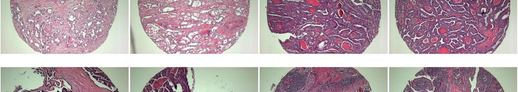
A201(VI)- Various cancer tissues
|
|
|
- Darleen Hoover
- 6 years ago
- Views:
Transcription
 Total spots: 88 Corresponding normal tissues with cancers: Diameter: 1.")
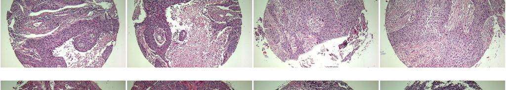
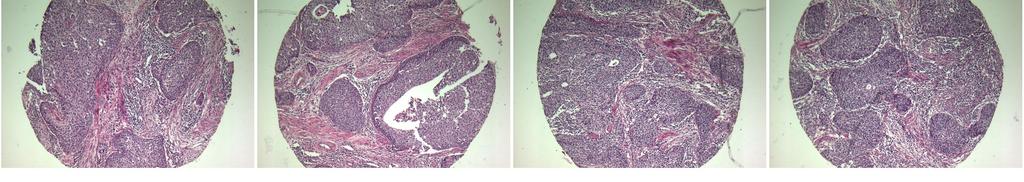
1 A201(VI)- Various cancer tissues (formalin fixed) For research use only Specifications: No. of cases: 44 Tissue type: Various cancer tissues No. of spots: 2 spots from each cancer case (88 spots) Total spots: 88 Corresponding normal tissues with cancers: Diameter: 1. 0 mm Documents: Product specification: layout, summary of tissue spots H&E stained images Detailed pathological information Layout: P T
 For research use only")
2 A201(VI)- Various cancer tissues Summary of tissue spots (formalin fixed) For research use only P T
 For research use only")
3 A201(VI)- Various cancer tissues Summary of tissue spots (formalin fixed) For research use only P T


4 A201(VI)- Various cancer tissues (formalin fixed) For research use only QC sheet_lot# Hematoxylin and Eosin staining A1 A2 A3 A4 A5 A6 A7 A8 A9 A10 A11 A12 B1 B2 B3 B4 B5 B6 B7 B8 B9 B10 B11 B12 P T


5 A201(VI)- Various cancer tissues (formalin fixed) For research use only QC sheet_lot# Hematoxylin and Eosin staining C1 C2 C3 C4 C5 C6 C7 C8 C9 C10 C11 C12 D1 D2 D3 D4 D5 D6 D7 D8 D9 D10 D11 D12 P T


6 A201(VI)- Various cancer tissues (formalin fixed) For research use only QC sheet_lot# Hematoxylin and Eosin staining E1 E2 E3 E4 E5 E6 E7 E8 E9 E10 E11 E12 F1 F2 F3 F4 F5 F6 F7 F8 F9 F10 F11 F12 P T


7 A201(VI)- Various cancer tissues (formalin fixed) For research use only QC sheet_lot# Hematoxylin and Eosin staining G1 G2 G3 G4 G5 G6 G7 G8 G9 G10 G11 G12 H1 H2 H3 H4 P T
8 1 1,2 f 40 brain: anaplastic astrocytoma Cerebral mass, left thalamus, Removal of tumor(fs): Glioblastoma. 2 3,4 m 8 brain: choroid plexus papillomabrain, intraventricular,left,removal: choroid plexus papilloma 3 5,6 m 64 A 4 7,8 f ,10 m ,12 m 37 esophagus : squamous cell esophagus :squamous cell larynx : squamous cell thymus:atypical carcinoid Esophagus and cardia of stomach, esophagectomy and node dissection: 1.Ulcerative squamous cell, poorly differentiated, 1) size: 3.3x1cm 2) extension to the outer muscle layer 3) lymphovascular permeation: Positive 2.Resection margins, proximal and distal: Free of tumor. 3.Regional lymph nodes: Free of tumor in all nodes(0/24), in detail, subcarinal(0/10), hilar(0/2), pretracheal(0/6),lower paratracheal(0/3), left paracardial(0/2), 8L(0/1),omental mass(free of tumor). Esophagus, resection: 1.Invasive squamous cell, moderately differentiated 1) size: 6x2.5cm 2) infiltrative growth 3) extension through the muscle layer to the adventitia but intact lateral resection margin 4) perineural and vascular invasion 5) resection margins, proximal and distal(stomach): Free of tumor. 6) regional lymph nodes:free of tumor in all 24 lymph(0/24),indetail,ipl(0/1),subcarinal(0/1),paraesophageal (0/5), pyloric(0/3), left hilar(0/5), celiac(0/1),right recurrent laryngeal(0/3), greater curvature(0/2), left ardial(0/3). larynx: squamous cell Thymus thymectomy: 1.Atypical carcinoid showing vascular invasion,penetrating the capsule 1) invassion into the thymic fat tissue and multifocal necrosis. *Note: immunohistochemical staining results ACTH, Synaptophysin,Chromogranin NSE:positive
9 7 1,2 m ,4 f ,6 m ,8 f 53 thyroid gland:papillary cacinoma thyroid gland:papillary cacinoma thyroid gland:papillary cacinoma thyroid gland : papillary Thyroid gland, total thyroidectomy with neck node dissection: 1.Papillary 1) involving bilateral whole lobe(size: 6.5x3.7cm) 2) extension to perithyroidal fat tissue 2. Regional lymph node: Tumor metastasis to 4 out of 20(4/20), in detail, level II(1/2), level III(3/13), level IV(0/5). Thyroid, total thyroidectomy:papillary 1) size: 4.5x3x2.5cm 2) extensive extension to perithyroidal soft tissue and surrounding skeletal muscle. 3) lymphovascular invasion. 4) multiple metastasis to surrounding soft tissue and lymph nodes (29/62: level 2,3,4,5(12/35), level 2(2/10),No.6(1/1), perithyroidal(14/16)). 5) intact jugular vein. 6) nodular goiter Thyroid gland, right, thyroidectomy: Papillary with extension beyond thyroid capsule. Thyroid gland, site unstated, thyroidectomy: Papillary extending beyond extrathyroidal tissue, in the background of Hashimoto's thyroiditis. B 11 9,10 f 57 breast: infiltrating ductal Breast, left, palliative mastectomy: 1.Infiltrating ductal 1) Histologic grade: II. 2) Black's nuclear grade: I 3) Extensive lymphatic invasion. 4) Intraductal component: less than 10%. 5) Multinodularity. 2.Nipple and areola: Free of tumor. 3.Overlying skin: Superficial dermal invasion by the tumor. 4.nderlying fascia: Presence of tumor, focal. 5.Lymph nodes, level I(9/23) and level II(2/2): Metastasis in 11 out of 25 lymph nodes (pn1biv). 6. Result of immunohistochemical staining: ER(+), PR(-), C erb B2(-).
10 12 11,12 f 47 breast: infiltrating ductal Breast, left, modified radical mastectomy: 1.Infiltrating ductal 1) size: 8x7x2cm 2) Modified Bloom-Richardson's histological grade: II (moderately differentiated) (tubule formation: III, nuclear pleomorphism: II, mitosis: II) 3) Black's nuclear grade: 2(moderately differentiated) 4) extensive lymphovascular invasion 5) extension to the anterior(fibroadipose) margin. 2.Nipple and areola: Focal direct invasion to the subdermal layer. 3.Overlying skin: Free of tumor. 4.Underlying fascia: Tumor's involvement. 5.Regional lymph nodes: Metastatic in 3 out of 8 lymph nodes (3/8), in detail, level II(0/1), level III(0/1), axillary lymph nodes(3/6). 6.Result of immunohistochemical staining: ER(+), PR(-), C erb B2(+). 13 1,2 f 72 stomach : adeno Stomach, pancreas and spleen, radical total gastrectomy, splenectomy and pancreatectomy: 1.Stomach: Ulceroinfiltrative adeno, moderately differentiated, 1) size: 10.7x9cm 2) extension to the serosa and pancreas parenchyme 3) lymphovascular permeation: positive 4) Intestinal type by Lauren's classification 2. Resection margins, proximal and distal: Free of tumor. Resection margin, pancreas: Free of tumor. 3. Regional lymph nodes: Tumor metastasis to 2 out of 47(2/47), in detail, N1(0/5), N3(0/5), N4(0/8), N5(0/2), N6(1/16), N7(0/2), N8(1/4), N9(0/2), N10(0/3). 4. Pancreas: Tumor extension. 5. Spleen: Congestion.
11 14 3,4 f 73 stomach : adeno Stomach with omentums, total gastrectomy: 1.Polypoid and infiltrative adeno, well to poorly differentiated, in antrum and body 1) size: 12.5x10.5cm 2) mixed type (by Lauren classification) and infiltrative growth type (by Ming classification) 3) extension to outer proper muscle layer 4) frequent vascular and perineural invasions 5) metastasis to 23 out of 47 omental lymph nodes (No.3(11/16), No.4(2/6), No.5(2/2), No.7(2/7), No.8(0/1) and No.9(0/2)) 6) non-neoplastic mucosa showing multifocal atrophic gastritis with intestinal metaplasia. 15 5,6 f 56 soft tissue: fibromatosis Soft tissue, buttock, left, excision: Fibromatosis, extraabdominal. 16 C 7,8 f 73 soft tissue:ganlioneuroma Soft tissue, mediastinum, excision: Ganglioneuroma. 17 9,10 m 71 tongue: squamous cell Tongue, tonsil, salivary gland and regional lymph nodes, left hemiglossectomy and radical neck dissection: Invasive squamous cell, moderately differentiated 1) size: 6x4.3x3.5cm 2) infiltrative growth 3) extension to underlying skeletal muscle 4) intact anterior, posterior, lateral and medial resection margins 5) intact but very close to basal resection margin (safety resection margin: less than 1mm) 6) metastasis to 1 out of 49 regional lymph nodes (right level II(0/10), right level III(0/3), right level IV (0/13), left level II(1/8), left level III(0/0), left level IV(0/7), and left level V(0/8)) Salivary gland, submandibular: Free from tumor extension.
12 18 11,12 m 58 tongue: squamous cell Tongue and lymph node, wide excision and dissection: 1. Tongue: Squamous cell, well differentiated 1) size: 2.7x2.2cm 2) extension to the sublingual gland 3) perineural invasion(+) 2. Resection margins, anterior, posterior, lateral side and basal 1) margin: Free of tumor. 3. Salivary gland, submandibular: Free of tumor. 4. Skeletal muscle: Free of tumor. 5. Regional lymph nodes: Free of tumor in all nodes(0/53), in detail, right level IIb(0/10), right level IIa(0/8), right level III (0/2), right level IV(0/4), right level Ib(0/5), left level Ia (0/4), left level Ib(0/5), left level III(0/6), left level IIa (0/3), left level IIb(0/6). 19 1,2 m 77 lung: non-small cell Lung, right upper lobe, lobectomy: 1.Poorly differentiated, non-small cell type. 1) confined with the lung parenchyme and no visceral pleura involvement. 2) two separate masses (3.9x3.2cm and 2.6x2cm) with same histology. 3) lymphatic permeation: positive. 2.Resection margin, bronchial: Free of tumor. 3.Surrounding lung: Emphysema. 4.Regional lymph nodes: Tumor metastasis to 1 out of 37 (1/37),in detail, left hilar(0/2), subcarinal(0/13), lower paratracheal(0/2), hilar(0/3), peribronchial(0/4), IPL(0/2), regional, attached(1/11). 5.Results of histochemical and immunohistochemical staining: PAS(-), Mucicarmine(-), NSE(-), Synaptophysin(-), Chromogranin(-) Lung, right upper lobe, lobectomy: 1.Poorly differentiated, non-small cell type, probably adeno 1) confined within the lung parenchyme and no visceral pleura involvement 2) two completely separate masses (3.9x3.2cm and 2.6x2cm) with same histology (T4) 3) lymphatic permeation: Positive 2. Resection margin, bronchial: Tumor positive.
13 20 3,4 m 57 lung: adeno D 21 5,6 m 82 lung :squamous cell 22 7,8 f 64 lung :lage cell 23 9,10 f ,12 m 71 lymph node: metastatic papillary lymph node: metastatic Lung, LLL, lobectomy: 1. Adeno, acinic and papillary 1) marked mucin production 2) size: 4.7x4.6cm 3) extension to visceral pleura 2. Bronchial resection margin: Free of tumor. 3. Regional lymph node, peribronchial: Tumor metastasis (1/1). Lung, LLL, lobectomy: 1. Squamous cell, poorly differentiated 1) size: 3.5x3cm 2) extension to the visceral pleura 2. Bronchial resection margin: Free of tumor. Lung, right lower lobe, lobectomy: 1) squamous cell, moderately differentiated - size: 7x5cm - extension to the visceral pleura - associated obstructive pneumonitis 2) bronchial resection margin: free of tumor. Lymph nodes, neck, right, internal jugular vein, upper (4/12) and lower(0/8), modified radical neck dissection: Metastatic papillary of the thyroid gland in 4 out of 20 nodes. Lymph nodes and submandibular gland, left, radical neck dissection: 1. Lymph nodes, level I(3/5), level IIa(1/2), level IIb(0/7), level III(0/2), level IV(4/7) and level V(1/4):(9/37): Metastatic poorly differentiated. 2. Submandibular gland: Free of tumor. 3. Oral mass, separately submitted: Involvement of. 4. Immunohistochemical stain results: Cytokeratin: positive, NSE, Synaptophysin, Chromogranin, CD56, leukocyte common antigen: negative.
14 25 1,2 m 76 liver :cholangio 26 3,4 m 70 liver :metatstatic 27 5,6 m 60 liver :hepatocellular Liver, right lobe, lobectomy: 1. Cholangio, moderately differentiated, peripheral type 1) size: 11.5x9.5x7.0cm 2) expanding growth and extensive necrosis 3) penetration of hepatic capsule and invasion to soft tissue adjacent to diaphragm 4) vascular invasion (but not portal vein) 5) intrahepatic metastasis 6) intact intrahepatic resection margin and diaphragm 7) nonneoplastic liver showing reactive hepatitis 2. Gallbladder, cholecystectomy: Free from tumor extension. Liver, right lobe, lobectomy: Metastatic, multiple, originated from large cell undifferentiated of lung(s ) 1) size: 5.7x5.5x4.3cm and 4.0x3.5x3.5cm 2) frequent vascular invasion 3) intact intrahepatic resection margin 4) nonneoplastic liver showing reactive hepatitis Liver, right lobe, lobectomy: Hepatocellular 1) size: 8x8x6.8cm 2) Edmondson-Steiner grade III 3) clear cell(majority) and trabecular histologic type 4) single nodular type with perinodal spreading growth 5) necrosis: less than 10% of total volume 6) microvascular invasion 7) intact intrahepatic resection margin 8) nonneoplastic liver showing: Chronic hepatitis, B viral, with mild inflammatory activity (Grade II) and early cirrhosis(stage III).
15 E 28 7,8 f 66 liver :hepatocellular Liver, right lobe, lobectomy: 1. Hepatocellular 1) size: 10.5x7.4x5cm 2) mixed histologic patterns including trabecular, adenoid and clear cel types 3) Edmondson grade II+III 4) single nodular type with perinodal spread 5) expanding but focal infiltrating growth pattern 6) microvascular invasion 7) no significant necrosis(less than 20% of total volume) 8) resection margin: Free of tumor. 2. Gallbladder, cholecystectomy: Autolytic changes. <Additional report > nonneoplastic liver: Chronic hepatitis, B-viral, with minimal inflammatory activity(grade I) and septal fibrosis(stage III). 29 9,10 m 53 kidney :renal cell Kidney, left, nephrectomy: 1. Renal cell, clear cell type 1) size: 4x4cm 2) confined within the renal capsule 3) nuclear grade II 2. Renal artery and vein: Free of tumor. 3. Ureter resection margin: Free of tumor. 4. Adrenal gland: Free of tumor.
16 30 11,12 f ,2 f ,4 f 54 kidney :renal cell ovary :malignant germ cell tumor ovary :serous cystadeno 33 5,6 f 44 ovary :clear cell Adrenal gland and kidney, left, radical adrenalectomy and nephrectomy: 1. Adrenal gland: Adrenal cortical, 1) size: 12x10cm 2) extension to the capsule and adjacent pancreas 2. Kidney: Free of tumor. 3. Ureter: Free of tumor. 4. Separate mass: Adrenal cortical, 5. Regional lymph node: Tumor metastasis(5/6), in detail, retroperitoneal(4/4), splenic(1/2) 6. Retroperitoneal resection margin: Free of tumor. 7.Spleen, splenectomy: Free of tumor. Ovary and fallopian tube, right, right salpingooophorectomy: 1.Ovary: Malignant germ cell tumor, probably yolk sac tumor (endodermal sinus tumor). 2. Fallopian tube: Paraovarian cyst. 3. Result of special stain: AFP(+), HCG(-), PAS(+), D-PAS(-). Uterus with right adnexa, total abdominal hysterectomy with right salpingooophorectomy: 1. Cervix: Chronic nonspecific inflammation. 2. Endometrium: Secretory phase. 3. Myometrium : 1) Leiomyomas, intramural. 2) Adenomyosis. 3) Tumor involvement at the outer wall of myometrium. 4.Ovary, right: Serous cystadeno. 5.Fallopian tube: Tumor involvement at the serosa wall. 6.Lymph nodes: Tumor metastasis to 4 out of 8(4/8), in detail, right pelvic(1/4), left pelvic(3/4). 7.Mass of right infundibulopelvic ligament: Tumor involvement. 8.Omentum: Free of tumor. Ovary, right, right salpingoophorectomy: Clear cell adeno, well differentiated, with extraovarian spread. Fallopian tube: Free of tumor.
17 34 7,8 f 33 ovary : mucinous cystadeno Ovary and fallopian tube, left, salpingooophorectomy: Ovary: Mucinous cystadeno confined within the capsule. Fallopian tube: Free of tumor. Ovary, right, partial oophorectomy: Mesothelial hyperplasia without tumor. Soft tissue, right and left, paracolic gutter, right retroperitoneum: Chronic nonspecific inflammation without tumor. Omentum, partial omentectomy: Chroni nonspecific inflammation with mesothelial hyperplasia. F 35 9,10 f 68 uterine cervix:squamous cell Uterus with both adnexae, radical abdominal hysterectomy with bilateral salpingooophorectomy, BPLND: 1) cervix: Invasive squamous cell, moderately differentiated - large cell, keratinizing - size: 3.3x3cm - extension to the nearly whole depth(invasion depth: 1.7cm) - lateral margin: Free of tumor - lymphovascular permeation: positive 2) endometrium: atrophy 3) myometrium : leiomyoma, subserosal 4) ovaries, bilateral: no pathological diagnosis 5) fallopian tubes, bilateral: no pathological diagnosis 6) parametrium, bilateral: free of tumor 7) vaginal cuff: free of tumor 8) regional lymph nodes: free of tumor in all nodes(0/16), in detail, right pelvic node(0/9), left pelvic node(0/5), common iliac node(0/2).
18 36 11,12 f 58 uterine cervix: invasive squamous cell Uterus with both adnexae, radical abdominal hysterectomy with bilateral salpingooophorectomy: 1.Cervix: Invasive squamous cell, large cell partly keratinizing 1) size: 3.2x3.8cm 2) entire cervix and lower endometrium involvement 3) extension through the whole depth (2.2cm in depth), but lateral margin, free of tumor. 4) ymphovascular permeation, suspicious 2.Endometrium: Atrophy. 3.Myometrium: Leiomyoma, intramural. 4.Ovaries, bilateral: No pathological diagnosis. 5.Fallopian tubes, bilateral: No pathological diagnosis. 6.Parametrium, bilateral: Free of tumor. 7.Vaginal cuff: Free of tumor. 8. Regional lymph nodes: Free of tumor in all nodes (0/20), in detail, right pelvic node(0/10), left pelvic node(0/10). 37 1,2 m 32 testis:seminoma 38 3,4 m 38 tests:seminoma Testis, pelvic, orchiectomy: Seminoma, classic, with multifocal necrosis and extensive vascular invasion. Spermatic cord: No pathological Testis, left, orchiectomy: Seminoma with invasion beyond tunica albuginea (pt2) and associated with intratubular germ cell neoplasia. Spermatic cord: Free of tumor.
19 39 5,6 m 74 colon :adeno Colon, sigmoid, radical sigmoid colectomy: 1. Ulcero-fungating adeno, poorly differentiated 1) size: 4.2cm in length 2) extension to the pericolic fat tissue (pt3c) 3) lateral margin: Free of tumor. 4) lymphovascular permeation: present 2. Resection margins, proximal and distal: Free of tumor. 3. Regional lymph nodes: Tumor metastasis to 2 out of 17(2/17), in detail, principal(0/8), regional(2/9). MSI(Microsatellite instability) test Marker Name bat26 D5S346 bat25 D17S250 D2S123 판독 Microsatellite No No No No No MSS instability Allelic uninformative uninformative No(1.15) uninformative Imbalance 40 7,8 m 47 G colon :adeno Colon, rectum, lower anterior resection: 1. Colon: Ulcerofungating adeno, moderately differentiated, 1) size: 5.5x4.5cm 2) extension to the pericolic fat tissue(pt3c) 3) lateral margin: Free of tumor 4) lymphovascular permeation: Positive 2. Urinary bladder: Free of tumor. 3. Resection margins, proximal and distal: Free of tumor. 4. Regional lymph nodes: Tumor metastasis to 4 out of 11(4/11), in detail, regional(4/8), principal(0/3).
20 41 9,10 m 46 rectum :adeno Rectum, low anterior resection: Ulcerofungating adeno, moderately differentiated 1) size: 6.2x5.2cm 2) infiltrative growth 3) extension to perirectal soft tissue but intact lateral resection margin 4) no vascular and perineural invasion 5) intact proximal and distal resection margins 6) metastasis to 5 out of 29 perirectal lymph nodes (principal(0/9) and perirectal(5/20)) MSI(Microsatellite instability) test Marker Name bat26 D5S346 bat25 D17S250 D2S123 판독 Microsatellite No No No No No MSS instability 42 11,12 f 79 rectum :adeno Allelic uninformative uninformative No(0.79) No(1.28) Imbalance Rectum, low anterior resection: 1. Ulcerative adeno, moderately differentiated, 1) size: 3.5x3.7cm 2) extension to the outer muscle layer(pt2) 3) lymphovascular permeation, positive 4) lateral margin: Free of tumor 2. Resection margins, proximal and distal: Free of tumor. 3. Separate mass at distal resection margin: Adenoma with low grade dysplasia. 4. Regional lymph nodes: Tumor metastasis to 1 out of 11(1/11), in detail, principal(0/2), regional(1/9). ** MSI(Microsatellite instability) test Marker Name bat26 D5S346 bat25 D17S250 D2S123 판독 Microsatellite No No No No No MSS instability Allelic uninformative No(0.86) uninformative No(0.97) No(0.88) Imbalance
 extension to the subcutaneous fat tissue (invasion depth: about 3cm).")
21 Skin, lower back, wide excision: 43 1,2 m 55 skin :dermatofibrosarcoma Dermatofibrosarcoma protuberans. Resection margin: Free of tumor. H 44 3,4 m 40 skin : squamous cell Skin, scalp, excision: Invasive squamous cell, well-differentiated with 1) extension to the subcutaneous fat tissue (invasion depth: about 3cm). Resection margins, all circumference and base: Free of tumor. Lymph nodes, level 2a(0/36), level 2b(0/11), level III(0/25), level IV(0/13) and level V(0/11):(0/96): Free of tumor.
A301 VI- Various cancer tissues with corresponding normal tissues
 A301 VI- Various cancer tissues with (formalin fixed) For research use only Specifications: No. of cases: 28 Tissue type: Various cancer tissues with No. of spots: 2 spots from each cancer case (56 spots)
A103(9)- Normal tissues, more than single spots
- Normal tissues, more than single spots") A103(9)- Normal tissues, more than single spots (formalin fixed) For research use only Specifications: No. of cases: 45 Tissue type: Normal tissues, more than single spots No. of spots: 2 spots from each
A103(9)- Normal tissues, more than single spots (formalin fixed) For research use only Specifications: No. of cases: 45 Tissue type: Normal tissues, more than single spots No. of spots: 2 spots from each
(formalin fixed) 6 non-neoplastic spots (6 spots) Corresponding normal tissues with cancers: Yes Diameter: 1. 0 mm
 6 non-neoplastic spots (6 spots) Corresponding normal tissues with cancers: Yes Diameter: 1. 0 mm") CBA729-Test slide, Head and neck cancer tissues (formalin fixed) For research use only Specifications: No. of cases: 6 Tissue type: Test slide, Head and neck cancer tissues No. of spots: 6 spots from each
CBA729-Test slide, Head and neck cancer tissues (formalin fixed) For research use only Specifications: No. of cases: 6 Tissue type: Test slide, Head and neck cancer tissues No. of spots: 6 spots from each
A218 : Esophagus cancer tissues. (formalin fixed)
") (formalin fixed) For research use only Specifications: No. of cases: 40 Tissue type: Esophagus cancer tissues No. of spots: 2 spots from each cancer case (80 spots) 4 non-neoplastic spots (4 spots) Total
(formalin fixed) For research use only Specifications: No. of cases: 40 Tissue type: Esophagus cancer tissues No. of spots: 2 spots from each cancer case (80 spots) 4 non-neoplastic spots (4 spots) Total
A916: rectum: adenocarcinoma
 General facts of colorectal cancer The colon has cecum, ascending, transverse, descending and sigmoid colon sections. Cancer can start in any of the r sections or in the rectum. The wall of each of these
A712(19)- Test slide, Breast cancer tissues with corresponding normal tissues
- Test slide, Breast cancer tissues with corresponding normal tissues") A712(19)- Test slide, Breast cancer tissues with corresponding normal tissues (formalin fixed) For research use only Specifications: No. of cases: 12 Tissue type: Breast cancer tissues with corresponding
A712(19)- Test slide, Breast cancer tissues with corresponding normal tissues (formalin fixed) For research use only Specifications: No. of cases: 12 Tissue type: Breast cancer tissues with corresponding
A712(18)- Test slide, Breast cancer tissues with corresponding normal tissues
- Test slide, Breast cancer tissues with corresponding normal tissues") A712(18)- Test slide, Breast cancer tissues with corresponding normal tissues (formalin fixed) For research use only Specifications: No. of cases: 12 Tissue type: Breast cancer tissues with corresponding
A712(18)- Test slide, Breast cancer tissues with corresponding normal tissues (formalin fixed) For research use only Specifications: No. of cases: 12 Tissue type: Breast cancer tissues with corresponding
A215- Urinary bladder cancer tissues
 A215- Urinary bladder cancer tissues (formalin fixed) For research use only Specifications: No. of cases: 45 Tissue type: Urinary bladder cancer tissues No. of spots: 2 spots from each cancer case (90
A220: Larynx cancer tissues. (formalin fixed)
") A220: Larynx cancer tissues (formalin fixed) For research use only Specifications: No. of cases: 45 Tissue type: Larynx cancer tissues No. of spots: 2 spots from each cancer case (90 spots) 4 non-neoplastic
A220: Larynx cancer tissues (formalin fixed) For research use only Specifications: No. of cases: 45 Tissue type: Larynx cancer tissues No. of spots: 2 spots from each cancer case (90 spots) 4 non-neoplastic
Gastric Cancer Histopathology Reporting Proforma
 Gastric Cancer Histopathology Reporting Proforma Mandatory questions (i.e. protocol standards) are in bold (e.g. S1.01). S1.01 Identification Family name Given name(s) Date of birth Sex Male Female Intersex/indeterminate
Definition of Synoptic Reporting
 Definition of Synoptic Reporting The CAP has developed this list of specific features that define synoptic reporting formatting: 1. All required cancer data from an applicable cancer protocol that are
Bladder Case 1 SURGICAL PATHOLOGY REPORT. Procedure: Cystoscopy, transurethral resection of bladder tumor (TURBT)
") Bladder Case 1 February 17, 2007 Specimen (s) received: Bladder Tumor Pre-operative Diagnosis: Bladder Cancer Post operative Diagnosis: Bladder Cancer Procedure: Cystoscopy, transurethral resection of
Bladder Case 1 February 17, 2007 Specimen (s) received: Bladder Tumor Pre-operative Diagnosis: Bladder Cancer Post operative Diagnosis: Bladder Cancer Procedure: Cystoscopy, transurethral resection of
Take Home Quiz 1 Please complete the quiz below prior to the session. Use the Multiple Primary and Histology Rules
 Take Home Quiz 1 Please complete the quiz below prior to the session. Use the Multiple Primary and Histology Rules Case 1 72 year old white female presents with a nodular thyroid. This was biopsied in
Quiz. b. 4 High grade c. 9 Unknown
 Quiz 1. 10/11/12 CT scan abdomen/pelvis: Metastatic liver disease with probable primary colon malignancy. 10/17/12 Colonoscopy with polypectomy: Adenocarcinoma of sigmoid colon measuring at least 6 mm
Case Scenario 1. Pathology report Specimen from mediastinoscopy Final Diagnosis : Metastatic small cell carcinoma with residual lymphatic tissue
 Case Scenario 1 Oncology Consult: Patient is a 51-year-old male with history of T4N3 squamous cell carcinoma of tonsil status post concurrent chemoradiation finished in October two years ago. He was hospitalized
MT09 - Normal Human Tissue Microarray, FDA
 Reveal Biosciences offers Histochemical Staining, Immunohistochemistry (IHC), In Situ Hybridization (ISH), Whole Slide Imaging, and Quantitative Image Analysis on any TMA MT09 - Normal Human Tissue Microarray,
SHN-1 Human Digestive Panel Test results
 SHN-1 Human Digestive Panel Test results HN-30 tongue HN-24 salivary gland HN-12 larynx HN-28 esophagus HN-29 stomach HN-20 pancreas HN-13 liver HN-14 gall bladder HN-27-1 duodenum HN-27-2 ileum HN-27-3
MALIGNANT NEOPLASMS OF THE BREAST MALIGNANT NEOPLASMS OF FEMALE GENITAL ORGANS
 MALIGNANT NEOPLASMS OF THE (INC. PAGET S DISEASE) 0 - Nipple and areola 1 - Central portion 2 - Upper-inner quadrant 3 - Lower-inner quadrant 4 - Upper-outer quadrant 5 - Lower-outer quadrant 6 - Axillary
NAACCR Webinar Series 1 Q&A. Fabulous Prizes. Collecting Cancer Data: Ovary 11/3/2011. Collecting Cancer Data: Ovary
 NAACCR 2011 2012 Webinar Series Collecting Cancer Data: Ovary Q&A Please submit all questions concerning webinar content through the Q&A panel. Reminder: If you have participants watching this webinar
Kidney Case 1 SURGICAL PATHOLOGY REPORT
 Kidney Case 1 Surgical Pathology Report February 9, 2007 Clinical History: This 45 year old woman was found to have a left renal mass. CT urography with reconstruction revealed a 2 cm medial mass which
Checklist; Anus: Excisional Biopsy Anus: Excisional Biopsy 1/1/ Checklist; Anus: Resection Anus: Resection 1/1/2005
 ChecklistTemplateVersions ChecklistTemplateVersion Ckey OfficialName VisibleText RevisionDate Checklist; Adrenal gland: 16.1000043 Resection Adrenal gland: Checklist; Ampulla of 17.1000043 Vater: Ampullectomy
Peritoneum: Def. : It is a thin serous membrane that lines the walls of the abdominal and pelvic cavities and clothes the viscera.
 Peritoneum: Def. : It is a thin serous membrane that lines the walls of the abdominal and pelvic cavities and clothes the viscera. Layers of the peritoneum: 1. Outer Layer ( Parietal Peritoneum) : lines
Index. Note: Page numbers of article titles are in boldface type.
 Note: Page numbers of article titles are in boldface type. A Adenocarcinoma, pancreatic ductal, laparoscopic distal pancreatectomy for, 61 Adrenal cortical carcinoma, laparoscopic adrenalectomy for, 114
Staging and Treatment Update for Gynecologic Malignancies
 Staging and Treatment Update for Gynecologic Malignancies Bunja Rungruang, MD Medical College of Georgia No disclosures 4 th most common new cases of cancer in women 5 th and 6 th leading cancer deaths
UICC TNM 8 th Edition Errata
 UICC TNM 8 th Edition Errata ions are in italics Head and Neck Tumours Pages 20, p27, p34, p38, p41, and p49 ly pn2a Metastasis in a single ipsilateral lymph node, less than 3cm in greatest dimension with
CAP Cancer Protocol and ecc Summary of Changes for August 2014 Thyroid Agile Release
 CAP Cancer Protocol and ecc Summary of Changes for August 2014 Thyroid Agile Release 2 REVISION HISTORY Date Author / Editor Comments 5/19/2014 Jaleh Mirza Created the document 8/12/2014 Samantha Spencer/Jaleh
Histology Coding ANSWERS
 Histology Coding ANSWERS 1.) Biopsy of a right thyroid nodule reveals papillary carcinoma. What is the ICD-O-3 code? a. 8050/3 - Papillary carcinoma b. 8260/3 - Papillary adenocarcinoma Rationale/comment:
Abstracting Upper GI Cancer Incidence and Treatment Data Quiz 1 Multiple Primary and Histologies Case 1 Final Pathology:
 Abstracting Upper GI Cancer Incidence and Treatment Data Quiz 1 Multiple Primary and Histologies Case 1 A 74 year old male with a history of GERD presents complaining of dysphagia. An esophagogastroduodenoscopy
UICC TNM 8 th Edition Errata
 UICC TNM 8 th Edition Errata ions are in italics Page 28 Oropharynx p16 positive Pathological Stage II,T2 N2 M0 T3 N0,N1 M0 Stage II,T2 N2 M0 T3,T4 N0,N1 M0 Page 61 Oesophagus Adenocarcinoma Pathological
Carcinoma of the Urinary Bladder Histopathology
 Carcinoma of the Urinary Bladder Histopathology Reporting Proforma (Radical & Partial Cystectomy, Cystoprostatectomy) Includes the International Collaboration on Cancer reporting dataset denoted by * Family
In the name ofgod. Abdomen 3. Dr. Zahiri
 In the name ofgod Abdomen 3 Dr. Zahiri Peritoneum Peritoneum It is the serous membrane(a type of loose connective tissue and is covered by mesothelium) that lines the abdominal cavity. Extensions of the
Case Scenario 1: Thyroid
 Case Scenario 1: Thyroid History and Physical Patient is an otherwise healthy 80 year old female with the complaint of a neck mass first noticed two weeks ago. The mass has increased in size and is palpable.
Radiology Pathology Conference
 Radiology Pathology Conference Nadia F. Yusaf, M.D. PGY-3 1/29/2010 Presentation material is for education purposes only. All rights reserved. 2010 URMC Radiology Page 1 of 90 Case 1 60 year- old man presents
8. The polyp in the illustration can be described as (circle all that apply) a. Exophytic b. Pedunculated c. Sessile d. Frank
 a. Exophytic b. Pedunculated c. Sessile d. Frank") Quiz 1 Overview 1. Beginning with the cecum, which is the correct sequence of colon subsites? a. Cecum, ascending, splenic flexure, transverse, hepatic flexure, descending, sigmoid. b. Cecum, ascending,
Quiz 1 Overview 1. Beginning with the cecum, which is the correct sequence of colon subsites? a. Cecum, ascending, splenic flexure, transverse, hepatic flexure, descending, sigmoid. b. Cecum, ascending,
UTERINE SARCOMA EXAMPLE OF A UTERINE SARCOMA USING PROPOSED TEMPLATE
 UTERINE SARCOMA EXAMPLE OF A UTERINE SARCOMA USING PROPOSED TEMPLATE Case: Adenosarcoma with heterologous elements and stromal overgrowth o TAH, BSO, omentectomy, staging biopsies of cul-de-sac, bladder
Cervical Cancer 3/25/2019. Abnormal vaginal bleeding
 Cervical Cancer Abnormal vaginal bleeding Postcoital, intermenstrual or postmenopausal Vaginal discharge Pelvic pain or pressure Asymptomatic In most patients who are not sexually active due to symptoms
82330 CALCIUM; IONIZED. ICD-10 Codes that Support Medical Necessity. ICD-10 Code. Description. A15.0 Tuberculosis of lung
 82330 CALCIUM; IONIZED ICD-10 Codes that Support Medical Necessity ICD-10 Code Description A15.0 Tuberculosis of lung A15.4 Tuberculosis of intrathoracic lymph nodes A15.5 Tuberculosis of larynx, trachea
Thyroid INTRODUCTION ANATOMY SUMMARY OF CHANGES
 AJC 7/14/06 1:19 PM Page 67 Thyroid C73.9 Thyroid gland SUMMARY OF CHANGES Tumor staging (T) has been revised and the categories redefined. T4 is now divided into T4a and T4b. Nodal staging (N) has been
Crosswalk File of ICD9 Diagnosis Codes to Risk Group Assignment 1-Apr-15
 1 1500 MALIGNANT NEOPLASM OF CERVICAL ESOPHAGUS 1 1501 MALIGNANT NEOPLASM OF THORACIC ESOPHAGUS 1 1502 MALIGNANT NEOPLASM OF ABDOMINAL ESOPHAGUS 1 1503 MALIGNANT NEOPLASM OF UPPER THIRD OF ESOPHAGUS 1
WLH Tumor Frequencies between cohort enrollment and 31-Dec Below the Women Lifestyle and Health tumor frequencies are tabulated according to:
 DESCRIPTION Below the Women Lifestyle and Health tumor frequencies are tabulated according to: Benign =171 (Cervix uteri) treated as not recorded =191 (non-melanoma skin cancer) treated as not recorded
Please complete prior to the webinar. HOSPITAL REGISTRY WEBINAR FEMALE REPRODUCTIVE SYSTEM EXERCISES CASE 1: FEMALE REPRODUCTIVE
 Please complete prior to the webinar. HOSPITAL REGISTRY WEBINAR FEMALE REPRODUCTIVE SYSTEM EXERCISES PHYSICAL EXAMINATION CASE 1: FEMALE REPRODUCTIVE 3/5 Patient presents through the emergency room with
155.2 Malignant neoplasm of liver not specified as primary or secondary. C22.9 Malignant neoplasm of liver, not specified as primary or secondary
 ICD-9 TO ICD-10 Reference ICD-9 150.9 Malignant neoplasm of esophagus unspecified site C15.9 Malignant neoplasm of esophagus, unspecified 151.9 Malignant neoplasm of stomach unspecified site C16.9 Malignant
Radiology Pathology Conference
 Radiology Pathology Conference Sharlin Johnykutty,, MD, Cytopathology Fellow Sara Majewski, MD, Radiology Resident Friday, August 28, 2009 Presentation material is for education purposes only. All rights
Case Scenario 1. 1/2/13 History: 64-year-old white female presented with right leg swelling and redness, abdominal pain.
 Case Scenario 1 1/2/13 History: 64-year-old white female presented with right leg swelling and redness, abdominal pain. 1/02/13 CT Abdomen/Pelvis: Abnormal area of nodular mesenteric and left anterior
Comprehensive Cancer Cover
 Comprehensive Cancer Cover Tech Spec Comprehensive Cancer Cover provides the life insured with cover for the diagnosis and treatment of defined malignant tumours. These tumours must be characterised either
WLH Tumor Frequencies between cohort enrollment and 31-Dec Below the Women Lifestyle and Health tumor frequencies are tabulated according to:
 WLH Tumor Frequencies between cohort enrollment and 31-Dec 2012 DESCRIPTION Below the Women Lifestyle and Health tumor frequencies are tabulated according to: Benign =171 (Cervix uteri) treated as not
New Cancer Cases By Site Breast 28% Lung 14% Colo-Rectal 10% Uterus 6% Thyroid 5% Lymphoma 4% Ovary 3%
 Uterine Malignancy New Cancer Cases By Site 2010 Breast 28% Lung 14% Colo-Rectal 10% Uterus 6% Thyroid 5% Lymphoma 4% Ovary 3% Cancer Deaths By Site 2010 Lung 26% Breast 15% Colo-Rectal 9% Pancreas 7%
Group B: Organ systems (digestive, respiratory, urinary, genital system, heart, glands and skin) green
 green") Group B: Organ systems (digestive, respiratory, urinary, genital system, heart, glands and skin) green Digestive system 1. Teeth Main points: external and internal structure of a tooth, fixation of a tooth
Group B: Organ systems (digestive, respiratory, urinary, genital system, heart, glands and skin) green Digestive system 1. Teeth Main points: external and internal structure of a tooth, fixation of a tooth
Quiz The main functions of the ovaries are a. To produce oocytes b. To produce estrogen c. To produce progesterone d.
 1. The main functions of the ovaries are a. To produce oocytes b. To produce estrogen c. To produce progesterone d. All of the above Quiz 1 2. Which part of the broad ligament suspends the ovaries? a.
Summary Stage 2018 (SS2018)
") Summary Stage 2018 (SS2018) NAACCR October Webinar October 24, 2018 General Information 2 Summary Stage 2018 1 General Summary Stage is ANATOMICALLY based Unlike AJCC, it does not use the following in
Summary Stage 2018 (SS2018) NAACCR October Webinar October 24, 2018 General Information 2 Summary Stage 2018 1 General Summary Stage is ANATOMICALLY based Unlike AJCC, it does not use the following in
Presentation material is for education purposes only. All rights reserved URMC Radiology Page 1 of 98
 Presentation material is for education purposes only. All rights reserved. 2011 URMC Radiology Page 1 of 98 Radiology / Pathology Conference February 2011 Brooke Koltz, Cytopathology Resident Presentation
UICC 8 th Edition Errata 25 th of May 2018
 UICC 8 th Edition Errata 25 th of May 2018 ions are in italics Head and Neck Tumours Page 19 Oral Cavity T2 T3 T4a Tumour 2 cm or less in greatest dimension and more than 5 mm but no more than 10 mm depth
Cancer Association of South Africa (CANSA)
") Cancer Association of South Africa (CANSA) Fact Sheet on ICD-10 Coding of Neoplasms Introduction The International Statistical Classification of Diseases and Related Health Problems, 10 th Revision (ICD-10)
Cancer Association of South Africa (CANSA) Fact Sheet on ICD-10 Coding of Neoplasms Introduction The International Statistical Classification of Diseases and Related Health Problems, 10 th Revision (ICD-10)
B REAST STAGING FORM. PATHOLOGIC Extent of disease through completion of definitive surgery. CLINICAL Extent of disease before any treatment
 B REAST STAGING FORM CLINICAL Extent of disease before any treatment y clinical staging completed after neoadjuvant therapy but before subsequent surgery (DCIS) (LCIS) (Paget s) mi c a b c d TUMOR SIZE:
Endometrial Stromal Sarcoma
 May 26, 2011 By Sushila Ladumor, MD [1] Endometrial stromal sarcoma (ESS) is a rare malignant tumor of the endometrium, occurring in the age group of 40-50 years. History The 50-year-old, female patient
B REAST STAGING FORM. PATHOLOGIC Extent of disease through completion of definitive surgery. CLINICAL Extent of disease before any treatment
 B REAST STAGING FORM Extent of disease before any treatment y clinical staging completed after neoadjuvant therapy but before subsequent surgery (DCIS) (LCIS) (Paget s) mi a b c a b c d TUMOR SIZE: S TAGE
Neoplasia literally means "new growth.
 NEOPLASIA Neoplasia literally means "new growth. A neoplasm, defined as "an abnormal mass of tissue the growth of which exceeds and is uncoordinated with that of the normal tissues and persists in the
Is Hepatic Resection Needed in the Patients with Peritoneal Side T2 Gallbladder Cancer?
 Is Hepatic Resection Needed in the Patients with Peritoneal Side T2 Gallbladder Cancer? Lee H, Park JY, Youn S, Kwon W, Heo JS, Choi SH, Choi DW Department of Surgery, Samsung Medical Center Sungkyunkwan
DUSTURBANCES OF GROWTH. MLS Basic histological diagnosis MLS HIST 422 Semester 8- batch 7 L8 Uz: Musa
 DUSTURBANCES OF GROWTH MLS Basic histological diagnosis MLS HIST 422 Semester 8- batch 7 L8 Uz: Musa Agnesia: means complete absence of an organ (Kidney). Aplasia: s defined in general as "defective development
*OPERATIVE PROCEDURE. Serum tumour markers within normal limits S1.04 PRINCIPAL CLINICIAN
 Neoplasia of the Testis - Orchidectomy Histopathology Reporting Proforma Includes the International Collaboration on Cancer reporting dataset denoted by * Family name Given name(s) Date of birth Indigenous
Staging Challenges in Lower GI Cancers. Disclosure of Relevant Financial Relationships. AJCC 8 th edition and CAP protocol updates
 Staging Challenges in Lower GI Cancers Sanjay Kakar, MD University of California, San Francisco March 05, 2017 Disclosure of Relevant Financial Relationships USCAP requires that all planners (Education
Uterine Cervix. Protocol applies to all invasive carcinomas of the cervix.
 Uterine Cervix Protocol applies to all invasive carcinomas of the cervix. Protocol revision date: January 2005 Based on AJCC/UICC TNM, 6 th edition and FIGO 2001 Annual Report Procedures Cytology (No Accompanying
2018 Implementation: SEER Summary Stage 2018
 2018 Implementation: SEER Summary Stage 2018 PRESENTED BY JENNIFER RUHL OCTOBER 24, 2018 10/23/2018 1 Q&A Please submit all questions concerning the content of the webinar through the Q&A panel Submit
Melanoma Case Scenario 1
 Melanoma Case Scenario 1 History and physical 11/5/16 Patient is a single, 48-year-old male in good health who presented to his primary physician for a yearly physical exam during which a 3.4 x 2.8 x 1.5
3 Circulatory Pathways
 40 Chapter 3 Circulatory Pathways Systemic Arteries -Arteries carry blood away from the heart to the various organs of the body. -The aorta is the longest artery in the body; it branches to give rise to
Colon and Rectum. Protocol revision date: January 2005 Based on AJCC/UICC TNM, 6th edition
 Colon and Rectum Protocol applies to all invasive carcinomas of the colon and rectum. Carcinoid tumors, lymphomas, sarcomas, and tumors of the vermiform appendix are excluded. Protocol revision date: January
MPH Quiz. 1. How many primaries are present based on this pathology report? 2. What rule is this based on?
 MPH Quiz Case 1 Surgical Pathology from hysterectomy performed July 11, 2007 Final Diagnosis: Uterus, resection: Endometrioid adenocarcinoma, Grade 1 involving most of endometrium, myometrial invasion
LUNG STAGING FORM LATERALITY: LEFT RIGHT BILATERAL
 LUNG STAGING FORM LATERALITY: LEFT RIGHT BILATERAL ( ) Tx Primary tumor cannot be assessed, or tumor proven by the presence of malignant cells in sputum or bronchial washings but not visualized by imaging
AJCC Staging of Head & Neck Cancer (7 th edition, 2010) -LIP & ORAL CAVITY-
 -LIP & ORAL CAVITY-") TX: primary tumor cannot be assessed T0: no evidence of primary tumor Tis: carcinoma in situ. T1: tumor is 2 cm or smaller AJCC Staging of Head & Neck Cancer (7 th edition, 2010) -LIP & ORAL CAVITY- T2:
TX: primary tumor cannot be assessed T0: no evidence of primary tumor Tis: carcinoma in situ. T1: tumor is 2 cm or smaller AJCC Staging of Head & Neck Cancer (7 th edition, 2010) -LIP & ORAL CAVITY- T2:
CEA (CARCINOEMBRYONIC ANTIGEN)
") (CARCINOEMBRYONIC ANTIGEN) 428 C15.3 Malignant neoplasm of upper third of esophagus C15.4 Malignant neoplasm of middle third of esophagus C15.5 Malignant neoplasm of lower third of esophagus C15.8 Malignant
(CARCINOEMBRYONIC ANTIGEN) 428 C15.3 Malignant neoplasm of upper third of esophagus C15.4 Malignant neoplasm of middle third of esophagus C15.5 Malignant neoplasm of lower third of esophagus C15.8 Malignant
Anatomical Considerations for Lab Practical II
 Anatomical Considerations for Lab Practical II For each of the following please be prepared to provide: Identification System Organ(s) or ducts to Function(s) location which it is attached Use your lecture
Epithelial tumors. Dr. F.F. Khuzin, PhD Dr. M.O. Mavlikeev
 Epithelial tumors Dr. F.F. Khuzin, PhD Dr. M.O. Mavlikeev Epithelial tumors Tumors from the epithelium are the most frequent among tumors. There are 2 group features of these tumors: The presence in most
Cancer Program Report 2014
 Cancer Program Report 2014 Queen of the Valley Hospital St Joseph Health Queen of the Valley Hospital - 2014 Site Table Site Total Class Sex Group Cases Analytic NonAn M F 0 I II ALL SITES 661 494 167
END-SEMESTER EXAM 2018 ANATOMY, HISTOLOGY AND EMBRYOLOGY FACULTY OF MEDICINE, 2 ND SEMESTER
 University of Szeged, Faculty of Medicine Department of Anatomy, Histology and Embryology Chairman: Prof. Antal Nógrádi MD, PhD, DSc Kossuth L. sgt. 40., H-6724 Szeged, Hungary Tel.: +36-62-545-665 P.
Melanoma Case Scenario 1
 Melanoma Case Scenario 1 History and physical 11/5/16 Patient is a single, 48-year-old male in good health who presented to his primary physician for a yearly physical exam during which a 3.4 x 2.8 x 1.5
Case Report Tumor-to-Tumor Metastasis: Lung Carcinoma Metastasizing to Thyroid Neoplasms
 Hindawi Publishing Corporation Volume 2015, Article ID 153932, 5 pages http://dx.doi.org/10.1155/2015/153932 Case Report Tumor-to-Tumor Metastasis: Lung Carcinoma Metastasizing to Thyroid Neoplasms Shiuan-Li
NEOPLASIA-I CANCER. Nam Deuk Kim, Ph.D.
 NEOPLASIA-I CANCER Nam Deuk Kim, Ph.D. 1 2 Tumor in the hieroglyphics of the Edwin Smith papyrus (1,600 B.C., Breasted s translation 1930) 3 War on Cancer (National Cancer Act, 1971) 4 Cancer Acts in Korea
FINALIZED SEER SINQ QUESTIONS
 0076 Source 1: WHO Class CNS Tumors pgs: 33 MP/H Rules/Histology--Brain and CNS: What is the histology code for a tumor originating in the cerebellum and extending into the fourth ventricle described as
Date Lab Pd. Lecture Notes (57)
") Name SECTION OBJECTIVES Describe the locations of the major body cavities List the organs located in each major body cavity Name the membranes associated with the thoracic and abdominopelvic cavities Name
Name SECTION OBJECTIVES Describe the locations of the major body cavities List the organs located in each major body cavity Name the membranes associated with the thoracic and abdominopelvic cavities Name
Cancer in Estonia 2014
 Cancer in Estonia 2014 Estonian Cancer Registry (ECR) is a population-based registry that collects data on all cancer cases in Estonia. More information about ECR is available at the webpage of National
Interventions for non-metastatic squamous cell carcinoma of the skin: a systematic review and pooled analysis of observational studies
 Web appendix 2: SEARCH STRATEGIES Interventions for non-metastatic squamous cell carcinoma of the skin: a systematic review and pooled analysis of observational studies MEDLINE 1. exp epidemiologic studies/
Neuroendocrine Carcinoma. Lebanon Neuroendocrine Neoplasms of H&N Nov /7/2011. Broad Classification:
 H&N Neuroendocrine Neoplasms: Classification and Diagnostic Considerations Adel K. El-Naggar, M.D., Ph.D. The University of Texas MD Anderson Cancer Center, Houston, Texas Broad Classification: A. Epithelial:
Thyroid and Adrenal Gland
 Thyroid and Adrenal Gland NAACCR 2011 2012 Webinar Series 12/1/11 Q&A Please submit all questions concerning webinar content through the Q&A panel. Reminder: If you have participants watching this webinar
Case Scenario 1 Discharge Summary Pathology Report Final Diagnosis: Oncology Consult
 Case Scenario 1 Discharge Summary A 31-year-old Brazilian male presented with a 6 month history of right-sided scrotal swelling. Backache was present for 2 months and a history of right epididymitis was
Multi-normal human tissues, FDA, 96 samples, 35 organs/sites from three individuals (1.5mm)
") ab178228 Multi-normal human tissues, FDA, 96 samples, 35 organs/sites from three individuals (1.5mm) Instructions for Use Designed for antibody cross reactivity analysis and IHC or ISH based protein or
ab178228 Multi-normal human tissues, FDA, 96 samples, 35 organs/sites from three individuals (1.5mm) Instructions for Use Designed for antibody cross reactivity analysis and IHC or ISH based protein or
Case Scenario 1 Worksheet. Primary Site C44.4 Morphology 8743/3 Laterality 0 Stage/ Prognostic Factors
 CASE SCENARIO 1 9/10/13 HISTORY: Patient is a 67-year-old white male and presents with lesion located 4-5cm above his right ear. The lesion has been present for years. No lymphadenopathy. 9/10/13 anterior
LOINC. Clinical information. RCPA code. Record if different to report header Operating surgeon name and contact details. Absent.
 Complete as narrative or use the structured format below 55752-0 17.02.28593 Clinical information 22027-7 17.02.30001 Record if different to report header Operating surgeon name and contact details 52101004
ACTIVITY 11: RESPIRATORY AND DIGESTIVE SYSTEMS RESPIRATORY SYSTEM
 ACTIVITY 11: RESPIRATORY AND DIGESTIVE SYSTEMS OBJECTIVES: 1) How to get ready: Read Chapters 25 and 26, McKinley et al., Human Anatomy, 4e. All text references are for this textbook. 2) Identify structures
MICROSTRUCTURES SMALL INTESTIN LARGE INTESTIN PANCREAS LIVER GALLBLADDER SALIVARY GLANDS ADRENALS THYROID AND PARATHYROID GLANDS
 MICROSTRUCTURES SMALL INTESTIN LARGE INTESTIN PANCREAS LIVER GALLBLADDER SALIVARY GLANDS ADRENALS THYROID AND PARATHYROID GLANDS HUMAN ANATOMY: MICROSTRUCTURES CLASSIFICATION: LOCATION AND BOUNDARIES,
study of body structure & Physiology study of body function
 study of body structure & Physiology study of body function Medical Words Made up of Latin, Greek, French, German, Dutch, Scandinavian, Italian, English, etc.. Common Medical Roots, Prefixes & Suffixes
OFCCR CLINICAL DIAGNOSIS AND TREATMENT FORM
 OFCCR CLINICAL DIAGNOSIS AND TREATMENT FORM Name: _, OFCCR # _ OCGN # _ OCR Group # _ HIN# Sex: MALE FEMALE UNKNOWN Date of Birth: DD MMM YYYY BASELINE DIAGNOSIS & TREATMENT 1. Place of Diagnosis: Name
The Lymphoid System Pearson Education, Inc.
 23 The Lymphoid System Introduction The lymphoid system consists of: Lymph Lymphatic vessels Lymphoid organs An Overview of the Lymphoid System Lymph consists of: Interstitial fluid Lymphocytes Macrophages
AJCC 7th Edition Handbook Errata as of 9/21/10
 5 81 Larynx ICD-O-3 Topography Codes Delete C32.3 Laryngeal cartilage 5 81 Larynx ICD-O-3 Topography Codes Add an asterisk after C32.8 5 81 Larynx ICD-O-3 Topography Codes Add an asterisk after C32.9 5
Esophageal Cancer Staging Essentials: The New TNM Staging System (7th edition) and Clinicoradiologic Implications
 and Clinicoradiologic Implications") Esophageal Cancer Staging Essentials: The New TNM Staging System (7th edition) and Clinicoradiologic Implications Poster No.: E-0060 Congress: ESTI 2012 Type: Scientific Exhibit Authors: K. Lee, T. J.
Esophageal Cancer Staging Essentials: The New TNM Staging System (7th edition) and Clinicoradiologic Implications Poster No.: E-0060 Congress: ESTI 2012 Type: Scientific Exhibit Authors: K. Lee, T. J.
What is endometrial cancer?
 Uterine cancer What is endometrial cancer? Endometrial cancer is the growth of abnormal cells in the lining of the uterus. The lining is called the endometrium. Endometrial cancer usually occurs in women
General history. Basic Data : Age :62y/o Date of admitted: Married status : Married
 General history Basic Data : Age :62y/o Date of admitted:940510 Married status : Married General history Chief Complain : bilateral ovarian cyst incidentally being found out during pap smear. Present Illness
Colorectal Cancer Structured Pathology Reporting Proforma DD MM YYYY
 Colorectal Cancer Structured Pathology Reporting Proforma Mandatory questions (i.e. protocol standards) are in bold (e.g. S1.03). Family name Given name(s) Date of birth DD MM YYYY S1.02 Clinical details
PRIMARY ADENOCARCINOMA OF THE FALLOPIAN TUBE - A CASE REPORT
 PRIMARY ADENOCARCINOMA OF THE FALLOPIAN TUBE - A CASE REPORT MANDAKINI BT, HAKEEM A, RAJASHREE P, SHAGUFTA R, PATTANKAR VL DEPARTMENT OF PATHOLOGY & OBSTETRICS AND GYNECOLOGY KHAJA BANDANAWAZ INSTITUTE