CenSSIS Multimode Cancer Imaging via Digital Breast Tomosynthesis
|
|
|
- Blake McDonald
- 5 years ago
- Views:
Transcription

1


2 CenSSIS Multimode Cancer Imaging via Digital Breast Tomosynthesis RPI Electrical Impedance Laboratory MGH Diffuse Optical Tomography Laboratory MGH Breast Imaging Laboratory Richard Moore Gordon-CenSSIS Engineering Leadership Program NTROI
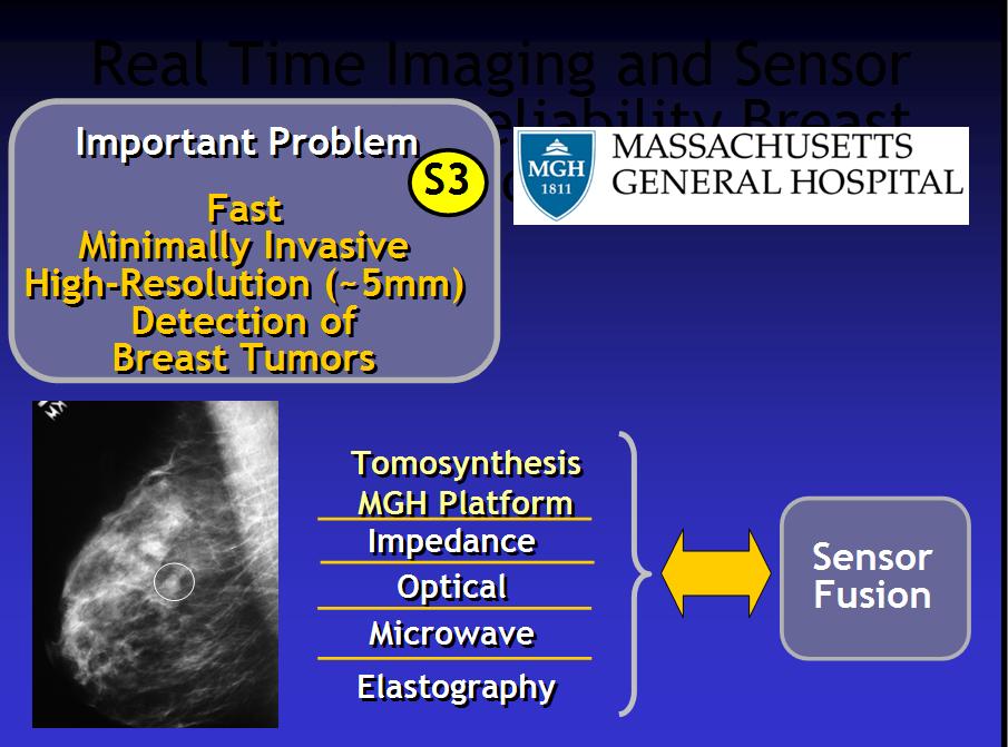
3 Aims What is Digital Breast Tomosynthesis (DBT)? Review how DBT was developed to solve the problem of superimposition of breast tissue commonly found using conventional 2-view mammography Highlight new vistas opened by CenSSIS Electrical Impedance Tomography (EIT) Diffuse Optical Tomography (DOT) Ultrafast Computing by Graphical Processing Unit

4 What is DBT? What is Conventional Mammography?


5 What is Conventional Mammography? L R L R Medial Lateral Oblique (MLO) Cranial - Caudal (CC) CM is 4 views per patient 4 compressions


6 Nature of the screening mammography: For each 1000 women screened with CTVM: About 80 are called back for additional imaging X-ray, US, MRI remember this number: 8% callbacks About 20 are recommended for some form of biopsy stereo core Bx, open surgical Bx, US guided Bx About 3-7 cancers will be discovered on pathology from these biopsies Overall this yields: Reduction in US breast cancer mortality: 30% Sensitivity: 75% - 90% Specificity: 90% - 95% Positive predictive value (biopsy): ~25%
, 23cm 18cm area, 100 m pixel size - 11")
7 What is DBT? Original Prototype Acquisition System - Detector: a-si (CsI), 23cm 18cm area, 100 m pixel size - 11 projections, 50 o (30 o actual) arc, 7sec acquisition time - MLO and CC views, patient seated, fixed height
")

 epitaxially")


8 Digital Breast Tomosynthesis (DBT) 2 nd generation GE prototype Detector: - amorphous-si (CsI) epitaxially grown scintillator - 300msec readout time - 23cm 19.2 cm area micron pixel size Acquisition: - 15 projections - 40 o arc - 15s acquisition - Mo and Rh anodes - same dose as CC+MLO o gantry rotation permits all standard views


9 Digital Breast Tomosynthesis What do you get? Conventional 2D DBT 3D


10 Digital Breast Tomosynthesis What do you get? 2D DBT 2D DBT Cancer Hammartoma Increase TPs Decrease FPs Direct clinical benefits of DBT for screening: Improved specificity, reducing callbacks by 42% Likely improved sensitivity in screening Reduced exam time and only two compressions per study Greater confidence in findings (conspicuity and location)
11 Digital Breast Tomosynthesis What do you get? By removing superimposed tissue, DBT provides more accurate diagnostic information about each finding -normal tissue configurations (some mimic lesions) -benign lesions -malignant lesions (Overall DBT n > 4100 to date) 3000-women NIH NCI-funded (5R33CA ) DBT screening trial, now completed: 42% callback reduction from 80/1000 to 51/1000 A 3D foundation platform on which to build functional imaging such as -Electrical Impedance Tomography (208 to date) -Diffuse Optical Tomography (203 to date)
12 Aims What is Digital Breast Tomosynthesis (DBT)? Review how DBT was developed to solve the problem of superimposition of breast tissue commonly found using conventional 2-view mammography Highlight new vistas opened by CenSSIS Electrical Impedance Tomography (EIT) Diffuse Optical Tomography (DOT) Ultrafast Computing by Graphical Processing Unit

13 Electrical Impedance Tomography (EIT)

14 Electrical Impedance Tomography (EIT)

 Left")


15 Electrical Impedance Tomography (EIT) Left Breast Ductal Carcinoma in-situ Admittance locus - carcinoma in-situ Hyalinized Fibroadenoma Normal Right Breast Normal Carcinoma Right breast carcinoma
16 Electrical Impedance Tomography (EIT) S3
17 Aims What is Digital Breast Tomosynthesis (DBT)? Review how DBT was developed to solve the problem of superimposition of breast tissue commonly found using conventional 2-view mammography Highlight new vistas opened by CenSSIS Electrical Impedance Tomography (EIT) Diffuse Optical Tomography (DOT) Ultrafast Computing by Graphical Processing Unit
 Water Spectroscopic light absorption measurements")
![give quantitative information about breast composition: Main absorbers: Oxy- and deoxy-hemoglobin [HbO] and [HbR] Blood volume and oxygen saturation With more wavelengths: lipid and water Optical](/docs-images/95/123631740/images/18-3.jpg "scattering coefficient related to cell sizes 3D reconstruction of quantitative physiological information ~1cm spatial resolution (limited by strong light scattering) Low-cost, non-invasive,")
18 Absorption coefficient (cm -1 ) Tomographic Optical Breast Imaging (TOBI) Detector array Emerging medical imaging modality based on: Illumination of the tissue with red and near-infrared laser light of different wavelengths Detection of the highly scattered and absorbed light that is remitted from the tissue Light Source array Wavelength (nm) Water Spectroscopic light absorption measurements give quantitative information about breast composition: Main absorbers: Oxy- and deoxy-hemoglobin [HbO] and [HbR] Blood volume and oxygen saturation With more wavelengths: lipid and water Optical scattering coefficient related to cell sizes 3D reconstruction of quantitative physiological information ~1cm spatial resolution (limited by strong light scattering) Low-cost, non-invasive, non-ionizing radiation


")

19 Why TOBI + X-Ray? X-Ray Mammography Clinical standard for screening of breast cancer. Provides structural information Very good spatial resolution (<1 mm) Near Infrared Diffuse Optical Tomography Provides complementary physiological information (blood volume, oxygen saturation, optical scattering) Relatively low spatial resolution (~ 1cm) We combined 3D X-ray and near infrared tomographic imaging in a single instrument, to improve the fusion of anatomic and physiologic data.
 TOBI TOMO Detector probe Detector")
20 Tomographic Optical Breast Imaging (TOBI) TOBI TOMO Detector probe Detector fibers


.")
21 Tomographic Optical Breast Imaging (TOBI) Source plate Source and detector probes mounted on the TOMO clear paddles Support paddle, with source plate to be inserted. The figure also shows the pressure sensors The optical probe sensor plate is not in direct contact with the breast (clear layer of the compressions paddle in-between). Both source and sensor can be removed from the system while the breast remains in fixed position for the X-ray mammogram


22 TOBI image reconstruction 3D image reconstruction of blood content and blood oxygenation Boundaries given by TOMO scan TOMO TOBI Absorption coefficient μ a at 830 nm Chest wall Fibro-glandular tissue (contour obtained from segmentation of the TOMO scan)


23 Tomographic Optical Breast Imaging (TOBI) -tumor
24 Tomographic Optical Breast Imaging (TOBI) Development of a combined X-Ray / Optical tomographic imager: - X-Ray Tomosynthesis Structure - Near infrared diffuse optical tomography Physiology X-Ray and optical images recorded with the breast in the same position: - Facilitates image comparison - Boundaries given by TOMO scan used in optical reconstruction 203 women imaged to date Pressure distribution due to the mammographic compression induces lower blood volume in the center of the breast Tumor was visible on TOBI scan as local higher absorption suggesting high vascularization
25 Aims What is Digital Breast Tomosynthesis (DBT)? Review how DBT was developed to solve the problem of superimposition of breast tissue commonly found using conventional 2-view mammography Highlight new vistas opened by CenSSIS Electrical Impedance Tomography (EIT) Diffuse Optical Tomography (DOT) Ultrafast Computing by Graphical Processing Unit
 120 Mbytes Update Back projection (n+1) Optimized Likelihood Function (end) In 2002 these were overwhelming data")
26 Reconstruction Method Iterative Maximum Likelihood Algorithm Initial 3-D Model Model volume 10Mbytes/ea = 800Mbytes Forward projection Measured Projections: P 10Mbytes/ea = 120 Mbytes Model volume 800 Mbytes Calculated Projections: P (n) 120 Mbytes Update Back projection (n+1) Optimized Likelihood Function (end) In 2002 these were overwhelming data spaces.
: - Dual processor 1.")
27 Reconstruction Computing Hardware for MLEM Ancient history (1997): - Pentium-4 single thread - had to reconstruct sections of datasets - 30 GB hard disk! - affordable at $ 3K - 28 hours per 8-iteration MLEM reconstruction MGH reconstructor (2000): - Dual processor 1.8 GHz, 2-GB of memory GB hard disk - affordable at $4K - ~5-9 hours per 8-iteration MLEM reconstruction - but at least it was the whole breast at once!
: - 32 cheap-ish computers on storage racks - 2.")
28 Reconstruction Computing Hardware for MLEM Cluster computing (generally): processors per cluster - robust toolsets - needs own power circuit! - affordable at $120K - 46 seconds per 8-iteration MLEM reconstruction MGH budget cluster (2004): - 32 cheap-ish computers on storage racks GHz, 800MHz FSB, 512 MB RAM, 40GB disk - 100Mbit Ethernet interconnect and switch - affordable at $48K for the cluster itself amp circuits and 2.5 tons extra AC - ~200 seconds per 8-iteration MLEM reconstruction - ~200 seconds to load/unload job

 - very affordable $600/ea - 46 seconds per")

29 Reconstruction Computing Hardware for MLEM GPU computing (today): processors per card, 2GB shared memory - NVIDIA CUDA - C programming environment with multiprocessor software GPU behavior simulation - still needs own special power circuit! (from PC power supply 750+ watts recommended) - very affordable $600/ea - 46 seconds per 8-iteration MLEM reconstruction Multi-GPU computing (today): - 4 GPU cards per PC processors per PC - affordable desk-side Supercomputing when problem is parallellizable ~ $4K - 11 seconds per 8-iteration reconstruction, so quite nearly real-time

30 Aims What is Digital Breast Tomosynthesis (DBT)? Review how DBT was developed to solve the problem of superimposition of breast tissue commonly found using conventional 2-view mammography Highlight new vistas opened by CenSSIS Electrical Impedance Tomography (EIT) Diffuse Optical Tomography (DOT) Ultrafast Computing by Graphical Processing Unit
 Diffuse Optical Tomography")
31 Multimode Digital Breast Tomosynthesis S3 DBT X-Ray (n > 4100) Electrical impedance Tomography (n = 208) Diffuse Optical Tomography (n = 203)
32 Multimode Digital Breast Tomosynthesis Looking forward Direct clinical benefits of DBT for screening: Improved specificity, reducing callbacks by 42% Likely improved sensitivity in screening Reduced exam time and only two compressions per study Greater confidence in findings (conspicuity and location) In addition to expected direct clinical benefits, we believe DBT can serve as a foundation for understanding new modalities by acquiring them in registration permitting functional assessment Fleshing out the domain of anatomically co-registered functional imaging is a monumental undertaking, complimenting recent advances in genetics and metabolic pathways


33

34 Thank you: Donna Burgess Jayne Cormier, RTRM Gordon Harris David Boody-Alter Lauren Ferrara Dianne Scourletis, RTRM Sarah Brown Daniel Kopans Tao Wu MaryAnne Chorlton, RTRM Anila Burimi R. Maxwell Flaherty Alex Stewart Edward Bullard Benjamin Brown Dianne Georgian-Smith Jeumin Zhang Avital Levy Chanelle Bray Gary Saulnier Michael Silevitch Jennifer Haggan Gwen Olsen Angie Burke Bret Kricun Eric Miller Dana Brooks Leslie Wojcik Brian Billman Diego Rivera David Issacson Jonathan Newell Greg Boverman Stephan Carp Tzu-Jen Kao Thomas L. Szabo Rujuta Kulkarni Dana Shaa Micha Moffe David Kaeli David Boas Juliette Selb Qianqian Fang The MGH Breast Imaging staff, administration and radiologists All women who volunteered for the 3000 women NIH-NBI screening trial General Electric Company Avon Center Martinos Center Dexela Ltd. Planar Imaging Systems nvidia Inc. CenSSIS Center for Subsurface Sensing and Imaging Systems-NSF US Department of Heath and Human Services NIH NCI NTROI Era of Hope Program-US ARMY Komen Foundation AVON Foundation NTROI
35
Contrast-Enhanced Breast Tomosynthesis: Combining the Best of Both Worlds for Better Breast-Cancer Diagnosis
 Contrast-Enhanced Breast Tomosynthesis: Combining the Best of Both Worlds for Better Breast-Cancer Diagnosis T Wu (twu2@partners.org), E Rafferty, R Moore, D Kopans, Massachusetts General Hospital, Boston,
Since its introduction in 2000, digital mammography has become
 Review Article Smith A, PhD email : Andrew.smith@hologic.com Since its introduction in 2000, digital mammography has become an accepted standard of care in breast cancer screening and has paved the way
Andrew Karellas, PhD
 Advanced Imaging for Breast Cancer: Screening, Diagnosis, and Assessing Response to Therapy The Role of Tomosynthesis Andrew Karellas, PhD Department of Radiology University of Massachusetts Medical School
Digital breast tomosynthesis
 GE Healthcare Digital breast tomosynthesis Daniel B. Kopans, M.D., F.A.C.R. Professor of Radiology Harvard Medical School Senior Radiologist - Breast Imaging Division Massachusetts General Hospital Since
CURRENTLY FDA APPROVED ARE FULL FIELD DIGITAL MAMMOGRAPHY SYSTEMS AND FILM SCREEN STILL BEING USED AT SOME INSTITUTIONS
 ABBY DUROJAYE,M.D CURRENTLY FDA APPROVED ARE FULL FIELD DIGITAL MAMMOGRAPHY SYSTEMS AND FILM SCREEN STILL BEING USED AT SOME INSTITUTIONS BOTH HAVE BEEN SHOWN TO BE EFFECTIVE TOOLS EARLY DETECTION OF BREAST
Breast Tomosynthesis. What is breast tomosynthesis?
 Scan for mobile link. Breast Tomosynthesis Breast tomosynthesis is an advanced form of mammography, a specific type of breast imaging that uses low-dose x-rays to detect cancer early when it is most treatable.
TOMOSYNTHESIS: WORTH ALL THE HYPE?
 X-Ray Associates of New Mexico, P.C. TOMOSYNTHESIS: WORTH ALL THE HYPE? MICHAEL N. LINVER, MD, FACR MAMMOGRAPHY: THE GOOD, THE PRETTY GOOD, & THE NOT SO GOOD MAMMOGRAPHY: THE GOOD, THE PRETTY GOOD, & THE
Mammography. What is Mammography?
 Scan for mobile link. Mammography Mammography is a specific type of breast imaging that uses low-dose x-rays to detect cancer early before women experience symptoms when it is most treatable. Tell your
Mammography is a most effective imaging modality in early breast cancer detection. The radiographs are searched for signs of abnormality by expert
 Abstract Methodologies for early detection of breast cancer still remain an open problem in the Research community. Breast cancer continues to be a significant problem in the contemporary world. Nearly
EARLY DETECTION: MAMMOGRAPHY AND SONOGRAPHY
 EARLY DETECTION: MAMMOGRAPHY AND SONOGRAPHY Elizabeth A. Rafferty, M.D. Avon Comprehensive Breast Center Massachusetts General Hospital Harvard Medical School Breast Cancer Screening Early detection of
8/3/2016. DBT Physics Basic to Advanced: Primer On Tomosynthesis. Tomosynthesis Pedigree
 DBT Physics Basic to Advanced: Primer On Tomosynthesis Andrew D. A. Maidment, Ph.D. University of Pennsylvania Department of Radiology Acknowledgements of Support Research support from the Komen Foundation,
Fundamentals of Breast Tomosynthesis
 Fundamentals of Breast Tomosynthesis Improving the Performance of Mammography Andrew Smith, Ph.D. This white paper is one in a series of research overviws on advanced technologies in women s healthcare.
1762 IEEE TRANSACTIONS ON MEDICAL IMAGING, VOL. 27, NO. 12, DECEMBER /$ IEEE
 1762 IEEE TRANSACTIONS ON MEDICAL IMAGING, VOL. 27, NO. 12, DECEMBER 2008 Regional Admittivity Spectra With Tomosynthesis Images for Breast Cancer Detection: Preliminary Patient Study Tzu-Jen Kao*, Member,
A Breast Surgeon s Use of Three Dimensional Specimen Tomosynthesis
 A Breast Surgeon s Use of Three Dimensional Specimen Tomosynthesis Cary S. Kaufman MD, FACS Associate Clinical Professor of Surgery A Breast Surgeon s Use of Three Dimensional Specimen Tomosynthesis Cary
Breast positioning system for full field digital mammography and digital breast tomosynthesis system
 Breast positioning system for full field digital mammography and digital breast tomosynthesis system Mari Varjonen* a, Martti Pamilo b, Pirjo Hokka b, Riina Hokkanen a, Pekka Strömmer a a Planmed Oy Asentajankatu
Digital Breast Tomosynthesis from a first idea to clinical routine
 International Master Programm Biomedical Engineering Digital Breast Tomosynthesis from a first idea to clinical routine Historical background 2D imaging of 3D objects has important limitations Jörg Barkhausen
Update of Digital Breast Tomosynthesis. Susan Orel Roth, MD
 Update of Digital Breast Tomosynthesis Susan Orel Roth, MD NCI estimates that : Why DBT? Approximately 20% of breast cancers are missed at mammography screening Average recall rates approximately 10%
Policy Library Clinical Advantages of Digital Breast Tomosynthesis in Symptomatic Patients
 Policy Library Clinical Advantages of Digital Breast Tomosynthesis in Symptomatic Patients Version: 1 Approved by: Faculty of Clinical Radiology Council Date of approval: Click and type: day month and
Fast 3D Optical Mammography using ICG Dynamics for Reader-Independent Lesion Differentiation
 Fast 3D Optical Mammography using ICG Dynamics for Reader-Independent Lesion Differentiation Sophie Piper 1 P.Schneider 1, N. Volkwein 1, N.Schreiter 1, C.H. Schmitz 1,2, A.Poellinger 1 1 Charité University
Epworth Healthcare Benign Breast Disease Symposium. Sat Nov 12 th 2016
 Epworth Healthcare Benign Breast Disease Symposium Breast cancer is common Sat Nov 12 th 2016 Benign breast disease is commoner, and anxiety about breast disease commoner still Breast Care Campaign UK
Current Status of Supplementary Screening With Breast Ultrasound
 Current Status of Supplementary Screening With Breast Ultrasound Stephen A. Feig, M.D., FACR Fong and Jean Tsai Professor of Women s Imaging Department of Radiologic Sciences University of California,
THE DIAGNOSTIC WORKUP: THE TEAM APPROACH
 X-Ray Associates of New Mexico, P.C. THE DIAGNOSTIC WORKUP: THE TEAM APPROACH MICHAEL N. LINVER, MD, FACR DAWN DERENBURGER, RTRM Disclosure There are no conflicts of interest or relevant financial interests
Using Near-Infrared Light To Detect Breast Cancer
 Using Near-Infrared Light To Detect Breast Cancer Sergio Fantini, Erica L. Heffer, Horst Siebold and Oliver Schütz 24 Optics & Photonics News November 2003 1047-6938/03/11/0024/6-$0010 Optical Society
SOORYA PETER IVEN JOSE CHRIST UNIVERSITY FACULTY OF ENGINEERING. Excerpt from the Proceedings of the 2014 COMSOL Conference in Bangalore
 SOORYA PETER IVEN JOSE CHRIST UNIVERSITY FACULTY OF ENGINEERING Introduction: Cancer is one of the most dreaded diseases of the modern world. Breast cancer is the second leading cause (after lung cancer)
Patient Dosimetry in Mammography and Tomosynthesis:
 2013 ICTP/IAEA Training Course on Radiation Protection of Patients Trieste Patient Dosimetry in Mammography and Tomosynthesis: What to measure, why and how John M. Boone, Ph.D., FAAPM, FSBI, FACR Professor
Breast Cancer Imaging
 Breast Cancer Imaging I. Policy University Health Alliance (UHA) will cover breast imaging when such services meet the medical criteria guidelines (subject to limitations and exclusions) indicated below.
Mammography. What is Mammography? What are some common uses of the procedure?
 Mammography What is Mammography? Mammography is a specific type of imaging that uses a low-dose x-ray system to examine breasts. A mammography exam, called a mammogram, is used to aid in the early detection
Mammography limitations. Clinical performance of digital breast tomosynthesis compared to digital mammography: blinded multi-reader study
 Clinical performance of digital breast tomosynthesis compared to digital mammography: blinded multi-reader study G. Gennaro (1), A. Toledano (2), E. Baldan (1), E. Bezzon (1), C. di Maggio (1), M. La Grassa
Updates in Mammography. Dr. Yang Faridah A. Aziz Department of Biomedical Imaging University Malaya Medical Centre
 Updates in Mammography Dr. Yang Faridah A. Aziz Department of Biomedical Imaging University Malaya Medical Centre Updates in Mammography Breast Imaging Dr. Yang Faridah A. Aziz Department of Biomedical
INTRODUCTION. Breast cancer is an epidemic that affects more than 200,000 new patients each year. With greater than
 Advances in Simultaneous Dual-Breast Optical Mammography M.S. Katz 1, R.E. Hardin 1, N.A. Franco 1, D.P. Klemer 2, C.H. Schmitz 2, Y. Pei 2, H.L. Graber 3, W.B. Solomon 3, R.L. Barbour 3 1 SUNY Downstate
Clinical feasibility of co-registered opto-acoustic and ultrasonic imaging for differentiation of breast tumors
 Clinical feasibility of co-registered opto-acoustic and ultrasonic imaging for differentiation of breast tumors Pamela Otto 1, Kenneth Kist 1, N. Carol Dornbluth 1, Don Herzog 2, Bryan Clingman 2, Sergey
Emerging Techniques in Breast Imaging: Contrast-Enhanced Mammography and Fast MRI
 Emerging Techniques in Breast Imaging: Contrast-Enhanced Mammography and Fast MRI Lilian Wang, M.D. Breast Imaging Section Department of Radiology Northwestern Medicine Overview Rationale for new imaging
EARLY DETECTION: MAMMOGRAPHY AND SONOGRAPHY
 EARLY DETECTION: MAMMOGRAPHY AND SONOGRAPHY Elizabeth A. Rafferty, M.D. Avon Comprehensive Breast Center Massachusetts General Hospital Harvard Medical School Breast Cancer Screening Early detection of
Radiation Dosimetry in Digital Breast Tomosynthesis. March, 2015 William J. O Connel, Dr. Ph, Senior Medical Physicist
 Radiation Dosimetry in Digital Breast Tomosynthesis March, 2015 William J. O Connel, Dr. Ph, Senior Medical Physicist Imagination at work. Syllabus 1. Introduction 2. Dosimetry in Mammography 3. Dosimetry
Contrast Enhanced Spectral Mammography (CESM) Updates
 Updates") Contrast Enhanced Spectral Mammography (CESM) Updates Georgeta Mihai, PhD, DABR Medical Physicist, BIDMC, Boston Assistant Professor, Harvard Medical School, Boston Disclosures None Acknowledgments: Da
Contrast Enhanced Spectral Mammography (CESM) Updates Georgeta Mihai, PhD, DABR Medical Physicist, BIDMC, Boston Assistant Professor, Harvard Medical School, Boston Disclosures None Acknowledgments: Da
Here are examples of bilateral analog mammograms from the same patient including CC and MLO projections.
 Good afternoon. It s my pleasure to be discussing Diagnostic Breast Imaging over the next half hour. I m Wei Yang, Professor of Diagnostic Radiology and Chief, the Section of Breast Imaging as well as
Molecular Breast Imaging: History and Recent Developments
 Molecular Breast Imaging: History and Recent Developments Associate Professor, Department of Imaging Physics The University of Texas MD Anderson Cancer Center, Houston, Texas Educational Objectives 1.
Mammography. Background and Perspective. Mammography Evolution. Background and Perspective. T.R. Nelson, Ph.D. x41433
 - 2015 Background and Perspective 2005 (in US) Women Men Mammography Invasive Breast Cancer Diagnosed 211,240 1,690 Noninvasive Breast Cancer Diagnosed 58,940 Deaths from Breast Cancer 40,410 460 T.R.
Contrast-Enhanced Spectral Mammography
 Contrast-Enhanced Spectral Mammography Illuminating Breast Cancer Detection SenoBright HD TM gehealthcare.com/senobright Mammography is the most reliable imaging technique for breasts, but limitations
What s New in Breast Imaging. Jennifer A. Harvey, M.D., FACR Professor of Radiology University of Virginia
 What s New in Breast Imaging Jennifer A. Harvey, M.D., FACR Professor of Radiology University of Virginia Disclosure Hologic, Inc. Shareholder and research agreement. Volpara Solutions, Ltd. Shareholder
FDA Executive Summary
 Meeting of the Radiological Devices Advisory Panel On October 24, 22, the panel will discuss, make recommendations, and vote on a premarket approval application supplement (P83/S) to expand the indications
Opportunities and Innovations in Digital Mammography John M. Sandrik, Ph.D. GE Healthcare Milwaukee, WI
 Opportunities and Innovations in Digital Mammography John M. Sandrik, Ph.D. GE Healthcare Milwaukee, WI john.sandrik@med.ge.com with many thanks to Vince Polkus, Advanced Applications Product Mgr. 1 Content
Tomosynthesis and breast imaging update. Dr Michael J Michell Consultant Radiologist King's College Hospital NHS Foundation Trust
 Tomosynthesis and breast imaging update Dr Michael J Michell Consultant Radiologist King's College Hospital NHS Foundation Trust Breast imaging new technology BREAST CANCER FLT PET shows different grades
#46: DIGITAL TOMOSYNTHESIS: What is the Data Really Showing? TERMS (AKA) WHAT IS TOMOSYNTHESIS? 3/3/2014. Digital breast tomosynthesis =
 WHAT IS TOMOSYNTHESIS? 3/3/2014. Digital breast tomosynthesis =") #46: DIGITAL TOMOSYNTHESIS: What is the Data Really Showing? January K. Lopez, MD Hoag Breast Care Center Newport Beach, CA Disclosures: None TERMS (AKA) Digital breast tomosynthesis = DBT Tomo 3D Full
#46: DIGITAL TOMOSYNTHESIS: What is the Data Really Showing? January K. Lopez, MD Hoag Breast Care Center Newport Beach, CA Disclosures: None TERMS (AKA) Digital breast tomosynthesis = DBT Tomo 3D Full
Breast Health and Imaging Glossary
 Contact: Lorna Vaughan HerSpace Breast Imaging & Biopsy Associates 300 State Route 35 South W. Long Branch, NJ 07764 732-571-9100, ext. 104 lorna@breast-imaging.com Breast Health and Imaging Glossary Women
3D Ultrasound Tomography for Breast Cancer Diagnosis
 3D Ultrasound Tomography for Breast Cancer Diagnosis N. V. Ruiter, M. Zapf, T. Hopp and H. Gemmeke INSTITUTE FOR DATA PROCESSING AND ELECTRONICS 1 KIT University of the State of Baden-Wuerttemberg and
Financial Disclosures
 Financial Disclosures 3D Mammography: The Latest Developments in the Breast Imaging Arena I have no financial disclosures Dr. Katharine Lampen-Sachar Breast and Body Radiologist Radiology Associates of
In vivo optical imaging : revealing endogeneous optical contrast at depth
 In vivo optical imaging : revealing endogeneous optical contrast at depth Anne PLANAT-CHRÉTIEN, Jean-Marc DINTEN CEA-LETI, MINATEC, Grenoble Jérôme GATEAU Université Paris Descartes, Paris 1 Why using
Clinically Available Optical Topography System
 Clinically Available Optical Topography System Clinically Available Optical Topography System 18 Fumio Kawaguchi Noriyoshi Ichikawa Noriyuki Fujiwara Yûichi Yamashita Shingo Kawasaki OVERVIEW: Progress
Breast tomosynthesis reduces radiologist performance variability compared to digital mammography
 Breast tomosynthesis reduces radiologist performance variability compared to digital mammography Andrew Smith 1, Elizabeth Rafferty 2, Loren Niklason 1 1 Hologic, Inc., Bedford MA, USA 2 Massachusetts
WHAT TO EXPECT. Genius 3D Mammography Exam. The most exciting advancement in mammography in over 30 years
 WHAT TO EXPECT Genius 3D Mammography Exam The most exciting advancement in mammography in over 30 years Screening for breast cancer Doctors and scientists agree that early detection is the best defense
CT Laser Mammography CTLM
 CT Laser Mammography CTLM Scanning for life IMAGINE...A breast imaging method that is painless, non-invasive, does not touch the breast, uses no harmful ionizing radiation, can be repeated as often as
SenoBright Contrast Enhanced Spectral Mammography Technology. Ann-Katherine Carton Sylvie Saab-Puong Matt Suminski
 SenoBright Contrast Enhanced Spectral Mammography Technology Ann-Katherine Carton Sylvie Saab-Puong Matt Suminski White Paper October 2012 SenoBright Contrast Enhanced Spectral Mammography Technology Ann-Katherine
Outline. Digital Breast Tomosynthesis: Update and Pearls for Implementation. Tomosynthesis Dataset: 2D/3D (Hologic Combo Acquisition)
") Outline Digital Breast Tomosynthesis (DBT) the new standard of care Digital Breast Tomosynthesis: Update and Pearls for Implementation Emily F. Conant, M.D. Professor, Chief of Breast Imaging Department
Outline Digital Breast Tomosynthesis (DBT) the new standard of care Digital Breast Tomosynthesis: Update and Pearls for Implementation Emily F. Conant, M.D. Professor, Chief of Breast Imaging Department
Ana Sofia Preto 19/06/2013
 Ana Sofia Preto 19/06/2013 Understanding the underlying pathophysiologic processes leading to the various types of calcifications Description and illustration of the several types of calcifications, according
Breast CT and Dosimetry
 2013 ICTP/IAEA Training Course on Radiation Protection of Patients Trieste Breast CT and Dosimetry John M. Boone, Ph.D., FAAPM, FSBI, FACR Professor and Vice Chair (Research) of Radiology Professor of
Contrast-Enhanced Digital Mammography
 2015 ARRS Breast Symposium Contrast-Enhanced Digital Mammography John Lewin, M.D. Diversified Radiology of Colorado CEDM - Outline History Technique Literature Review / Cases Clinical Status Inexpensive,
arxiv: v2 [cs.cv] 8 Mar 2018
![arxiv: v2 [cs.cv] 8 Mar 2018](/thumbs/87/97094636.jpg "arxiv: v2 [cs.cv] 8 Mar 2018") Automated soft tissue lesion detection and segmentation in digital mammography using a u-net deep learning network Timothy de Moor a, Alejandro Rodriguez-Ruiz a, Albert Gubern Mérida a, Ritse Mann a, and
Automated soft tissue lesion detection and segmentation in digital mammography using a u-net deep learning network Timothy de Moor a, Alejandro Rodriguez-Ruiz a, Albert Gubern Mérida a, Ritse Mann a, and
Correlation between lesion type and the additional value of digital breast tomosynthesis
 Correlation between lesion type and the additional value of digital breast tomosynthesis Poster No.: C-1604 Congress: ECR 2011 Type: Scientific Exhibit Authors: C. Van Ongeval, L. Cockmartin, A. Van Steen,
Breast Imaging & You
 Breast Imaging & You What s Inside: Breast Imaging... 2 Digital Breast Tomosynthesis (DBT) mammograms... 4 Breast cancer screening... 6 Dense breast tissue... 8 Automated Breast Ultrasound (ABUS)... 9
PET in Radiation Therapy. Outline. Tumor Segmentation in PET and in Multimodality Images for Radiation Therapy. 1. Tumor segmentation in PET
 Tumor Segmentation in PET and in Multimodality Images for Radiation Therapy Wei Lu, Ph.D. Department of Radiation Oncology Mallinckrodt Institute of Radiology Washington University in St. Louis Outline
The latest developments - Automated Breast Volume Scanning. Dr. med. M. Golatta
 The latest developments - Automated Breast Volume Scanning Dr. med. M. Golatta Automated Breast Volume US: Why? o Mammography is limited in dense breasts: high false negative rate o Many of these tumors
Molecular Imaging and Breast Cancer
 Molecular Imaging and Breast Cancer Breast cancer forms in tissues of the breast usually in the ducts, tubes that carry milk to the nipple, and lobules, the glands that make milk. It occurs in both men
WHAT TO EXPECT. Genius 3D MAMMOGRAPHY Exam. The most exciting advancement in mammography in over 30 years
 WHAT TO EXPECT Genius 3D MAMMOGRAPHY Exam The most exciting advancement in mammography in over 30 years 91% of patients agree the quality of care provided by the facility was better with a Genius 3D MAMMOGRAPHY
Breast Imaging & You
 Breast Imaging & You What s Inside: Breast Imaging... 2 Digital Breast Tomosynthesis (DBT) mammograms... 4 Breast cancer screening... 6 Dense breast tissue... 8 Automated breast ultrasound (ABUS)... 9
the one name in cancer care.
 the one name in cancer care. Landmark study evaluating close to half a million mammography exams published in the Journal of the American Medical Association (JAMA) 1 Hologic 3D Mammography Significantly
Melissa Hartman, DO Women s Health Orlando VA Medical Center
 Melissa Hartman, DO Women s Health Orlando VA Medical Center Most common non-skin cancer and Second deadliest cancer in women Majority are diagnosed by abnormal screening study An approach to breast cancer
Volume 14 - Issue 3, Matrix
 Volume 14 - Issue 3, 2014 - Matrix Digital Breast Tomosynthesis for Screening and Diagnosis of Breast Cancer Author ECRI ECRI Institute 29 Broadwater Road Suite 104 Welwyn Garden City AL7 3BQ United Kingdom
Standard Breast Imaging Modalities. Lilian Wang, M.D. Breast Imaging Section Department of Radiology Northwestern Medicine
 Standard Breast Imaging Modalities Lilian Wang, M.D. Breast Imaging Section Department of Radiology Northwestern Medicine Overview Standard breast imaging modalities Mammography Ultrasound MRI Imaging
Breast Cancer Screening and Diagnosis
 Breast Cancer Screening and Diagnosis Priya Thomas, MD Assistant Professor Clinical Cancer Prevention and Breast Medical Oncology University of Texas MD Anderson Cancer Center Disclosures Dr. Thomas has
RSNA, /radiol Appendix E1. Methods
 RSNA, 2016 10.1148/radiol.2016151097 Appendix E1 Methods US and Near-infrared Data Acquisition Four optical wavelengths (740 nm, 780 nm, 808 nm, and 830 nm) were used to sequentially deliver the light
Introduction 1. Executive Summary 5
 Roman_pages 20-09-2005 21:01 Pagina IX Table of contents Introduction 1 Executive Summary 5 1. Epidemiological guidelines for quality assurance in breast cancer screening 15 1.10 Introduction 17 1.20 Local
Profound understanding of anatomy
 ENGLISH Profound understanding of anatomy The unique Planmeca ProMax 3D product family offers equipment for all maxillofacial imaging. All volume sizes from the smallest special cases to whole head images
Look differently. Invenia ABUS. Automated Breast Ultrasound
 Look differently. Invenia ABUS Automated Breast Ultrasound InveniaTM ABUS from GE Healthcare offers a view beyond mammography, with breast screening technology that looks differently. 40 % The unseen risk.
Breast Tomosynthesis An additional screening tool in the fight against breast cancer
 What to Expect Breast Tomosynthesis An additional screening tool in the fight against breast cancer Every woman over 40 should be examined for breast cancer once a year. American Cancer Society What to
WHAT TO EXPECT. Breast Tomosynthesis An additional screening tool in the fight against breast cancer HOLOGIC. The Women's Health Company
 WHAT TO EXPECT Breast Tomosynthesis An additional screening tool in the fight against breast cancer HOLOGIC The Women's Health Company ...,. Screening for breast cancer Doctors and scientists agree that
Corporate Medical Policy
 Corporate Medical Policy File Name: Origination: Last CAP Review: Next CAP Review: Last Review: digital_breast_tomosynthesis 3/2011 6/2016 6/2017 11/2016 Description of Procedure or Service Conventional
CHAPTER 2 MAMMOGRAMS AND COMPUTER AIDED DETECTION
 9 CHAPTER 2 MAMMOGRAMS AND COMPUTER AIDED DETECTION 2.1 INTRODUCTION This chapter provides an introduction to mammogram and a description of the computer aided detection methods of mammography. This discussion
Breast Imaging Update: Old Dog New Tricks
 Breast Imaging Update: Old Dog New Tricks Claire McKay, DO M&S Imaging Assoc. San Antonio, TX cmckayhart@juno.com Goals Describe modalities available, old and new Provide understanding of pros and cons
3D Mammography. The most exciting advancement in mammography in over 30 years
 What to Expect 3D Mammography The most exciting advancement in mammography in over 30 years Screening for breast cancer Doctors and scientists agree that early detection is the best defense against breast
WHAT TO EXPECT. Genius 3D MAMMOGRAPHY Exam. The most exciting advancement in mammography in over 30 years
 WHAT TO EXPECT Genius 3D MAMMOGRAPHY Exam The most exciting advancement in mammography in over 30 years 91% of patients agree the quality of care provided by the facility was better with a Genius 3D MAMMOGRAPHY
Innovations and Applications of Tomosynthesis. Andrew D. A. Maidment, Ph.D. University of Pennsylvania Department of Radiology
 Innovations and Applications of Tomosynthesis Andrew D. A. Maidment, Ph.D. University of Pennsylvania Department of Radiology Acknowledgements of Support Grant support from the Komen Foundation, DOD, NIH,
Hacia la imagenología tomográfica de mama
 Hacia la imagenología tomográfica de mama Futuro y presente Ioannis Sechopoulos, Ph.D., DABR Advanced X ray Tomographic Imaging (AXTI) Lab Department of Radiology and Nuclear Medicine Radboud University
Breast Cancer Screening and High Risk
 Breast Cancer Screening and High Risk Mary Freyvogel, DO Breast Surgeon Clinical Assistant Professor of Surgery University Hospitals Case Medical Center St. John Medical Center / Elyria Medical Center
Optical Imaging: Technology and Applications for Radiology
 September 2004 Optical Imaging: Technology and Applications for Radiology Rima Arnaout Harvard Medical School Year III Our Patient: 61 yo female, yearly mammogram Suspicious right breast mass: spiculated,,
Profound understanding of anatomy
 ENGLISH Profound understanding of anatomy The unique Planmeca ProMax 3D product family offers equipment for all maxillofacial imaging. All volumes sizes from the smallest special cases to whole head images
In-Room Radiographic Imaging for Localization
 In-Room Radiographic Imaging for Localization Fang-Fang Yin, Zhiheng Wang, Sua Yoo, Devon Godfrey, Q.-R. Jackie Wu Department of Radiation Oncology Duke University Medical Center Durham, North Carolina
Profound understanding of anatomy
 ENGLISH Profound understanding of anatomy Planmeca ProMax 3D, the intelligent and multipurpose X-ray unit, is designed to obtain complete information on patient anatomy in the minutest detail. The unit
Simulation of spiculated breast lesions
 Simulation of spiculated breast lesions Premkumar Elangovan* a, Faisal Alrehily b, R Ferrari Pinto a, Alaleh Rashidnasab a,c, David R Dance b,d, Kenneth C Young b,d, Kevin Wells a a Centre for Vision,
Diagnostic benefits of ultrasound-guided. CNB) versus mammograph-guided biopsy for suspicious microcalcifications. without definite breast mass
 versus mammograph-guided biopsy for suspicious microcalcifications. without definite breast mass") Volume 118 No. 19 2018, 531-543 ISSN: 1311-8080 (printed version); ISSN: 1314-3395 (on-line version) url: http://www.ijpam.eu ijpam.eu Diagnostic benefits of ultrasound-guided biopsy versus mammography-guided
Volume 118 No. 19 2018, 531-543 ISSN: 1311-8080 (printed version); ISSN: 1314-3395 (on-line version) url: http://www.ijpam.eu ijpam.eu Diagnostic benefits of ultrasound-guided biopsy versus mammography-guided
Photoacoustic Imaging and Therapy in Biomedicine. Nicholas Tobey and Grace Yook. Optical Engineering. Dr. Kasra Daneshvar
 Photoacoustic Imaging 1 Photoacoustic Imaging and Therapy in Biomedicine Nicholas Tobey and Grace Yook Optical Engineering Dr. Kasra Daneshvar July 16, 2010 Photoacoustic Imaging 2 Abstract When a pulsed
The Radiology Aspects
 REQUIREMENTS FOR INTERNATIONAL ACCREDITATION OF BREAST CENTERS/UNITS The Radiology Aspects Miri Sklair-Levy, Israel RADIOLOGY GUIDELINES FOR QUALITY ASSURANCE IN BREAST CANCER SCREENING AND DIAGNOSIS Radiologists
Breast Cancer Detection by Determination of Optical Properties of Non- Malignant and Malignant Breast Tissues
 Research Article Breast Cancer Detection by Determination of Optical Properties of Non- Malignant and Malignant Breast Tissues El-Sharkawy YH * Department of Biomedical Engineering, Military Technical
Development and application of novel spectroscopic tools for breast cancer diagnosis
 Development and application of novel spectroscopic tools for breast cancer diagnosis LBRC researchers: Ramachandra Dasari, Jeon Woong Kang, Niyom Lue, Rishikesh Pandey, Nicolas Spegazzini External technology
FACT: FACT: Breast Cancer Staging. For cancer to occur, something must damage nucleus of the cell. Stage I. Stage II 10/9/2018
 Digital Breast Tomosynthesis (DBT) Pathology Findings: Case Studies and Beyond For cancer to occur, something must damage nucleus of the cell. Advanced Health Education Center www.aheconline.com Copyright
Compression-induced changes in the physiological state of the breast as observed through frequency domain photon migration measurements
 Journal of Biomedical Optics 116, 064016 November/December 2006 Compression-induced changes in the physiological state of the breast as observed through frequency domain photon migration measurements Stefan
The power and promise of breast tomosynthesis is here. Selenia Dimensions system with Acquisition Workstation 5000
 WOMEN S HEALTH BREAST SOLUTIONS HEALTH The power and promise of breast tomosynthesis is here Selenia Dimensions system with Acquisition Workstation 5000 3D mammography: A new dimension in early breast
Why Choose Breast Radiology?
 Why Choose Breast Radiology? Hannah Gay ST6, St George s Hospital BSBR exec committee trainee representative. Introduction Breast cancer is the most common cancer in the UK. Lifetime risk of 1 in 8 for
Multi-Modality Breast Cancer Assessment Tools Using Diffuse Optical and Electrical Impedance Spectroscopy
 Multi-Modality Breast Cancer Assessment Tools Using Diffuse Optical and Electrical Impedance Spectroscopy by Majid Shokoufi M.Sc., Iran University of Science & Technology, 2002 Thesis Submitted in Partial
Diagnostic Medical Physicist Via Christi Hospitals Wichita, Wichita, KS
 Digital Breast Tomosynthesis SWAAPM Meeting 30 Mar 2012 Jerry A. Thomas, MS, FAAPM, DABR, CHP, DABSNM Diagnostic Medical Physicist Via Christi Hospitals Wichita, Wichita, KS Talk Overview Breast Cancer