Hacia la imagenología tomográfica de mama
|
|
|
- Paula Cummings
- 5 years ago
- Views:
Transcription


")


1 Hacia la imagenología tomográfica de mama Futuro y presente Ioannis Sechopoulos, Ph.D., DABR Advanced X ray Tomographic Imaging (AXTI) Lab Department of Radiology and Nuclear Medicine Radboud University Medical Center and LRCB Dutch Reference Centre for Screening Nijmegen, the Netherlands advanced x-ray tomographic imaging 1


2 2

 (Is more always better?")

3 Towards Tomographic Breast Imaging 2 D 2+ D 2.2 D 3 D Standard Mammography Stereoscopic Mammography Digital Tomosynthesis Dedicated Breast CT (If your optical system can handle it!) (Is more always better?) Digital Mammography Improvements Digital Mammography Improvements Detection in some patient subgroups (DMIST) Workflow 3


4 Digital Mammography Improvements Digital Mammography Improvements Contrast enhanced imaging Advanced imaging techniques 29% of cancers missed by overlying tissue STEREOSCOPIC DIGITAL MAMMOGRAPHY Birdwell et al, Radiology 219, (2001). 4


5 Translated X ray source X ray beam limits 6 Lesions of Interest Detector These two images are shown separately to each eye Benign Mass Half silvered mirror Cross polarized images Note shift Cross polarized lenses 5
6 High Risk Screening Study N = 1298 cases Mammo Stereo Sensitivity 12/19 13/19 Recall rate 12.9% 9.6%* Stereoscopic Mammography Promising Results D Orsi et al, Radiology, 2013 Stereoscopic Mammography Cheap! (and df fast!) Stereoscopic Mammography Too little, too late? 6


7 Stereoscopic Mammography DIGITAL BREAST TOMOSYNTHESIS Translated X ray source Recall X ray beam Lesions of Interest Detector 27 This information is used to reconstruct the volume 28 Courtesy of Hologic Inc. 7


8 Recall CC view.idc Courtesy of Hologic Inc. 30 Courtesy of Hologic Inc. FFDM System Tomo System Find the 8 differences Courtesy of Hologic Inc. 31 8


 Screening")


9 Benefits Mammography++ System Workflow Interpretation Dose but with some discrimination of vertical position! 33 (Some) Screening Trials Trial il N Cancer Det Recall Rate Rate STORM 2 9,672 DM: +35% Synth: +40% DM: +16% Synth: +30% OSLO 25, % 13% MALMÖ 14, % +43% slice 7/14 = 5.46 mm thick slice 36/72 9


10 An adjunct to mammography? Implementation? + DBT DM Or a replacement of mammography? Do we need two views? + CC MLO DBT DM


 Lång et al 2016 1v")


11 Or is one view enough? One view breast tomo: feasible? CC MLO Retrospective studies Gennaro 2010 Svane 2011 Svahn 2012 Wallis v tomo 2v DM 1v tomo 2v DM 1v tomo > 2v DM 1v tomo 2v DM Malmö Breast Tomosynthesis Screening Trial (MBTST) Lång et al v tomo > 2v DM +43 % increase in cancer detection rate Wide angle system Study design 6 readers from 3 different institutions Does 1 view experience matter? 3 readers 3 readers < 3 years > 3 years


12 Sensitivity 100% p < 0.05 * Less experienced, significant increase respect to 1v tomo, p< 0.05 Specificity 100% p < 0.05 * Less experienced, significant decrease respect to 1v tomo, p< % 90% 80% 70% 60% * * * All < 3y exp > 3y exp 80% 70% 60% * All < 3y exp > 3y exp 50% MLO DBT MLO DBT + CC DM 2v DBT + 2v DM 2v DM 50% MLO DBT MLO DBT + CC DM 2v DBT + 2v DM 2v DM Case based ROC AUC Average all readers 1v tomo not significantly different JAFROC analysis FoM NO statistical difference between 1v DBT and the rest of modalities, p = x 48 T Chakraborty et al


13 Limitations Conclusion Not screening population 1/3 were recalls seen with 2 view mammo One vendor Perhaps enough Mammogram Orig. Synthetic Tomo Slice SYNTHETIC MAMMOGRAMS Gur et al, Academic Radiology, Vol 19, No 2,
14 Recall Rates False Positive Rate DBT + FFDM % Detected Cancers DBT + Synthetic False Positive Rate % Detected Cancers 1 st Generation nd Generation Synthetic Mammograms Current synthetic 2D image can replace FFDM in combination with DBT Skaane et al, Radiology, Vol 271(3), Ratios Dose DBT/DM from Clinical Studies RADIATION DOSE 1 view DBT vs DM = view DBT vs DM = view DBT + DM vs DM = view DBT + DM vs DM = Svahn et al, The Breast, 24 (2015)
15 Synthetic (?) 1 view DBT vs DM = view DBT vs DM = TO BE ANSWERED x Svahn et al, The Breast, 24 (2015) T Remaining questions in DBT Remaining questions in DBT What is a screening DBT exam? Reading time 15
16 Remaining questions in DBT Reading strategy False negatives: mammo vs tomo Tomo + Tomo Mammo Mammo + Visibility 13 0 Radiographic appearance 3 1 Interpretative 3 6 error Lång et al, Br J Radiol 2014;87: Remaining questions in DBT Remaining questions in DBT What after a tomo screen? Multiple rounds? 16


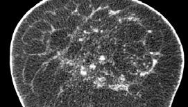
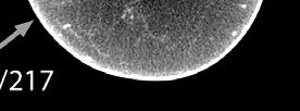
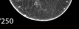
17 DEDICATED BREAST COMPUTED TOMOGRAPHY 17


18 Albion 2003 Bodega 2007 Courtesy of Koning Corp. Cambria 2011 Courtesy of John Boone, Ph.D. Doheny 2014 Fully 3D SPECT CT: CT source down Fully 3D SPECT CT: CT source up Spiral BCT White arrow points to 16x20cm 2 CZT based SPECT camera; orange arrows point to x ray CT source, with opposed 40x30cm 2 flat panel detector; phantom on radiopaque bed. Courtesy of Dr. Martin Tornai of the Duke Multi Modality Imaging Lab Kalender et al, Eur Radiol,


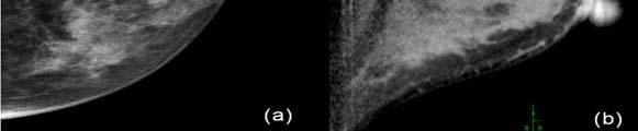
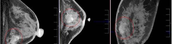
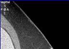
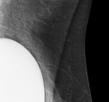
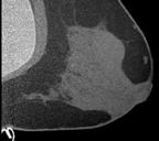
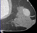
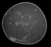
19 Coronal CLINICAL IMAGES Transverse 19
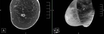

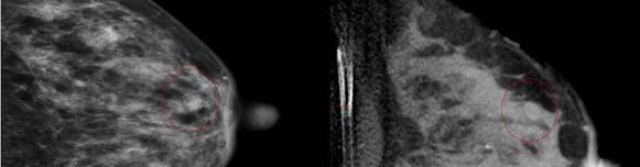
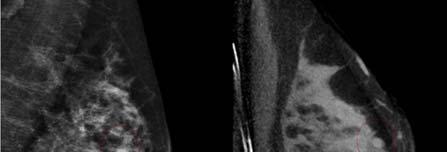
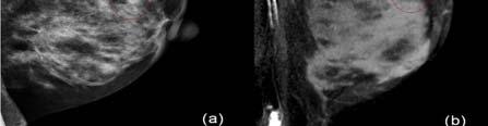
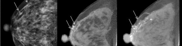
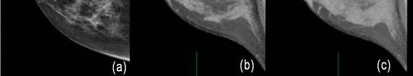
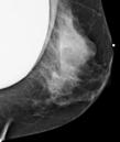
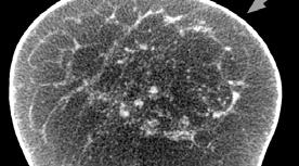
20 BCT Mammo Comparison Mammo BCT: Fibroadenoma He et al, Eur Radiol, 2016 Mammo BCT: Fibroadenoma Mammo BCT: Microcalcifications He et al, Eur Radiol, 2016 He et al, Eur Radiol,


21 Mammo BCT: Microcalcifications BREAST DOSE He et al, Eur Radiol, 2016 AGD Equivalent to 2 view Mammo Breast CT vs. Mammography Boone et al, Medical Physics, 2005; 32(12), 3767 Sechopoulos et al, Medical Physics, 2010; 37(8),


 2.5 2.0 1.5 1.")

22 Breast CT vs. Mammography BODY DOSE O Connell et al, J Clinic Imaging Science, 2012 Anthropomorphic Phantom Dose Variation with Projection Angle Dose Ratio (%) Lung (IL) Breast (CL) Stomach Projection Angle (deg) Sechopoulos et al, Radiology, 2008 Sechopoulos et al, Radiology,

23 Relative Organ Dose Organ 50 kvp 80 kvp Heart 1.75%/0.79% 3.08%/1.58% Lung (IL) 1.79%/2.03% 2.93%/3.25% Thymus 1.27% 2.35% Thyroid 0.08% 0.19% Uterus/Fetus 0.010% 0.026% Clavicle (IL) 1.57% 2.80% Rib Cage 4.14% 5.56% Sternum 5.16% 7.74% CLINICAL PERFORMANCE Sechopoulos et al, Radiology, 2008 First Results (N=69) Preliminary Results Diagnostic Work up 16 BI RADS 4 or 5 after work up Bx: 8 malignant lesions 8 benign lesions Breast CT Mammo 15 adequate BCT studies x Lindfors et al, Radiology, 2008 T 23
 270/332 288/332 279/332 81.3 % 86.7 % 84.0 % Specificity (76.7 85.4) (82.6 90.2) (79.6 87.8) AUROC 0.829 (0.783 0.875) 0.856 (0.820 0.888) 0.861 (0.825 0.")
24 Preliminary Results Diagnostic Work up Breast CT: 8 malignant 8 biopsy 8 benign 5 biopsy 3 no biopsy All patients (N = 442 lesions) Sensitivity MG US BCBCT C 93/ % ( ) 93/ % ( ) 97/ % ( ) 270/ / / % 86.7 % 84.0 % Specificity ( ) ( ) ( ) AUROC ( ) ( ) ( ) He et al, Eur Radiol, 2016 BI RADS c and d density breasts (n = 270 lesions) MG US BCBCT CE BCBCT Sensitivity 78.4 ( ) 81.1 ( ) 89.2 ( ) 98.7 ( ) Specificity 70.1 ( ) AUROC ( ) 82.7 ( ) ( ) 80.1 ( ) ( ) ( ) ( ) CLINICAL IMPLEMENTATION QUESTIONS He et al, Eur Radiol,


25 Questions in BCT Questions in BCT Screening or Work up? Tissue coverage? Questions in BCT 01%of 0.1% tumors in Tail of Spence (N=839) Questions in BCT Reading time? (reading strategy) Ampil et al, Anticancer Research,


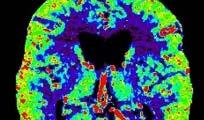
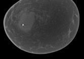
26 PET / CT for dedicated breast imaging MORFOLOGIA Courtesy of John Boone Contrast Enhanced BCT Contrast Enhanced BCT Malignant benign He et al, Eur Radiol, 2016 Courtesy of John Boone, Ph.D. 26


27 Contrast Enhanced BCT Subtraction Invasive Mammary Carcinoma Pre injection Post injection Registered Subtraction 5 Courtesy of John Boone, Ph.D. Coronal SPECT CT Volume Rendered SPECT CT Towards Tomographic Breast Imaging 2 D 2+ D 2.2 D 3 D SPECT CT Standard Mammography Stereoscopic Mammography Digital Tomosynthesis Dedicated Breast CT Large arrows point to the same location of a surgically confirmed DCIS lesion in 48yo patient. Small arrow points to posteriorly located biopsy clip. Blobs posterior to breast are chest wall signals from myocardial uptake of 99m Tc sestamibi. (If your optical system can handle it!) (Is more always better?) Courtesy of Dr. Martin Tornai of the Duke Multi Modality Imaging Lab 27
28 Towards Tomographic Breast Imaging 2 D 3 D 4 D 4D Breast CT Tumor Biology Profiling Standard Mammography Dedicated Breast CT (Is more always better?) Dedicated Breast CT FUNCTIONAL!! 4D Breast CT Neoadjuvant chemotherapy treatment planning 4D Breast CT Response prediction/monitoring 28
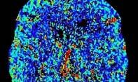


29 Correct Quantification 4D Noise Filtering Ramamurthy et al, PMB, Courtesy of Dr. Mathias Prokop Motion Correction PreCA PreCA registered onto CA CA Motion Correction Difference to CA Images Courtesy of Dr. Marc Kachelriess Courtesy of Dr. Marc Kachelriess 29


30 Patient BCT Classification 4D Breast Phantom Caballo et al, IEEE TMI, under review Caballo et al, to be presented at RSNA 2017 Image based phenotyping pcr prediction Courtesy of Dr. Despina Kontos Courtesy of Dr. Despina Kontos 30
31 Response Monitoring FUTURO Y PRESENTE (?) Courtesy of Dr. Despina Kontos Towards Tomographic Breast Imaging 2 D 2+ D 2.2 D 3 D Standard Mammography Stereoscopic Mammography Digital Tomosynthesis Dedicated Breast CT What if CC+MLO CC+MLO+ML 31


32 Methods Methods Diagnostic work up with mammo views Screening with DBT Combo mode Results 842 work-ups after screening DBT in previous 30 days 266 ML/LM view during work-up 28 BI-RADS 3 after work-up and excluded 133 biopsied cases 105 nonbiopsied cases 11 patients excluded due to breast size 121 patients in final cohort Breast Density Non dense 42% (51/121) Dense 58% (70/121) 0 patients excluded for breast implants 106 patients excluded followup unavailable 32
33 Abnormality Calcifications 31% (38/121) Soft Tissue 64% (78/121) Both 4% (5/121) ROC results Overall 3-View DM DBT P - value ( ) ( ) ROC results Secondary No significantdifference between: Attendings vs. fellows Microcalcifications vs. soft tissue Non dense vs. dense breasts Abnormality 3-View Digital Digital Breast Mammography (%) Tomosynthesis (%) Benign recall rate (43 84) (44 76) Malignant recall rate (50 90) (59 89) P - value


34 Reading time 3 view DM: 49.4seconds DBT: 73.7 p < Limitations Biased sampling: Screening with tomo Unilateral exams No location Towards Tomographic Breast Imaging 2 D 2+ D 2.2 D 3 D Standard Mammography Stereoscopic Mammography Digital Tomosynthesis Dedicated Breast CT Courtesy of Dr. Patrice Heid 34

35 Courtesy of Dr. Patrice Heid Courtesy of Dr. Patrice Heid Courtesy of Dr. Patrice Heid Warren et al, Medical Physics,


36 CDMAM Phantom Gold disc detection Warren et al, Medical Physics, 2012 Gold disc detection QUALITY CONTROL Warren et al, Medical Physics,


37 Screening stopped: 2016: 12 times 2015: 15 times 2014: 5 times Reasons Grid lines clearly visible Compression paddle height indication off Lines caused by mechanical shocks during readout Accidental dose increase by service engineer (70%!) Courtesy of Ruben van Engen Courtesy of Ruben van Engen Thickness indication off Thickness 63 mm W/Rh 30 kv Thickness 139 mm W/Ag 39 kv Thickness indication off Thickness 63 mm Thickness 139 mm W/Rh 30 kv W/Ag 39 kv Courtesy of Ruben van Engen Courtesy of Ruben van Engen 37


38 Lines due to mechanical shocks Example of grid lines visible Courtesy of Ruben van Engen Courtesy of Ruben van Engen Example of detector problem Step 1: Optimal Mammography Courtesy of Ruben van Engen 38


39 Advancing Human Health Courtesy of Dr. Ehsan Samei Education Clinical Service Research and Development Culture Summary Clinical need to reduce superposition Stereoscopic mammography Digital breast tomosynthesis Dedicatedbreast CT Summary Perhaps. 3 view mammography 39


40 Summary Definitely. Optimized technology at peak performance 40
Update of Digital Breast Tomosynthesis. Susan Orel Roth, MD
 Update of Digital Breast Tomosynthesis Susan Orel Roth, MD NCI estimates that : Why DBT? Approximately 20% of breast cancers are missed at mammography screening Average recall rates approximately 10%
Since its introduction in 2000, digital mammography has become
 Review Article Smith A, PhD email : Andrew.smith@hologic.com Since its introduction in 2000, digital mammography has become an accepted standard of care in breast cancer screening and has paved the way
Tomosynthesis and breast imaging update. Dr Michael J Michell Consultant Radiologist King's College Hospital NHS Foundation Trust
 Tomosynthesis and breast imaging update Dr Michael J Michell Consultant Radiologist King's College Hospital NHS Foundation Trust Breast imaging new technology BREAST CANCER FLT PET shows different grades
#46: DIGITAL TOMOSYNTHESIS: What is the Data Really Showing? TERMS (AKA) WHAT IS TOMOSYNTHESIS? 3/3/2014. Digital breast tomosynthesis =
 WHAT IS TOMOSYNTHESIS? 3/3/2014. Digital breast tomosynthesis =") #46: DIGITAL TOMOSYNTHESIS: What is the Data Really Showing? January K. Lopez, MD Hoag Breast Care Center Newport Beach, CA Disclosures: None TERMS (AKA) Digital breast tomosynthesis = DBT Tomo 3D Full
#46: DIGITAL TOMOSYNTHESIS: What is the Data Really Showing? January K. Lopez, MD Hoag Breast Care Center Newport Beach, CA Disclosures: None TERMS (AKA) Digital breast tomosynthesis = DBT Tomo 3D Full
Outline. Digital Breast Tomosynthesis: Update and Pearls for Implementation. Tomosynthesis Dataset: 2D/3D (Hologic Combo Acquisition)
") Outline Digital Breast Tomosynthesis (DBT) the new standard of care Digital Breast Tomosynthesis: Update and Pearls for Implementation Emily F. Conant, M.D. Professor, Chief of Breast Imaging Department
Outline Digital Breast Tomosynthesis (DBT) the new standard of care Digital Breast Tomosynthesis: Update and Pearls for Implementation Emily F. Conant, M.D. Professor, Chief of Breast Imaging Department
Digital Breast Tomosynthesis from a first idea to clinical routine
 International Master Programm Biomedical Engineering Digital Breast Tomosynthesis from a first idea to clinical routine Historical background 2D imaging of 3D objects has important limitations Jörg Barkhausen
Breast tomosynthesis reduces radiologist performance variability compared to digital mammography
 Breast tomosynthesis reduces radiologist performance variability compared to digital mammography Andrew Smith 1, Elizabeth Rafferty 2, Loren Niklason 1 1 Hologic, Inc., Bedford MA, USA 2 Massachusetts
Contrast-Enhanced Digital Mammography
 2015 ARRS Breast Symposium Contrast-Enhanced Digital Mammography John Lewin, M.D. Diversified Radiology of Colorado CEDM - Outline History Technique Literature Review / Cases Clinical Status Inexpensive,
Fremtidens rolle for tomosyntese
 Dansk Radiologisk Selskab 9.årsmøde Odense 3.januar, 214 Fremtidens rolle for tomosyntese Professor dr. med. Per Skaane Oslo University Hospital Ullevaal Breast Imaging Center Oslo / Norway PERSKA@ous-hf.no
Digital Breast Tomosynthesis Ready for Routine Screening?
 Digital Breast Tomosynthesis Ready for Routine Screening? Sophia Zackrisson MD, PhD, Assoc Prof of Radiology Skåne University Healthcare, Lund University, Sweden 1 Mammography screening 20% reduced breast
Contrast Enhanced Spectral Mammography (CESM) Updates
 Updates") Contrast Enhanced Spectral Mammography (CESM) Updates Georgeta Mihai, PhD, DABR Medical Physicist, BIDMC, Boston Assistant Professor, Harvard Medical School, Boston Disclosures None Acknowledgments: Da
Contrast Enhanced Spectral Mammography (CESM) Updates Georgeta Mihai, PhD, DABR Medical Physicist, BIDMC, Boston Assistant Professor, Harvard Medical School, Boston Disclosures None Acknowledgments: Da
Mammography limitations. Clinical performance of digital breast tomosynthesis compared to digital mammography: blinded multi-reader study
 Clinical performance of digital breast tomosynthesis compared to digital mammography: blinded multi-reader study G. Gennaro (1), A. Toledano (2), E. Baldan (1), E. Bezzon (1), C. di Maggio (1), M. La Grassa
Financial Disclosures
 Financial Disclosures 3D Mammography: The Latest Developments in the Breast Imaging Arena I have no financial disclosures Dr. Katharine Lampen-Sachar Breast and Body Radiologist Radiology Associates of
TOMOSYNTHESIS: WORTH ALL THE HYPE?
 X-Ray Associates of New Mexico, P.C. TOMOSYNTHESIS: WORTH ALL THE HYPE? MICHAEL N. LINVER, MD, FACR MAMMOGRAPHY: THE GOOD, THE PRETTY GOOD, & THE NOT SO GOOD MAMMOGRAPHY: THE GOOD, THE PRETTY GOOD, & THE
8/3/2016. DBT Physics Basic to Advanced: Primer On Tomosynthesis. Tomosynthesis Pedigree
 DBT Physics Basic to Advanced: Primer On Tomosynthesis Andrew D. A. Maidment, Ph.D. University of Pennsylvania Department of Radiology Acknowledgements of Support Research support from the Komen Foundation,
Disclosures. Outline. Learning Objectives. Introduction. Introduction. Stereotactic Breast Biopsy vs Mammography: Image Quality and Dose.
 Disclosures Stereotactic Biopsy vs Mammography: and Dose None Vikas Patel, PhD, DABR Upstate Medical Physics 2014 Annual Meeting The American Association of Physicists in Medicine Austin, TX Learning Objectives
What s New in Breast Imaging. Jennifer A. Harvey, M.D., FACR Professor of Radiology University of Virginia
 What s New in Breast Imaging Jennifer A. Harvey, M.D., FACR Professor of Radiology University of Virginia Disclosure Hologic, Inc. Shareholder and research agreement. Volpara Solutions, Ltd. Shareholder
Opportunities and Innovations in Digital Mammography John M. Sandrik, Ph.D. GE Healthcare Milwaukee, WI
 Opportunities and Innovations in Digital Mammography John M. Sandrik, Ph.D. GE Healthcare Milwaukee, WI john.sandrik@med.ge.com with many thanks to Vince Polkus, Advanced Applications Product Mgr. 1 Content
Breast CT and Dosimetry
 2013 ICTP/IAEA Training Course on Radiation Protection of Patients Trieste Breast CT and Dosimetry John M. Boone, Ph.D., FAAPM, FSBI, FACR Professor and Vice Chair (Research) of Radiology Professor of
Breast Imaging Update: Old Dog New Tricks
 Breast Imaging Update: Old Dog New Tricks Claire McKay, DO M&S Imaging Assoc. San Antonio, TX cmckayhart@juno.com Goals Describe modalities available, old and new Provide understanding of pros and cons
EARLY DETECTION: MAMMOGRAPHY AND SONOGRAPHY
 EARLY DETECTION: MAMMOGRAPHY AND SONOGRAPHY Elizabeth A. Rafferty, M.D. Avon Comprehensive Breast Center Massachusetts General Hospital Harvard Medical School Breast Cancer Screening Early detection of
CURRENTLY FDA APPROVED ARE FULL FIELD DIGITAL MAMMOGRAPHY SYSTEMS AND FILM SCREEN STILL BEING USED AT SOME INSTITUTIONS
 ABBY DUROJAYE,M.D CURRENTLY FDA APPROVED ARE FULL FIELD DIGITAL MAMMOGRAPHY SYSTEMS AND FILM SCREEN STILL BEING USED AT SOME INSTITUTIONS BOTH HAVE BEEN SHOWN TO BE EFFECTIVE TOOLS EARLY DETECTION OF BREAST
EARLY DETECTION: MAMMOGRAPHY AND SONOGRAPHY
 EARLY DETECTION: MAMMOGRAPHY AND SONOGRAPHY Elizabeth A. Rafferty, M.D. Avon Comprehensive Breast Center Massachusetts General Hospital Harvard Medical School Breast Cancer Screening Early detection of
FDA Executive Summary
 Meeting of the Radiological Devices Advisory Panel On October 24, 22, the panel will discuss, make recommendations, and vote on a premarket approval application supplement (P83/S) to expand the indications
TOMOSYNTHESIS. Daniela Bernardi. U.O. Senologia Clinica e Screening mammografico APSS Trento, Italy
 TOMOSYNTHESIS Daniela Bernardi U.O. Senologia Clinica e Screening mammografico APSS Trento, Italy BACKGROUND early detection through screening MAMMOGRAPHY is associated with reduced breast cancer morbidity
Fundamentals of Breast Tomosynthesis
 Fundamentals of Breast Tomosynthesis Improving the Performance of Mammography Andrew Smith, Ph.D. This white paper is one in a series of research overviws on advanced technologies in women s healthcare.
Radiation Dosimetry in Digital Breast Tomosynthesis. March, 2015 William J. O Connel, Dr. Ph, Senior Medical Physicist
 Radiation Dosimetry in Digital Breast Tomosynthesis March, 2015 William J. O Connel, Dr. Ph, Senior Medical Physicist Imagination at work. Syllabus 1. Introduction 2. Dosimetry in Mammography 3. Dosimetry
Breast Tomosynthesis
 Breast Tomosynthesis The Use of Breast Tomosynthesis in a Clinical Setting 2 What s Inside Introduction... 1 Initial Hologic Clinical Trial Purpose and Methodology... 1 Clinical Trial Results... 2 Improved
Breast Tomosynthesis
 Breast Tomosynthesis The Use of Breast Tomosynthesis in a Clinical Setting 2 What s Inside Introduction... 1 Initial Hologic Clinical Trial Purpose and Methodology... 1 Clinical Trial Results... 2 Improved
Corporate Medical Policy
 Corporate Medical Policy File Name: Origination: Last CAP Review: Next CAP Review: Last Review: digital_breast_tomosynthesis 3/2011 6/2016 6/2017 11/2016 Description of Procedure or Service Conventional
Breast Tomosynthesis An additional screening tool in the fight against breast cancer
 What to Expect Breast Tomosynthesis An additional screening tool in the fight against breast cancer Every woman over 40 should be examined for breast cancer once a year. American Cancer Society What to
WHAT TO EXPECT. Breast Tomosynthesis An additional screening tool in the fight against breast cancer HOLOGIC. The Women's Health Company
 WHAT TO EXPECT Breast Tomosynthesis An additional screening tool in the fight against breast cancer HOLOGIC The Women's Health Company ...,. Screening for breast cancer Doctors and scientists agree that
Patient Dosimetry in Mammography and Tomosynthesis:
 2013 ICTP/IAEA Training Course on Radiation Protection of Patients Trieste Patient Dosimetry in Mammography and Tomosynthesis: What to measure, why and how John M. Boone, Ph.D., FAAPM, FSBI, FACR Professor
Mammography. What is Mammography?
 Scan for mobile link. Mammography Mammography is a specific type of breast imaging that uses low-dose x-rays to detect cancer early before women experience symptoms when it is most treatable. Tell your
arxiv: v2 [cs.cv] 8 Mar 2018
![arxiv: v2 [cs.cv] 8 Mar 2018](/thumbs/87/97094636.jpg "arxiv: v2 [cs.cv] 8 Mar 2018") Automated soft tissue lesion detection and segmentation in digital mammography using a u-net deep learning network Timothy de Moor a, Alejandro Rodriguez-Ruiz a, Albert Gubern Mérida a, Ritse Mann a, and
Automated soft tissue lesion detection and segmentation in digital mammography using a u-net deep learning network Timothy de Moor a, Alejandro Rodriguez-Ruiz a, Albert Gubern Mérida a, Ritse Mann a, and
Current Status of Supplementary Screening With Breast Ultrasound
 Current Status of Supplementary Screening With Breast Ultrasound Stephen A. Feig, M.D., FACR Fong and Jean Tsai Professor of Women s Imaging Department of Radiologic Sciences University of California,
Molecular Breast Imaging: History and Recent Developments
 Molecular Breast Imaging: History and Recent Developments Associate Professor, Department of Imaging Physics The University of Texas MD Anderson Cancer Center, Houston, Texas Educational Objectives 1.
A comparison of the accuracy of film-screen mammography, full-field digital mammography, and digital breast tomosynthesis
 Clinical Radiology xxx (2012) 1e6 Contents lists available at SciVerse ScienceDirect Clinical Radiology journal homepage: www.clinicalradiologyonline.net A comparison of the accuracy of film-screen mammography,
Emerging Technologies in Breast Imaging
 June 30, 2016 Emerging Technologies in Breast Imaging Jay A. Baker, M.D. Division of Breast Imaging New Technologies in Breast Imaging Full Field Digital Mammography (FFDM) X-ray/Mammography Computer-Aided
Standard Breast Imaging Modalities. Lilian Wang, M.D. Breast Imaging Section Department of Radiology Northwestern Medicine
 Standard Breast Imaging Modalities Lilian Wang, M.D. Breast Imaging Section Department of Radiology Northwestern Medicine Overview Standard breast imaging modalities Mammography Ultrasound MRI Imaging
Detection and Classification of Calcifications on Digital Breast Tomosynthesis and 2D Digital Mammography: A Comparison
 Women s Imaging Original Research Spangler et al. Digital Breast Tomosynthesis Versus 2D Digital Mammography Women s Imaging Original Research FOCUS ON: M. Lee Spangler 1 Margarita L. Zuley 2 Jules H.
Digital Breast Tomosynthesis
 Digital Breast Tomosynthesis Policy Number: Original Effective Date: MM.05.012 06/28/2013 Line(s) of Business: Current Effective Date: HMO; PPO; QUEST 06/28/2013 Section: Radiology Place(s) of Service:
Digital Breast Tomosynthesis in the Diagnostic Environment: A Subjective Side-by-Side Review
 Women s Imaging Original Research Hakim et al. Digital Breast Tomosynthesis Women s Imaging Original Research Christiane M. Hakim 1 Denise M. Chough 1 Marie A. Ganott 1 Jules H. Sumkin 1 Margarita L. Zuley
Medical Policy An independent licensee of the Blue Cross Blue Shield Association
 Digital Breast Tomosynthesis Page 1 of 31 Medical Policy An independent licensee of the Blue Cross Blue Shield Association Title: Digital Breast Tomosynthesis Professional Institutional Original Effective
The latest developments - Automated Breast Volume Scanning. Dr. med. M. Golatta
 The latest developments - Automated Breast Volume Scanning Dr. med. M. Golatta Automated Breast Volume US: Why? o Mammography is limited in dense breasts: high false negative rate o Many of these tumors
Supplemental Screening for Dense Breasts. Reagan Leverett, MD, MS
 Supplemental Screening for Dense Breasts Reagan Leverett, MD, MS Outline Anatomy and Density Risk of dense breasts Theory of Supplemental Screening Options for supplemental screening Tomosynthesis Ultrasound
Breast Tomosynthesis. What is breast tomosynthesis?
 Scan for mobile link. Breast Tomosynthesis Breast tomosynthesis is an advanced form of mammography, a specific type of breast imaging that uses low-dose x-rays to detect cancer early when it is most treatable.
Assessment of extent of disease: digital breast tomosynthesis (DBT) versus full-field digital mammography (FFDM)
 versus full-field digital mammography (FFDM)") Assessment of extent of disease: digital breast tomosynthesis (DBT) versus full-field digital mammography (FFDM) Poster No.: C-1237 Congress: ECR 2012 Type: Scientific Paper Authors: N. Seo 1, H. H. Kim
Assessment of extent of disease: digital breast tomosynthesis (DBT) versus full-field digital mammography (FFDM) Poster No.: C-1237 Congress: ECR 2012 Type: Scientific Paper Authors: N. Seo 1, H. H. Kim
Updates in Mammography. Dr. Yang Faridah A. Aziz Department of Biomedical Imaging University Malaya Medical Centre
 Updates in Mammography Dr. Yang Faridah A. Aziz Department of Biomedical Imaging University Malaya Medical Centre Updates in Mammography Breast Imaging Dr. Yang Faridah A. Aziz Department of Biomedical
High Risk Screening: A Multimodality Approach
 High Risk Screening: A Multimodality Approach John Lewin, M.D., FACR, FSBI The Women s Imaging Center Denver, Colorado Disclosures Consultant to Hologic Previously received research funds from Hologic
WHAT TO EXPECT. Genius 3D Mammography Exam. The most exciting advancement in mammography in over 30 years
 WHAT TO EXPECT Genius 3D Mammography Exam The most exciting advancement in mammography in over 30 years Screening for breast cancer Doctors and scientists agree that early detection is the best defense
Pitfalls and Limitations of Breast MRI. Susan Orel Roth, MD Professor of Radiology University of Pennsylvania
 Pitfalls and Limitations of Breast MRI Susan Orel Roth, MD Professor of Radiology University of Pennsylvania Objectives Review the etiologies of false negative breast MRI examinations Discuss the limitations
Challenges to Delivery of High Quality Mammography
 Challenges to Delivery of High Quality Mammography Overview of Current Challenges Barbara Monsees, Washington University Geographic Access, Equity and Impact on Quality Tracy Onega, Dartmouth Medical School
Here are examples of bilateral analog mammograms from the same patient including CC and MLO projections.
 Good afternoon. It s my pleasure to be discussing Diagnostic Breast Imaging over the next half hour. I m Wei Yang, Professor of Diagnostic Radiology and Chief, the Section of Breast Imaging as well as
Emerging Techniques in Breast Imaging: Contrast-Enhanced Mammography and Fast MRI
 Emerging Techniques in Breast Imaging: Contrast-Enhanced Mammography and Fast MRI Lilian Wang, M.D. Breast Imaging Section Department of Radiology Northwestern Medicine Overview Rationale for new imaging
Breast Tomosynthesis
 Breast Tomosynthesis Considerations for Routine Clinical Use Breast Tomosynthesis Considerations for routine clinical use Andrew Smith, Ph.D., Vice President - Imaging Sciences, Hologic This white paper
New Imaging Modalities for better Screening and Diagnosis
 New Imaging Modalities for better Screening and Diagnosis Miri Sklair-Levy, MD Department of Diagnostic Imaging Sheba Medical Center, Sackler School of Medicine, Tel Aviv University Department of Diagnostic
Improving Reading Time of Digital Breast Tomosynthesis with Concurrent Computer Aided Detection
 White Paper Improving Reading Time of Digital Breast Tomosynthesis with Concurrent Computer Aided Detection WHITE PAPER 2 3 Abstract PowerLook Tomo Detection, a concurrent computer-aided detection (CAD)
Implementation of a New Tomosynthesis Program: A Physicists Perspective
 Implementation of a New Tomosynthesis Program: A Physicists Perspective Bill Geiser, MS DABR Senior Medical Physicist wgeiser@mdanderson.org 1 Conflict of Interest None of the authors nor their immediate
Mammographic imaging of nonpalpable breast lesions. Malai Muttarak, MD Department of Radiology Chiang Mai University Chiang Mai, Thailand
 Mammographic imaging of nonpalpable breast lesions Malai Muttarak, MD Department of Radiology Chiang Mai University Chiang Mai, Thailand Introduction Contents Mammographic signs of nonpalpable breast cancer
Innovations and Applications of Tomosynthesis. Andrew D. A. Maidment, Ph.D. University of Pennsylvania Department of Radiology
 Innovations and Applications of Tomosynthesis Andrew D. A. Maidment, Ph.D. University of Pennsylvania Department of Radiology Acknowledgements of Support Grant support from the Komen Foundation, DOD, NIH,
Clinical Policy: Digital Breast Tomosynthesis Reference Number: CP.MP.90
 Clinical Policy: Reference Number: CP.MP.90 Effective Date: 01/18 Last Review Date: 12/17 Coding Implications Revision Log See Important Reminder at the end of this policy for important regulatory and
Mammography. Background and Perspective. Mammography Evolution. Background and Perspective. T.R. Nelson, Ph.D. x41433
 - 2015 Background and Perspective 2005 (in US) Women Men Mammography Invasive Breast Cancer Diagnosed 211,240 1,690 Noninvasive Breast Cancer Diagnosed 58,940 Deaths from Breast Cancer 40,410 460 T.R.
The value of the craniocaudal mammographic view in breast cancer detection: A preliminary study
 The value of the craniocaudal mammographic view in breast cancer detection: A preliminary study P D Trieu 1, Prof. P C Brennan 1, Dr. W Lee 2, Dr. E Ryan 1, Dr. W Reed 1, Dr. M Pietrzyk 1 1. Medical Image
Mammography. What is Mammography? What are some common uses of the procedure?
 Mammography What is Mammography? Mammography is a specific type of imaging that uses a low-dose x-ray system to examine breasts. A mammography exam, called a mammogram, is used to aid in the early detection
Breast Cancer Screening and Diagnosis
 Breast Cancer Screening and Diagnosis Priya Thomas, MD Assistant Professor Clinical Cancer Prevention and Breast Medical Oncology University of Texas MD Anderson Cancer Center Disclosures Dr. Thomas has
Melissa Hartman, DO Women s Health Orlando VA Medical Center
 Melissa Hartman, DO Women s Health Orlando VA Medical Center Most common non-skin cancer and Second deadliest cancer in women Majority are diagnosed by abnormal screening study An approach to breast cancer
SCREENING FOR BREAST CANCER BREAST IMAGING
 SCREENING FOR BREAST CANCER BREAST IMAGING Liane Philpotts, MD, FSBI, FACR Professor, Radiology and Biomedical Imaging Division Chief, Breast Imaging Dec. 5, 2017 Warner, E. NEJM 2011 Screening for
Breast positioning system for full field digital mammography and digital breast tomosynthesis system
 Breast positioning system for full field digital mammography and digital breast tomosynthesis system Mari Varjonen* a, Martti Pamilo b, Pirjo Hokka b, Riina Hokkanen a, Pekka Strömmer a a Planmed Oy Asentajankatu
Proven clinical effectiveness at low radiation dose
 MicroDose Mammography Solutions Proven clinical effectiveness at low radiation dose Several studies provide evidence that Philips MicroDose Mammography* can provide outstanding image quality at 18% to
Dense Breasts. A Breast Cancer Risk Factor and Imaging Challenge
 Dense Breasts A Breast Cancer Risk Factor and Imaging Challenge Renee Pinsky, MD University of Michigan Department of Radiology Division of Breast Imaging No Disclosures QUIZ: ARE YOU DENSE? a. Breast
Screening Options in Dense Breasts. Donna Plecha, M.D. Co-Director UHCMC Breast Centers Associate Professor of Radiology Director of Breast Imaging
 Screening Options in Dense Breasts Donna Plecha, M.D. Co-Director UHCMC Breast Centers Associate Professor of Radiology Director of Breast Imaging Dense Breasted Women Decreased sensitivity of mammography
2/14/2019. Advances in Breast Imaging. Outline. Jiali Wang, Ph.D., DABR Medical Physicist. February 23, 2019
 Advances in Breast Imaging SCPMG Medical Imaging Technology & Informatics Jiali Wang, Ph.D., DABR Medical Physicist February 23, 2019 Outline History of breast imaging Advances in breast imaging a) Full-Field
BREAST IMAGING and NEW IMAGING MODALITIES- A Surgeons view
 BREAST IMAGING and NEW IMAGING MODALITIES- A Surgeons view DR CHANTEL THORNTON SPECIALIST BREAST CANCER SURGEON BMSc (hons) MBBS (hons) FRACS Epworth Hospital, Richmond- Agora Centre for Women s Health
Diagnostic Medical Physicist Via Christi Hospitals Wichita, Wichita, KS
 Digital Breast Tomosynthesis SWAAPM Meeting 30 Mar 2012 Jerry A. Thomas, MS, FAAPM, DABR, CHP, DABSNM Diagnostic Medical Physicist Via Christi Hospitals Wichita, Wichita, KS Talk Overview Breast Cancer
Policy Library Clinical Advantages of Digital Breast Tomosynthesis in Symptomatic Patients
 Policy Library Clinical Advantages of Digital Breast Tomosynthesis in Symptomatic Patients Version: 1 Approved by: Faculty of Clinical Radiology Council Date of approval: Click and type: day month and
Andrew Karellas, PhD
 Advanced Imaging for Breast Cancer: Screening, Diagnosis, and Assessing Response to Therapy The Role of Tomosynthesis Andrew Karellas, PhD Department of Radiology University of Massachusetts Medical School
Physics of MBI (~10 slides)
") Physics of MBI (~10 slides) Molecular Breast Imaging (MBI) physics and performance testing JW Hugg, BR Simrak, PD Smith, BE Patt Gamma Medica, Inc., Northridge, CA Molecular Breast Imaging (MBI) is an
Physics of MBI (~10 slides) Molecular Breast Imaging (MBI) physics and performance testing JW Hugg, BR Simrak, PD Smith, BE Patt Gamma Medica, Inc., Northridge, CA Molecular Breast Imaging (MBI) is an
PhD Title: Improving QA/QC in mammography screening and breast in Montenegro
 3rd Doctoral Coordinated Research Project (CRP) on Advances in medical imaging techniques (E2.40.19) MNE BEL PhD Title: Improving QA/QC in mammography screening and breast in Montenegro MNE BEL PhD Candidate:
Breast Cancer Imaging
 Breast Cancer Imaging I. Policy University Health Alliance (UHA) will cover breast imaging when such services meet the medical criteria guidelines (subject to limitations and exclusions) indicated below.
Moderne mammadiagnostikk hvor står vi og hvor går vi?
 NBCG Oslo 15. juni 2018 Moderne mammadiagnostikk hvor står vi og hvor går vi? Professor dr.med. emeritus Per Skaane Oslo University Hospital Ullevaal Breast Imaging Center Oslo / Norway PERSKA@ous-hf.no
WHAT TO EXPECT. Genius 3D MAMMOGRAPHY Exam. The most exciting advancement in mammography in over 30 years
 WHAT TO EXPECT Genius 3D MAMMOGRAPHY Exam The most exciting advancement in mammography in over 30 years 91% of patients agree the quality of care provided by the facility was better with a Genius 3D MAMMOGRAPHY
Women s Imaging Original Research
 Women s Imaging Original Research Brandt et al. DBT for Screening Recalls Without Calcifications Women s Imaging Original Research FOCUS ON: Kathleen R. Brandt 1 Daniel A. Craig 1 Tanya L. Hoskins 2 Tara
Acknowledgments. A Specific Diagnostic Task: Lung Nodule Detection. A Specific Diagnostic Task: Chest CT Protocols. Chest CT Protocols
 Personalization of Pediatric Imaging in Terms of Needed Indication-Based Quality Per Dose Acknowledgments Duke University Medical Center Ehsan Samei, PhD Donald Frush, MD Xiang Li PhD DABR Cleveland Clinic
9/3/2015. New Mammographic Modality Training. Mammographer. Qualified Radiologic Technologist. Tomosynthesis Training
 FDA Required Breast Tomosynthesis Training New Mammographic Modality Training On any new mammography technology, such as breast tomosynthesis, the Mammography Quality Standards Act (MQSA) (http:fda.gov/radiation-emitting
CZT Detectors for Medical Imaging and Plant Biology
 Small Business, High Tech Development CZT Detectors for Medical Imaging and Plant Biology Haris Kudrolli Inc. Workshop 2018 January 11, 2018 Outline Scientific Section CZT detectors Plant Imaging Applications
Dose reduction in Hologic Selenia FFDM units through AEC optimization, without compromising diagnostic image quality.
 Dose reduction in Hologic Selenia FFDM units through AEC optimization, without compromising diagnostic image quality. Poster No.: C-1636 Congress: ECR 2015 Type: Authors: Keywords: DOI: Scientific Exhibit
Digital breast tomosynthesis
 GE Healthcare Digital breast tomosynthesis Daniel B. Kopans, M.D., F.A.C.R. Professor of Radiology Harvard Medical School Senior Radiologist - Breast Imaging Division Massachusetts General Hospital Since
Contrast-Enhanced Breast Tomosynthesis
 Contrast-Enhanced Breast Tomosynthesis AIP Industrial Physics Forum 2009 Andrew D. A. Maidment, Ph.D. Chief, Physics Section Department of Radiology University of Pennsylvania Acknowledgements of Support
ACCREDITATION DOCUMENT THE RADIOGRAPHER
 ACCREDITATION DOCUMENT THE RADIOGRAPHER Nijmegen, October 2012 1. Introduction An optimal quality of mammography is one of the fundamental requirements for successful breast cancer screening programmes.
Screening with Abbreviated Breast MRI (AB-MR)
") Screening with Abbreviated Breast MRI (AB-MR) Christopher Comstock, MD, FACR, FSBI Department of Radiology Memorial Sloan-Kettering Cancer Center New York, NY Outline History of our approach to screening
Screening with Abbreviated Breast MRI (AB-MR) Christopher Comstock, MD, FACR, FSBI Department of Radiology Memorial Sloan-Kettering Cancer Center New York, NY Outline History of our approach to screening
THE FUTURE OF MEDICAL IMAGING A. D. A. Maidment* Department of Radiology, University of Pennsylvania, 3400 Spruce Street, Philadelphia, PA 19104, USA
 Radiation Protection Dosimetry (2010), Vol. 139, No. 1 3, pp. 3 7 Advance Access publication 18 March 2010 doi:10.1093/rpd/ncq090 A. D. A. Maidment* Department of Radiology, University of Pennsylvania,
Accreditation Case Review: Mammography and Stereotactic Biopsy
 Accreditation Case Review: Mammography and Stereotactic Biopsy Brett T. Parkinson MD Breast Imaging Director Breast Care Services Intermountain Healthcare Chair, ACR Committee on Mammography Accreditation
CT Laser Mammography CTLM
 CT Laser Mammography CTLM Scanning for life IMAGINE...A breast imaging method that is painless, non-invasive, does not touch the breast, uses no harmful ionizing radiation, can be repeated as often as
Imaging Systems. Nooshin Kiarashi. Department of Electrical and Computer Engineering Duke University. Date: Approved: Loren W. Nolte, Co- Supervisor
 Towards Realizing Virtual Clinical Trials for Optimization and Evaluation of Breast Imaging Systems by Nooshin Kiarashi Department of Electrical and Computer Engineering Duke University Date: Approved:
Session 4: Test instruments to assess interpretive performance challenges and opportunities Overview of Test Set Design and Use
 Session 4: Test instruments to assess interpretive performance challenges and opportunities Overview of Test Set Design and Use Robert A. Smith, PhD American Cancer Society Test Sets vs. Audits Benefits
The Radiology Aspects
 REQUIREMENTS FOR INTERNATIONAL ACCREDITATION OF BREAST CENTERS/UNITS The Radiology Aspects Miri Sklair-Levy, Israel RADIOLOGY GUIDELINES FOR QUALITY ASSURANCE IN BREAST CANCER SCREENING AND DIAGNOSIS Radiologists
Multicenter Trial, Phase I Assessment of 2-D FFDM Versus Combo of 2-D and 3-D Tomosynthesis in Breast Cancer Screening
 ACRIN PA 4006: Comparison of Full Field Digital Mammography with Digital Breast Tomosynthesis Image Acquisition in Relation to Screening Call-Back Rate Imaging Manual Multicenter Trial, Phase I Assessment
Kish chakrabarti, Ph.D. Senior Physicist CDRH/FDA
 Facility Certification Extension Requirements, Quality Assurance and Medical Physicists role for Hologic Selenia Dimensions Digital Breast Tomosynthesis (DBT) System Kish chakrabarti, Ph.D. Senior Physicist
Mammography: Analysis and Advanced Techniques $50. Special Price. 8 credit seminar October 28, 2017 Knoxville, TN. a division of Herzing University
 Mammography: Analysis and Advanced Techniques $50 Special Price 8 credit seminar October 28, 2017 Knoxville, TN a division of Herzing University Seminar Schedule 8:00 am Registration and Coffee 8:30 am