MicroRNAs: molecular features and role in cancer.
|
|
|
- Gabriel Shaw
- 6 years ago
- Views:
Transcription


1 MicroRNAs: molecular features and role in cancer. Elodie Lages, Hélène Ipas, Audrey Guttin, Houssam Nesr, François Berger, Jean-Paul Issartel To cite this version: Elodie Lages, Hélène Ipas, Audrey Guttin, Houssam Nesr, François Berger, et al.. MicroRNAs: molecular features and role in cancer.. Frontiers in Bioscience, Frontiers in Bioscience, 2012, 17, pp <inserm > HAL Id: inserm Submitted on 20 Sep 2012 HAL is a multi-disciplinary open access archive for the deposit and dissemination of scientific research documents, whether they are published or not. The documents may come from teaching and research institutions in France or abroad, or from public or private research centers. L archive ouverte pluridisciplinaire HAL, est destinée au dépôt et à la diffusion de documents scientifiques de niveau recherche, publiés ou non, émanant des établissements d enseignement et de recherche français ou étrangers, des laboratoires publics ou privés.
2 MicroRNAs: molecular features and role in cancer Elodie Lages 1,2, Hélène Ipas 1,2, Audrey Guttin 1,2,3, Houssam Nesr 1,2, François Berger 1,2, Jean-Paul Issartel 1,2,3,4 1 INSERM, U836, Team7 Nanomedicine and Brain, BP 170, Grenoble Cedex 9, F-38042, France 2 Université Joseph Fourier, Institut des Neurosciences, BP 170, Grenoble Cedex 9, F-38042, France 3 Clinical Transcriptomics and Proteomics Facilities, Institut de Biologie et Pathologie - Centre Hospitalier Universitaire and Grenoble Institut des Neurosciences, Grenoble, France. 4 CNRS, France. TABLE OF CONTENTS 1. Abstract Introduction mirna: nomenclature, biogenesis and functions mirna nomenclature Interfering RNAs: similarities and differences mirna biogenesis Localization and structure of mirna coding genes Transcription of primary mirna transcript Maturation of pri-mirnas into mirnas Formation of the pre-mirna in the nucleus Pre-miRNAs export from the nucleus to the cytoplasm Mature mirna production in the cytoplasm Gene expression regulation by mirnas mirisc (mirna-induced silencing complex) formation Interaction between mirnas and mrna targets Mechanisms of interaction Categories of target sites Biocomputational tools for the prediction of target sites Molecular mechanisms of translation regulation of mrna targets by mirnas mrna maturation, stability, and translation: basic features Translation initiation repression by mirnas Repression of the postinitiation step of translation by mirnas Regulation by degradation of the mrna targets Translation activation by mirnas mirnas in tumorigenesis and tumor development processes Modifications of mirna expression levels in cancer mirna expression profiles in tumors Deregulation of mirna expression by defects in the biogenesis mechanism Deregulation of mirna expression by chromosomal rearrangements Deregulation of mirna expression due to point mutations in coding genes Modifications of mirna expression due to transcriptional regulation Modifications of the interactions between mirnas and their mrna targets Deregulation by chromosomal modification Deregulation by point mutations Oncogene and tumor suppressor mirnas involved in molecular pathways of tumor development Apoptosis and cellular proliferation processes Angiogenesis process Exosomal mirnas: new potential tumor biomarkers mirnas in cancer therapy Conclusion Acknowledgements References Abstract micrornas (mirnas) are small noncoding endogenously produced RNAs that play key roles in controlling the expression of many cellular proteins. Once they are recruited and incorporated into a ribonucleoprotein complex mirisc, they can target specific mrnas in a mirna sequence-dependent process and interfere in the translation into proteins of the targeted mrnas via several mechanisms. Consequently, mirnas can regulate many cellular pathways and processes. Dysregulation of their physiological roles may largely contribute to disease. In particular, in cancer, mirnas can be involved in the deregulation of the expression of important genes that play key roles in tumorigenesis, tumor development, and angiogenesis and have oncogenic or tumor suppressor roles. This review focuses on the biogenesis and maturation of mirnas, their mechanisms of gene regulation, and the way their expression is deregulated in cancer. The involvement of mirnas in several oncogenic pathways such as angiogenesis and apoptosis, and in the inter-cellular dialog mediated by mirna-loaded exosomes as well as the development of new therapeutical strategies based on mirnas will be discussed. 1
3 2. Introduction mirnas were described in Caenorhabditis elegans at the beginning of the 1990s (1) as small noncoding endogenously produced RNAs that inhibit mrna translation. Nevertheless, the existence in eukaryotic cells of RNA molecules called tcrnas (translational control RNAs), that were able to inhibit the translation of mrnas, had been previously suggested by Heywood et al. (2). The first mirna described in C. elegans, lin-4, is a small RNA containing complementary sequences for repeated elements of the 3 UTR region of the lin-14 and lin-28 mrnas. Lee et al. proposed a lin-4-dependent regulation of the translation of the lin-14 and lin-28 mrnas due to a sense antisense RNA interaction. This regulation model was validated by Wightman et al. who used reporter genes and demonstrated that the 3 untranslated region (UTR) of lin-14 mrna was essential for the regulation of its translation by lin-4 (3). A second small RNA, let-7, which, similar to lin-4, comes from the family of heterochronic switch genes, and controls the larval development of C. elegans was then discovered by Reinhart et al. (4). let-7 plays a role in the transition of the larval L3/L4 stage to the adult stage, repressing the expression of LIN-41 by interacting with the 3 UTR region of the mrna. lin-4 and let-7 were first called small temporal RNAs (strnas) (5) due to their role in the control of development and were renamed mirnas in 2001 (6) (Figure 1). Afterwards, several mirnas were described in other organisms such as Drosophila melanogaster, mice, and plants as well as in human cell lines (7-13). 3. mirna: nomenclature, biogenesis and functions 3.1. mirna nomenclature mirnas are identified either by cloning and sequencing studies or informatics approaches using prediction programs based on conservation of mirna within species and on the detection of the stem-loop structures typical of mirna precursors. All mirnas named in scientific publications and generally found using cloning and sequencing techniques are listed in the database called mirbase ( (14)). mirbase version 18 (November 2011) now classifies 1527 mirnas for humans. mirna-encoding genes are first transcribed in stem-loop structured primary transcripts called pri-mirnas, which are then excised by RNAse III Drosha and its cofactor DGCR8, freeing a precursor called pre-mirna. These precursor molecules are then exported from the nucleus to the cytoplasm with exportin-5. They are then matured by the RNAse III Dicer to free a duplex mirna-mirna*. One strand, called mature, is then incorporated into the RISC complex, a complex containing essentially Argonaute-type proteins. (Figure 2). While genes or pre-mirnas are noted mir-x, mature mirnas are noted mir-x where X is a numeric value. A three-letter code is added as a prefix to refer to the organism they originate from. One letter stands for the organism genus and the two others for the species, e.g., hsa for Homo sapiens or mmu for Mus musculus. When mature mirnas differ by a few bases, a letter is added as a suffix, reflecting the difference: e.g., mir-181a and mir- 181b: - mir-181a: aacauucaacgcugucggugagu - mir-181b: aacauucauugcugucggugggu When both strands of a pre-mirna are processed as mature mirnas, an indication is given to specify which arm of the hairpin structure of the pre-mirna generates one or the other of the two mirnas. Several nomenclatures can be used: - mir-x and mir-x*: mir-x* is the less abundant of the two mature mirnas in the cells. - mir-x-5p and mir-x-3p: 5p and 3p stand for the 5 and 3 arms of the stem-loop structure of the pre-mirna, respectively. This nomenclature is mainly used when the respective abundance of the two mirnas is still unknown (15). - mir-x-s (5 arm) and mir-x-as (3 arm). Moreover, a mature mirna can result from the transcription and maturation of transcripts from separate genomic loci. A numeral suffix is added to the names of all the multiloci mirnas, e.g., mir-181a-1 and mir-181a-2 (mir-181a-1 originates from a locus on chromosome 1 and mir-181a-2 from a locus on chromosome 9). Nevertheless, some mirnas, such as hsa-let-7 and cel-lin-4, do not follow the above-described nomenclature Interfering RNAs: similarities and differences RNA interference (RNAi) is mainly responsible for the post-transcriptional regulation of gene expression (mrna cleavage or decay, translation repression or activation) but is also involved in transcriptional regulation as described by various studies (16-19). Mainly two classes of small RNAs interfere in the RNA interference process: mirnas and sirnas (short interfering RNA). These RNAs share similarities such as their short size (about 22 nucleotides), their maturation by the ribonuclease III Dicer, and their implication in the RISC complex for post-transcriptional repression. Nevertheless, there are a number of important differences, such as the following: - mirnas are exclusively endogenously produced. They come from intrinsic genes of the organism they interfere in, whereas sirnas may originate from viruses, transposable elements, perfectly paired endogenous double-stranded RNA resulting from an antisense transcription of various loci (20), or transfected exogenous and double-stranded RNAs. 2
4 - mirnas play a role in the regulation of genes that do not generate them, whereas sirnas mainly regulate the genes that produce them. This point is critical in order to secure host cell defense against parasite RNAs such as viruses or transposons. - In mammals, mirnas have a varying degree of complementarity with their target, whereas sirna always have a perfect complementarity with their targets. When the complementarity is perfect, the target mrna is degraded in the presence of the Ago2 protein, which is the sole protein of the four Argonaute family proteins responsible for endonucleolytic cleavage. In plants, the difference between mirnas and sirnas is very small because the pairing between mirna and mrna is often perfect (21). In addition to mirnas and sirnas, there are other small RNAs that contribute to maintaining the genome integrity: the pirnas (piwi-interacting RNAs). They are longer (26 31 nucleotides), abundant in germinal cells but can also be found in somatic tissues (22-24) (Figure 3). These RNAs, formerly called repeat-associated sirna (rasirna), were discovered in D. melanogaster (25). In 2006, several studies showed the presence of pirnas in mammals (25-28). Contrary to sirna and mirna, it seems that they come from long single-stranded RNA precursors and their biogenesis does not include the RNAse III Dicer. Once matured, pirnas associate themselves with PIWI proteins, a subfamily of Argonaute proteins, principally to target transposons and preserve genome integrity (22) mirna biogenesis Localization and structure of mirna coding genes These genes are widespread on all chromosomes except for chromosome Y and approximately 50% of the described mirnas are located in clusters. When these clusterized mirnas are under the control of a single promoter, they are dubbed polycistronic mirnas. mirna coding genes can be independent gene units and mirnas are then called intergenic; they can also be gene units coding for pre-mrna that either code for proteins or do not code for them. In this case, mirnas can be intronic or exonic (Figure 4) (29-31) Transcription of primary mirna transcript Most of the mirna coding genes are transcribed by the RNA polymerase II, producing long primary transcripts (pri-mirnas). These pri-mirnas have a typical structure composed of a stem (about 33 nucleotides), a loop connecting the two strands of the stem, and single-stranded flanking sequences (Figure 5). Like mrnas, these sequences contain a 7- methyl-guanosine (m7g) cap at their 5 end and a 3 polya tail (32, 33). With the transcription performed by the RNA polymerase II, the regulation of this mechanism could be similar to the regulation of the transcription of protein-coding genes. The localization of promoter regions of mirna coding genes was predicted in silico by genomic mapping of the transcription start sites (TSS) and by mapping of transcription factor binding motifs. This was reported in Arabidopsis thaliana, C. elegans, and H. sapiens (34-36). Furthermore, two studies highlighted the localization and characteristics of TSS sequences of mirnas in human using high-throughput genomic analyses such as chromatin immunoprecipitation (37, 38) Maturation of pri-mirnas into mirnas Formation of the pre-mirna in the nucleus In human, primary transcripts are processed in the nucleus by a protein complex called microprocessor that is composed of the ribonuclease Drosha associated with its cofactor DGCR8 (DiGeorge syndrome Critical Region gene 8) (39). The Drosha complex also contains several other factors such as EWSR1, FUS, numerous heterogeneous nuclear ribonucleoproteins (hnrnps), p68 (DDX5) and p72 (DDX17) DEAD-box helicases. The roles of these accessory components in mirna maturation is largely unknown, some hnrnps and p68/p72 may promote the fidelity and activity of the Drosha processing (40, 41). DGCR8, which interacts with double-stranded RNA, is required to allow the binding of the Drosha/DGCR8 complex to the pri-mirna at the junction between the flanking sequences and the stem-loop. Once the complex is bound to the pri-mirna, DGCR8 specifies the Drosha cleavage distance (~11 bp) away from the junction between the flanking sequences and the stem-loop. The Drosha enzyme is a class 2 ribonuclease III, i.e., it contains two RNAse III domains and one binding domain for double-stranded RNA. This structure allows an asymmetric cleavage of the double-stranded RNA and the release of a shortened pri-mirna stem-loop (42). This shortened stem-loop is called premirna and contains two unpaired nucleotides at the 3 end and a phosphate group at the 5 end (43) (Figure 5). Another pre-mirna biogenesis pathway is the mirtron pathway, which is connected to the mrna splicing mechanism. After splicing and lariat release, the intron structures itself in a stem-loop to generate a pre-mirna. In this case, the mechanism of mirtron maturation bypasses cleavage by the pre-mirna generating enzyme Drosha. The pre-mirna 5 and 3 ends correspond to the donor splicing site (GU) and the acceptor splicing site (AG), respectively. This mirtron pathway, first described in the invertebrates D. melanogaster et C. elegans (44, 45), was then evidenced in mammals (such as mir-877 and mir-1224) (46). The main difference between the invertebrate and mammal mirtron pathways seems to rely on the fact that the mature mirna is usually located on the 3 strand of the stem-loop in invertebrates while it is located on the 5 strand in mammals (Figure 6) Pre-miRNAs export from the nucleus to the cytoplasm The export of RNAs (messenger, ribosomal, and transfer RNAs) is made possible by the action of receptors of the karyopherin family, such as CRM1 and exportin-t (Exp-t). Nevertheless, nuclear-export of pre-mirnas mostly relies on 3
5 another receptor called exportin-5 (Exp-5) (47, 48). This receptor, discovered in 2002, belongs to the karyopherin β family. Like all the other receptors of this family, it binds directly and specifically to the RAN-GTP protein (RAS-related nuclear protein with bound GTP) in the nucleus and then binds to nucleoporins to allow the pre-mirna transfer from the nucleus to the cytoplasm (49). Exp-5 specifically interacts with double-stranded RNAs longer than 14 bp that contain a protruding 3 end (50), hence its binding specificity for pre-mirnas (51). Once the pre-mirna/exp-5/ran-gtp complex is assembled and is bound to nucleoporins, it goes through the nuclear pores. Then the RAN-GAP enzyme (RAN-GTPase activating protein), located on the cytoplasmic side of the pores, activates the hydrolysis of the RAN-bound GTP into RAN-bound GDP (52), leading to the release of the pre-mirna in the cytoplasm (Figure 7) Mature mirna production in the cytoplasm In the cytoplasm, pre-mirnas become the substrates of the ribonuclease III Dicer, which completes their processing. Previously described for sirna processing (53), Dicer was found to be involved in the final cleavage of premirnas into 21-nucleotide mirnas in C. elegans (54, 55). Dicer is a class 3 type III ribonuclease composed of two RNAse III domains, one double-stranded RNA (dsrna) binding domain, one PAZ signature (Piwi/Argonaute/Zwille), and a helicase domain. The PAZ domain allows Dicer to recognize the single-stranded 3 overhangs of the pre-mirna (56, 57). In addition, the dsrna binding domain allows the recognition of the stem by Dicer (58, 59) and the cleavage by the RNAse III domains of the dsrna about 21 nt away from the PAZ-bound end. This cleavage generates another single-stranded 3 overhang (Figure 8) Gene expression regulation by mirnas mirisc (mirna-induced silencing complex) formation During the maturation step catalyzed by Dicer, two other proteins, TRBP (TAR (HIV-1) RNA binding protein or TARBP2) and Ago2 (Argonaute 2), were found to be involved in the formation of the RISC loading complex (RLC) with Dicer (60-63). Most of the studies carried out in vitro with recombinant proteins revealed that the association of only three proteins Dicer, TRBP, and Ago2 makes an equistoichiometric complex that is able to process the pre-mirna and to select one of the two strands to form the mirisc. The dimeric TRBP (64) first dissociates into monomers to form the RLC. In drosophila, the assembly of the Dicer/TRBP/Ago complex requires ATP (65), whereas in human, assembly of the complex can be either ATP-independent (61-63) or ATP-dependent (66). The RNA duplex obtained after maturation contains 2-nt 3 overhang ends, resulting from the action of the two type III RNases, Drosha and Dicer. In some rare cases, the two duplex strands can accumulate in the cell (6, 67) with both strands acting as mature strands to guide the miriscs to the mrna targets (68). Nevertheless, most of the time only one of the two strands is selected according to asymmetric rules (69, 70). The unselected strand is called the passenger strand and noted mir-x* (see nomenclature Part 3.1). The selected strand is called the guide strand or mature strand and noted mir-x. This strand has the less thermodynamically stable 5 end, i.e., with the weakest 4-bp-long duplex binding energy (70). Indeed, this thermodynamic instability can facilitate the unwinding of the duplex by helicases, such as Gemin3 (67) or RCK/p54 (71). Unwinding of the duplex may also be promoted by Dicer s helicase domain, since it was shown that it can be performed by the in vitro associated RLC (only containing Dicer/TRBP/Ago2) (63). ATP does not seem to be necessary for the dissociation of the two strands by a helicase. Indeed, the energy released after the cleavage of the two phosphodiester bonds by Dicer could provide energy to achieve this dissociation step (63). Another recent study suggests that Ago1 and Ago2 may participate in the separation of the duplex s two strands (72). For sirna duplexes, after the dissociation of the two strands, the passenger strand is degraded by cleavage by Argonaute proteins (73, 74). As for the mirnas duplexes, the fate of the passenger strand seems less clearly identified. According to Matranga et al., degradation by cleavage does not occur because of the imperfect complementarity between the two strands (75). According to Shin et al., the passenger strand of a mirna duplex may be degraded if strong complementarity is observed in the middle part of the duplex (76). Another pathway has also been proposed by Diederichs and Haber, who claim that the passenger strand is cleaved by Ago2 before Dicer s action (77). RLC allows in fine the loading of the mature mirna into the mirisc after the dissociation of the two strands. Among the three proteins that take part in the RLC (Dicer, TRBP, and Ago), only the Ago component is required for RISC activity in the mirisc (Figure 9). Argonaute proteins are composed of two main regions: one containing the N-terminal and the PAZ domains, the other one containing the Mid and PIWI domains (78). Although the functions of these domains are poorly defined, it seems that the three domains PAZ, Mid, and PIWI play an important role in the RNA interference phenomenon as they are required for the interaction with the mirna. Indeed, the PAZ domain, which is similar to the Dicer domain, binds to RNA duplexes containing two unpaired nucleotides on the 3 end and therefore recognizes the 3 end of mature mirnas (56, 57, 79). The Mid domain recognizes the phosphate 5 end of mature mirnas and also mrna m7g capping (80, 81). Finally, the PIWI domain contains a tertiary structure specific of the RNase H family providing an endonucleolytic activity to RISC (82). In drosophila, only Ago1 is specialized in the loading of mirnas (Ago2 is specialized in loading sirnas) (83). In human, there are four Argonaute proteins (Ago1 4). All four forms can load mirnas, but only Ago2 has an endonucleolytic activity (84, 85) Interaction between mirnas and mrna targets Mechanisms of interaction The main interaction between mirnas and target messenger RNAs occurs at the level of the mrna 3 UTR and generates duplexes. In plants, most mirnas show a perfect complementarity with their mrna targets and perfect duplex 4
6 formation is associated with the degradation of the targets by endonucleolytic cleavage (86, 87). On the contrary, in metazoans, a perfect complementarity between mirnas and their mrna targets is not always the rule. Several specific features of duplex formation have been described which were used to define algorithms to predict mirna targets. In 2003, Lewis et al. reported that the hybridization between mirna and its mrna target has to be perfectly complementary between nucleotides 2 and 7 from the 5 end of the mirna; this region was called the seed region (88). Any mismatch in this region and in particular any G:U type mismatch would more or less severely reduce the mirna efficiency to interact with a given mrna target, depending on the size of the seed region (89, 90). For example, with a 6-nt seed region, a G:U type mismatch leads to a complete loss of the mirna activity. In addition, the presence of an A residue in position 1 and/or an A/U residue in position 9 from the 5 end of the mirna (i.e., on both sides of the seed region) may improve the interaction between mirna and the targeted mrna (91). Another feature is the presence of a mismatched bulge in the middle region of the mirna mrna duplex, which prohibits the endonucleolytic cleavage catalyzed by Ago2 (92). In addition, a sequence complementarity on the 3 end of the mirna stabilizes the mirna mrna duplex, especially when it occurs between nucleotides 13 and 16 from the 5 end of the mirna (93). Finally, other factors can improve the mirna efficiency such as AU-repeat mrna sequences close to the mirna binding site, or the proximity of the mirna site to the stop codon or to the polya tail of the target, especially for mrna with long 3 UTR (>1300 nt). However, for a distance between the mirna site and the stop codon shorter than 15 nucleotides a severe drop in mirna efficiency is expected. Proximity to the stop codon and to the polya tail of the mirna site increases the accessibility of the mirisc. Moreover, the binding of several different mirnas to the 3 UTR promotes the cooperative repressing phenomenon (94). All these structural features are summarized in Figure 10. Besides the above-mentioned canonical mechanisms of mirna gene regulation through 3 UTR interactions, other non-canonical mirna-mediated mechanisms of mrna expression modulation have been reported. Some mirnas have been shown to bind to the open reading frame (95) or to the 5 UTR of the target genes (96) Categories of target sites Several categories of target sites have been described according to their structural features (91, 97). There are two main categories, which can be called canonical or marginal sites, and more rarely, atypical sites (Figure 11). Canonical sites are characterized by strong pairing in the seed region and contain three types of sites (8mer, 7mer-m8, and 7mer-A1). The 8mer sites have an interaction on 8 nucleotides including the 6 seed region nucleotides (nucleotides 2 7), and nucleotides in positions 1 and 8. The 7mer-m8 and 7mer-A1 sites include the 6 seed region nucleotides and, respectively, any nucleotide in position 8 (m8) or the A residue in position 1. Marginal sites are characterized by a weaker pairing in the 5 end of the mirna, with pairing of only 6 nucleotides located inside the seed region (6mer) or located between nucleotides 3 and 8 (6mer offset). Finally, atypical sites are characterized by an increased number of complementary nucleotides at the 3 end Biocomputational tools for the prediction of target sites Several tools and databases have been set up to assist in the identification of the mrnas that are putatively targeted by mirnas in mammals. Target identification is based on different criteria such as the binding duplex stringency characterized by the length of the pairing region and the thermodynamic stability of this binding, the conservation of the target sites between species, or the number of mirnas targeting a specific 3 UTR part. The main biocomputational tools are referenced in Table 1. TargetScanS is considered the simplified and improved version of TargetScan. Indeed, in this version, the necessary pairing is only 6 nucleotides in the seed region and the target site number on a single mrna is not required in this version. The three biocomputational tools, TargetScanS, PicTar, and EIMMo have many common putative target sites because the engines look for sites with strong base-pairing in the seed region. However, there is no perfect overlapping between the results obtained with these three engines that use different UTR databases and/or mirna sequence files (105). Moreover, these algorithms have some intrinsic differences in the criteria for mrna target prediction. For example, TargetScanS requires that the mrna target site ends by an A residue located in complementarity to the first mirna 5 nucleotide, whereas the two other algorithms allow any Watson-Crick base pairing in this position (97). The reliability of these algorithms is sometimes questionable (see for example (106)). In order to reduce falsepositive or false-negative targets of mirnas, a useful tool, TargetCombo ( has been developed to combine many data sets. In particular it is possible to query files that combine all the targets predicted by at least one of the databases, Diana-microT, TargetScanS, miranda and PicTar, or the targets all predicted by TargetScanS, miranda and PicTar, or finally the results obtained with TarBase (105). The biocomputational tool TarBase ( compiles all the experimentally validated targets (107, 108) Molecular mechanisms of translation regulation of mrna targets by mirnas mrna maturation, stability, and translation: basic features After transcription, mrnas are processed by addition of a cap m7g at the 5 end, a polya extension at the 3 end, and by appropriate exon splicing. Cap and polya tail strengthen mrna stability since they interact with specific protein complexes that prevent exonuclease activity due to steric hindrance. mrna stability is also regulated by the AU-rich elements binding proteins (ARE-BP), which bind to AU-rich domains located in the 3 UTR of mrnas. 5
7 In the cytoplasm, mrna translation requires three major steps: initiation, elongation, and termination (109, 110). The initiation occurs at the level of the cap, with first association of the 40S ribosomal subunit, methionine initiator trna, protein factors to aggregate in a 43S preinitiation complex, and involvement of the eif4f complex. The 43S complex then shifts to the initiation codon AUG and partially dissociates, while a 80S is assembled by binding the 60S ribosomal subunit prior to translation. Alternatively, translation initiation may rely on the recruitment of ribosomes by an internal ribosome entry site (IRES) and does not need the loading of the initiation protein complex at the mrna cap structure (111). The IRES sequences are characterized by stem-loop secondary structures in the mrna 5 UTR that contain an initiation codon at their 3 ends. The 40S ribosomal subunit binds directly to the initiation codon and initiates translation. After translation initiation, the ribosome moves codon by codon and recruits amino acid-loaded trnas. At the level of a stop codon, the action of termination factors promotes ribosome dissociation and translation stops. Translation initiation is a key step for translation regulation (110), and it is frequently repressed by mirnas Translation initiation repression by mirnas Several studies carried out on cultured cells demonstrated that the functional m7g cap is required for translation inhibition by mirnas to take place. Indeed, mirna inhibition of translation is impeded when mrnas have a nonfunctional cap structure or when they are translated from IRES (112, 113). Some mrnas have a bicistronic feature, with one cistron translated in a cap-dependent way and another one translated from IRES. In this case, only the translation of the first cistron is repressed by mirna (113). The importance of the m7g cap structure in the regulation of translation initiation is also illustrated by the fact that the central domain sequences of Ago proteins share some similarities with the central domain of the eif4e factor, the cap-associated factor essential for translation initiation. In the Ago2 central domain, there are two phenylalanine residues that have proved to be necessary for binding Ago2 at the cap. Consequently, these proteins may inhibit initiation translation by competition with cap-associated factors such as eif4e for binding to the cap ( ). In addition, translation initiation repression mediated by mirna may rely on interaction between the mirisc complex and the eif6 factor. This factor prevents the association between the 60S ribosomal subunit and the 40S ribosomal subunit and blocks translation initiation (115). The different mechanisms of the regulation of mrna target expression by mirnas are shown in Figure Repression of the postinitiation step of translation by mirnas Studies conducted on C. elegans highlighted that lin-14 and lin-28 mrnas, the targets of the lin-4 mirna, are associated with polysomes, while they are poorly translated into proteins (116, 117). mrna association with polysomes is usually a sign of strong translation activity. A low level of lin-14 and lin-28 mrna translation could then be taken as an indication that mirnas can play a role in translation inhibition at a postinitiation step. Similar situations were also observed in mammals. Indeed, Petersen et al. (118) reported that IRES-dependent translation of mrna (a process that does not rely on a cap-dependent initiation step) can also be repressed by mirnas. They found that mirnas and their mrna targets are present in translation-active polysomes and, in the presence of puromycin, which aborts the translation elongation step, mirnas and mrnas are finally found in the translation-inactive polysomes. It was then concluded that the postinitiation repression mechanism is due to a premature release of ribosomes (called drop off) during the elongation step (118). Postinitiation repression was also observed in HeLa cells in Kras mrna, a let-7 target (119). Let-7 leads to inhibition of Kras expression and both the mirna and its mrna target were found associated with translation-active polysomes. However, the authors suggested that in this case, the mirna-mediated postinitiation repression mechanism stems from a decrease in the speed of translational elongation. Finally, Nottrot et al. (120) also found mirnas and mrnas associated in polysomes, and concluded that translation repression by mirna could be the result of a degradation of the newly synthesized polypeptide Regulation by degradation of the mrna targets Several studies reported that mirnas repress protein expression with limited effects on the levels of the target mrnas. Nevertheless, other studies reported a mirna-mediated decrease in stability of the mrnas. For example, in C. elegans let-7 decreases lin-41 mrna stability and lin-4 reduces the stability of both lin-14 and lin-28 mrnas (121). In human, lin-28 mrna stability is lowered by mir-125a and mir-125b (122). Moreover, in mammals mirnas were reported to reduce protein output mainly by destabilization of target mrnas (123). Accelerated deadenylation of the polya-tailed target mrna was reported to be a key step in mirna-mediated mrna decay (124). In eucaryotes, deadenylation is first initiated by the PARN2/PARN3 complex and then requires a protein complex composed of NOT and two deadenylases, CAF1 and CCR4 (125). Once deadenylated, mrnas can be degraded by exonucleases from their two ends. The 5 -to-3 end degradation is performed by the Xrn1 exonuclease after cap removal. In human, this step involves a complex made of DCP2 and several cofactors such as DCP1, EDC3, Ge-1, and RCK/p54 (126). All these proteins involved in the 5 -to-3 end degradation are located in cytoplasmic granules called p-bodies (processing bodies) (125, 127, 128), while the 3 -to-5 end mrna degradation is carried out by protein complexes called exosomes (129). These p-body cytoplasmic granules, also important in RNA interference phenomena, contain the GW182 protein, a 182-kDa protein containing glycine-tryptophane (GW) repeated elements (130). This protein is able to bind to Argonaute proteins, present in p-bodies associated with mirnas and mrna targets (131). Stress granules are another kind of cytoplasmic granule produced by eucaryote cells subjected to stress, such as oxidative stress, thermic shock, or UV irradiation (132). Using immunofluorescent techniques, stress granules and p-bodies were proved to be two independent and distinct structures (133, 134). These cytoplasmic granules are a storage place for nontranslated mrnas. 6
8 Translation activation by mirnas In most of cases, mirnas play a role in negative regulation of gene expression by mrna target translation repression or by mrna degradation. Nevertheless, in a few cases positive regulation by translation activation has been observed and especially in conditions of cellular stress. ARE sequences (AU-rich elements) located in the mrna 3 UTR are critical elements for translation activation. It was shown that Ago2 and FXR1 bind to ARE sequences in order to activate the translation of the TNFα factor in cultured cells under serum privation and to stop the cell cycle (135). mir-369 which has a seed region complementary to two target sites of the ARE sequence of the TNFα coding mrna was shown to recruit Ago2 and FXR1 and to activate TNFα translation under serum privation conditions. It has been concluded that mirnas may play a role of translation repressors in proliferating cells and a role of translation activators in cells blocked at the G0/G1 stage. Consequently, Ago2 may be involved in either activation or repression of mrna translation (135). Interestingly, mrna translation activation by mirna was also reported to proceed through an indirect action of mirna by Eiring et al. (136). In the specific case of the chronic myelogenous leukemia, mir-328 was found acting as a decoy by binding to heterogeneous ribonucleoprotein hnrnp E2 in a mirna s seed sequence independent way and this relieves the hnrnp E2 dependent translational repression of the CEBPA mrna involved in myeloid cell differentiation. mirnas play an important role in translation regulation since it is assumed that up to 60% of the protein coding genes can be under the control of mirnas (98). They are strongly involved in many cell processes such as development, differentiation, proliferation, and apoptosis (21, ), all processes that are often deregulated in tumors. mirna are believed to provide major contributions in tumorigenesis and description of these pathological roles at a molecular level are now increasingly documented. 4. mirnas in tumorigenesis and tumor development processes 4.1. Modifications of mirna expression levels in cancer mirna expression profiles in tumors Different techniques can be used to assess the mirna expression level in tumor samples such as Northern blot analysis, quantitative PCR, oligonucleotide microarrays, SAGE and RAKE techniques, and quantitative flow cytometry or Next Generation Sequencing. Most of these techniques are useful for large-scale studies, involving large numbers of samples to be analyzed, or large numbers of different mirnas to assay, or both. Two of the early large-scale studies assayed many mirnas in several types of tumors, using a quantitative flow cytometry technique based on the use of beads (141) or a microarray technique and Northern blots (142). Approximately 25% of the mirnas studied were found to be significantly deregulated in at least one tumor type. The first study mainly revealed that mirnas are underexpressed in tumor tissues in comparison to normal tissues, whereas the second one uncovered that mirnas are overexpressed in tumor tissues. The different conclusions reported by these two studies conducted on similar types of tumor tissue (breast, colon, lung, pancreas, prostate, and stomach) probably resulted from the use of two different technical platforms or the sample size (142). mirna expression deregulation in several types of cancer were reassessed by taking into account the two abovementioned original studies and the analysis of only one type of tumor (143). This highlighted that a number of mirnas such as mir-21, mir-155, mir-221, and mir-222 appeared to be consistently overexpressed in many tumor tissues (brain, thyroid, gastrointestinal tract, liver, lung, and breast), whereas others such as mir-143, mir-145, and the let-7/mir-98 cluster were found to be underexpressed in tumor tissues. However, the expression of several mirnas is not always modified identically in all types of cancer. For example, the mir-17/92 cluster has been reported to be overexpressed in seven tumor types and underexpressed in three others. mirnas are also interesting diagnostic biomarkers distinguishing different subtypes of tumors from the same tissue, such as in gliomas (144), thyroid cancer (145) or renal cell carcinoma (146) Deregulation of mirna expression by defects in the biogenesis mechanism Altered expression of the proteins involved in the mirna biogenesis and maturation machinery has been observed in several cancers. For example, deregulation of Drosha expression is associated with ovarian cancer (147), skin epithelium cancer (148), and esophagus tumors (149). A high Drosha expression level is associated with a poor survival prognosis. Interestingly, when esophagus tumor cell lines were transfected by sirnas designed to target the Drosha coding mrna, a significant decrease in the cell proliferation was noted (149). Inactivating mutations in XPO5, coding for Exportin 5, have been discovered in several cancer cell lines with microsatellite instability (150). These mutations lead to trapping pre-mirnas in the nucleus, thus preventing mirna processing. Dicer was also found to be overexpressed in lung carcinoma and prostate adenocarcinoma (151, 152) and underexpressed in high-grade ovarian carcinoma, small-cell lung tumors with poor prognosis, and invasive lung adenocarcinoma (147, 152, 153). In addition, frameshift mutations in the TRBP2 genes were found to diminish the TRBP protein expression. This causes destabilization of the DICER complex and a defect in the processing of mirnas (154). 7
9 Expression and activity of some major components of the mirna processing pathway such as Drosha/DGCR8, Dicer/TRBP2 or Argonautes may be regulated by the transcription factors p53, p63, and p73 (155). The promoters of the genes coding for Drosha, DGCR8, Dicer and TRBP contain p53-re sequences indicating that they could be direct transcriptional targets of p53 and preferentially of p63/p73. Moreover, direct interactions between WW domains in p63 and p73 and PY domains in DGCR8 protein may influence mirna processing. In addition, the pro-apoptotic TA-p63 isoform may indirectly decrease the expression of Dicer, for example, by increasing the expression of let-7, a mirna that has been reported to reduce the expression of Dicer. Drosha, DGCR8, Dicer, and TRBP are also controlled by several mirnas whose expression is regulated by p53. This suggests that p53/p63/p73 could regulate the expression of the mirna processing components, at both the transcriptional and post-transcriptional levels Deregulation of mirna expression by chromosomal rearrangements Two studies highlighted that mirna genes are located in chromosomal regions that are frequently altered in cancers (156, 157). Alterations of the copy number for 37%, 73%, and 86% of 283 mirna coding genes were pinpointed in ovarian cancer, breast cancer, and melanoma, respectively. Nearly 40 mirna coding genes are altered in common in these three types of tumor (157). Modifications of the expression of mirna coding genes may result from chromosomal translocation, leading, for example, to overexpression of the gene coding for mir-125b, due to translocation t(2;11) in some types of leukemia (158). They may also be linked to amplification or deletion of genomic regions. This is exemplified in the 17q23 region, which was found to be amplified in neuroblastoma (159). This region hosts the gene coding for mir-21, which is usually upregulated in tumors. Another example is the presence of a deletion in the 13q14 region in several cases of B-cell chronic lymphoid leukemia. Genes coding for mir-15a and mir-16-1, underexpressed in 68% of these types of tumor, are located in this region (160) Deregulation of mirna expression due to point mutations in coding genes The number of point mutations or SNPs (single nucleotide polymorphisms) in human pri-mirna or pre-mirna sequences has been reported to be usually very low (161). Nevertheless, several studies have indicated that SNPs may explain changes in mirna expression between tumor and normal tissues. One SNP (G:C) in the pre-mir-146a sequence (60 nucleotides from the first nucleotide) has been revealed and associated with a decrease in the expression of mature mir-146a in thyroid papillary carcinoma (162) and in prostate cancer (163). Moreover, a G/C heterozygous status in tissues is associated with a higher risk of thyroid papillary carcinoma in comparison to G/G or C/C homozygous status (162). In prostate cancer, a C/C homozygous status was reported to correlate to halving the risk of cancer in comparison to G/G or G/C genotypes (163). Another SNP (G:U), identified in the seed region of mir-125a located 8 nt from the 5 end, could have an impact on the interaction between mirna-125a and its mrna target (164). A G/U heterozygous status was observed in some cases of breast cancer (165). In non-small-cell lung cancer, an SNP (U:C) in the pre-mir-196a-2 (78 nucleotides from the first nucleotide) has been identified and patients with a C/C homozygous status had a shorter survival rate compared to patients with a U/U or U/C genotype (166). Calin et al. highlighted mutations in five out of 42 mirnas in 15% of chronic lymphocytic leukemia (CLL) samples (167). In another type of lymphocytic leukemia, MLL-AF4 acute lymphocytic leukemia, mir-128b has been found to be underexpressed in tumor samples, due to a mutation in the primary transcript located 13 bp from the 3 end of mature mir-128b (168). This mutation also contributes to glucocorticoid resistance of tumor cells Modifications of mirna expression due to transcriptional regulation Transcription of mirna encoding genes by RNA polymerase II can be regulated by several transcriptional factors. In the flanking regions of mirna-coding genes in C. elegans and in humans the presence of patterns involved in transcriptional regulation has been studied (169). A number of transcriptional factors such as p53, E2F, and Myc have been reported to play a role in this regulation. p53, known to be a tumor suppressor, has been highlighted as a transcriptional regulator of several mirnas (170). It acts as a transcription activator of the mir-34a gene, as it can bind to the promoter region of this gene ( ). It may also impair the expression of several mirnas by inhibition of the transcription factor E2F1 (174). Activation by transcription factors belonging to the E2F family has been reported to increase the expression of some mirnas (175). Another transcription factor, Myc, which has oncogenic properties, has been described as an expression modulator for a number of mirnas. Indeed, by binding to promoter regions, Myc has been shown to be an expression activator of the mir- 17/92 cluster, for example (176), but also an expression repressor for many mirnas involved in proliferation processes (177). Transcription regulation, which can account for a modulation of mirna expression levels, can also result from epigenetic modifications at the promoter regions of specific mirna coding genes. One important epigenetic modification is DNA methylation, which is a reversible phenomenon and involves only cytosines belonging to CpG dinucleotide motifs. CpG-rich genomic domains, called CpG islands, can be located in several promoter regions. DNA methylation of CpG islands in promoters is a phenomenon that controls the expression level of downstream genes, as this modifies the chromatin structure and consequently the binding of transcription factors; in addition it favors MBD (methyl-dna binding) protein fixation (178). The role of DNA methylation in repression of mirna expression was confirmed by using DNA demethylation drugs on cultured tumor cells that promote mirna overexpression ( ). Several mirnas have been studied more specifically, such as mir-124 (181) and mir-34a (182), for which the lower expression in cell lines or in tumor tissues is due to a hypermethylation of CpG islands, and mir-128 (183) and let-7a-3 (184), which are overexpressed due to a hypomethylation of CpG islands. 8
10 4.2. Modifications of the interactions between mirnas and their mrna targets Deregulation by chromosomal modification As mentioned above, several genomic alterations, such as chromosomal rearrangements or point mutations, may rationalize the differences observed in mirna expression between tumor and normal tissues. These alterations are important events in the tumor genesis process, as they prevent proper regulation of normal cellular processes by mirnas. In addition, and similarly, genomic alterations can also affect the 3 UTR sequences of mrna targets and lead to gene regulation modifications by impairment of the mirna/mrna target hybridizations. One study demonstrated that a 4-nt TTCA insertion in the 3 UTR of the IL-1 alpha mrna significantly disrupts a binding site for mir-122 and mir-378 and has a substantial influence on IL-1 alpha expression regulation (185). Studies conducted on mantle cell lymphoma have shown high expression of CCND1 in these tumors due to deletions and mutations in the CCND1 mrna 3 UTR (186). These sequence modifications impair the interaction between CCND1 mrna and mir- 16, increasing the expression of the CCND1 protein (187). Genomic translocation can delete the normal 3 UTR part of the mrna coding for HMGA2, making let-7 unable to bind to this region and to restrain HMGA2 expression (188). This protein has been shown to be involved in the tumorigenesis of many malignant tumors such as lung cancer ( ) and pancreas cancer (192). In the case of ABCG2 (a ubiquitous ATP-binding cassette (ABC) transporter, playing a role in absorption, distribution, and elimination of drugs), a shortening of the 3 UTR mrna has been detected in drug-resistant colon cancer cell lines, leading to a loss of a mir-519c binding site. This relieves the repression of ABCG2 translation, contributes to the overexpression of the transporter, and confers drug resistance to the tumor cells (193) Deregulation by point mutations Yu et al. (194) investigated SNPs in the mrna 3 UTR sequence complementary to several mirnas. They noted fewer SNPs in the regions that are complementary to the mirna seed region than in the other part of the 3 UTRs (194). This was taken as evidence of a selection pressure and the importance of the regulatory mechanism by mirnas. SNPs located in mirna complementary regions have been studied in a tumor context in an increasing number of reports. Two SNPs in the 3 UTR of the KIT mrna (tyrosine kinase receptor) in thyroid papillary carcinoma have been described. These SNPs (G:A and G:C) affect the binding of mir-221/222 and mir-146a/146b mirnas to the KIT mrna, favor expression of the protein, and consequently increase the risk of cancer (195). In CD86 mrna 3 UTR, a (G:C) SNP was found associated with a higher risk of sporadic colorectal cancer. This site is the target of five mirnas; three experience a decrease in their binding affinity with the mrna, contrary to the other two (196). Another study was conducted on the mrna 3 UTR region coding for KRAS in non-small-cell lung cancer in moderate smokers. This mrna contains ten target sites in the 3 UTR for the mirna let-7 family. The authors described only one SNP per target site and most of these SNPs are infrequently found in tumors (197). Only one SNP (U:G) was found in about 20% of tumors and therefore can be associated with a higher risk of developing these tumors, and this type of mutation leads to an increase in KRAS mrna expression in vitro (198). Another study was conducted on breast cancer and most particularly on integrins, proteins involved in the control of cellular attachment to the extracellular matrix. SNPs were found in the 3 UTR of integrin coding mrnas, in particular ITGB4. A strong correlation was found between the presence of the polymorphic allele and the negative status of hormonal receptors in breast cancer (199) Oncogene and tumor suppressor mirnas involved in molecular pathways of tumor development mirnas can be considered as oncogenes when an increase in their expression contributes to a malignant transformation of normal cells, or on the contrary, as tumor suppressors in the reciprocal. The status of oncogenes or tumor suppressors is generally given to mirnas that have been found to be deregulated in many tumor types. Several mirnas have been described as oncogenes (frequently called oncomirs). A few specific examples are given hereafter. mir-372/373 are two mirnas that have been described as playing a role in the development of testicular germ cell tumors in presence of active wild-type p53 and oncogenic RAS (200). The polycistronic cluster mir17-92 also seems to have an oncogenic role such as in lung cancer and B-cell lymphoma (201, 202). Indeed, overexpression of this cluster seems to accelerate tumor development of B-cell lymphoma in mice expressing c-myc. mir-155 is also described as an oncogenic mirna, as it is overexpressed in B-cell lymphoma (142, ). Some mirnas, such as mir-21, are also considered as oncogenes when they can repress the expression of tumor suppressor proteins. mir-21 is overexpressed in many tumor types and targets the mrnas of tumor suppressors PDCD4 and TPM1 (143, ). On the contrary, other mirnas have been described as tumor suppressors: let-7 (156, 189, ) as well as mir-15a and mir-16-1 (160, ). mir-34a is also considered a tumor suppressor repressing several oncogenic genes, such as c-met in brain tumors (221) and uveal melanoma (222) or Bcl-2 in neuroblastoma (223). For a more comprehensive list of oncogenic or tumor suppressor mirnas, the reader is referred to the mir 2 Disease database ( (224)). mirnas involved in tumor development can control many key signalization pathways such as apoptosis, cellular proliferation, and angiogenesis. 9
11 Apoptosis and cellular proliferation processes The apoptosis process can be activated via two pathways, the intrinsic and the extrinsic pathways (Figure 13). In the intrinsic one, several pro-apoptotic and anti-apoptotic members of the Bcl-2 family are involved and their actions are antagonized to regulate mitochondrial membrane permeability. The anti-apoptotic proteins Bcl-2 and Bcl-xL are often overexpressed in tumors ( ) and prevent mitochondrial permeabilization by inhibition of the cytosolic pro-apoptotic Bax and Bak (228). After a cellular stress, expression of pro-apoptotic members of the Bcl-2 family such as Puma and Noxa is enhanced by p53-dependent transcriptional activation. Subsequent activation of Bax and Bak proteins then allow their migration to the mitochondrial membrane where they concur to increase the permeability of the mitochondrial membrane. This permeabilization is associated with a release of cytochrome c into the cytoplasm (229), which binds to the APAF-1 protein (apoptotic protease-activating factor-1) and to two procaspases-9 to form the apoptosome. Then this protein complex promotes the activation of caspase-9, an apoptosis-initiating caspase (230). Activated caspase-9 cleaves procaspase-3, -6, or - 7. In normal conditions, activation of these caspases is inhibited by proteins called IAPs (inhibitor of apoptosis protein). IAPs are inhibited by Smac/DIABLO proteins that are released into the cytoplasm after membrane permeabilization of mitochondria (231, 232). As for the extrinsic apoptotic pathway, it starts outside of the cell with an activation of the pro-apoptotic receptors at the surface of the cell by ligands such as CD95L (FAS-L). Ligand binding to the extracellular domain receptors promotes intracellular domain binding to the FADD protein (Fas-associated death domain) (233). Then this complex recruits procaspase-8 or -10 and activates it. Activated caspases-8 and -10 then participate in the downstream steps of the intrinsic apoptotic pathway as they activate activator caspase-3, -6, or -7 and can also cleave and activate the Bid protein. The truncated protein tbid allows the relocalization of Bax and Bak (234). A number of mirnas have been shown to be able to regulate the apoptotic pathways at different stages (235). Several proteins of the Bcl-2 family are regulated by mirnas. The Bcl-2 mrna is the target of mir-15a/16 and it was demonstrated that both mirnas negatively regulate Bcl-2 at a post-transcriptional level and induce apoptosis (236). The Bcl-xL protein is inhibited by the pro-apoptotic protein Bim whose expression level is downregulated by certain mirnas of the mir-17/92 and mir-106b/25 clusters. Indeed, the mir-17/92 cluster is essential for B cell development, and absence of mir-17/92 leads to increased levels of the pro-apoptotic protein Bim and inhibits B cell development (237). mir-17/92 was found to be overexpressed in many cancers such as B-cell lymphoma. The mir-106b/25 cluster was found to be overexpressed in many gastric and prostate cancers. Some mirnas, most particularly mir-106b, are assumed to target Bim but also p21, which is involved in cell cycle control, and E2F1 (238, 239). mir-34a is also an important regulator of the initiation of the apoptosis pathway, as this mirna lowers the expression of the deacetylase SIRT1. This leads to the acetylation of p53, the increase in its transcriptional activity, and the induction of the pro-apoptotic protein Puma (240). A recent study showed that Puma is the target of two mirnas, mir-221/222, in glioblastoma cells (241). These two mirnas also play an important role in cellular proliferation by regulation of p27 Kip1 expression (242). The second part of the intrinsic pathway, with apoptosome formation and activation of caspases, is also regulated by mirnas. For example, mir-1 and mir-133 are co-transcribed from the same polycistron, but they have antagonistic roles in apoptosis, as mir-1 has a pro-apoptotic effect, whereas mir-133 is an anti-apoptotic factor. In fact, mir-1 targets the antiapoptotic proteins HSP60 and HSP70, two proteins that prevent apoptosome formation, while mir-133 represses caspase-9 expression (243). mir-21 also plays an anti-apoptotic role initially observed in glioblastoma cells (244), since this mirna regulates the expression of APAF-1, caspase-3, and PDCD4 (245). In addition, PDCD4 also regulates caspase-3 activity (246). The extrinsic pathway can be the target of mir-21 and mir-182, which control the expression of FAS-L (247) and protein FADD (248), respectively Angiogenesis process Growth of new blood vessels from preexisting vessels, a process called angiogenesis, is a key step in tumor progression (249, 250). The control of this process by mirnas has been evidenced to occur after the experimental knockdown of Dicer, by the transfection of specific sirnas in endothelial cells. Indeed, the reduction of the expression of Dicer led to a decrease in angiogenesis, by a reduction of capillary sprouting of endothelial cells and tube-forming activity (251). After Dicer knockdown, Suarez et al. have shown an alteration in the expression of key genes in angiogenesis such as IL-8, angiopoietin receptor Tie-2, and VEGFR2 (252). Several studies analyzed the expression levels of mirnas in endothelial cells and highlighted their pro-angiogenic properties in vivo (mir-126, mir-378, mir-296, and the mir-17/92 cluster), or in vitro (mir-210, let-7f, mir-27b, and mir-130a), or their anti-angiogenic properties (mir-221/222, mir-15b, and mir-16) (253). mir-126, the only endothelial cell-specific mirna, was studied in vivo in a mouse model in which a drop in the expression of this mirna was correlated with a decrease in angiogenesis and vascular integrity (254). The same conclusion was drawn in zebrafish in which knockdown of mir-126 leads to the loss of vascular integrity. This mirna plays a role in the repression of negative regulators of the VEGF pathway, such as SPRED1 and PIK3R2, and it also seems that mir-126 regulates the expression of EGFL7 (255). This protein is also involved in many processes and particularly in angiogenic sprouting (256). Other mirnas, nonspecific of endothelial cells, such as mir-378, mir-296, and the mir-17/92 cluster, also have pro-angiogenic properties. The overexpression of mir-378 in glioblastoma U87 cells leads to angiogenesis of tumors after injection of the cells in nude mice by repression of the expression of tumor suppressors Fus-1 and SuFu (257). An inhibition of mir-296 in the glioblastoma U87 cells injected in nude mice reduces angiogenesis (258). Concerning the mir-17/92 cluster, overexpression in cells promotes the development of better-perfused tumors (259). 10
12 mir-210, a hypoxia-inducible mirna, stimulates the cellular migration and the formation of vessel-like structures by downregulating Ephrin-A3 expression (260). mir-130a shows pro-angiogenic properties by targeting mrnas coding for the anti-angiogenic proteins Gax and Hoxa5 (261). On the contrary, some mirnas are more likely anti-angiogenic factors such as mir-221/222, whose overexpression in vitro in endothelial cells leads to a decrease in cellular migration and the formation of vessel-like structures by repression of c-kit (262). The mirnas mir-15b and mir-16 target the VEGF-coding mrna and thus have anti-angiogenic properties (263) Exosomal mirnas: new potential tumor biomarkers Quantitative analyses of mirna expression in tumor tissues revealed that mirnas can be regarded as appropriate biomarkers that can help discriminate tumor types. mirna profiling for tumor typing appears to be an attractive approach for diagnosis as a complement to pathology methods (144). This can be performed directly on tumor tissue specimens, but other less invasive approaches are currently under investigation. These approaches are based on the fact that mirnas can be found in detectable amounts in small membrane vesicles (about 100 nm in diameter) called exosomes (264). Exosomes offer mirnas a protection against RNases (265) and are secreted by a wide range of cell types such as hematopoietic cells, dendritic cells, mast cells, neurons, and tumor cells ( ). Exosome purification and analysis of mirna content may give new opportunities for diagnosis. In addition, exosomes and their RNase-protected mirna are present in several biological fluids. Indeed, their presence has been evidenced in blood ( ), urine (273, 274), saliva (275, 276) and amniotic fluid (277). Assaying exosomal mirnas in body fluids could be useful for cancer diagnosis. In fact, tumor-derived exosomes were proved to contain numerous tumor-specific mirnas. For instance, exosomes can be enriched either in the let-7 mirna family if derived from metastatic gastric cancer (278) or in various pre-mirnas if derived from mesenchymal stem cells (279). mirnas derived from glioma exosomes can therefore be envisioned for use as diagnosis biomarkers or surrogate markers for therapy follow-up (268, 269). For example, the exosomal mirnas mir-21 and mir-155 extracted from blood have been reported to be valuable biomarkers for diagnosis of patients with lung adenocarcinomas (271). In ovarian cancer, exosomal circulating mirnas can also be informative to distinguish various tumor stages (280). In prostate cancer cells, irradiation-induced premature senescence is associated with an increased release of exosomes. Thus, a quantitative assessment of the tumor exosomes released in body fluids should constitute an easy means for therapy follow-up (281). It should be noted that exosomes may also have considerable implications in cancer development, given that these vesicles have been reported to be involved in an intercellular transfer of the mrnas and mirnas they contain between glioma cells (282), or from tumor cells into normal cells in the tissues (264, 283). Also, exosomes derived from cancer stem cells, which seem to be responsible for the increase in tumor growth, invasion, or vascularization, could establish a premetastatic niche away from the tumor tissue (284) mirnas in cancer therapy With the discovery of the role of mirnas in controlling the expression of many proteins, and moreover that they behave as oncogenic or tumor suppressor components, it soon became clear that mirnas can be involved or may be used in new therapeutic approaches. It goes without saying that taking advantage of mirnas in new antitumor developments requires that a number of hurdles be overcome (285). Briefly, mirnas intrinsically have a number of attractive features for therapy. Their mechanism of action guides the therapeutic process. In comparison to small inhibitory molecules that reduce the activity of targeted enzymes but leave the amount of cellular enzyme intact, mirnas can be used to reduce the cellular concentration of targeted proteins. mirnas are natural cellular components and they include an intrinsic signature that may ensure specificity to target. These may have important consequences in the control of side effects. Nevertheless, accurate mirna-target recognition is not perfectly managed (see above) and off-target effects are still dreaded. On the other hand, one single mirna can target several oncogenic proteins and the opportunity to simultaneously repress several oncogenes with only a single mirna appears very attractive in the search to improve anticancer therapy, while limiting possible side effects. An example of this multitarget therapy can be found with mir-34a, which represses c-met, CDK4 (cyclin-dependent kinase 4), and Bcl-2 (B-cell lymphoma 2), all involved in separate oncogenic pathways (286). mirna-based therapies can be tentatively classified according to two different criteria: the antitumoral effects that are strategically expected and the mode of action triggered. First and most specifically, several antitumoral effects can be putatively promoted using mirna-based strategies (287). Indeed, given the numerous roles played by mirnas in the tumor physiopathology mentioned above, modulation of mirna expression in tumor tissues may have direct effects on several biochemical pathways and proliferation, survival, differentiation, and migration of tumor cells. Hence therapeutic strategies can include direct tumor suppressive effects, antiangiogenic effects, antimetastatic effects, suppression of immune evasion of tumors, or sensitization of tumor cells to traditional anticancer treatments such as radiotherapy or chemotherapy. Second, mirna-based therapies can be based on the development of mirna mimicking compounds to increase the cellular concentration of any given mirna or on the contrary of mirna antagonists to reduce their level. Upregulation of mirna cellular concentration can be obtained by transfection of the cells with exogenous mirna, i.e., pre-mirna or mature mirna, whereas antisense-mediated inhibition is based on the use of complementary antisense oligonucleotides of mature mirnas to downregulate mirnas (287). A third attractive therapeutic approach called indirect mirna-based therapy takes advantage of the fact that some mirnas are expressed in reduced amounts in cancer cells but are present in high concentration in normal cells. The introduction of specific vectors or constructs containing a mirna-regulated suicide 11
13 gene in cancer cells may result in the expression of the given suicide gene only in the cancer cells that are devoid of the suicide gene-regulating mirna. This strategy may be very safe and leave all normal cells unaffected. mirna replacement is the approach based on the administration of synthetic mirnas so as to increase the cellular concentration of tumor suppressor mirnas whose levels are frequently lowered in tumors. For example, in the case of let-7, which is a tumor suppressor downregulated in several cancer types, an intratumoral introduction of the let-7 oligonucleotide into a mouse model of non-small-cell lung cancer led to a reduction of the tumor size (288). Exogenous delivery of a mir-34 mimic by transfection or use of lentivirus constructs resulted in a reduction of the tumor in gastric cancer (289). mir-based strategies are under investigation at a preclinical stage for development of mirna replacement therapies against cancers. For example, several are based on the delivery of mimics of mir-34 mir-16 or let-7, which are said to be tumor suppressors in tumors (Mirna Therapeutics, Austin, TX, USA) (286). Antagonists of mirnas are required to reduce the cellular levels of oncogenic mirnas that are overexpressed in tumors. This can be achieved using antisense oligonucleotides (ASO) that can hybridize to mirnas and then prevent mirna interactions with the mrna targets. Chemical modifications are needed to protect ASOs from nuclease degradation and to improve their affinity for mirnas (290). The main modifications are the use of 2 -O-methyl-RNA nucleotides or locked nucleic acid (LNA ) nucleotides. In the locked nucleic acid nucleotides, the ribose ring is modified by a methylene bridge connecting the 2 -O atom and the 4 -C atom. A recent study has successfully used 2 -O-methyl- and DNA/LNA-mixed oligonucleotides to specifically knock down mir-21 and to investigate the potential contribution of this mirna in the regulation of apoptosis-associated genes in glioblastoma cell lines (244). In these cell lines, Papagiannakopoulos et al. compared these two mir-21 downregulating types of ASOs and observed greater knockdown using LNA-complementary antisense mir-21 than the 2 -O-methyl-complementary antisense mir-21, increasing the levels of major components of the apoptotic pathway (245). mir-122 is a mirna expressed in liver that is essential for replication of the hepatitis C virus (HCV) RNA genome, causing liver disease and increased risk of hepatocellular carcinoma. Therapeutic silencing of mir-122 using a locked nucleic acid (LNA) -modified oligonucleotide (SPC3649) complementary antisense to mir-122 was proved to suppress viremia in primates (291). The antgomir-122 (SPC3649 or Miravirsen - Santaris Pharma A/S, Hørsholm, Denmark) is currently being evaluated in Phase 2a clinical trials. Regulus Therapeutics (San Diego, CA, USA) is developing proprietary chemically modified anti-mir oligonucleotides delivered systemically to target mir-21, which is overexpressed in many cancer types (142) and can promote tumor progression and metastasis (292). Regulus s anti-mir-21 downregulates mir-21 in the liver and reduces hepatocellular carcinoma (HCC). Sensitization of tumors to chemotherapy or radiotherapy by mirna-based therapy also seems to be a valuable approach according to a number of published results. For example, the oncogenic cluster mir-17/92 has been shown to be involved in the radioresistance of tumor cells. Indeed, overexpression of these clustered mirnas in mantle cell lymphoma cells decreases the radiosensitivity of these cells by targeting PTEN and activating the PI3K/Akt signal pathway (293). In a similar way, mir-221/222 are also described as being involved in the radioresistance of tumor cells in gastric cancer. A knockdown of these mirnas in cancer cell lines using complementary antisense oligonucleotides (2 -O-methyloligonucleotides) increases PTEN expression and increases the radiosensitivity of the tumoral cells (294). The role played by mirnas in the chemoresistance process ((295) for review) and as biomarkers of chemoresistance has been demonstrated. Ranade et al. found that mir-92*, mir-147, and mir-574-5p are overexpressed in small-cell lung cancer and are potential biomarkers of chemoresistance (296). In other types of cancer such as colon cancer or ovarian cancer, several other mirnas have been described as being involved in chemoresistance. It has been shown that mir-140 and mir-215 are overexpressed in drug-resistant colorectal cancer cell lines and the targeted proteins involved in this resistance may be HDAC4 and DHFR, respectively (297, 298). In ovarian cancer, several mirnas are involved in drug resistance, such as mir-30c, mir-130a, and mir-335, which are downregulated in drug-resistant cell lines and promote upregulation of M-CSF (macrophage colony-stimulating factor), a previously described resistance factor for ovarian cancers (299). Collectively, all these studies show that appropriate control of specific mirna concentrations in tumors may have beneficial impacts on tumor initiation and promotion. One main obstacle to these therapeutic strategies, as pointed by Purow (285), probably relates to the correct delivery of the mirna mimics or antagonists to tumor cells. 5. Conclusion mirna appears to be an original class of noncoding RNAs. Approximately 1000 different mirnas are expressed in human. This rather limited number may facilitate the understanding of their global physiological or pathological roles in cells. However, the large number of mrnas that they can individually target makes the analysis extremely complicated. Moreover, additional factors or events probably remain to be identified to achieve a complete appraisal of the way mrna expression is finely tuned in cells under the control of mirnas. Study of mirnas in cancer obviously provides new opportunities for diagnosis. On the other hand, given their critical role in the control of gene expression and in their possible involvement in molecular dialogs between cells driven by exosomes, research programs in which mirnas are regarded as targets for new therapeutic drugs or approaches seem perfectly warranted and look promising. 12
14 6. Acknowledgements This study was supported by grants from the Ligue contre le Cancer (comités départementaux Isère, Drôme and Ardèche and Ligue Nationale) ( the Région Rhône-Alpes ( the Lyon, Auvergne, Rhône-Alpes Cancéropole ( We thank Ms. L. Northrup (PhD, ELS; English Solutions) for manuscript editing. 7. References 1. Lee, R. C., R. L. Feinbaum & V. Ambros: The C. elegans heterochronic gene lin-4 encodes small RNAs with antisense complementarity to lin-14. Cell, 75, (1993) 2. Heywood, S. M., D. S. Kennedy & A. J. Bester: Separation of specific initiation factors involved in the translation of myosin and myoglobin messenger RNAs and the isolation of a new RNA involved in translation. Proc Natl Acad Sci U S A, 71, (1974) 3. Wightman, B., I. Ha & G. Ruvkun: Posttranscriptional regulation of the heterochronic gene lin-14 by lin-4 mediates temporal pattern formation in C. elegans. Cell, 75, (1993) 4. Reinhart, B. J., F. J. Slack, M. Basson, A. E. Pasquinelli, J. C. Bettinger, A. E. Rougvie, H. R. Horvitz & G. Ruvkun: The 21-nucleotide let-7 RNA regulates developmental timing in Caenorhabditis elegans. Nature, 403, (2000) 5. Pasquinelli, A. E., B. J. Reinhart, F. Slack, M. Q. Martindale, M. I. Kuroda, B. Maller, D. C. Hayward, E. E. Ball, B. Degnan, P. Müller, J. Spring, A. Srinivasan, M. Fishman, J. Finnerty, J. Corbo, M. Levine, P. Leahy, E. Davidson & G. Ruvkun: Conservation of the sequence and temporal expression of let-7 heterochronic regulatory RNA. Nature, 408, (2000) 6. Lagos-Quintana, M., R. Rauhut, W. Lendeckel & T. Tuschl: Identification of novel genes coding for small expressed RNAs. Science, 294, (2001) 7. Lagos-Quintana, M., R. Rauhut, A. Yalcin, J. Meyer, W. Lendeckel & T. Tuschl: Identification of tissue-specific micrornas from mouse. Curr Biol, 12, (2002) 8. Reinhart, B. J., E. G. Weinstein, M. W. Rhoades, B. Bartel & D. P. Bartel: MicroRNAs in plants. Genes Dev, 16, (2002) 9. Aravin, A. A., M. Lagos-Quintana, A. Yalcin, M. Zavolan, D. Marks, B. Snyder, T. Gaasterland, J. Meyer & T. Tuschl: The small RNA profile during Drosophila melanogaster development. Dev Cell, 5, (2003) 10. Dostie, J., Z. Mourelatos, M. Yang, A. Sharma & G. Dreyfuss: Numerous micrornps in neuronal cells containing novel micrornas. RNA, 9, (2003) 11. Houbaviy, H. B., M. F. Murray & P. A. Sharp: Embryonic stem cell-specific MicroRNAs. Dev Cell, 5, (2003) 12. Lagos-Quintana, M., R. Rauhut, J. Meyer, A. Borkhardt & T. Tuschl: New micrornas from mouse and human. RNA, 9, (2003) 13. Lim, L. P., M. E. Glasner, S. Yekta, C. B. Burge & D. P. Bartel: Vertebrate microrna genes. Science, 299, (2003) 14. Griffiths-Jones, S., H. K. Saini, S. van Dongen & A. J. Enright: mirbase: tools for microrna genomics. Nucleic Acids Res, 36, D D (2008) 15. Griffiths-Jones, S.: The microrna Registry. Nucleic Acids Res, 32, D D (2004) 16. Place, R. F., L. C. Li, D. Pookot, E. J. Noonan & R. Dahiya: MicroRNA-373 induces expression of genes with complementary promoter sequences. Proc Natl Acad Sci U S A, 105, (2008) 17. Gonzalez, S., D. G. Pisano & M. Serrano: Mechanistic principles of chromatin remodeling guided by sirnas and mirnas. Cell Cycle, 7, (2008) 18. Kim, D. H., P. Saetrom, O. Snove, Jr. & J. J. Rossi: MicroRNA-directed transcriptional gene silencing in mammalian cells. Proc Natl Acad Sci U S A, 105, (2008) 19. Khraiwesh, B., M. A. Arif, G. I. Seumel, S. Ossowski, D. Weigel, R. Reski & W. Frank: Transcriptional control of gene expression by micrornas. Cell, 140, (2010) 20. Watanabe, T., Y. Totoki, A. Toyoda, M. Kaneda, S. Kuramochi-Miyagawa, Y. Obata, H. Chiba, Y. Kohara, T. Kono, T. Nakano, M. A. Surani, Y. Sakaki & H. Sasaki: Endogenous sirnas from naturally formed dsrnas regulate transcripts in mouse oocytes. Nature, 453, (2008) 21. Bartel, D. P.: MicroRNAs: genomics, biogenesis, mechanism, and function. Cell, 116, (2004) 22. Malone, C. D., J. Brennecke, M. Dus, A. Stark, W. R. McCombie, R. Sachidanandam & G. J. Hannon: Specialized pirna pathways act in germline and somatic tissues of the Drosophila ovary. Cell, 137, (2009) 23. Li, C., V. V. Vagin, S. Lee, J. Xu, S. Ma, H. Xi, H. Seitz, M. D. Horwich, M. Syrzycka, B. M. Honda, E. L. Kittler, M. L. Zapp, C. Klattenhoff, N. Schulz, W. E. Theurkauf, Z. Weng & P. D. Zamore: Collapse of germline pirnas in the absence of Argonaute3 reveals somatic pirnas in flies. Cell, 137, (2009) 24. Yan, Z., H. Y. Hu, X. Jiang, V. Maierhofer, E. Neb, L. He, Y. Hu, H. Hu, N. Li, W. Chen & P. Khaitovich: Widespread expression of pirna-like molecules in somatic tissues. Nucleic Acids Res, 39, (2011) 25. Aravin, A., D. Gaidatzis, S. Pfeffer, M. Lagos-Quintana, P. Landgraf, N. Iovino, P. Morris, M. J. Brownstein, S. Kuramochi-Miyagawa, T. Nakano, M. Chien, J. J. Russo, J. Ju, R. Sheridan, C. Sander, M. Zavolan & T. Tuschl: A novel class of small RNAs bind to MILI protein in mouse testes. Nature, 442, (2006) 26. Girard, A., R. Sachidanandam, G. J. Hannon & M. A. Carmell: A germline-specific class of small RNAs binds mammalian Piwi proteins. Nature, 442, (2006) 27. Grivna, S. T., E. Beyret, Z. Wang & H. Lin: A novel class of small RNAs in mouse spermatogenic cells. Genes Dev, 20, (2006) 28. Watanabe, T., A. Takeda, T. Tsukiyama, K. Mise, T. Okuno, H. Sasaki, N. Minami & H. Imai: Identification and characterization of two novel classes of small RNAs in the mouse germline: retrotransposon-derived sirnas in oocytes and germline small RNAs in testes. Genes Dev, 20, (2006) 13
15 29. Rodriguez, A., S. Griffiths-Jones, J. L. Ashurst & A. Bradley: Identification of mammalian microrna host genes and transcription units. Genome Res, 14, (2004) 30. Saini, H. K., A. J. Enright & S. Griffiths-Jones: Annotation of mammalian primary micrornas. BMC Genomics, 9, (2008) 31. Kim, V. N., J. Han & M. C. Siomi: Biogenesis of small RNAs in animals. Nat Rev Mol Cell Biol, 10, (2009) 32. Cai, X., C. H. Hagedorn & B. R. Cullen: Human micrornas are processed from capped, polyadenylated transcripts that can also function as mrnas. RNA, 10, (2004) 33. Lee, Y., M. Kim, J. Han, K.-H. Yeom, S. Lee, S. H. Baek & V. N. Kim: MicroRNA genes are transcribed by RNA polymerase II. EMBO J, 23, (2004) 34. Megraw, M., V. Baev, V. Rusinov, S. T. Jensen, K. Kalantidis & A. G. Hatzigeorgiou: MicroRNA promoter element discovery in Arabidopsis. RNA, 12, (2006) 35. Saini, H. K., S. Griffiths-Jones & A. J. Enright: Genomic analysis of human microrna transcripts. Proc Natl Acad Sci U S A, 104, (2007) 36. Zhou, X., J. Ruan, G. Wang & W. Zhang: Characterization and identification of microrna core promoters in four model species. PLoS Comput Biol, 3, e37-e37 (2007) 37. Marson, A., S. S. Levine, M. F. Cole, G. M. Frampton, T. Brambrink, S. Johnstone, M. G. Guenther, W. K. Johnston, M. Wernig, J. Newman, J. M. Calabrese, L. M. Dennis, T. L. Volkert, S. Gupta, J. Love, N. Hannett, P. A. Sharp, D. P. Bartel, R. Jaenisch & R. A. Young: Connecting microrna genes to the core transcriptional regulatory circuitry of embryonic stem cells. Cell, 134, (2008) 38. Ozsolak, F., L. L. Poling, Z. Wang, H. Liu, X. S. Liu, R. G. Roeder, X. Zhang, J. S. Song & D. E. Fisher: Chromatin structure analyses identify mirna promoters. Genes Dev, 22, (2008) 39. Gregory, R. I., K.-P. Yan, G. Amuthan, T. Chendrimada, B. Doratotaj, N. Cooch & R. Shiekhattar: The Microprocessor complex mediates the genesis of micrornas. Nature, 432, (2004) 40. Guil, S. & J. F. Caceres: The multifunctional RNA-binding protein hnrnp A1 is required for processing of mir-18a. Nat Struct Mol Biol, 14, (2007) 41. Fukuda, T., K. Yamagata, S. Fujiyama, T. Matsumoto, I. Koshida, K. Yoshimura, M. Mihara, M. Naitou, H. Endoh, T. Nakamura, C. Akimoto, Y. Yamamoto, T. Katagiri, C. Foulds, S. Takezawa, H. Kitagawa, K. Takeyama, B. W. O'Malley & S. Kato: DEAD-box RNA helicase subunits of the Drosha complex are required for processing of rrna and a subset of micrornas. Nat Cell Biol, 9, (2007) 42. Lee, Y., C. Ahn, J. Han, H. Choi, J. Kim, J. Yim, J. Lee, P. Provost, O. Rådmark, S. Kim & V. N. Kim: The nuclear RNase III Drosha initiates microrna processing. Nature, 425, (2003) 43. Han, J., Y. Lee, K.-H. Yeom, J.-W. Nam, I. Heo, J.-K. Rhee, S. Y. Sohn, Y. Cho, B.-T. Zhang & V. N. Kim: Molecular basis for the recognition of primary micrornas by the Drosha-DGCR8 complex. Cell, 125, (2006) 44. Okamura, K., J. W. Hagen, H. Duan, D. M. Tyler & E. C. Lai: The mirtron pathway generates microrna-class regulatory RNAs in Drosophila. Cell, 130, (2007) 45. Ruby, J. G., C. H. Jan & D. P. Bartel: Intronic microrna precursors that bypass Drosha processing. Nature, 448, (2007) 46. Berezikov, E., W.-J. Chung, J. Willis, E. Cuppen & E. C. Lai: Mammalian mirtron genes. Mol Cell, 28, (2007) 47. Lund, E., S. Güttinger, A. Calado, J. E. Dahlberg & U. Kutay: Nuclear export of microrna precursors. Science, 303, (2004) 48. Yi, R., B. P. Doehle, Y. Qin, I. G. Macara & B. R. Cullen: Overexpression of exportin 5 enhances RNA interference mediated by short hairpin RNAs and micrornas. RNA, 11, (2005) 49. Brownawell, A. M. & I. G. Macara: Exportin-5, a novel karyopherin, mediates nuclear export of double-stranded RNA binding proteins. J Cell Biol, 156, (2002) 50. Gwizdek, C., B. Ossareh-Nazari, A. M. Brownawell, A. Doglio, E. Bertrand, I. G. Macara & C. Dargemont: Exportin-5 mediates nuclear export of minihelix-containing RNAs. J Biol Chem, 278, (2003) 51. Zeng, Y. & B. R. Cullen: Structural requirements for pre-microrna binding and nuclear export by Exportin 5. Nucleic Acids Res, 32, (2004) 52. Kehlenbach, R. H., A. Dickmanns, A. Kehlenbach, T. Guan & L. Gerace: A role for RanBP1 in the release of CRM1 from the nuclear pore complex in a terminal step of nuclear export. J Cell Biol, 145, (1999) 53. Bernstein, E., A. A. Caudy, S. M. Hammond & G. J. Hannon: Role for a bidentate ribonuclease in the initiation step of RNA interference. Nature, 409, (2001) 54. Hutvágner, G., J. McLachlan, A. E. Pasquinelli, E. Bálint, T. Tuschl & P. D. Zamore: A cellular function for the RNAinterference enzyme Dicer in the maturation of the let-7 small temporal RNA. Science, 293, (2001) 55. Ketting, R. F., S. E. Fischer, E. Bernstein, T. Sijen, G. J. Hannon & R. H. Plasterk: Dicer functions in RNA interference and in synthesis of small RNA involved in developmental timing in C. elegans. Genes Dev, 15, (2001) 56. Song, J.-J., J. Liu, N. H. Tolia, J. Schneiderman, S. K. Smith, R. A. Martienssen, G. J. Hannon & L. Joshua-Tor: The crystal structure of the Argonaute2 PAZ domain reveals an RNA binding motif in RNAi effector complexes. Nat Struct Biol, 10, (2003) 57. Ma, J.-B., K. Ye & D. J. Patel: Structural basis for overhang-specific small interfering RNA recognition by the PAZ domain. Nature, 429, (2004) 58. Zhang, H., F. A. Kolb, V. Brondani, E. Billy & W. Filipowicz: Human Dicer preferentially cleaves dsrnas at their termini without a requirement for ATP. EMBO J, 21, (2002) 59. Zhang, H., F. A. Kolb, L. Jaskiewicz, E. Westhof & W. Filipowicz: Single processing center models for human Dicer and bacterial RNase III. Cell, 118, (2004) 60. Chendrimada, T. P., R. I. Gregory, E. Kumaraswamy, J. Norman, N. Cooch, K. Nishikura & R. Shiekhattar: TRBP recruits the Dicer complex to Ago2 for microrna processing and gene silencing. Nature, 436, (2005) 61. Gregory, R. I., T. P. Chendrimada, N. Cooch & R. Shiekhattar: Human RISC couples microrna biogenesis and posttranscriptional gene silencing. Cell, 123, (2005) 14
16 62. Maniataki, E. & Z. Mourelatos: A human, ATP-independent, RISC assembly machine fueled by pre-mirna. Genes Dev, 19, (2005) 63. MacRae, I. J., E. Ma, M. Zhou, C. V. Robinson & J. A. Doudna: In vitro reconstitution of the human RISC-loading complex. Proc Natl Acad Sci U S A, 105, (2008) 64. Cosentino, G. P., S. Venkatesan, F. C. Serluca, S. R. Green, M. B. Mathews & N. Sonenberg: Double-stranded-RNAdependent protein kinase and TAR RNA-binding protein form homo- and heterodimers in vivo. Proc Natl Acad Sci U S A, 92, (1995) 65. Kawamata, T., H. Seitz & Y. Tomari: Structural determinants of mirnas for RISC loading and slicer-independent unwinding. Nat Struct Mol Biol, 16, (2009) 66. Yoda, M., T. Kawamata, Z. Paroo, X. Ye, S. Iwasaki, Q. Liu & Y. Tomari: ATP-dependent human RISC assembly pathways. Nat Struct Mol Biol, 17, (2010) 67. Mourelatos, Z., J. Dostie, S. Paushkin, A. Sharma, B. Charroux, L. Abel, J. Rappsilber, M. Mann & G. Dreyfuss: mirnps: a novel class of ribonucleoproteins containing numerous micrornas. Genes Dev, 16, (2002) 68. Okamura, K., M. D. Phillips, D. M. Tyler, H. Duan, Y.-t. Chou & E. C. Lai: The regulatory activity of microrna* species has substantial influence on microrna and 3' UTR evolution. Nat Struct Mol Biol, 15, (2008) 69. Schwarz, D. S., G. Hutvágner, T. Du, Z. Xu, N. Aronin & P. D. Zamore: Asymmetry in the assembly of the RNAi enzyme complex. Cell, 115, (2003) 70. Khvorova, A., A. Reynolds & S. D. Jayasena: Functional sirnas and mirnas exhibit strand bias. Cell, 115, (2003) 71. Chu, C.-y. & T. M. Rana: Translation repression in human cells by microrna-induced gene silencing requires RCK/p54. PLoS Biol, 4, e210-e210 (2006) 72. Wang, B., S. Li, H. H. Qi, D. Chowdhury, Y. Shi & C. D. Novina: Distinct passenger strand and mrna cleavage activities of human Argonaute proteins. Nat Struct Mol Biol, 16, (2009) 73. Rand, T. A., S. Petersen, F. Du & X. Wang: Argonaute2 cleaves the anti-guide strand of sirna during RISC activation. Cell, 123, (2005) 74. Leuschner, P. J. F., S. L. Ameres, S. Kueng & J. Martinez: Cleavage of the sirna passenger strand during RISC assembly in human cells. EMBO Rep, 7, (2006) 75. Matranga, C., Y. Tomari, C. Shin, D. P. Bartel & P. D. Zamore: Passenger-strand cleavage facilitates assembly of sirna into Ago2-containing RNAi enzyme complexes. Cell, 123, (2005) 76. Shin, C.: Cleavage of the star strand facilitates assembly of some micrornas into Ago2-containing silencing complexes in mammals. Mol Cells, 26, (2008) 77. Diederichs, S. & D. A. Haber: Dual role for argonautes in microrna processing and posttranscriptional regulation of microrna expression. Cell, 131, (2007) 78. Yuan, Y.-R., Y. Pei, J.-B. Ma, V. Kuryavyi, M. Zhadina, G. Meister, H.-Y. Chen, Z. Dauter, T. Tuschl & D. J. Patel: Crystal structure of A. aeolicus argonaute, a site-specific DNA-guided endoribonuclease, provides insights into RISCmediated mrna cleavage. Mol Cell, 19, (2005) 79. Lingel, A., B. Simon, E. Izaurralde & M. Sattler: Nucleic acid 3'-end recognition by the Argonaute2 PAZ domain. Nat Struct Mol Biol, 11, (2004) 80. Boland, A., F. Tritschler, S. Heimstädt, E. Izaurralde & O. Weichenrieder: Crystal structure and ligand binding of the MID domain of a eukaryotic Argonaute protein. EMBO Rep, 11, (2010) 81. Djuranovic, S., M. K. Zinchenko, J. K. Hur, A. Nahvi, J. L. Brunelle, E. J. Rogers & R. Green: Allosteric regulation of Argonaute proteins by mirnas. Nat Struct Mol Biol, 17, (2010) 82. Song, J.-J., S. K. Smith, G. J. Hannon & L. Joshua-Tor: Crystal structure of Argonaute and its implications for RISC slicer activity. Science, 305, (2004) 83. Okamura, K., A. Ishizuka, H. Siomi & M. C. Siomi: Distinct roles for Argonaute proteins in small RNA-directed RNA cleavage pathways. Genes Dev, 18, (2004) 84. Liu, J., M. A. Carmell, F. V. Rivas, C. G. Marsden, J. M. Thomson, J.-J. Song, S. M. Hammond, L. Joshua-Tor & G. J. Hannon: Argonaute2 is the catalytic engine of mammalian RNAi. Science, 305, (2004) 85. Meister, G., M. Landthaler, A. Patkaniowska, Y. Dorsett, G. Teng & T. Tuschl: Human Argonaute2 mediates RNA cleavage targeted by mirnas and sirnas. Mol Cell, 15, (2004) 86. Rhoades, M. W., B. J. Reinhart, L. P. Lim, C. B. Burge, B. Bartel & D. P. Bartel: Prediction of plant microrna targets. Cell, 110, (2002) 87. Jones-Rhoades, M. W., D. P. Bartel & B. Bartel: MicroRNAS and their regulatory roles in plants. Annu Rev Plant Biol, 57, (2006) 88. Lewis, B. P., I. h. Shih, M. W. Jones-Rhoades, D. P. Bartel & C. B. Burge: Prediction of mammalian microrna targets. Cell, 115, (2003) 89. Doench, J. G. & P. A. Sharp: Specificity of microrna target selection in translational repression. Genes Dev, 18, (2004) 90. Brennecke, J., A. Stark, R. B. Russell & S. M. Cohen: Principles of microrna-target recognition. PLoS Biol, 3, e85-e85 (2005) 91. Lewis, B. P., C. B. Burge & D. P. Bartel: Conserved seed pairing, often flanked by adenosines, indicates that thousands of human genes are microrna targets. Cell, 120, (2005) 92. Elbashir, S. M., J. Martinez, A. Patkaniowska, W. Lendeckel & T. Tuschl: Functional anatomy of sirnas for mediating efficient RNAi in Drosophila melanogaster embryo lysate. EMBO J, 20, (2001) 93. Grimson, A., K. K.-H. Farh, W. K. Johnston, P. Garrett-Engele, L. P. Lim & D. P. Bartel: MicroRNA targeting specificity in mammals: determinants beyond seed pairing. Mol Cell, 27, (2007) 94. Filipowicz, W., S. N. Bhattacharyya & N. Sonenberg: Mechanisms of post-transcriptional regulation by micrornas: are the answers in sight? Nat Rev Genet, 9, (2008) 95. Forman, J. J. & H. A. Coller: The code within the code: micrornas target coding regions. Cell Cycle, 9, (2010) 15
17 96. Lytle, J. R., T. A. Yario & J. A. Steitz: Target mrnas are repressed as efficiently by microrna-binding sites in the 5' UTR as in the 3' UTR. Proc Natl Acad Sci U S A, 104, (2007) 97. Bartel, D. P.: MicroRNAs: target recognition and regulatory functions. Cell, 136, (2009) 98. Friedman, R. C., K. K.-H. Farh, C. B. Burge & D. P. Bartel: Most mammalian mrnas are conserved targets of micrornas. Genome Res, 19, (2009) 99. Krek, A., D. Grün, M. N. Poy, R. Wolf, L. Rosenberg, E. J. Epstein, P. MacMenamin, I. da Piedade, K. C. Gunsalus, M. Stoffel & N. Rajewsky: Combinatorial microrna target predictions. Nat Genet, 37, (2005) 100. John, B., A. J. Enright, A. Aravin, T. Tuschl, C. Sander & D. S. Marks: Human MicroRNA targets. PLoS Biol, 2, e363- e363 (2004) 101. Betel, D., M. Wilson, A. Gabow, D. S. Marks & C. Sander: The microrna.org resource: targets and expression. Nucleic Acids Res, 36, D D (2008) 102. Kertesz, M., N. Iovino, U. Unnerstall, U. Gaul & E. Segal: The role of site accessibility in microrna target recognition. Nat Genet, 39, (2007) 103. Gaidatzis, D., E. van Nimwegen, J. Hausser & M. Zavolan: Inference of mirna targets using evolutionary conservation and pathway analysis. BMC Bioinformatics, 8, (2007) 104. Kiriakidou, M., P. T. Nelson, A. Kouranov, P. Fitziev, C. Bouyioukos, Z. Mourelatos & A. Hatzigeorgiou: A combined computational-experimental approach predicts human microrna targets. Genes Dev, 18, (2004) 105. Sethupathy, P., M. Megraw & A. G. Hatzigeorgiou: A guide through present computational approaches for the identification of mammalian microrna targets. Nat Meth, 3, (2006) 106. Lam, L. T., X. Lu, H. Zhang, R. Lesniewski, S. Rosenberg & D. Semizarov: A microrna screen to identify modulators of sensitivity to BCL2 inhibitor ABT-263 (navitoclax). Mol Cancer Ther, 9, (2010) 107. Sethupathy, P., B. Corda & A. G. Hatzigeorgiou: TarBase: A comprehensive database of experimentally supported animal microrna targets. RNA, 12, (2006) 108. Papadopoulos, G. L., M. Reczko, V. A. Simossis, P. Sethupathy & A. G. Hatzigeorgiou: The database of experimentally supported targets: a functional update of TarBase. Nucleic Acids Res, 37, D D (2009) 109. Pestova, T. V., V. G. Kolupaeva, I. B. Lomakin, E. V. Pilipenko, I. N. Shatsky, V. I. Agol & C. U. Hellen: Molecular mechanisms of translation initiation in eukaryotes. Proc Natl Acad Sci U S A, 98, (2001) 110. Gebauer, F. & M. W. Hentze: Molecular mechanisms of translational control. Nat Rev Mol Cell Biol, 5, (2004) 111. Kean, K. M.: The role of mrna 5'-noncoding and 3'-end sequences on 40S ribosomal subunit recruitment, and how RNA viruses successfully compete with cellular mrnas to ensure their own protein synthesis. Biol Cell, 95, (2003) 112. Humphreys, D. T., B. J. Westman, D. I. K. Martin & T. Preiss: MicroRNAs control translation initiation by inhibiting eukaryotic initiation factor 4E/cap and poly(a) tail function. Proc Natl Acad Sci U S A, 102, (2005) 113. Pillai, R. S., S. N. Bhattacharyya, C. G. Artus, T. Zoller, N. Cougot, E. Basyuk, E. Bertrand & W. Filipowicz: Inhibition of translational initiation by Let-7 MicroRNA in human cells. Science, 309, (2005) 114. Kiriakidou, M., G. S. Tan, S. Lamprinaki, M. De Planell-Saguer, P. T. Nelson & Z. Mourelatos: An mrna m7g cap binding-like motif within human Ago2 represses translation. Cell, 129, (2007) 115. Chendrimada, T. P., K. J. Finn, X. Ji, D. Baillat, R. I. Gregory, S. A. Liebhaber, A. E. Pasquinelli & R. Shiekhattar: MicroRNA silencing through RISC recruitment of eif6. Nature, 447, (2007) 116. Olsen, P. H. & V. Ambros: The lin-4 regulatory RNA controls developmental timing in Caenorhabditis elegans by blocking LIN-14 protein synthesis after the initiation of translation. Dev Biol, 216, (1999) 117. Seggerson, K., L. Tang & E. G. Moss: Two genetic circuits repress the Caenorhabditis elegans heterochronic gene lin- 28 after translation initiation. Dev Biol, 243, (2002) 118. Petersen, C. P., M.-E. Bordeleau, J. Pelletier & P. A. Sharp: Short RNAs repress translation after initiation in mammalian cells. Mol Cell, 21, (2006) 119. Maroney, P. A., Y. Yu, J. Fisher & T. W. Nilsen: Evidence that micrornas are associated with translating messenger RNAs in human cells. Nat Struct Mol Biol, 13, (2006) 120. Nottrott, S., M. J. Simard & J. D. Richter: Human let-7a mirna blocks protein production on actively translating polyribosomes. Nat Struct Mol Biol, 13, (2006) 121. Bagga, S., J. Bracht, S. Hunter, K. Massirer, J. Holtz, R. Eachus & A. E. Pasquinelli: Regulation by let-7 and lin-4 mirnas results in target mrna degradation. Cell, 122, (2005) 122. Wu, L. & J. G. Belasco: Micro-RNA regulation of the mammalian lin-28 gene during neuronal differentiation of embryonal carcinoma cells. Mol Cell Biol, 25, (2005) 123. Guo, H., N. T. Ingolia, J. S. Weissman & D. P. Bartel: Mammalian micrornas predominantly act to decrease target mrna levels. Nature, 466, (2010) 124. Wu, L., J. Fan & J. G. Belasco: MicroRNAs direct rapid deadenylation of mrna. Proc Natl Acad Sci U S A, 103, (2006) 125. Parker, R. & H. Song: The enzymes and control of eukaryotic mrna turnover. Nat Struct Mol Biol, 11, (2004) 126. Eulalio, A., I. Behm-Ansmant & E. Izaurralde: P bodies: at the crossroads of post-transcriptional pathways. Nat Rev Mol Cell Biol, 8, 9-22 (2007) 127. Ingelfinger, D., D. J. Arndt-Jovin, R. Lührmann & T. Achsel: The human LSm1-7 proteins colocalize with the mrnadegrading enzymes Dcp1/2 and Xrnl in distinct cytoplasmic foci. RNA, 8, (2002) 128. Sheth, U. & R. Parker: Decapping and decay of messenger RNA occur in cytoplasmic processing bodies. Science, 300, (2003) 129. Brengues, M., D. Teixeira & R. Parker: Movement of eukaryotic mrnas between polysomes and cytoplasmic processing bodies. Science, 310, (2005) 130. Eystathioy, T., E. K. L. Chan, S. A. Tenenbaum, J. D. Keene, K. Griffith & M. J. Fritzler: A phosphorylated cytoplasmic autoantigen, GW182, associates with a unique population of human mrnas within novel cytoplasmic speckles. Mol Biol Cell, 13, (2002) 16
18 131. Liu, J., F. V. Rivas, J. Wohlschlegel, J. R. Yates, R. Parker & G. J. Hannon: A role for the P-body component GW182 in microrna function. Nat Cell Biol, 7, (2005) 132. Kedersha, N. & P. Anderson: Stress granules: sites of mrna triage that regulate mrna stability and translatability. Biochem Soc Trans, 30, (2002) 133. Cougot, N., S. Babajko & B. Séraphin: Cytoplasmic foci are sites of mrna decay in human cells. J Cell Biol, 165, (2004) 134. Kedersha, N., G. Stoecklin, M. Ayodele, P. Yacono, J. Lykke-Andersen, M. J. Fritzler, D. Scheuner, R. J. Kaufman, D. E. Golan & P. Anderson: Stress granules and processing bodies are dynamically linked sites of mrnp remodeling. J Cell Biol, 169, (2005) 135. Vasudevan, S. & J. A. Steitz: AU-rich-element-mediated upregulation of translation by FXR1 and Argonaute 2. Cell, 128, (2007) 136. Eiring, A. M., J. G. Harb, P. Neviani, C. Garton, J. J. Oaks, R. Spizzo, S. Liu, S. Schwind, R. Santhanam, C. J. Hickey, H. Becker, J. C. Chandler, R. Andino, J. Cortes, P. Hokland, C. S. Huettner, R. Bhatia, D. C. Roy, S. A. Liebhaber, M. A. Caligiuri, G. Marcucci, R. Garzon, C. M. Croce, G. A. Calin & D. Perrotti: mir-328 functions as an RNA decoy to modulate hnrnp E2 regulation of mrna translation in leukemic blasts. Cell, 140, (2010) 137. Brennecke, J., D. R. Hipfner, A. Stark, R. B. Russell & S. M. Cohen: bantam encodes a developmentally regulated microrna that controls cell proliferation and regulates the proapoptotic gene hid in Drosophila. Cell, 113, (2003) 138. Chen, C.-Z., L. Li, H. F. Lodish & D. P. Bartel: MicroRNAs modulate hematopoietic lineage differentiation. Science, 303, (2004) 139. Harfe, B. D.: MicroRNAs in vertebrate development. Curr Opin Genet Dev, 15, (2005) 140. Pasquinelli, A. E., S. Hunter & J. Bracht: MicroRNAs: a developing story. Curr Opin Genet Dev, 15, (2005) 141. Lu, J., G. Getz, E. A. Miska, E. Alvarez-Saavedra, J. Lamb, D. Peck, A. Sweet-Cordero, B. L. Ebert, R. H. Mak, A. A. Ferrando, J. R. Downing, T. Jacks, H. R. Horvitz & T. R. Golub: MicroRNA expression profiles classify human cancers. Nature, 435, (2005) 142. Volinia, S., G. A. Calin, C. G. Liu, S. Ambs, A. Cimmino, F. Petrocca, R. Visone, M. Iorio, C. Roldo, M. Ferracin, R. L. Prueitt, N. Yanaihara, G. Lanza, A. Scarpa, A. Vecchione, M. Negrini, C. C. Harris & C. M. Croce: A microrna expression signature of human solid tumors defines cancer gene targets. Proc Natl Acad Sci U S A, 103, (2006) 143. Wang, Y. & C. G. L. Lee: MicroRNA and cancer--focus on apoptosis. J Cell Mol Med, 13, (2009) 144. Lages, E., A. Guttin, M. El Atifi, C. Ramus, H. Ipas, I. Dupre, D. Rolland, C. Salon, C. Godfraind, F. defraipont, M. Dhobb, L. Pelletier, D. Wion, E. Gay, F. Berger & J. P. Issartel: MicroRNA and target protein patterns reveal physiopathological features of glioma subtypes. PLoS One, 6, e20600 (2011) 145. Kitano, M., R. Rahbari, E. E. Patterson, Y. Xiong, N. B. Prasad, Y. Wang, M. A. Zeiger & E. Kebebew: Expression Profiling of Difficult-to-diagnose Thyroid Histologic Subtypes Shows Distinct Expression Profiles and Identify Candidate Diagnostic micrornas. Ann Surg Oncol, 18, (2011) 146. Youssef, Y. M., N. M. White, J. Grigull, A. Krizova, C. Samy, S. Mejia-Guerrero, A. Evans & G. M. Yousef: Accurate molecular classification of kidney cancer subtypes using microrna signature. Eur Urol, 59, (2011) 147. Merritt, W. M., Y. G. Lin, L. Y. Han, A. A. Kamat, W. A. Spannuth, R. Schmandt, D. Urbauer, L. A. Pennacchio, J.-F. Cheng, A. M. Nick, M. T. Deavers, A. Mourad-Zeidan, H. Wang, P. Mueller, M. E. Lenburg, J. W. Gray, S. Mok, M. J. Birrer, G. Lopez-Berestein, R. L. Coleman, M. Bar-Eli & A. K. Sood: Dicer, Drosha, and outcomes in patients with ovarian cancer. N Engl J Med, 359, (2008) 148. Sand, M., T. Gambichler, M. Skrygan, D. Sand, N. Scola, P. Altmeyer & F. G. Bechara: Expression levels of the microrna processing enzymes Drosha and dicer in epithelial skin cancer. Cancer Investig, 28, (2010) 149. Sugito, N., H. Ishiguro, Y. Kuwabara, M. Kimura, A. Mitsui, H. Kurehara, T. Ando, R. Mori, N. Takashima, R. Ogawa & Y. Fujii: RNASEN regulates cell proliferation and affects survival in esophageal cancer patients. Clin Cancer Res, 12, (2006) 150. Melo, S. A., C. Moutinho, S. Ropero, G. A. Calin, S. Rossi, R. Spizzo, A. F. Fernandez, V. Davalos, A. Villanueva, G. Montoya, H. Yamamoto, S. Schwartz, Jr. & M. Esteller: A genetic defect in exportin-5 traps precursor micrornas in the nucleus of cancer cells. Cancer Cell, 18, (2010) 151. Chiosea, S., E. Jelezcova, U. Chandran, M. Acquafondata, T. McHale, R. W. Sobol & R. Dhir: Up-regulation of dicer, a component of the MicroRNA machinery, in prostate adenocarcinoma. Am J Pathol, 169, (2006) 152. Chiosea, S., E. Jelezcova, U. Chandran, J. Luo, G. Mantha, R. W. Sobol & S. Dacic: Overexpression of Dicer in precursor lesions of lung adenocarcinoma. Cancer Res, 67, (2007) 153. Karube, Y., H. Tanaka, H. Osada, S. Tomida, Y. Tatematsu, K. Yanagisawa, Y. Yatabe, J. Takamizawa, S. Miyoshi, T. Mitsudomi & T. Takahashi: Reduced expression of Dicer associated with poor prognosis in lung cancer patients. Cancer Sci, 96, (2005) 154. Melo, S. A., S. Ropero, C. Moutinho, L. A. Aaltonen, H. Yamamoto, G. A. Calin, S. Rossi, A. F. Fernandez, F. Carneiro, C. Oliveira, B. Ferreira, C. G. Liu, A. Villanueva, G. Capella, S. Schwartz, Jr., R. Shiekhattar & M. Esteller: A TARBP2 mutation in human cancer impairs microrna processing and DICER1 function. Nat Genet, 41, (2009) 155. Boominathan, L.: The tumor suppressors p53, p63, and p73 are regulators of microrna processing complex. PloS One, 5, e10615-e10615 (2010) 156. Calin, G. A., C. Sevignani, C. D. Dumitru, T. Hyslop, E. Noch, S. Yendamuri, M. Shimizu, S. Rattan, F. Bullrich, M. Negrini & C. M. Croce: Human microrna genes are frequently located at fragile sites and genomic regions involved in cancers. Proc Natl Acad Sci U S A, 101, (2004) 157. Zhang, L. & G. Coukos: MicroRNAs: a new insight into cancer genome. Cell Cycle, 5, (2006) 158. Bousquet, M., C. Quelen, R. Rosati, V. Mansat-De Mas, R. La Starza, C. Bastard, E. Lippert, P. Talmant, M. Lafage- Pochitaloff, D. Leroux, C. Gervais, F. Viguié, J.-L. Lai, C. Terre, B. Beverlo, C. Sambani, A. Hagemeijer, P. Marynen, G. Delsol, N. Dastugue, C. Mecucci & P. Brousset: Myeloid cell differentiation arrest by mir-125b-1 in myelodysplastic syndrome and acute myeloid leukemia with the t(2;11)(p21;q23) translocation. J Exp Med, 205, (2008) 17
19 159. Saito-Ohara, F., I. Imoto, J. Inoue, H. Hosoi, A. Nakagawara, T. Sugimoto & J. Inazawa: PPM1D is a potential target for 17q gain in neuroblastoma. Cancer Res, 63, (2003) 160. Calin, G. A., C. D. Dumitru, M. Shimizu, R. Bichi, S. Zupo, E. Noch, H. Aldler, S. Rattan, M. Keating, K. Rai, L. Rassenti, T. Kipps, M. Negrini, F. Bullrich & C. M. Croce: Frequent deletions and down-regulation of micro- RNA genes mir15 and mir16 at 13q14 in chronic lymphocytic leukemia. Proc Natl Acad Sci U S A, 99, (2002) 161. Saunders, M. A., H. Liang & W.-H. Li: Human polymorphism at micrornas and microrna target sites. Proc Natl Acad Sci U S A, 104, (2007) 162. Jazdzewski, K., E. L. Murray, K. Franssila, B. Jarzab, D. R. Schoenberg & A. de la Chapelle: Common SNP in premir-146a decreases mature mir expression and predisposes to papillary thyroid carcinoma. Proc Natl Acad Sci U S A, 105, (2008) 163. Xu, B., N.-H. Feng, P.-C. Li, J. Tao, D. Wu, Z.-D. Zhang, N. Tong, J.-F. Wang, N.-H. Song, W. Zhang, L.-X. Hua & H.-F. Wu: A functional polymorphism in Pre-miR-146a gene is associated with prostate cancer risk and mature mir-146a expression in vivo. Prostate, 70, (2010) 164. Duan, R., C. Pak & P. Jin: Single nucleotide polymorphism associated with mature mir-125a alters the processing of pri-mirna. Hum Mol Genet, 16, (2007) 165. Li, W., R. Duan, F. Kooy, S. L. Sherman, W. Zhou & P. Jin: Germline mutation of microrna-125a is associated with breast cancer. J Med Genet, 46, (2009) 166. Hu, Z., J. Chen, T. Tian, X. Zhou, H. Gu, L. Xu, Y. Zeng, R. Miao, G. Jin, H. Ma, Y. Chen & H. Shen: Genetic variants of mirna sequences and non-small cell lung cancer survival. J Clin Invest, 118, (2008) 167. Calin, G. A., M. Ferracin, A. Cimmino, G. Di Leva, M. Shimizu, S. E. Wojcik, M. V. Iorio, R. Visone, N. I. Sever, M. Fabbri, R. Iuliano, T. Palumbo, F. Pichiorri, C. Roldo, R. Garzon, C. Sevignani, L. Rassenti, H. Alder, S. Volinia, C. G. Liu, T. J. Kipps, M. Negrini & C. M. Croce: A MicroRNA signature associated with prognosis and progression in chronic lymphocytic leukemia. N Engl J Med, 353, (2005) 168. Kotani, A., D. Ha, D. Schotte, M. L. den Boer, S. A. Armstrong & H. F. Lodish: A novel mutation in the mir-128b gene reduces mirna processing and leads to glucocorticoid resistance of MLL-AF4 acute lymphocytic leukemia cells. Cell Cycle, 9, (2010) 169. Ohler, U., S. Yekta, L. P. Lim, D. P. Bartel & C. B. Burge: Patterns of flanking sequence conservation and a characteristic upstream motif for microrna gene identification. RNA, 10, (2004) 170. Xi, Y., R. Shalgi, O. Fodstad, Y. Pilpel & J. Ju: Differentially regulated micro-rnas and actively translated messenger RNA transcripts by tumor suppressor p53 in colon cancer. Clin Cancer Res, 12, (2006) 171. Chang, T.-C., E. A. Wentzel, O. A. Kent, K. Ramachandran, M. Mullendore, K. H. Lee, G. Feldmann, M. Yamakuchi, M. Ferlito, C. J. Lowenstein, D. E. Arking, M. A. Beer, A. Maitra & J. T. Mendell: Transactivation of mir-34a by p53 broadly influences gene expression and promotes apoptosis. Mol Cell, 26, (2007) 172. Raver-Shapira, N., E. Marciano, E. Meiri, Y. Spector, N. Rosenfeld, N. Moskovits, Z. Bentwich & M. Oren: Transcriptional activation of mir-34a contributes to p53-mediated apoptosis. Mol Cell, 26, (2007) 173. Tarasov, V., P. Jung, B. Verdoodt, D. Lodygin, A. Epanchintsev, A. Menssen, G. Meister & H. Hermeking: Differential regulation of micrornas by p53 revealed by massively parallel sequencing: mir-34a is a p53 target that induces apoptosis and G1-arrest. Cell Cycle, 6, (2007) 174. Brosh, R., R. Shalgi, A. Liran, G. Landan, K. Korotayev, G. H. Nguyen, E. Enerly, H. Johnsen, Y. Buganim, H. Solomon, I. Goldstein, S. Madar, N. Goldfinger, A.-L. Børresen-Dale, D. Ginsberg, C. C. Harris, Y. Pilpel, M. Oren & V. Rotter: p53-repressed mirnas are involved with E2F in a feed-forward loop promoting proliferation. Mol Syst Biol, 4, (2008) 175. Bueno, M. J., M. Gómez de Cedrón, U. Laresgoiti, J. Fernández-Piqueras, A. M. Zubiaga & M. Malumbres: Multiple E2F-induced micrornas prevent replicative stress in response to mitogenic signaling. Mol Cell Biol, 30, (2010) 176. O'Donnell, K. A., E. A. Wentzel, K. I. Zeller, C. V. Dang & J. T. Mendell: c-myc-regulated micrornas modulate E2F1 expression. Nature, 435, (2005) 177. Chang, T.-C., D. Yu, Y.-S. Lee, E. A. Wentzel, D. E. Arking, K. M. West, C. V. Dang, A. Thomas-Tikhonenko & J. T. Mendell: Widespread microrna repression by Myc contributes to tumorigenesis. Nat Genet, 40, (2008) 178. Tate, P. H. & A. P. Bird: Effects of DNA methylation on DNA-binding proteins and gene expression. Curr Opin Genet Dev, 3, (1993) 179. Saito, Y. & P. A. Jones: Epigenetic activation of tumor suppressor micrornas in human cancer cells. Cell Cycle, 5, (2006) 180. Saito, Y., G. Liang, G. Egger, J. M. Friedman, J. C. Chuang, G. A. Coetzee & P. A. Jones: Specific activation of microrna-127 with downregulation of the proto-oncogene BCL6 by chromatin-modifying drugs in human cancer cells. Cancer Cell, 9, (2006) 181. Lujambio, A., S. Ropero, E. Ballestar, M. F. Fraga, C. Cerrato, F. Setién, S. Casado, A. Suarez-Gauthier, M. Sanchez- Cespedes, A. Git, A. Gitt, I. Spiteri, P. P. Das, C. Caldas, E. Miska & M. Esteller: Genetic unmasking of an epigenetically silenced microrna in human cancer cells. Cancer Res, 67, (2007) 182. Lodygin, D., V. Tarasov, A. Epanchintsev, C. Berking, T. Knyazeva, H. Körner, P. Knyazev, J. Diebold & H. Hermeking: Inactivation of mir-34a by aberrant CpG methylation in multiple types of cancer. Cell Cycle, 7, (2008) 183. Mi, S., J. Lu, M. Sun, Z. Li, H. Zhang, M. B. Neilly, Y. Wang, Z. Qian, J. Jin, Y. Zhang, S. K. Bohlander, M. M. Le Beau, R. A. Larson, T. R. Golub, J. D. Rowley & J. Chen: MicroRNA expression signatures accurately discriminate acute lymphoblastic leukemia from acute myeloid leukemia. Proc Natl Acad Sci U S A, 104, (2007) 184. Brueckner, B., C. Stresemann, R. Kuner, C. Mund, T. Musch, M. Meister, H. Sültmann & F. Lyko: The human let-7a-3 locus contains an epigenetically regulated microrna gene with oncogenic function. Cancer Res, 67, (2007) 185. Gao, Y., Y. He, J. Ding, K. Wu, B. Hu, Y. Liu, Y. Wu, B. Guo, Y. Shen, D. Landi, S. Landi, Y. Zhou & H. Liu: An insertion/deletion polymorphism at mirna-122-binding site in the interleukin-1alpha 3' untranslated region confers risk for hepatocellular carcinoma. Carcinogenesis, 30, (2009) 18
20 186. Wiestner, A., M. Tehrani, M. Chiorazzi, G. Wright, F. Gibellini, K. Nakayama, H. Liu, A. Rosenwald, H. K. Muller- Hermelink, G. Ott, W. C. Chan, T. C. Greiner, D. D. Weisenburger, J. Vose, J. O. Armitage, R. D. Gascoyne, J. M. Connors, E. Campo, E. Montserrat, F. Bosch, E. B. Smeland, S. Kvaloy, H. Holte, J. Delabie, R. I. Fisher, T. M. Grogan, T. P. Miller, W. H. Wilson, E. S. Jaffe & L. M. Staudt: Point mutations and genomic deletions in CCND1 create stable truncated cyclin D1 mrnas that are associated with increased proliferation rate and shorter survival. Blood, 109, (2007) 187. Chen, R. W., L. T. Bemis, C. M. Amato, H. Myint, H. Tran, D. K. Birks, S. G. Eckhardt & W. A. Robinson: Truncation in CCND1 mrna alters mir-16-1 regulation in mantle cell lymphoma. Blood, 112, (2008) 188. Mayr, C., M. T. Hemann & D. P. Bartel: Disrupting the pairing between let-7 and Hmga2 enhances oncogenic transformation. Science, 315, (2007) 189. Sarhadi, V. K., H. Wikman, K. Salmenkivi, E. Kuosma, T. Sioris, J. Salo, A. Karjalainen, S. Knuutila & S. Anttila: Increased expression of high mobility group A proteins in lung cancer. J Pathol, 209, (2006) 190. Meyer, B., S. Loeschke, A. Schultze, T. Weigel, M. Sandkamp, T. Goldmann, E. Vollmer & J. Bullerdiek: HMGA2 overexpression in non-small cell lung cancer. Mol Carcinog, 46, (2007) 191. Di Cello, F., J. Hillion, A. Hristov, L. J. Wood, M. Mukherjee, A. Schuldenfrei, J. Kowalski, R. Bhattacharya, R. Ashfaq & L. M. S. Resar: HMGA2 participates in transformation in human lung cancer. Mol Cancer Res, 6, (2008) 192. Abe, N., T. Watanabe, Y. Suzuki, N. Matsumoto, T. Masaki, T. Mori, M. Sugiyama, G. Chiappetta, A. Fusco & Y. Atomi: An increased high-mobility group A2 expression level is associated with malignant phenotype in pancreatic exocrine tissue. Br J Cancer, 89, (2003) 193. To, K. K., Z. Zhan, T. Litman & S. E. Bates: Regulation of ABCG2 expression at the 3' untranslated region of its mrna through modulation of transcript stability and protein translation by a putative microrna in the S1 colon cancer cell line. Mol Cell Biol, 28, (2008) 194. Yu, Z., Z. Li, N. Jolicoeur, L. Zhang, Y. Fortin, E. Wang, M. Wu & S.-H. Shen: Aberrant allele frequencies of the SNPs located in microrna target sites are potentially associated with human cancers. Nucleic Acids Res, 35, (2007) 195. He, H., K. Jazdzewski, W. Li, S. Liyanarachchi, R. Nagy, S. Volinia, G. A. Calin, C.-G. Liu, K. Franssila, S. Suster, R. T. Kloos, C. M. Croce & A. de la Chapelle: The role of microrna genes in papillary thyroid carcinoma. Proc Natl Acad Sci U S A, 102, (2005) 196. Landi, D., F. Gemignani, A. Naccarati, B. Pardini, P. Vodicka, L. Vodickova, J. Novotny, A. Försti, K. Hemminki, F. Canzian & S. Landi: Polymorphisms within micro-rna-binding sites and risk of sporadic colorectal cancer. Carcinogenesis, 29, (2008) 197. Chin, L. J., E. Ratner, S. Leng, R. Zhai, S. Nallur, I. Babar, R.-U. Muller, E. Straka, L. Su, E. A. Burki, R. E. Crowell, R. Patel, T. Kulkarni, R. Homer, D. Zelterman, K. K. Kidd, Y. Zhu, D. C. Christiani, S. A. Belinsky, F. J. Slack & J. B. Weidhaas: A SNP in a let-7 microrna complementary site in the KRAS 3' untranslated region increases non-small cell lung cancer risk. Cancer Res, 68, (2008) 198. Paranjape, T., H. Heneghan, R. Lindner, F. K. Keane, A. Hoffman, A. Hollestelle, J. Dorairaj, K. Geyda, C. Pelletier, S. Nallur, J. W. Martens, M. J. Hooning, M. Kerin, D. Zelterman, Y. Zhu, D. Tuck, L. Harris, N. Miller, F. Slack & J. Weidhaas: A 3'-untranslated region KRAS variant and triple-negative breast cancer: a case-control and genetic analysis. Lancet Oncol, 12, (2011) 199. Brendle, A., H. Lei, A. Brandt, R. Johansson, K. Enquist, R. Henriksson, K. Hemminki, P. Lenner & A. Försti: Polymorphisms in predicted microrna-binding sites in integrin genes and breast cancer: ITGB4 as prognostic marker. Carcinogenesis, 29, (2008) 200. Voorhoeve, P. M., C. le Sage, M. Schrier, A. J. M. Gillis, H. Stoop, R. Nagel, Y.-P. Liu, J. van Duijse, J. Drost, A. Griekspoor, E. Zlotorynski, N. Yabuta, G. De Vita, H. Nojima, L. H. J. Looijenga & R. Agami: A genetic screen implicates mirna-372 and mirna-373 as oncogenes in testicular germ cell tumors. Cell, 124, (2006) 201. Hayashita, Y., H. Osada, Y. Tatematsu, H. Yamada, K. Yanagisawa, S. Tomida, Y. Yatabe, K. Kawahara, Y. Sekido & T. Takahashi: A polycistronic microrna cluster, mir-17-92, is overexpressed in human lung cancers and enhances cell proliferation. Cancer Res, 65, (2005) 202. He, L., J. M. Thomson, M. T. Hemann, E. Hernando-Monge, D. Mu, S. Goodson, S. Powers, C. Cordon-Cardo, S. W. Lowe, G. J. Hannon & S. M. Hammond: A microrna polycistron as a potential human oncogene. Nature, 435, (2005) 203. Tam, W., S. H. Hughes, W. S. Hayward & P. Besmer: Avian bic, a gene isolated from a common retroviral site in avian leukosis virus-induced lymphomas that encodes a noncoding RNA, cooperates with c-myc in lymphomagenesis and erythroleukemogenesis. J Virol, 76, (2002) 204. Eis, P. S., W. Tam, L. Sun, A. Chadburn, Z. Li, M. F. Gomez, E. Lund & J. E. Dahlberg: Accumulation of mir-155 and BIC RNA in human B cell lymphomas. Proc Natl Acad Sci U S A, 102, (2005) 205. Kluiver, J., S. Poppema, D. de Jong, T. Blokzijl, G. Harms, S. Jacobs, B.-J. Kroesen & A. van den Berg: BIC and mir- 155 are highly expressed in Hodgkin, primary mediastinal and diffuse large B cell lymphomas. J Pathol, 207, (2005) 206. Costinean, S., N. Zanesi, Y. Pekarsky, E. Tili, S. Volinia, N. Heerema & C. M. Croce: Pre-B cell proliferation and lymphoblastic leukemia/high-grade lymphoma in E(mu)-miR155 transgenic mice. Proc Natl Acad Sci U S A, 103, (2006) 207. Chen, Y., T. Knösel, G. Kristiansen, A. Pietas, M. E. Garber, S. Matsuhashi, I. Ozaki & I. Petersen: Loss of PDCD4 expression in human lung cancer correlates with tumour progression and prognosis. J Pathol, 200, (2003) 208. Mudduluru, G., F. Medved, R. Grobholz, C. Jost, A. Gruber, J. H. Leupold, S. Post, A. Jansen, N. H. Colburn & H. Allgayer: Loss of programmed cell death 4 expression marks adenoma-carcinoma transition, correlates inversely with phosphorylated protein kinase B, and is an independent prognostic factor in resected colorectal cancer. Cancer, 110, (2007) 209. Zhu, S., M.-L. Si, H. Wu & Y.-Y. Mo: MicroRNA-21 targets the tumor suppressor gene tropomyosin 1 (TPM1). J Biol Chem, 282, (2007) 19
21 210. Asangani, I. A., S. A. K. Rasheed, D. A. Nikolova, J. H. Leupold, N. H. Colburn, S. Post & H. Allgayer: MicroRNA-21 (mir-21) post-transcriptionally downregulates tumor suppressor Pdcd4 and stimulates invasion, intravasation and metastasis in colorectal cancer. Oncogene, 27, (2008) 211. Iorio, M. V., M. Ferracin, C.-G. Liu, A. Veronese, R. Spizzo, S. Sabbioni, E. Magri, M. Pedriali, M. Fabbri, M. Campiglio, S. Ménard, J. P. Palazzo, A. Rosenberg, P. Musiani, S. Volinia, I. Nenci, G. A. Calin, P. Querzoli, M. Negrini & C. M. Croce: MicroRNA gene expression deregulation in human breast cancer. Cancer Res, 65, (2005) 212. Johnson, S. M., H. Grosshans, J. Shingara, M. Byrom, R. Jarvis, A. Cheng, E. Labourier, K. L. Reinert, D. Brown & F. J. Slack: RAS is regulated by the let-7 microrna family. Cell, 120, (2005) 213. Akao, Y., Y. Nakagawa & T. Naoe: let-7 microrna functions as a potential growth suppressor in human colon cancer cells. Biol Pharm Bull, 29, (2006) 214. Yanaihara, N., N. Caplen, E. Bowman, M. Seike, K. Kumamoto, M. Yi, R. M. Stephens, A. Okamoto, J. Yokota, T. Tanaka, G. A. Calin, C.-G. Liu, C. M. Croce & C. C. Harris: Unique microrna molecular profiles in lung cancer diagnosis and prognosis. Cancer Cell, 9, (2006) 215. Lee, Y. S. & A. Dutta: The tumor suppressor microrna let-7 represses the HMGA2 oncogene. Genes Dev, 21, (2007) 216. Sampson, V. B., N. H. Rong, J. Han, Q. Yang, V. Aris, P. Soteropoulos, N. J. Petrelli, S. P. Dunn & L. J. Krueger: MicroRNA let-7a down-regulates MYC and reverts MYC-induced growth in Burkitt lymphoma cells. Cancer Res, 67, (2007) 217. Dong, J. T.: Chromosomal deletions and tumor suppressor genes in prostate cancer. Cancer Metastasis Rev, 20, (2001) 218. Calin, G. A., A. Cimmino, M. Fabbri, M. Ferracin, S. E. Wojcik, M. Shimizu, C. Taccioli, N. Zanesi, R. Garzon, R. I. Aqeilan, H. Alder, S. Volinia, L. Rassenti, X. Liu, C.-G. Liu, T. J. Kipps, M. Negrini & C. M. Croce: MiR-15a and mir-16-1 cluster functions in human leukemia. Proc Natl Acad Sci U S A, 105, (2008) 219. Nasser, M. W., J. Datta, G. Nuovo, H. Kutay, T. Motiwala, S. Majumder, B. Wang, S. Suster, S. T. Jacob & K. Ghoshal: Down-regulation of micro-rna-1 (mir-1) in lung cancer. Suppression of tumorigenic property of lung cancer cells and their sensitization to doxorubicin-induced apoptosis by mir-1. J Biol Chem, 283, (2008) 220. Reddy, S. D. N., K. Ohshiro, S. K. Rayala & R. Kumar: MicroRNA-7, a homeobox D10 target, inhibits p21-activated kinase 1 and regulates its functions. Cancer Res, 68, (2008) 221. Guessous, F., Y. Zhang, A. Kofman, A. Catania, Y. Li, D. Schiff, B. Purow & R. Abounader: microrna-34a is tumor suppressive in brain tumors and glioma stem cells. Cell Cycle, 9, (2010) 222. Yan, R. T., L. He & S. Z. Wang: Pro-photoreceptor activity of chick neurogenin1. Invest Ophthalmol Vis Sci, 50, (2009) 223. Cole, K. A., E. F. Attiyeh, Y. P. Mosse, M. J. Laquaglia, S. J. Diskin, G. M. Brodeur & J. M. Maris: A functional screen identifies mir-34a as a candidate neuroblastoma tumor suppressor gene. Mol Cancer Res, 6, (2008) 224. Jiang, Q., Y. Wang, Y. Hao, L. Juan, M. Teng, X. Zhang, M. Li, G. Wang & Y. Liu: mir2disease: a manually curated database for microrna deregulation in human disease. Nucleic Acids Res, 37, D (2009) 225. Igney, F. H. & P. H. Krammer: Death and anti-death: tumour resistance to apoptosis. Nat Rev Cancer, 2, (2002) 226. Bush, J. A. & G. Li: The role of Bcl-2 family members in the progression of cutaneous melanoma. Clin Exp Metastasis, 20, (2003) 227. Coultas, L. & A. Strasser: The role of the Bcl-2 protein family in cancer. Semin Cancer Biol, 13, (2003) 228. Reed, J. C., J. M. Jurgensmeier & S. Matsuyama: Bcl-2 family proteins and mitochondria. Biochim Biophys Acta, 1366, (1998) 229. Henry-Mowatt, J., C. Dive, J.-C. Martinou & D. James: Role of mitochondrial membrane permeabilization in apoptosis and cancer. Oncogene, 23, (2004) 230. Li, P., D. Nijhawan, I. Budihardjo, S. M. Srinivasula, M. Ahmad, E. S. Alnemri & X. Wang: Cytochrome c and datpdependent formation of Apaf-1/caspase-9 complex initiates an apoptotic protease cascade. Cell, 91, (1997) 231. Du, C., M. Fang, Y. Li, L. Li & X. Wang: Smac, a mitochondrial protein that promotes cytochrome c-dependent caspase activation by eliminating IAP inhibition. Cell, 102, (2000) 232. Verhagen, A. M., P. G. Ekert, M. Pakusch, J. Silke, L. M. Connolly, G. E. Reid, R. L. Moritz, R. J. Simpson & D. L. Vaux: Identification of DIABLO, a mammalian protein that promotes apoptosis by binding to and antagonizing IAP proteins. Cell, 102, (2000) 233. Chinnaiyan, A. M., K. O'Rourke, M. Tewari & V. M. Dixit: FADD, a novel death domain-containing protein, interacts with the death domain of Fas and initiates apoptosis. Cell, 81, (1995) 234. Eskes, R., S. Desagher, B. Antonsson & J. C. Martinou: Bid induces the oligomerization and insertion of Bax into the outer mitochondrial membrane. Mol Cell Biol, 20, (2000) 235. Vecchione, A. & C. M. Croce: Apoptomirs: small molecules have gained the license to kill. Endocr-Relat Cancer, 17, F37-50-F37-50 (2010) 236. Cimmino, A., G. A. Calin, M. Fabbri, M. V. Iorio, M. Ferracin, M. Shimizu, S. E. Wojcik, R. I. Aqeilan, S. Zupo, M. Dono, L. Rassenti, H. Alder, S. Volinia, C.-G. Liu, T. J. Kipps, M. Negrini & C. M. Croce: mir-15 and mir-16 induce apoptosis by targeting BCL2. Proc Natl Acad Sci U S A, 102, (2005) 237. Ventura, A., A. G. Young, M. M. Winslow, L. Lintault, A. Meissner, S. J. Erkeland, J. Newman, R. T. Bronson, D. Crowley, J. R. Stone, R. Jaenisch, P. A. Sharp & T. Jacks: Targeted deletion reveals essential and overlapping functions of the mir-17 through 92 family of mirna clusters. Cell, 132, (2008) 238. Ambs, S., R. L. Prueitt, M. Yi, R. S. Hudson, T. M. Howe, F. Petrocca, T. A. Wallace, C.-G. Liu, S. Volinia, G. A. Calin, H. G. Yfantis, R. M. Stephens & C. M. Croce: Genomic profiling of microrna and messenger RNA reveals deregulated microrna expression in prostate cancer. Cancer Res, 68, (2008) 239. Petrocca, F., R. Visone, M. R. Onelli, M. H. Shah, M. S. Nicoloso, I. de Martino, D. Iliopoulos, E. Pilozzi, C.-G. Liu, M. Negrini, L. Cavazzini, S. Volinia, H. Alder, L. P. Ruco, G. Baldassarre, C. M. Croce & A. Vecchione: E2F1-regulated micrornas impair TGFbeta-dependent cell-cycle arrest and apoptosis in gastric cancer. Cancer Cell, 13, (2008) 20
22 240. Yamakuchi, M., M. Ferlito & C. J. Lowenstein: mir-34a repression of SIRT1 regulates apoptosis. Proc Natl Acad Sci U S A, 105, (2008) 241. Zhang, C.-Z., J.-X. Zhang, A.-L. Zhang, Z.-D. Shi, L. Han, Z.-F. Jia, W.-D. Yang, G.-X. Wang, T. Jiang, Y.-P. You, P.- Y. Pu, J.-Q. Cheng & C.-S. Kang: MiR-221 and mir-222 target PUMA to induce cell survival in glioblastoma. Mol Cancer, 9, (2010) 242. le Sage, C., R. Nagel, D. A. Egan, M. Schrier, E. Mesman, A. Mangiola, C. Anile, G. Maira, N. Mercatelli, S. A. Ciafrè, M. G. Farace & R. Agami: Regulation of the p27(kip1) tumor suppressor by mir-221 and mir-222 promotes cancer cell proliferation. EMBO J, 26, (2007) 243. Xu, C., Y. Lu, Z. Pan, W. Chu, X. Luo, H. Lin, J. Xiao, H. Shan, Z. Wang & B. Yang: The muscle-specific micrornas mir-1 and mir-133 produce opposing effects on apoptosis by targeting HSP60, HSP70 and caspase-9 in cardiomyocytes. J Cell Sci, 120, (2007) 244. Chan, J. A., A. M. Krichevsky & K. S. Kosik: MicroRNA-21 is an antiapoptotic factor in human glioblastoma cells. Cancer Res, 65, (2005) 245. Papagiannakopoulos, T., A. Shapiro & K. S. Kosik: MicroRNA-21 targets a network of key tumor-suppressive pathways in glioblastoma cells. Cancer Res, 68, (2008) 246. Bitomsky, N., N. Wethkamp, R. Marikkannu & K. H. Klempnauer: sirna-mediated knockdown of Pdcd4 expression causes upregulation of p21(waf1/cip1) expression. Oncogene, 27, (2008) 247. Sayed, D., M. He, C. Hong, S. Gao, S. Rane, Z. Yang & M. Abdellatif: MicroRNA-21 is a downstream effector of AKT that mediates its antiapoptotic effects via suppression of Fas ligand. J Biol Chem, 285, (2010) 248. Ovcharenko, D., K. Kelnar, C. Johnson, N. Leng & D. Brown: Genome-scale microrna and small interfering RNA screens identify small RNA modulators of TRAIL-induced apoptosis pathway. Cancer Res, 67, (2007) 249. Fischer, C., M. Schneider & P. Carmeliet: Principles and therapeutic implications of angiogenesis, vasculogenesis and arteriogenesis. Handb Exp Pharmacol (2006) 250. Sullivan, D. C. & R. Bicknell: New molecular pathways in angiogenesis. Br J Cancer, 89, (2003) 251. Kuehbacher, A., C. Urbich, A. M. Zeiher & S. Dimmeler: Role of Dicer and Drosha for endothelial microrna expression and angiogenesis. Circ Res, 101, (2007) 252. Suárez, Y., C. Fernández-Hernando, J. S. Pober & W. C. Sessa: Dicer dependent micrornas regulate gene expression and functions in human endothelial cells. Circ Res, 100, (2007) 253. Wang, S. & E. N. Olson: AngiomiRs--key regulators of angiogenesis. Curr Opin Genet Dev, 19, (2009) 254. Wang, S., A. B. Aurora, B. A. Johnson, X. Qi, J. McAnally, J. A. Hill, J. A. Richardson, R. Bassel-Duby & E. N. Olson: The endothelial-specific microrna mir-126 governs vascular integrity and angiogenesis. Dev Cell, 15, (2008) 255. Fish, J. E., M. M. Santoro, S. U. Morton, S. Yu, R. F. Yeh, J. D. Wythe, K. N. Ivey, B. G. Bruneau, D. Y. Stainier & D. Srivastava: mir-126 regulates angiogenic signaling and vascular integrity. Dev Cell, 15, (2008) 256. Nikolic, I., K. H. Plate & M. H. Schmidt: EGFL7 meets mirna-126: an angiogenesis alliance. J Angiogenes Res, 2, 9 (2010) 257. Lee, D. Y., Z. Deng, C.-H. Wang & B. B. Yang: MicroRNA-378 promotes cell survival, tumor growth, and angiogenesis by targeting SuFu and Fus-1 expression. Proc Natl Acad Sci U S A, 104, (2007) 258. Würdinger, T., B. A. Tannous, O. Saydam, J. Skog, S. Grau, J. Soutschek, R. Weissleder, X. O. Breakefield & A. M. Krichevsky: mir-296 regulates growth factor receptor overexpression in angiogenic endothelial cells. Cancer Cell, 14, (2008) 259. Dews, M., A. Homayouni, D. Yu, D. Murphy, C. Sevignani, E. Wentzel, E. E. Furth, W. M. Lee, G. H. Enders, J. T. Mendell & A. Thomas-Tikhonenko: Augmentation of tumor angiogenesis by a Myc-activated microrna cluster. Nat Genet, 38, (2006) 260. Fasanaro, P., Y. D'Alessandra, V. Di Stefano, R. Melchionna, S. Romani, G. Pompilio, M. C. Capogrossi & F. Martelli: MicroRNA-210 modulates endothelial cell response to hypoxia and inhibits the receptor tyrosine kinase ligand Ephrin-A3. J Biol Chem, 283, (2008) 261. Chen, Y. & D. H. Gorski: Regulation of angiogenesis through a microrna (mir-130a) that down-regulates antiangiogenic homeobox genes GAX and HOXA5. Blood, 111, (2008) 262. Poliseno, L., A. Tuccoli, L. Mariani, M. Evangelista, L. Citti, K. Woods, A. Mercatanti, S. Hammond & G. Rainaldi: MicroRNAs modulate the angiogenic properties of HUVECs. Blood, 108, (2006) 263. Hua, Z., Q. Lv, W. Ye, C.-K. A. Wong, G. Cai, D. Gu, Y. Ji, C. Zhao, J. Wang, B. B. Yang & Y. Zhang: MiRNAdirected regulation of VEGF and other angiogenic factors under hypoxia. PloS One, 1, e116-e116 (2006) 264. Valadi, H., K. Ekstrom, A. Bossios, M. Sjostrand, J. J. Lee & J. O. Lotvall: Exosome-mediated transfer of mrnas and micrornas is a novel mechanism of genetic exchange between cells. Nat Cell Biol, 9, (2007) 265. Kosaka, N., H. Iguchi & T. Ochiya: Circulating microrna in body fluid: a new potential biomarker for cancer diagnosis and prognosis. Cancer Sci, 101, (2010) 266. Thery, C., L. Duban, E. Segura, P. Veron, O. Lantz & S. Amigorena: Indirect activation of naive CD4+ T cells by dendritic cell-derived exosomes. Nat Immunol, 3, (2002) 267. Keller, S., M. P. Sanderson, A. Stoeck & P. Altevogt: Exosomes: from biogenesis and secretion to biological function. Immunol Lett, 107, (2006) 268. Skog, J., T. Wurdinger, S. van Rijn, D. H. Meijer, L. Gainche, M. Sena-Esteves, W. T. Curry, Jr., B. S. Carter, A. M. Krichevsky & X. O. Breakefield: Glioblastoma microvesicles transport RNA and proteins that promote tumour growth and provide diagnostic biomarkers. Nat Cell Biol, 10, (2008) 269. Hunter, M. P., N. Ismail, X. Zhang, B. D. Aguda, E. J. Lee, L. Yu, T. Xiao, J. Schafer, M. L. Lee, T. D. Schmittgen, S. P. Nana-Sinkam, D. Jarjoura & C. B. Marsh: Detection of microrna expression in human peripheral blood microvesicles. PLoS One, 3, e3694 (2008) 270. Caby, M.-P., D. Lankar, C. Vincendeau-Scherrer, G. Raposo & C. Bonnerot: Exosomal-like vesicles are present in human blood plasma. Int Immunol, 17, (2005) 21
23 271. Rabinowits, G., C. Gercel-Taylor, J. M. Day, D. D. Taylor & G. H. Kloecker: Exosomal microrna: a diagnostic marker for lung cancer. Clin Lung Cancer, 10, 42-6 (2009) 272. Taylor, D. D., C. Gercel-Taylor & L. P. Parker: Patient-derived tumor-reactive antibodies as diagnostic markers for ovarian cancer. Gynecol Oncol, 115, (2009) 273. Pisitkun, T., R. F. Shen & M. A. Knepper: Identification and proteomic profiling of exosomes in human urine. Proc Natl Acad Sci U S A, 101, (2004) 274. Gonzales, P. A., T. Pisitkun, J. D. Hoffert, D. Tchapyjnikov, R. A. Star, R. Kleta, N. S. Wang & M. A. Knepper: Largescale proteomics and phosphoproteomics of urinary exosomes. J Am Soc Nephrol, 20, (2009) 275. Michael, A., S. D. Bajracharya, P. S. Yuen, H. Zhou, R. A. Star, G. G. Illei & I. Alevizos: Exosomes from human saliva as a source of microrna biomarkers. Oral Dis, 16, 34-8 (2010) 276. Palanisamy, V., S. Sharma, A. Deshpande, H. Zhou, J. Gimzewski & D. T. Wong: Nanostructural and transcriptomic analyses of human saliva derived exosomes. PloS One, 5, e8577-e8577 (2010) 277. Keller, S., C. Rupp, A. Stoeck, S. Runz, M. Fogel, S. Lugert, H. D. Hager, M. S. Abdel-Bakky, P. Gutwein & P. Altevogt: CD24 is a marker of exosomes secreted into urine and amniotic fluid. Kidney Int, 72, (2007) 278. Ohshima, K., K. Inoue, A. Fujiwara, K. Hatakeyama, K. Kanto, Y. Watanabe, K. Muramatsu, Y. Fukuda, S. Ogura, K. Yamaguchi & T. Mochizuki: Let-7 microrna family is selectively secreted into the extracellular environment via exosomes in a metastatic gastric cancer cell line. PLoS One, 5, e13247 (2010) 279. Chen, T. S., R. C. Lai, M. M. Lee, A. B. Choo, C. N. Lee & S. K. Lim: Mesenchymal stem cell secretes microparticles enriched in pre-micrornas. Nucleic Acids Res, 38, (2010) 280. Taylor, D. D. & C. Gercel-Taylor: MicroRNA signatures of tumor-derived exosomes as diagnostic biomarkers of ovarian cancer. Gynecol Oncol, 110, (2008) 281. Lehmann, B. D., M. S. Paine, A. M. Brooks, J. A. McCubrey, R. H. Renegar, R. Wang & D. M. Terrian: Senescenceassociated exosome release from human prostate cancer cells. Cancer Res, 68, (2008) 282. Katakowski, M., B. Buller, X. Wang, T. Rogers & M. Chopp: Functional microrna is transferred between glioma cells. Cancer Res, 70, (2010) 283. Collino, F., M. C. Deregibus, S. Bruno, L. Sterpone, G. Aghemo, L. Viltono, C. Tetta & G. Camussi: Microvesicles derived from adult human bone marrow and tissue specific mesenchymal stem cells shuttle selected pattern of mirnas. PLoS One, 5, e11803 (2010) 284. Grange, C., M. Tapparo, F. Collino, L. Vitillo, C. Damasco, M. C. Deregibus, C. Tetta, B. Bussolati & G. Camussi: Microvesicles released from human renal cancer stem cells stimulate angiogenesis and formation of lung premetastatic niche. Cancer Res, 71, (2011) 285. Purow, B.: The elephant in the room: do microrna-based therapies have a realistic chance of succeeding for brain tumors such as glioblastoma? J Neurooncol, 103, (2011) 286. Lammers, P. & A. G. Bader: The Therapeutic Potential of micrornas. Innovations in Pharmaceutical Technology, (2011) 287. Bader, A. G., D. Brown, J. Stoudemire & P. Lammers: Developing therapeutic micrornas for cancer. Gene Ther (2011) 288. Trang, P., P. P. Medina, J. F. Wiggins, L. Ruffino, K. Kelnar, M. Omotola, R. Homer, D. Brown, A. G. Bader, J. B. Weidhaas & F. J. Slack: Regression of murine lung tumors by the let-7 microrna. Oncogene, 29, (2010) 289. Ji, Q., X. Hao, Y. Meng, M. Zhang, J. Desano, D. Fan & L. Xu: Restoration of tumor suppressor mir-34 inhibits human p53-mutant gastric cancer tumorspheres. BMC Cancer, 8, (2008) 290. Esau, C. C.: Inhibition of microrna with antisense oligonucleotides. Methods (San Diego, Calif.), 44, (2008) 291. Lanford, R. E., E. S. Hildebrandt-Eriksen, A. Petri, R. Persson, M. Lindow, M. E. Munk, S. Kauppinen & H. Orum: Therapeutic silencing of microrna-122 in primates with chronic hepatitis C virus infection. Science, 327, (2010) 292. Medina, P. P., M. Nolde & F. J. Slack: OncomiR addiction in an in vivo model of microrna-21-induced pre-b-cell lymphoma. Nature, 467, (2010) 293. Jiang, P., E. Y. Rao, N. Meng, Y. Zhao & J. J. Wang: MicroRNA significantly enhances radioresistance in human mantle cell lymphoma cells. Radiat Oncol, 5, 100 (2010) 294. Chun-Zhi, Z., H. Lei, Z. An-Ling, F. Yan-Chao, Y. Xiao, W. Guang-Xiu, J. Zhi-Fan, P. Pei-Yu, Z. Qing-Yu & K. Chun- Sheng: MicroRNA-221 and microrna-222 regulate gastric carcinoma cell proliferation and radioresistance by targeting PTEN. BMC Cancer, 10, 367 (2010) 295. Allen, K. E. & G. J. Weiss: Resistance may not be futile: microrna biomarkers for chemoresistance and potential therapeutics. Mol Cancer Ther, 9, (2010) 296. Ranade, A. R., D. Cherba, S. Sridhar, P. Richardson, C. Webb, A. Paripati, B. Bowles & G. J. Weiss: MicroRNA 92a- 2*: a biomarker predictive for chemoresistance and prognostic for survival in patients with small cell lung cancer. J Thorac Oncol, 5, (2010) 297. Song, B., Y. Wang, Y. Xi, K. Kudo, S. Bruheim, G. I. Botchkina, E. Gavin, Y. Wan, A. Formentini, M. Kornmann, O. Fodstad & J. Ju: Mechanism of chemoresistance mediated by mir-140 in human osteosarcoma and colon cancer cells. Oncogene, 28, (2009) 298. Song, B., Y. Wang, M. A. Titmus, G. Botchkina, A. Formentini, M. Kornmann & J. Ju: Molecular mechanism of chemoresistance by mir-215 in osteosarcoma and colon cancer cells. Mol Cancer, 9, 96 (2010) 299. Sorrentino, A., C. G. Liu, A. Addario, C. Peschle, G. Scambia & C. Ferlini: Role of micrornas in drug-resistant ovarian cancer cells. Gynecol Oncol, 111, (2008) 22
24 Abbreviations: mirna: microrna; sirnas: short interfering RNA; MiRISC: mirna-induced silencing complex. Key words: oncogenes, tumor suppressors, exosome, biomarkers, cancer therapy. Send correspondence to: Dr. Jean-Paul Issartel INSERM U836, Grenoble Institut des Neurosciences, Equipe 7 Bâtiment EJ Safra, Chemin Fortuné Ferrini La Tronche, France Phone: (33) fax: (33) jean-paul.issartel@ujf-grenoble.fr 23
25 Table 1: Biocomputational tools for prediction of mrna target sites in metazoans. Biocomputational tools Criteria Websites References Targetscan a; d; e; f; g (88, 98) TargetscanS a; b (6-nt pairing necessary); d; f; g (91) PicTar a; c; d; e (99) MiRanda a; b; d; e (100, 101) Microcosm targets PITA Top Based on MiRanda software a; b; d; e; g; h mir07_prediction.html EIMMo a; d; e (103) Diana-microT a; b; c; d; f (104, 105) (14) (102) a. Strong pairing in the seed region b. Moderate pairing in the seed region c. Thermodynamic stability d. Interspecies preservation of target sites e. Number of target sites on the 3 UTR of a given mrna f. Sequence context (location of the mirna sites with regard to the stop codon and to the polya tail or presence of AU-rich clusters) g. Accessibility of the target site to the RISC complex h. Possibility of tuning the search parameters Figure 1. Time-dependent expression of lin-4 and let-7 micrornas during C. elegans larval development and their impact on the expression of key proteins involved in larvae stage transitions. Figure 2. The biogenesis mechanism of micrornas. The transcription of a mirna coding gene by a polymerase (yellow symbol) or the splicing of an intron generates a primary transcript called pri-mirna. The cleavage of this transcript by the RNAse Drosha associated with DGCR8 (orange) generates an RNA precursor called pre-mirna. This precursor is then exported to the cytoplasm via the exportin-5 (purple) where it will be excised by the Drosha enzyme associated with the TRBP protein (blue). Only one of the two duplex strands of the mirna, called the mature strand, is incorporated into the RISC complex, mainly composed of Argonaute proteins (green). Figure 3. Comparison of mirnas, sirnas, and pirnas. mirnas are processed according to the pathway described in Figure 2. sirnas come from exogenous double-stranded RNA (Exo-dsRNA; viral infection) or endogenous double-stranded RNA (Endo-dsRNA; reverse transcription or transposons). Like mirnas, sirnas are matured by Dicer associated with TRBP and then incorporated into the RISC complex mainly composed of Ago2. sirnas play a role in cell defense after a viral infection, in the inhibition of transposon propagation, or in the simple regulation of gene expression. pirnas are different from mirnas and sirnas mainly because of their origin and their maturation; they are derived from single-stranded RNAs and are not processed by Dicer. They are also a few nucleotides longer than mirnas and sirnas, are mainly present in germinal cell lines, and contribute to maintaining the genome integrity targeting transposable elements. Figure 4. Localization of mirna coding genes located in gene units coding for pre-mrnas (31). mirnas can be located in transcription units not coding for proteins (A) or coding for proteins (B). mirnas can also be located in either the intron (1) or exon (2). Figure 5: Maturation of pri-mirna into pre-mirna by the Drosha/DGCR8 complex (43). DGCR8 binds to double-stranded RNA, recognizes the pri-mirna flanking sequences, and allows the stem-loop cleavage by the ribonuclease Drosha, approximately 11 bp from the junction between the flanking sequences and the stem-loop. This asymmetric cleavage leaves two unpaired nucleotides at the 3 OH end. Figure 6: mirtron pathway (44,45). In the nucleus, short introns with hairpin potential, termed mirtrons, can be spliced and debranched into pre-mirna hairpins. Hairpin potential is due to the complementarity of the sequences of the 5 and 3 domains of the introns (red and blue domains of the intron in the figure, respectively). The intron donor splicing site (GU) and the acceptor splicing site (AG) constitute the 5 end and the 3 end of the pre-mirtron, respectively. Figure 7: Mechanism of pre-mirna export from the nucleus to the cytoplasm (48). 24
26 The Exp-5/RAN-GTP/pre-miRNA complex binds to nucleoporins in the nucleus and goes through the nuclear pores. The release of pre-mirnas into the cytoplasm is linked to the hydrolysis of RAN-bound GTP by the RAN-GTPase-activating protein. Exp-5 is then transported back into the nucleus and RAN-GTP is regenerated with RCC1 (Ran guanine nucleotide exchange factor). Figure 8: Dicer-dependent cleavage of pre-mirna (59). The Dicer PAZ domain recognizes the single-stranded 2 nucleotide-long 3 overhang of the pre-mirna. The dsrna binding domain (dsrbd) allows the positioning of the RNase III domains and the cleavage of the pre-mirna. Figure 9: mirisc formation (77). The RLC is composed of Dicer, TRBP, and Ago2. Two pathways may lead to the formation of mirisc. One (left) includes dissociation of the two strands of the duplex after cleavage of the pre-mirna by Dicer. The second (right) first relies on the cleavage of the complementary strand by Ago2, allowing the formation of a transitory mirna called Ac-pre-miRNA (Ago2- clived pre-mirna) before cleavage by Dicer. In this model, the passenger strand is then degraded. Figure 10: Schematic model of the interaction between mirnas and target mrnas in metazoans (94). The major structural features are the following. mirnas target mrnas in the 3 UTR at a distance from the stop codon greater than 15 nt. A perfect complementarity is found in the seed region between nucleotides 2 and 7 from the 5 end of the mirna (pink rectangle). A central bulge prevents endonucleolytic cleavage mediated by Ago2 (a major difference with the mirna target mrna interaction in plants). A few nucleotide matches in the mirna 3 end (especially between nucleotides 13 and 16) is necessary for the best stabilization of the mirna/mrna target duplex (green rectangle). The presence in the mrna sequence of an A residue in position 1 and/or an A or U residue in position 9 could increase mirna efficiency. Figure 11: The three different categories of target sites: canonical, marginal, and atypical sites (97). Canonical sites are composed of complementary sites of 7 or 8 nucleotides at the mirna 5 end (6 seed region nucleotides + position 1 and/or 8). Marginal sites encompass complementarity sites for only 6 nucleotides in the seed region or 6 nucleotides between positions 3 and 8). Atypical sites show strand complementarity at the 3 end of mirna to compensate for weak base-pairing in the 5 region. m8: nucleotide in position 8 A1: nucleotide A in position 1 Figure 12: Mechanisms of regulation of mrna target translation by mirna. (A) Regulation by translation initiation repression. This regulation can occur either by inhibition of eif4e fixation at the cap because of a competition with Ago proteins or by inhibition of the recruitment of the 60S ribosomal subunit. (B) Regulation by repression at the translation postinitiation step. Three mechanisms have been described: regulation by premature release of ribosomes (drop-off), by decrease in the elongation kinetics, or by degradation of the newly synthesized peptide. (C) Regulation by mrna degradation. A complex containing two deadenylases (CCR4 and CAF1) and NOT protein degrades the polya tail. A decapping complex removes the cap at the 5 end and mrna degradation can take place from 5 to 3 with the endonuclease Xrn1 or from 3 to 5 with exosomes. Figure 13: Implication of mirnas in the apoptosis process (235). In the intrinsic pathway, the pro-apoptotic protein Puma is the target of mir-221/222 and is regulated by p53, target of the protein SIRT1, itself regulated by mir-34a. Anti-apoptotic protein Bcl-2 is the direct target of mir-15a/16, and Bcl-xL is the indirect target of some members of the mir-17/92 and mir-106b/25 clusters that regulate the pro-apoptotic protein Bim. Several mirnas repress the apoptosome proteins, such as mir-21 targeting APAF-1 and mir-133 targeting caspase-9. mir- 1 is a pro-apoptotic mirna targeting HSP60 and HSP70, proteins that inhibit apoptosome formation. In the extrinsic pathway, concerning the CD95 receptor and its ligand CD95L (or FAS-L), mir-21 acts as a repressor of this ligand, whereas mir-182 lowers FADD expression. mir-21 also plays a role in caspase-3 regulation. Running title: MicroRNAs: molecular features and role in cancer 25
27
28
29
30
31
32
33
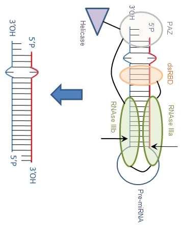
34
mirna Dr. S Hosseini-Asl
 mirna Dr. S Hosseini-Asl 1 2 MicroRNAs (mirnas) are small noncoding RNAs which enhance the cleavage or translational repression of specific mrna with recognition site(s) in the 3 - untranslated region
MicroRNA and Male Infertility: A Potential for Diagnosis
 Review Article MicroRNA and Male Infertility: A Potential for Diagnosis * Abstract MicroRNAs (mirnas) are small non-coding single stranded RNA molecules that are physiologically produced in eukaryotic
MicroRNA in Cancer Karen Dybkær 2013
 MicroRNA in Cancer Karen Dybkær RNA Ribonucleic acid Types -Coding: messenger RNA (mrna) coding for proteins -Non-coding regulating protein formation Ribosomal RNA (rrna) Transfer RNA (trna) Small nuclear
MicroRNAs and Cancer
 MicroRNAs and Cancer MicroRNAs are an abundant class of small (20-25 nucleotides) non-protein coding RNAs that function as negative gene regulators. The human genome contains up to 1000 micrornas which
Chapter 10 - Post-transcriptional Gene Control
 Chapter 10 - Post-transcriptional Gene Control Chapter 10 - Post-transcriptional Gene Control 10.1 Processing of Eukaryotic Pre-mRNA 10.2 Regulation of Pre-mRNA Processing 10.3 Transport of mrna Across
MicroRNA dysregulation in cancer. Systems Plant Microbiology Hyun-Hee Lee
 MicroRNA dysregulation in cancer Systems Plant Microbiology Hyun-Hee Lee Contents 1 What is MicroRNA? 2 mirna dysregulation in cancer 3 Summary What is MicroRNA? What is MicroRNA? MicroRNAs (mirnas) -
V16: involvement of micrornas in GRNs
 What are micrornas? V16: involvement of micrornas in GRNs How can one identify micrornas? What is the function of micrornas? Elisa Izaurralde, MPI Tübingen Huntzinger, Izaurralde, Nat. Rev. Genet. 12,
Molecular Biology (BIOL 4320) Exam #2 May 3, 2004
 Exam #2 May 3, 2004") Molecular Biology (BIOL 4320) Exam #2 May 3, 2004 Name SS# This exam is worth a total of 100 points. The number of points each question is worth is shown in parentheses after the question number. Good
Molecular Biology (BIOL 4320) Exam #2 May 3, 2004 Name SS# This exam is worth a total of 100 points. The number of points each question is worth is shown in parentheses after the question number. Good
Alternative RNA processing: Two examples of complex eukaryotic transcription units and the effect of mutations on expression of the encoded proteins.
 Alternative RNA processing: Two examples of complex eukaryotic transcription units and the effect of mutations on expression of the encoded proteins. The RNA transcribed from a complex transcription unit
Ch. 18 Regulation of Gene Expression
 Ch. 18 Regulation of Gene Expression 1 Human genome has around 23,688 genes (Scientific American 2/2006) Essential Questions: How is transcription regulated? How are genes expressed? 2 Bacteria regulate
Bi 8 Lecture 17. interference. Ellen Rothenberg 1 March 2016
 Bi 8 Lecture 17 REGulation by RNA interference Ellen Rothenberg 1 March 2016 Protein is not the only regulatory molecule affecting gene expression: RNA itself can be negative regulator RNA does not need
reported to underlie diverse aspects of biology, including developmental timing, differentiation, proliferation, cell death, and metabolism.
 1100 MicroRNA in Cancer: The Involvement of Aberrant MicroRNA Biogenesis Regulatory Pathways Genes & Cancer 1(11) 1100 1114 The Author(s) 2011 Reprints and permission: sagepub.com/journalspermissions.nav
10/31/2017. micrornas and cancer. From the one gene-one enzyme hypothesis to. microrna DNA RNA. Transcription factors.
 micrornas and cancer Cellular and Molecular Biology of Cancer (PATH G4500-001) November 1 st, 2017 -Katia Basso- Columbia University Katia Basso, PhD Office: ICRC RM506 E-mail: kb451@cumc.columbia.edu
Circular RNAs (circrnas) act a stable mirna sponges
 act a stable mirna sponges") Circular RNAs (circrnas) act a stable mirna sponges cernas compete for mirnas Ancestal mrna (+3 UTR) Pseudogene RNA (+3 UTR homolgy region) The model holds true for all RNAs that share a mirna binding
Circular RNAs (circrnas) act a stable mirna sponges cernas compete for mirnas Ancestal mrna (+3 UTR) Pseudogene RNA (+3 UTR homolgy region) The model holds true for all RNAs that share a mirna binding
Overview: Conducting the Genetic Orchestra Prokaryotes and eukaryotes alter gene expression in response to their changing environment
 Overview: Conducting the Genetic Orchestra Prokaryotes and eukaryotes alter gene expression in response to their changing environment In multicellular eukaryotes, gene expression regulates development
MicroRNAs, RNA Modifications, RNA Editing. Bora E. Baysal MD, PhD Oncology for Scientists Lecture Tue, Oct 17, 2017, 3:30 PM - 5:00 PM
 MicroRNAs, RNA Modifications, RNA Editing Bora E. Baysal MD, PhD Oncology for Scientists Lecture Tue, Oct 17, 2017, 3:30 PM - 5:00 PM Expanding world of RNAs mrna, messenger RNA (~20,000) trna, transfer
Regulation of Gene Expression in Eukaryotes
 Ch. 19 Regulation of Gene Expression in Eukaryotes BIOL 222 Differential Gene Expression in Eukaryotes Signal Cells in a multicellular eukaryotic organism genetically identical differential gene expression
Transcriptional control in Eukaryotes: (chapter 13 pp276) Chromatin structure affects gene expression. Chromatin Array of nuc
 Chromatin structure affects gene expression. Chromatin Array of nuc") Transcriptional control in Eukaryotes: (chapter 13 pp276) Chromatin structure affects gene expression Chromatin Array of nuc 1 Transcriptional control in Eukaryotes: Chromatin undergoes structural changes
Transcriptional control in Eukaryotes: (chapter 13 pp276) Chromatin structure affects gene expression Chromatin Array of nuc 1 Transcriptional control in Eukaryotes: Chromatin undergoes structural changes
MicroRNA expression profiling and functional analysis in prostate cancer. Marco Folini s.c. Ricerca Traslazionale DOSL
 MicroRNA expression profiling and functional analysis in prostate cancer Marco Folini s.c. Ricerca Traslazionale DOSL What are micrornas? For almost three decades, the alteration of protein-coding genes
THE EFFECT OF HSA-MIR-105 ON PROSTATE CANCER GROWTH
 THE EFFECT OF HSA-MIR-105 ON PROSTATE CANCER GROWTH by David Rice Honeywell Bachelor of Science, Queen s University, 2010 THESIS SUBMITTED IN PARTIAL FULFILLMENT OF THE REQUIREMENTS FOR THE DEGREE OF MASTER
Molecular Biology (BIOL 4320) Exam #2 April 22, 2002
 Exam #2 April 22, 2002") Molecular Biology (BIOL 4320) Exam #2 April 22, 2002 Name SS# This exam is worth a total of 100 points. The number of points each question is worth is shown in parentheses after the question number. Good
Molecular Biology (BIOL 4320) Exam #2 April 22, 2002 Name SS# This exam is worth a total of 100 points. The number of points each question is worth is shown in parentheses after the question number. Good
Genetics and Genomics in Medicine Chapter 6 Questions
 Genetics and Genomics in Medicine Chapter 6 Questions Multiple Choice Questions Question 6.1 With respect to the interconversion between open and condensed chromatin shown below: Which of the directions
Human Genome: Mapping, Sequencing Techniques, Diseases
 Human Genome: Mapping, Sequencing Techniques, Diseases Lecture 4 BINF 7580 Fall 2005 1 Let us review what we talked about at the previous lecture. Please,... 2 The central dogma states that the transfer
Eukaryotic mrna is covalently processed in three ways prior to export from the nucleus:
 RNA Processing Eukaryotic mrna is covalently processed in three ways prior to export from the nucleus: Transcripts are capped at their 5 end with a methylated guanosine nucleotide. Introns are removed
Genetics. Instructor: Dr. Jihad Abdallah Transcription of DNA
 Genetics Instructor: Dr. Jihad Abdallah Transcription of DNA 1 3.4 A 2 Expression of Genetic information DNA Double stranded In the nucleus Transcription mrna Single stranded Translation In the cytoplasm
The Biology and Genetics of Cells and Organisms The Biology of Cancer
 The Biology and Genetics of Cells and Organisms The Biology of Cancer Mendel and Genetics How many distinct genes are present in the genomes of mammals? - 21,000 for human. - Genetic information is carried
Cross species analysis of genomics data. Computational Prediction of mirnas and their targets
 02-716 Cross species analysis of genomics data Computational Prediction of mirnas and their targets Outline Introduction Brief history mirna Biogenesis Why Computational Methods? Computational Methods
Novel RNAs along the Pathway of Gene Expression. (or, The Expanding Universe of Small RNAs)
") Novel RNAs along the Pathway of Gene Expression (or, The Expanding Universe of Small RNAs) Central Dogma DNA RNA Protein replication transcription translation Central Dogma DNA RNA Spliced RNA Protein
Novel RNAs along the Pathway of Gene Expression (or, The Expanding Universe of Small RNAs) Central Dogma DNA RNA Protein replication transcription translation Central Dogma DNA RNA Spliced RNA Protein
RNA Processing in Eukaryotes *
 OpenStax-CNX module: m44532 1 RNA Processing in Eukaryotes * OpenStax This work is produced by OpenStax-CNX and licensed under the Creative Commons Attribution License 3.0 By the end of this section, you
We are IntechOpen, the world s leading publisher of Open Access books Built by scientists, for scientists. International authors and editors
 We are IntechOpen, the world s leading publisher of Open Access books Built by scientists, for scientists 4,000 116,000 120M Open access books available International authors and editors Downloads Our
1. Identify and characterize interesting phenomena! 2. Characterization should stimulate some questions/models! 3. Combine biochemistry and genetics
 1. Identify and characterize interesting phenomena! 2. Characterization should stimulate some questions/models! 3. Combine biochemistry and genetics to gain mechanistic insight! 4. Return to step 2, as
he micrornas of Caenorhabditis elegans (Lim et al. Genes & Development 2003)
") MicroRNAs: Genomics, Biogenesis, Mechanism, and Function (D. Bartel Cell 2004) he micrornas of Caenorhabditis elegans (Lim et al. Genes & Development 2003) Vertebrate MicroRNA Genes (Lim et al. Science
MicroRNAs: Genomics, Biogenesis, Mechanism, and Function (D. Bartel Cell 2004) he micrornas of Caenorhabditis elegans (Lim et al. Genes & Development 2003) Vertebrate MicroRNA Genes (Lim et al. Science
Phenomena first observed in petunia
 Vectors for RNAi Phenomena first observed in petunia Attempted to overexpress chalone synthase (anthrocyanin pigment gene) in petunia. (trying to darken flower color) Caused the loss of pigment. Bill Douherty
microrna Presented for: Presented by: Date:
 microrna Presented for: Presented by: Date: 2 micrornas Non protein coding, endogenous RNAs of 21-22nt length Evolutionarily conserved Regulate gene expression by binding complementary regions at 3 regions
Improved annotation of C. elegans micrornas by deep sequencing reveals structures associated with processing by Drosha and Dicer
 BIOINFORMATICS Improved annotation of C. elegans micrornas by deep sequencing reveals structures associated with processing by Drosha and Dicer M. BRYAN WARF, 1 W. EVAN JOHNSON, 2 and BRENDA L. BASS 1
Supplementary Figure 1
 Supplementary Figure 1 Asymmetrical function of 5p and 3p arms of mir-181 and mir-30 families and mir-142 and mir-154. (a) Control experiments using mirna sensor vector and empty pri-mirna overexpression
RECAP (1)! In eukaryotes, large primary transcripts are processed to smaller, mature mrnas.! What was first evidence for this precursorproduct
! In eukaryotes, large primary transcripts are processed to smaller, mature mrnas.! What was first evidence for this precursorproduct") RECAP (1) In eukaryotes, large primary transcripts are processed to smaller, mature mrnas. What was first evidence for this precursorproduct relationship? DNA Observation: Nuclear RNA pool consists of
RECAP (1) In eukaryotes, large primary transcripts are processed to smaller, mature mrnas. What was first evidence for this precursorproduct relationship? DNA Observation: Nuclear RNA pool consists of
Mechanisms of alternative splicing regulation
 Mechanisms of alternative splicing regulation The number of mechanisms that are known to be involved in splicing regulation approximates the number of splicing decisions that have been analyzed in detail.
Pre-mRNA has introns The splicing complex recognizes semiconserved sequences
 Adding a 5 cap Lecture 4 mrna splicing and protein synthesis Another day in the life of a gene. Pre-mRNA has introns The splicing complex recognizes semiconserved sequences Introns are removed by a process
TRANSCRIPTION. DNA à mrna
 TRANSCRIPTION DNA à mrna Central Dogma Animation DNA: The Secret of Life (from PBS) http://www.youtube.com/watch? v=41_ne5ms2ls&list=pl2b2bd56e908da696&index=3 Transcription http://highered.mcgraw-hill.com/sites/0072507470/student_view0/
RNA (Ribonucleic acid)
") RNA (Ribonucleic acid) Structure: Similar to that of DNA except: 1- it is single stranded polunucleotide chain. 2- Sugar is ribose 3- Uracil is instead of thymine There are 3 types of RNA: 1- Ribosomal
RNA (Ribonucleic acid) Structure: Similar to that of DNA except: 1- it is single stranded polunucleotide chain. 2- Sugar is ribose 3- Uracil is instead of thymine There are 3 types of RNA: 1- Ribosomal
L I F E S C I E N C E S
 1a L I F E S C I E N C E S 5 -UUA AUA UUC GAA AGC UGC AUC GAA AAC UGU GAA UCA-3 5 -TTA ATA TTC GAA AGC TGC ATC GAA AAC TGT GAA TCA-3 3 -AAT TAT AAG CTT TCG ACG TAG CTT TTG ACA CTT AGT-5 NOVEMBER 2, 2006
Bio 111 Study Guide Chapter 17 From Gene to Protein
 Bio 111 Study Guide Chapter 17 From Gene to Protein BEFORE CLASS: Reading: Read the introduction on p. 333, skip the beginning of Concept 17.1 from p. 334 to the bottom of the first column on p. 336, and
RECAP (1)! In eukaryotes, large primary transcripts are processed to smaller, mature mrnas.! What was first evidence for this precursorproduct
! In eukaryotes, large primary transcripts are processed to smaller, mature mrnas.! What was first evidence for this precursorproduct") RECAP (1) In eukaryotes, large primary transcripts are processed to smaller, mature mrnas. What was first evidence for this precursorproduct relationship? DNA Observation: Nuclear RNA pool consists of
RECAP (1) In eukaryotes, large primary transcripts are processed to smaller, mature mrnas. What was first evidence for this precursorproduct relationship? DNA Observation: Nuclear RNA pool consists of
Prokaryotes and eukaryotes alter gene expression in response to their changing environment
 Chapter 18 Prokaryotes and eukaryotes alter gene expression in response to their changing environment In multicellular eukaryotes, gene expression regulates development and is responsible for differences
RAS Genes. The ras superfamily of genes encodes small GTP binding proteins that are responsible for the regulation of many cellular processes.
 ۱ RAS Genes The ras superfamily of genes encodes small GTP binding proteins that are responsible for the regulation of many cellular processes. Oncogenic ras genes in human cells include H ras, N ras,
1. Investigate the structure of the trna Synthase in complex with a trna molecule. (pdb ID 1ASY).
.") Problem Set 11 (Due Nov 25 th ) 1. Investigate the structure of the trna Synthase in complex with a trna molecule. (pdb ID 1ASY). a. Why don t trna molecules contain a 5 triphosphate like other RNA molecules
Problem Set 11 (Due Nov 25 th ) 1. Investigate the structure of the trna Synthase in complex with a trna molecule. (pdb ID 1ASY). a. Why don t trna molecules contain a 5 triphosphate like other RNA molecules
BIO360 Fall 2013 Quiz 1
 BIO360 Fall 2013 Quiz 1 1. Examine the diagram below. There are two homologous copies of chromosome one and the allele of YFG carried on the light gray chromosome has undergone a loss-of-function mutation.
Chapter 11 How Genes Are Controlled
 Chapter 11 How Genes Are Controlled PowerPoint Lectures for Biology: Concepts & Connections, Sixth Edition Campbell, Reece, Taylor, Simon, and Dickey Copyright 2009 Pearson Education, Inc. Lecture by Mary
Utility of Circulating micrornas in Cardiovascular Disease
 Utility of Circulating micrornas in Cardiovascular Disease Pil-Ki Min, MD, PhD Cardiology Division, Gangnam Severance Hospital, Yonsei University College of Medicine Introduction Biology of micrornas Circulating
micrornas (mirna) and Biomarkers
 and Biomarkers") micrornas (mirna) and Biomarkers Small RNAs Make Big Splash mirnas & Genome Function Biomarkers in Cancer Future Prospects Javed Khan M.D. National Cancer Institute EORTC-NCI-ASCO November 2007 The Human
micrornas (mirna) and Biomarkers Small RNAs Make Big Splash mirnas & Genome Function Biomarkers in Cancer Future Prospects Javed Khan M.D. National Cancer Institute EORTC-NCI-ASCO November 2007 The Human
Polyomaviridae. Spring
 Polyomaviridae Spring 2002 331 Antibody Prevalence for BK & JC Viruses Spring 2002 332 Polyoma Viruses General characteristics Papovaviridae: PA - papilloma; PO - polyoma; VA - vacuolating agent a. 45nm
Removal of Shelterin Reveals the Telomere End-Protection Problem
 Removal of Shelterin Reveals the Telomere End-Protection Problem DSB Double-Strand Breaks causate da radiazioni stress ossidativo farmaci DSB e CROMATINA Higher-order chromatin packaging is a barrier to
Translation. Host Cell Shutoff 1) Initiation of eukaryotic translation involves many initiation factors
 Initiation of eukaryotic translation involves many initiation factors") Translation Questions? 1) How does poliovirus shutoff eukaryotic translation? 2) If eukaryotic messages are not translated how can poliovirus get its message translated? Host Cell Shutoff 1) Initiation
Translation Questions? 1) How does poliovirus shutoff eukaryotic translation? 2) If eukaryotic messages are not translated how can poliovirus get its message translated? Host Cell Shutoff 1) Initiation
MOLECULAR CELL BIOLOGY
 1 Lodish Berk Kaiser Krieger scott Bretscher Ploegh Matsudaira MOLECULAR CELL BIOLOGY SEVENTH EDITION CHAPTER 13 Moving Proteins into Membranes and Organelles Copyright 2013 by W. H. Freeman and Company
Eukaryotic Gene Regulation
 Eukaryotic Gene Regulation Chapter 19: Control of Eukaryotic Genome The BIG Questions How are genes turned on & off in eukaryotes? How do cells with the same genes differentiate to perform completely different,
RNA interference (RNAi)
") RN interference (RNi) Natasha aplen ene Silencing Section Office of Science and Technology Partnerships Office of the Director enter for ancer Research National ancer Institute ncaplen@mail.nih.gov Plants
RN interference (RNi) Natasha aplen ene Silencing Section Office of Science and Technology Partnerships Office of the Director enter for ancer Research National ancer Institute ncaplen@mail.nih.gov Plants
MCB Chapter 11. Topic E. Splicing mechanism Nuclear Transport Alternative control modes. Reading :
 MCB Chapter 11 Topic E Splicing mechanism Nuclear Transport Alternative control modes Reading : 419-449 Topic E Michal Linial 14 Jan 2004 1 Self-splicing group I introns were the first examples of catalytic
Protein Synthesis
 Protein Synthesis 10.6-10.16 Objectives - To explain the central dogma - To understand the steps of transcription and translation in order to explain how our genes create proteins necessary for survival.
Santosh Patnaik, MD, PhD! Assistant Member! Department of Thoracic Surgery! Roswell Park Cancer Institute!
 Santosh Patnaik, MD, PhD Assistant Member Department of Thoracic Surgery Roswell Park Cancer Institute MicroRNA biology, techniques and applications History Biogenesis Nomenclature Tissue specificity Mechanisms
NOVEL FUNCTION OF mirnas IN REGULATING GENE EXPRESSION. Ana M. Martinez
 NOVEL FUNCTION OF mirnas IN REGULATING GENE EXPRESSION Ana M. Martinez Switching from Repression to Activation: MicroRNAs can Up-Regulate Translation. Shoba Vasudevan, Yingchun Tong, Joan A. Steitz AU-rich
Research Article Base Composition Characteristics of Mammalian mirnas
 Journal of Nucleic Acids Volume 2013, Article ID 951570, 6 pages http://dx.doi.org/10.1155/2013/951570 Research Article Base Composition Characteristics of Mammalian mirnas Bin Wang Department of Chemistry,
General Biology 1004 Chapter 11 Lecture Handout, Summer 2005 Dr. Frisby
 Slide 1 CHAPTER 11 Gene Regulation PowerPoint Lecture Slides for Essential Biology, Second Edition & Essential Biology with Physiology Presentation prepared by Chris C. Romero Neil Campbell, Jane Reece,
Removal of Shelterin Reveals the Telomere End-Protection Problem
 Removal of Shelterin Reveals the Telomere End-Protection Problem DSB Double-Strand Breaks causate da radiazioni stress ossidativo farmaci DSB e CROMATINA Higher-order chromatin packaging is a barrier to
Biochemistry 2000 Sample Question Transcription, Translation and Lipids. (1) Give brief definitions or unique descriptions of the following terms:
 Give brief definitions or unique descriptions of the following terms:") (1) Give brief definitions or unique descriptions of the following terms: (a) exon (b) holoenzyme (c) anticodon (d) trans fatty acid (e) poly A tail (f) open complex (g) Fluid Mosaic Model (h) embedded
(1) Give brief definitions or unique descriptions of the following terms: (a) exon (b) holoenzyme (c) anticodon (d) trans fatty acid (e) poly A tail (f) open complex (g) Fluid Mosaic Model (h) embedded
High AU content: a signature of upregulated mirna in cardiac diseases
 https://helda.helsinki.fi High AU content: a signature of upregulated mirna in cardiac diseases Gupta, Richa 2010-09-20 Gupta, R, Soni, N, Patnaik, P, Sood, I, Singh, R, Rawal, K & Rani, V 2010, ' High
DSB. Double-Strand Breaks causate da radiazioni stress ossidativo farmaci
 DSB Double-Strand Breaks causate da radiazioni stress ossidativo farmaci DSB e CROMATINA Higher-order chromatin packaging is a barrier to the detection and repair of DNA damage DSBs induce a local decrease
Cellular MicroRNA and P Bodies Modulate Host-HIV-1 Interactions. 指導教授 : 張麗冠博士 演講者 : 黃柄翰 Date: 2009/10/19
 Cellular MicroRNA and P Bodies Modulate Host-HIV-1 Interactions 指導教授 : 張麗冠博士 演講者 : 黃柄翰 Date: 2009/10/19 1 MicroRNA biogenesis 1. Pri mirna: primary mirna 2. Drosha: RNaseIII 3. DCR 1: Dicer 1 RNaseIII
Processing of RNA II Biochemistry 302. February 13, 2006
 Processing of RNA II Biochemistry 302 February 13, 2006 Precursor mrna: introns and exons Intron: Transcribed RNA sequence removed from precursor RNA during the process of maturation (for class II genes:
Molecular Cell Biology - Problem Drill 10: Gene Expression in Eukaryotes
 Molecular Cell Biology - Problem Drill 10: Gene Expression in Eukaryotes Question No. 1 of 10 1. Which of the following statements about gene expression control in eukaryotes is correct? Question #1 (A)
Introduction retroposon
 17.1 - Introduction A retrovirus is an RNA virus able to convert its sequence into DNA by reverse transcription A retroposon (retrotransposon) is a transposon that mobilizes via an RNA form; the DNA element
Computational Biology I LSM5191
 Computational Biology I LSM5191 Aylwin Ng, D.Phil Lecture Notes: Transcriptome: Molecular Biology of Gene Expression II TRANSLATION RIBOSOMES: protein synthesizing machines Translation takes place on defined
number Done by Corrected by Doctor Maha Shomaf
 number 19 Done by Waseem Abo-Obeida Corrected by Abdullah Zreiqat Doctor Maha Shomaf Carcinogenesis: the molecular basis of cancer. Non-lethal genetic damage lies at the heart of carcinogenesis and leads
Dottorato di Ricerca in Genetica e Biologia Cellulare. MUTANT P53 INHIBITS microrna BIOGENESIS BY INTERFERING WITH THE MICROPROCESSOR COMPLEX
 Dipartimento di Ecologia e Biologia Dottorato di Ricerca in Genetica e Biologia Cellulare Ciclo XXVII Settore scientifico-disciplinare BIO/11 MUTANT P53 INHIBITS microrna BIOGENESIS BY INTERFERING WITH
Alternative splicing. Biosciences 741: Genomics Fall, 2013 Week 6
 Alternative splicing Biosciences 741: Genomics Fall, 2013 Week 6 Function(s) of RNA splicing Splicing of introns must be completed before nuclear RNAs can be exported to the cytoplasm. This led to early
Regulation of microrna biogenesis and turnover by animals and their viruses
 Cell. Mol. Life Sci. (2013) 70:3525 3544 DOI 10.1007/s00018-012-1257-1 Cellular and Molecular Life Sciences REVIEW Regulation of microrna biogenesis and turnover by animals and their viruses Valentina
Investigating the regulation of mirna biogenesis and Argonaute2 by RNA binding proteins
 Investigating the regulation of mirna biogenesis and Argonaute2 by RNA binding proteins Patrick Peter Connerty Supervisor: Gyorgy Hutvagner Thesis Submitted for the Degree of Doctor of Philosophy (Science)
Molecular biology :- Cancer genetics lecture 11
 Molecular biology :- Cancer genetics lecture 11 -We have talked about 2 group of genes that is involved in cellular transformation : proto-oncogenes and tumour suppressor genes, and it isn t enough to
number Done by Corrected by Doctor Ashraf
 number 4 Done by Nedaa Bani Ata Corrected by Rama Nada Doctor Ashraf Genome replication and gene expression Remember the steps of viral replication from the last lecture: Attachment, Adsorption, Penetration,
Campbell Biology 10. A Global Approach. Chapter 18 Control of Gene Expression
 Lecture on General Biology 2 Campbell Biology 10 A Global Approach th edition Chapter 18 Control of Gene Expression Chul-Su Yang, Ph.D., chulsuyang@hanyang.ac.kr Infection Biology Lab., Dept. of Molecular
Gene Regulation. Bacteria. Chapter 18: Regulation of Gene Expression
 Chapter 18: Regulation of Gene Expression A Biology 2013 1 Gene Regulation rokaryotes and eukaryotes alter their gene expression in response to their changing environment In multicellular eukaryotes, gene
Objectives: Prof.Dr. H.D.El-Yassin
 Protein Synthesis and drugs that inhibit protein synthesis Objectives: 1. To understand the steps involved in the translation process that leads to protein synthesis 2. To understand and know about all
Small RNAs and how to analyze them using sequencing
 Small RNAs and how to analyze them using sequencing Jakub Orzechowski Westholm (1) Long- term bioinforma=cs support, Science For Life Laboratory Stockholm (2) Department of Biophysics and Biochemistry,
Introduction. Cancer Biology. Tumor-suppressor genes. Proto-oncogenes. DNA stability genes. Mechanisms of carcinogenesis.
 Cancer Biology Chapter 18 Eric J. Hall., Amato Giaccia, Radiobiology for the Radiologist Introduction Tissue homeostasis depends on the regulated cell division and self-elimination (programmed cell death)
Processing of RNA II Biochemistry 302. February 14, 2005 Bob Kelm
 Processing of RNA II Biochemistry 302 February 14, 2005 Bob Kelm What s an intron? Transcribed sequence removed during the process of mrna maturation (non proteincoding sequence) Discovered by P. Sharp
REGULATED SPLICING AND THE UNSOLVED MYSTERY OF SPLICEOSOME MUTATIONS IN CANCER
 REGULATED SPLICING AND THE UNSOLVED MYSTERY OF SPLICEOSOME MUTATIONS IN CANCER RNA Splicing Lecture 3, Biological Regulatory Mechanisms, H. Madhani Dept. of Biochemistry and Biophysics MAJOR MESSAGES Splice
Bioinformation Volume 5
 Mi-DISCOVERER: A bioinformatics tool for the detection of mi-rna in human genome Saadia Arshad, Asia Mumtaz, Freed Ahmad, Sadia Liaquat, Shahid Nadeem, Shahid Mehboob, Muhammad Afzal * Department of Bioinformatics,
Intrinsic cellular defenses against virus infection
 Intrinsic cellular defenses against virus infection Detection of virus infection Host cell response to virus infection Interferons: structure and synthesis Induction of antiviral activity Viral defenses
CRS4 Seminar series. Inferring the functional role of micrornas from gene expression data CRS4. Biomedicine. Bioinformatics. Paolo Uva July 11, 2012
 CRS4 Seminar series Inferring the functional role of micrornas from gene expression data CRS4 Biomedicine Bioinformatics Paolo Uva July 11, 2012 Partners Pharmaceutical company Fondazione San Raffaele,
Transcriptome-wide analysis of microrna expression in the malaria mosquito Anopheles gambiae
 Biryukova et al. BMC Genomics 2014, 15:557 RESEARCH ARTICLE Open Access Transcriptome-wide analysis of microrna expression in the malaria mosquito Anopheles gambiae Inna Biryukova 1*, Tao Ye 2 and Elena
Introduction to Cancer Biology
 Introduction to Cancer Biology Robin Hesketh Multiple choice questions (choose the one correct answer from the five choices) Which ONE of the following is a tumour suppressor? a. AKT b. APC c. BCL2 d.
Regulation of Gene Expression
 Chapter 18 Regulation of Gene Expression PowerPoint Lecture Presentations for Biology Eighth Edition Neil Campbell and Jane Reece Lectures by Chris Romero, updated by Erin Barley with contributions from
Alan Weiner BIOCHEM 530 Friday, MEB 248 October 23, 2015 RNA structure, the ribosome, structure-based drug design
 Alan Weiner BIOCHEM 530 Friday, MEB 248 October 23, 2015 RNA structure, the ribosome, structure-based drug design Crick's "Central Dogma"? DNA makes RNA makes protein Crick's "Central Dogma"? DNA makes
Insulin mrna to Protein Kit
 Insulin mrna to Protein Kit A 3DMD Paper BioInformatics and Mini-Toober Folding Activity Student Handout www.3dmoleculardesigns.com Insulin mrna to Protein Kit Contents Becoming Familiar with the Data...
Chapter 18 Regulation of Gene Expression
 Chapter 18 Regulation of Gene Expression Differential Expression of Genes Prokaryotes and eukaryotes precisely regulate gene expression in response to environmental conditions In multicellular eukaryotes,
DNA codes for RNA, which guides protein synthesis.
 Section 3: DNA codes for RNA, which guides protein synthesis. K What I Know W What I Want to Find Out L What I Learned Vocabulary Review synthesis New RNA messenger RNA ribosomal RNA transfer RNA transcription
Post-transcriptional regulation of an intronic microrna
 Post-transcriptional regulation of an intronic microrna Carl Novina Dana-Farber Cancer Institute Harvard Medical School Broad Institute of Harvard and MIT Qiagen Webinar 05-17-11 Outline 1. The biology
Chapter 11. How Genes Are Controlled. Lectures by Edward J. Zalisko
 Chapter 11 How Genes Are Controlled PowerPoint Lectures for Campbell Essential Biology, Fifth Edition, and Campbell Essential Biology with Physiology, Fourth Edition Eric J. Simon, Jean L. Dickey, and
Lecture 2: Virology. I. Background
 Lecture 2: Virology I. Background A. Properties 1. Simple biological systems a. Aggregates of nucleic acids and protein 2. Non-living a. Cannot reproduce or carry out metabolic activities outside of a
Profiles of gene expression & diagnosis/prognosis of cancer. MCs in Advanced Genetics Ainoa Planas Riverola
 Profiles of gene expression & diagnosis/prognosis of cancer MCs in Advanced Genetics Ainoa Planas Riverola Gene expression profiles Gene expression profiling Used in molecular biology, it measures the