Anyone can get breast cancer BREAST MRI BREAST CANCER. The incidence of getting breast cancer is 1:19 in Malaysia
|
|
|
- Loreen Leonard
- 5 years ago
- Views:
Transcription


1 Anyone can get breast cancer BREAST MRI KATE Datin Dr Fatimah Moosa Sunway Medical Centre DATIN SERI ENDON KYLIE SIZE DOES NOT MAKE A DIFFERENCE BREAST CANCER The incidence of getting breast cancer is 1:19 in Malaysia Breast cancer - commonest cancer in all ethnic groups Year breast cancer cases were reported 31.0 % of newly diagnosed cases. Y mi voz Junto a las demás 1
2 MR Imaging for Breast Cancer Detection, Diagnosis, and Staging At the present time, mammography is the only imaging modality with proved efficacy for the detection of early, clinically occult breast cancer screening. Limitations in both the specificity and sensitivity of mammography have stimulated exploration into adjunctive imaging modalities which includes MRI breast Advances in surface coil technology, development of new imaging protocols and introduction of contrast enhanced MR imaging show promising tool for breast imaging modality MR Imaging for Breast Cancer Detection, Diagnosis, and Staging For MR imaging to be a viable modality for breast imaging, it must be demonstrated that this technology can provide clinically valuable information that cannot be obtained with conventional imaging methods Several clinical studies have now demonstrated the potential of MR imaging for greater specificity and sensitivity than is possible with mammography, ultrasound and physical examination Benefits of Breast MRI Can image breast implants and ruptures Highly sensitive to small abnormalities Used effectively in dense breast Can evaluate inverted nipples for evidence of cancer Can evaluate the extent of breast cancer multicenteric or multifocal lesions Sensitivity and Specificity of Examinations for Breast Cancer in a High Risk Population* Sensitivity Specificity Clinical Examination 18% 98% Mammography 40% 95% MRI 71% 90% *Women with genetic or familial predisposition to breast cancer Benefits of Breast MRI Can determine what type of surgery in indicated ( lumpectomy vs. mastectomy) May help detect breast recurrences, residual tumours from scar tissue after lumpectomy Can locate primary tumour in women whose cancer has spread to axillary LN Can spot or characterise small abnormalities misses by mammography Maybe useful in screening women at high risk for breast cancer Limitations of Breast MRI MRI takes min compared to min for screening mammography Cost of MRI is several times the cost of mammography MRI pt must tolerate claustrophobia MRI can be non specific, often cannot distinguish between cancerous and non-cancerous lesions 2
3 Limitations of Breast MRI Minimally invasive breast biopsy techniques need sofisticated software and techniques Cannot image micro calcifications Advanced MRI techniques are often not available at most centres Cannot always produce results cited in research studies Technical Requirements No universally accepted standard or optimal technique Multitude of imaging variables including magnetic field strength (high vs low), breast surface coil configuration, and imaging parameters ( i.e., two-dimentional vs. three-dimentional acquisition, imaging matrix, FOV, TOA and unilateral vs. bilateral examination Suggested Protocol Axial SE/FSE T1: Acts as localiser.thickslices/gap throught both breasts Sagital SE /FSE/PD/T2: Prescribed through the breast include the axillae Axial gradient echo T1 with contrast: dynamic pre and post contrast.use FAT SAT to distinguish enhancing pathology from fat Additional sequences: Sagital/coronal SE/FSE T1 post contrast STIR: coronal or axial Image Optimisation: Technical Issues Increase SNR due to recent developments in coil technology Phase array coils return highest and most uniform signal. Others can give glare at nipple and signal fall of nearer the chest FSE is a great advantage as it facilitates the acquisition of very fine matrices in relatively short scan times. Smallest FOV, thin slices and small gaps used to maintain spatial resolution further FAT SAT and other fat suppression tech. are useful in pts with fatty breast to distinguish lesion from surrounding breast tissue Artefact Problems Respiratory artefact: reduced in prone position Cardiac motion and flow within the mammillary vessels are often troublesome, hence swapping the phase encoding axis to S to I on sagital images and R to L on axial removes the artifact from the breast, but it can interfere with the breast. FC also reduces flow artifact but it increases the signal in vess. And the minimum TE available, not usually beneficial in T1W seq. SAT(presaturation pulse) brought into FOV and placed posteriorly over the heart are useful in reducing motion artefact 3


4 Aurora 1.5T Dedicated Breast MRI Differences 1 st FDA approved designed specifically for breast, dedicated and integrated with propriety CAD and biopsy capability Superior image at 4.5T quality and clearness Ability to see full coverage of both breast,chest wall and axillae in a single scan Feet first entry,patient comfort and greater access to breast for biopsy intervention Aurora Dedicated Breast MRI with 3-D Bilateral SpiralRODEO A more efficient manner for acquiring data 3X signal to noise, and 4 X resolution SpiralRODEO encodes signal acquisition during 46% of the scan time High resolution fat-suppressed imaging thru water excitation Magnetization transfer contrast (ductal tissue suppression) Faster scan times five 3 minute sequences Increased Temporal Resolution with the first time point at 90 seconds Isotropic Resolution 1.1 mm slice thickness for better detection of Ductal Carcinama In-Situ Dedicated Breast MRI System 3-D Image Acquisition Matrix 360x360x128 _ 3-D Image Reconstruction Matrix 512x512x160 slices _ 1mm 1.1mm slice thickness; no gap between slices _ Less than 3 minutes per scan _ Post-contrast time points obtained at 90-second, 4.5-minute, 7.5-minute and 10.5-minute intervals Importance of good Fat Suppression (to avoid snowman in snowstorm effect) A picture is worth a thousand words 4


5 Poor magnetic field homogeneity: hot spots Why Shim? Ellipsoid homogeneous field and image driven shimming provide homogeneous fat suppression of breasts and axillary regions Lesion Visibility: Suppression of Fat Signal In contrast to mammography, where lesion detectability is increased in a fatty background, on MR an enhancing lesions may not be detected because it becomes isointense to fat following contrast Hence signal from fat need to be eliminated Lesion Visibility: Suppression of Fat Signal One method is by post processing image subtraction (subtracting the precontrast from the post contrast sequences) Alternatively, fat suppression techniques use of a variation of chemical selective fat suppression where an extra RF pulse is used produces images of enhancing lesions that are easily identified on a dark background without any significant increase in imaging time 5


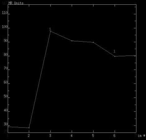
6 Fat suppression Sequences Breast MR Imaging for Lesion Characterisation One of the major limitations of mammogram is the overlap in the appearance of benign and malignant lesions MR imaging in the characterisation of mammographically and clinically detected lesions have reported improved specificity with MR imaging using differences in enhancement kinetics and morphologic features between malignant and benign lesions Breast MR Imaging for Lesion Characterisation Reported specificities of MR have been variable ranging from 37% to 97% This wide range is reflective of multiple factors including differences in magnetic field strength, imaging parameters, pt selection criteria, image interpretation, and histologic variation of benign and malignant lesions 6-10% of benign lesion do enhance Enhancement kinetics can be used to differentiate malignant lesions from benign, where malignant lesions demonstrate rapid increase in signal intensity followed by a wash-out of contrast in delayed images, whereas benign lesion exhibit a slower, progressive rise in signal intensity without wash-out of contrast 6
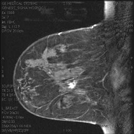
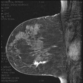
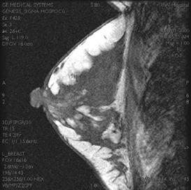
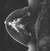
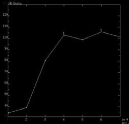
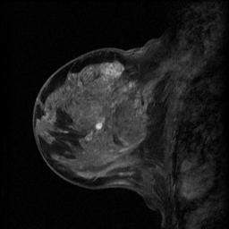
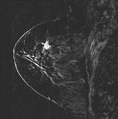
7 Malignant enhancement pattern 7


8 Breast MR Image-Guided Interventional System Allow largest access area to the breast for MRI-Interventional procedures Accommodate core biopsy or vacuum assisted biopsy devices Large apeture allows for increase access to breast for both medial and lateral approaches Core/Mammotome with MR Fast and simple set-up Hand held use Visual Targeting Z-lock and Imageable Aperture Grid System Maximize Breast Access Position the Cube Insert Targeting Set Note: Clean and anesthetize skin before making skin nick for targeting set insertion. Remove obturator and insert probe for biopsy Biopsy Take sample Activate Rotate 8

 Extracapsular")


")
9 Mark Biopsy Site Breast implants MR imaging of Breast Implants Intracapsular Rupture- contained by fibrous capsule with linguine sign(arrows) Extracapsular tear With collapse of the silicone shell Extensive Gel Bleed with focal rupture without complete collapse of implant shell- inverted teardrop sign Summary MR imaging has proved to be excellent imaging modality in locating free silicone and evaluating an implant rupture, with a sensitivity of approximately 94% and specificity of 97% The most reliable findings for detection of rupture include identification the collapse implant shell (linguine sign) and free silicone within breast parenchyma 9

10 Conclusion While MRI is en effective problemsolving technology, it has limitations as a screening tool for breast cancer MRI s limited availability, expense, and frequent non-specificity has slowed its widespread acceptance However there is a potential role for screening in young women with dense breast in the high risk group THANK YOU 10
Contrast-enhanced Breast MRI RSSA 2013
 Contrast-enhanced Breast MRI RSSA 2013 Prof. dr. Maurice van den Bosch University Medical Center Utrecht, the Netherlands Index 1) Breast cancer 2) Why MRI of the breast 3) Technique 4) Interpretation
BI-RADS and Breast MRI. Kathy Borovicka, M.D. Thursday February 15, 2018
 BI-RADS and Breast MRI Kathy Borovicka, M.D. Thursday February 15, 2018 Learning Objectives Be familiar with the Breast Imaging Reporting and Data System (BI-RADS) Understand the components of a breast
Standard Breast Imaging Modalities. Lilian Wang, M.D. Breast Imaging Section Department of Radiology Northwestern Medicine
 Standard Breast Imaging Modalities Lilian Wang, M.D. Breast Imaging Section Department of Radiology Northwestern Medicine Overview Standard breast imaging modalities Mammography Ultrasound MRI Imaging
MR Advance Techniques. Vascular Imaging. Class II
 MR Advance Techniques Vascular Imaging Class II 1 Vascular Imaging There are several methods that can be used to evaluate the cardiovascular systems with the use of MRI. MRI will aloud to evaluate morphology
Armed Forces Institute of Pathology.
 Armed Forces Institute of Pathology www.radpath.com Armed Forces Institute of Pathology Breast Disease www.radpath.org Armed Forces Institute of Pathology Interpretation of Breast MRI Leonard M. Glassman
Echelon Oval provides a robust suite of leading musculoskeletal imaging capabilities for detailed assessment of all anatomy for your most challenging
 Echelon Oval provides a robust suite of leading musculoskeletal imaging capabilities for detailed assessment of all anatomy for your most challenging cases. Hitachi Medical Systems America, Inc. 1959 Summit
Breast Health and Imaging Glossary
 Contact: Lorna Vaughan HerSpace Breast Imaging & Biopsy Associates 300 State Route 35 South W. Long Branch, NJ 07764 732-571-9100, ext. 104 lorna@breast-imaging.com Breast Health and Imaging Glossary Women
Breast Imaging Update: Old Dog New Tricks
 Breast Imaging Update: Old Dog New Tricks Claire McKay, DO M&S Imaging Assoc. San Antonio, TX cmckayhart@juno.com Goals Describe modalities available, old and new Provide understanding of pros and cons
Large health system benefits from multiple scanners for breast MRI
 Publication for the Philips MRI Community Issue 42 DECEMBER 2010 Large health system benefits from multiple scanners for breast MRI Breast MR and MR-guided biopsy are daily practice at WellStar Health
Why Talk About Technique? MRI of the Knee:
 Why Talk About Technique? MRI of the Knee: Part 1 - Imaging Techniques Mark Anderson, M.D. University of Virginia Health Sciences Center Charlottesville, Virginia Always had an interest teach our fellows
Pitfalls and Limitations of Breast MRI. Susan Orel Roth, MD Professor of Radiology University of Pennsylvania
 Pitfalls and Limitations of Breast MRI Susan Orel Roth, MD Professor of Radiology University of Pennsylvania Objectives Review the etiologies of false negative breast MRI examinations Discuss the limitations
Emerging Techniques in Breast Imaging: Contrast-Enhanced Mammography and Fast MRI
 Emerging Techniques in Breast Imaging: Contrast-Enhanced Mammography and Fast MRI Lilian Wang, M.D. Breast Imaging Section Department of Radiology Northwestern Medicine Overview Rationale for new imaging
MRI of breast implant: broken or not broken?
 MRI of breast implant: broken or not broken? Poster No.: C-1651 Congress: ECR 2014 Type: Educational Exhibit Authors: M. Pancot, A. Dal Col, C. Zuiani, M. Bazzocchi; Udine/IT Keywords: Breast, MR, Education,
Detailed Program of the second BREAST IMAGING AND INTERVENTIONS PROGRAM am am : Clinician s requirements from breast imaging
 Detailed Program of the second BREAST IMAGING AND INTERVENTIONS PROGRAM 2012 Day one, 2 nd November BREAST IMAGING AND INTERVENTIONS PROGRAM 2012 9.00 AM 9.10 am Introduction 9.10 am - 9.30 am : Clinician
RECENT ADVANCES IN CLINICAL MR OF ARTICULAR CARTILAGE
 In Practice RECENT ADVANCES IN CLINICAL MR OF ARTICULAR CARTILAGE By Atsuya Watanabe, MD, PhD, Director, Advanced Diagnostic Imaging Center and Associate Professor, Department of Orthopedic Surgery, Teikyo
Breast MRI Update. Jeffrey C. Weinreb, MD, FACR Yale University School of Medicine
 Breast MRI Update Jeffrey C. Weinreb, MD, FACR jeffrey.weinreb@yale.edu Yale University School of Medicine I disclose the following financial relationships with relevant commercial interests: Bracco Bayer
ACR Breast MRI Accreditation Program - DRAFT
 ACR Breast MRI Accreditation Program - DRAFT Donna M. Reeve, MS, DABR, DABMP Department of Imaging Physics Educational Objectives Provide an overview of the ACR Breast MRI Accreditation Program (BMRAP)
ACR MRI Accreditation Program. ACR MRI Accreditation Program Update. Educational Objectives. ACR accreditation. History. New Modular Program
 ACR MRI Accreditation Program Update Donna M. Reeve, MS, DABR, DABMP Department of Imaging Physics University of Texas M.D. Anderson Cancer Center Educational Objectives Present requirements of the new
Breast Cancer Imaging
 Breast Cancer Imaging I. Policy University Health Alliance (UHA) will cover breast imaging when such services meet the medical criteria guidelines (subject to limitations and exclusions) indicated below.
Improving Methods for Breast Cancer Detection and Diagnosis. The National Cancer Institute (NCI) is funding numerous research projects to improve
 is funding numerous research projects to improve") CANCER FACTS N a t i o n a l C a n c e r I n s t i t u t e N a t i o n a l I n s t i t u t e s o f H e a l t h D e p a r t m e n t o f H e a l t h a n d H u m a n S e r v i c e s Improving Methods for
CANCER FACTS N a t i o n a l C a n c e r I n s t i t u t e N a t i o n a l I n s t i t u t e s o f H e a l t h D e p a r t m e n t o f H e a l t h a n d H u m a n S e r v i c e s Improving Methods for
MRI in breast cancer: diagnosis and intervention. Dr Sue Barter Addenbrookes Hospital, Cambridge UK
 MRI in breast cancer: diagnosis and intervention Dr Sue Barter Addenbrookes Hospital, Cambridge UK Intervention will be discussed in High Risk Screening! Indications UK and Europe: Breast MRI is well established
Index. C Calcifications fat necrosis 1, 61 fat necrosis 4, 69 nipple/peri-areolar involvement 1, 165
 A ADH. See Atypical ductal hyperplasia (ADH) American College of Radiology (ACR), BI-RADS background parenchymal enhancement, 8, 9, 81, 82 fibroglandular tissue guidelines, 6 American Joint Committee on
NEW SUBTRACTION ALGORITHMS FOR EVALUATION OF BREAST LESIONS ON DYNAMIC CONTRAST ENHANCED MR MAMMOGRAPHY
 A-056 NEW SUBTRACTION ALGORITHMS FOR EVALUATION OF BREAST LESIONS ON DYNAMIC CONTRAST ENHANCED MR MAMMOGRAPHY So Hee Cho, M.D., Byung Gil Choi, M.D., Hak Hee Kim, M.D., Euy Neyng Kim, M.D., Bum-soo Kim,
Updates in Mammography. Dr. Yang Faridah A. Aziz Department of Biomedical Imaging University Malaya Medical Centre
 Updates in Mammography Dr. Yang Faridah A. Aziz Department of Biomedical Imaging University Malaya Medical Centre Updates in Mammography Breast Imaging Dr. Yang Faridah A. Aziz Department of Biomedical
Breast Cancer Diagnosis, Treatment and Follow-up
 Breast Cancer Diagnosis, Treatment and Follow-up What is breast cancer? Each of the body s organs, including the breast, is made up of many types of cells. Normally, healthy cells grow and divide to produce
Contrast-Enhanced Breast Tomosynthesis: Combining the Best of Both Worlds for Better Breast-Cancer Diagnosis
 Contrast-Enhanced Breast Tomosynthesis: Combining the Best of Both Worlds for Better Breast-Cancer Diagnosis T Wu (twu2@partners.org), E Rafferty, R Moore, D Kopans, Massachusetts General Hospital, Boston,
醫用磁振學 MRM 肌肉骨骼磁振造影簡介 肌肉骨骼磁振造影. 本週課程內容 General Technical Considerations 肌肉骨骼磁振造影簡介 盧家鋒助理教授國立陽明大學生物醫學影像暨放射科學系
 本週課程內容 http://www.ym.edu.tw/~cflu 肌肉骨骼磁振造影簡介 醫用磁振學 MRM 肌肉骨骼磁振造影 盧家鋒助理教授國立陽明大學生物醫學影像暨放射科學系 alvin4016@ym.edu.tw MRI of the musculoskeletal system (5th/6th edition) Editor: Thomas H. Berquist MD 2 General
ACR MRI Accreditation: Medical Physicist Role in the Application Process
 ACR MRI Accreditation: Medical Physicist Role in the Application Process Donna M. Reeve, MS, DABR, DABMP Department of Imaging Physics University of Texas M.D. Anderson Cancer Center Educational Objectives
Clinical Applications
 C H A P T E R 16 Clinical Applications In selecting pulse sequences and measurement parameters for a specific application, MRI allows the user tremendous flexibility to produce variations in contrast between
Successful Breast MRI Program : The ingredients
 Successful Breast MRI Program : The ingredients Dr. Smriti Hari Associate Professor Deptt. Of Radiology All India Institute of Medical Sciences New Delhi How to perform Breast MRI Breast MRI descriptors
Imaging in breast cancer. Mammography and Ultrasound Donya Farrokh.MD Radiologist Mashhad University of Medical Since
 Imaging in breast cancer Mammography and Ultrasound Donya Farrokh.MD Radiologist Mashhad University of Medical Since A mammogram report is a key component of the breast cancer diagnostic process. A mammogram
BREAST MRI PROTOCOLS
 BREAST MRI PROTOCOLS Updated 03/14/2018 Routine Bilateral w/wo Silicone Breast Implant Post Biopsy Additional View Right Unilateral w/wo Left Unilateral w/wo Saline Breast Implant Biopsy Breast (Bilateral
The latest developments - Automated Breast Volume Scanning. Dr. med. M. Golatta
 The latest developments - Automated Breast Volume Scanning Dr. med. M. Golatta Automated Breast Volume US: Why? o Mammography is limited in dense breasts: high false negative rate o Many of these tumors
Contrast-Enhanced Digital Mammography
 2015 ARRS Breast Symposium Contrast-Enhanced Digital Mammography John Lewin, M.D. Diversified Radiology of Colorado CEDM - Outline History Technique Literature Review / Cases Clinical Status Inexpensive,
Magnetic Resonance Imaging of the Female Breast
 January 2002 Magnetic Resonance Imaging of the Female Breast Its Role, Indication, Technique & Future. Christian Zechmann, Heidelberg Medical School Year, IV Dr. Introduction Breast cancer is the most
Magnetic Resonance Angiography
 Magnetic Resonance Angiography 1 Magnetic Resonance Angiography exploits flow enhancement of GR sequences saturation of venous flow allows arterial visualization saturation of arterial flow allows venous
CURRENT METHODS IN IMAGE GUIDED BREAST BIOPSY
 CURRENT METHODS IN IMAGE GUIDED BREAST BIOPSY Stuart Silver April 24, 2004 OBJECTIVES Review development of current techniques Discuss stereotactic breast biopsy Discuss US guided breast biopsy 1 OBJECTIVES
Prof. Dr. NAGUI M. ABDELWAHAB,M.D.; MARYSE Y. AWADALLAH, M.D. AYA M. BASSAM, Ms.C.
 Role of Whole-body Diffusion MR in Detection of Metastatic lesions Prof. Dr. NAGUI M. ABDELWAHAB,M.D.; MARYSE Y. AWADALLAH, M.D. AYA M. BASSAM, Ms.C. Cancer is a potentially life-threatening disease,
MRI Abdomen Protocol Pancreas/MRCP with Contrast
 MRI Abdomen Protocol Pancreas/MRCP with Contrast Reviewed By: Brett Mollard, MD; Anna Ellermeier, MD Last Reviewed: July 2018 Contact: (866) 761-4200 Standard uses: 1. Characterization of cystic and solid
Abdominal applications of DWI
 Postgraduate course, SPR San Antonio (Texas), May 14-15, 2013 Abdominal applications of DWI Rutger A.J. Nievelstein Wilhelmina Children s s Hospital, Utrecht (NL) Outline What is DWI? How to perform? Challenges
BREAST MRI. Elizabeth A. Rafferty, M.D. Avon Comprehensive Breast Center Massachusetts General Hospital Harvard Medical School
 BREAST MRI Elizabeth A. Rafferty, M.D. Avon Comprehensive Breast Center Massachusetts General Hospital Harvard Medical School BREAST MRI Any assessment of the breast parenchyma requires the administration
COPE Library Sample
 Breast Anatomy LOBULE LOBE ACINI (MILK PRODUCING UNITS) NIPPLE AREOLA COMPLEX ENLARGEMENT OF DUCT AND LOBE LOBULE SUPRACLAVICULAR NODES INFRACLAVICULAR NODES DUCT DUCT ACINI (MILK PRODUCING UNITS) 8420
Breast Tomosynthesis. What is breast tomosynthesis?
 Scan for mobile link. Breast Tomosynthesis Breast tomosynthesis is an advanced form of mammography, a specific type of breast imaging that uses low-dose x-rays to detect cancer early when it is most treatable.
ANNEX 1 OBJECTIVES. At the completion of the training period, the fellow should be able to:
 1 ANNEX 1 OBJECTIVES At the completion of the training period, the fellow should be able to: 1. Breast Surgery Evaluate and manage common benign and malignant breast conditions. Assess the indications
Breast Implants-a pictorial review of typical and atypical complications.
 Breast Implants-a pictorial review of typical and atypical complications. Poster No.: C-0898 Congress: ECR 2012 Type: Educational Exhibit Authors: N. B. Ibrahim, C. Kim; Burlington, MA/US Keywords: Ultrasound,
WHICH INDICATION FOR BREAST MRI?
 WHICH INDICATION FOR BREAST MRI? Dr. P. De Visschere, Prof. Dr. G. Villeirs Genitourinary Radiology and Mammography University Hospital Gent Symposium Belgian Menopause Society 13/03/2010 Which Indication
Diagnostic Dilemmas of Breast Imaging
 Diagnostic Dilemmas of Breast Imaging Common Causes of Error in Breast Cancer Detection By: Jason Cord, M.D. Mammography: Initial Imaging The standard for detection of breast cancer Screening mammography
Breast MR biopsy. I Thomassin-Naggara, A.Jalaguier-Coudray, J Chopier
 Breast MR biopsy I Thomassin-Naggara, A.Jalaguier-Coudray, J Chopier Background EUSOBI When a radiologist perform a MR breast imaging he has to be able to realize or to be apart of a network who is able
Current Status of Supplementary Screening With Breast Ultrasound
 Current Status of Supplementary Screening With Breast Ultrasound Stephen A. Feig, M.D., FACR Fong and Jean Tsai Professor of Women s Imaging Department of Radiologic Sciences University of California,
3D Conformal Radiation Therapy for Mucinous Carcinoma of the Breast
 1 Angela Kempen February Case Study February 22, 2012 3D Conformal Radiation Therapy for Mucinous Carcinoma of the Breast History of Present Illness: JE is a 45 year-old Caucasian female who underwent
Case Scenario 1 History and Physical 3/15/13 Imaging Pathology
 Case Scenario 1 History and Physical 3/15/13 The patient is an 84 year old white female who presented with an abnormal mammogram. The patient has a five year history of refractory anemia with ringed sideroblasts
Categorical Classification of Spiculated Mass on Breast MRI
 Categorical Classification of Spiculated Mass on Breast MRI Poster No.: C-1974 Congress: ECR 2013 Type: Authors: Scientific Exhibit Y. Kanda 1, S. Kanao 2, M. Kataoka 2, K. Togashi 2 ; 1 Kyoto City/JP,
Breast MRI: Friend or Foe?
 Breast MRI: Friend or Foe? UCSF Postgraduate Course May 18, 2013 Cheryl Ewing, MD Clinical Professor of Surgery UCSF Department of Surgery APPLEGATE HAS DOUBLE MASTECTOMY IN CANCER SCARE DIAGNOSED WITH
Mammographic imaging of nonpalpable breast lesions. Malai Muttarak, MD Department of Radiology Chiang Mai University Chiang Mai, Thailand
 Mammographic imaging of nonpalpable breast lesions Malai Muttarak, MD Department of Radiology Chiang Mai University Chiang Mai, Thailand Introduction Contents Mammographic signs of nonpalpable breast cancer
Abdominal MRI Techniques in Pediatric Oncology
 Abdominal MRI Techniques in Pediatric Oncology Jonathan R. Dillman, M.D. Assistant Professor Departments of Radiology & Urology Section of Pediatric Radiology C.S. Mott Children s Hospital Disclosures
BREAST MRI. Elizabeth A. Rafferty, M.D. Avon Comprehensive Breast Center Massachusetts General Hospital Harvard Medical School
 BREAST MRI Elizabeth A. Rafferty, M.D. Avon Comprehensive Breast Center Massachusetts General Hospital Harvard Medical School BREAST MRI Any assessment of the breast parenchyma requires the administration
Mammography. What is Mammography? What are some common uses of the procedure?
 Mammography What is Mammography? Mammography is a specific type of imaging that uses a low-dose x-ray system to examine breasts. A mammography exam, called a mammogram, is used to aid in the early detection
New Imaging Modalities for better Screening and Diagnosis
 New Imaging Modalities for better Screening and Diagnosis Miri Sklair-Levy, MD Department of Diagnostic Imaging Sheba Medical Center, Sackler School of Medicine, Tel Aviv University Department of Diagnostic
The Future of Breast MRI Improving Outcomes
 The Future of Breast MRI Improving Outcomes Connie Lehman MD PhD Professor of Radiology Harvard Medical School Director of Breast Imaging Massachusetts General Hospital Opportunities New technology provides
AMSER Case of the Month: November 2018
 AMSER Case of the Month: November 2018 52 year old female with an abnormal screening mammogram Areeg Rehman, MS 4 Nova Southeastern University Rebecca T. Sivarajah, MD Penn State University College of
Presented by: Lillian Erdahl, MD
 Presented by: Lillian Erdahl, MD Learning Objectives What is Breast Cancer Types of Breast Cancer Risk Factors Warning Signs Diagnosis Treatment Options Prognosis What is Breast Cancer? A disease that
FieldStrength. Achieva 3.0T enables cutting-edge applications, best-in-class MSK images
 FieldStrength Publication for the Philips MRI Community Issue 33 December 2007 Achieva 3.0T enables cutting-edge applications, best-in-class MSK images Palo Alto Medical Clinic Sports Medicine Center employs
Full ultrasound breast volumes. Faster scans. Streamlined workflow. ACUSON S2000 Automated Breast Volume Scanner. Answers for life.
 Full ultrasound breast volumes. Faster scans. Streamlined workflow. ACUSON S2000 Automated Breast Volume Scanner Answers for life. 1 ACQUIRE An automated whole breast solution. Reduced acquisition time.
EARLY DETECTION: MAMMOGRAPHY AND SONOGRAPHY
 EARLY DETECTION: MAMMOGRAPHY AND SONOGRAPHY Elizabeth A. Rafferty, M.D. Avon Comprehensive Breast Center Massachusetts General Hospital Harvard Medical School Breast Cancer Screening Early detection of
Mammography. What is Mammography?
 Scan for mobile link. Mammography Mammography is a specific type of breast imaging that uses low-dose x-rays to detect cancer early before women experience symptoms when it is most treatable. Tell your
OASIS 1.2T: MULTIPARAMETRIC MRI OF PROSTATE CANCER
 OASIS 1.2T: MULTIPARAMETRIC MRI OF PROSTATE CANCER By Dr. John Feller, MD, Radiologist Desert Medical Imaging, Palm Springs, CA MRI is clinically accepted as the best imaging modality for displaying anatomical
CT & MRI of Benign Liver Neoplasms Srinivasa R Prasad
 CT & MRI of Benign Liver Neoplasms Srinivasa R Prasad No financial disclosures Acknowledgements Many thanks to Drs. Heiken, Narra & Menias (MIR) Dr. Sahani (MGH) for sharing images Benign Liver Tumors:
Post & Pillar Biopsy Method With a vacuum assisted device
 Post & Pillar Biopsy Method With a vacuum assisted device * Images to the right will help demonstrate the steps and supplies needed. The images were taken on a BBC coil, using an apple in place of the
Invasive Coronary Imaging Modalities for Vulnerable Plaque Detection
 Invasive Coronary Imaging Modalities for Vulnerable Plaque Detection Gary S. Mintz, MD Cardiovascular Research Foundation New York, NY Greyscale IVUS studies have shown Plaque ruptures do not occur randomly
CT Imaging at the Point-of-Care
 ENGLISH True Dedication The new Planmed Verity Extremity CT Scanner revolutionizes extremity CT imaging. The compact unit brings 3D imaging at emergency departments, orthopedic clinics or trauma centers
Breast Imaging & You
 Breast Imaging & You What s Inside: Breast Imaging... 2 Digital Breast Tomosynthesis (DBT) mammograms... 4 Breast cancer screening... 6 Dense breast tissue... 8 Automated Breast Ultrasound (ABUS)... 9
Current Imaging Diagnosis of the Breast Tumors
 Breast Cancer Current Imaging Diagnosis of the Breast Tumors JMAJ 45(6): 258 264, 2002 Tokiko ENDO Director of the Department of Radiology, National Nagoya Hospital Abstract: Breast masses include the
Here are examples of bilateral analog mammograms from the same patient including CC and MLO projections.
 Good afternoon. It s my pleasure to be discussing Diagnostic Breast Imaging over the next half hour. I m Wei Yang, Professor of Diagnostic Radiology and Chief, the Section of Breast Imaging as well as
Lesion Imaging Characteristics Mass, Favoring Benign Circumscribed Margins Intramammary Lymph Node
 Lesion Imaging Characteristics Mass, Favoring Benign Circumscribed Margins Intramammary Lymph Node Oil Cyst Mass, Intermediate Concern Microlobulated Margins Obscured Margins Mass, Favoring Malignant Indistinct
Non Contrast MRA. Mayil Krishnam. Director, Cardiovascular and Thoracic Imaging University of California, Irvine
 Non Contrast MRA Mayil Krishnam Director, Cardiovascular and Thoracic Imaging University of California, Irvine No disclosures Non contrast MRA-Why? Limitations of CTA Radiation exposure Iodinated contrast
What is Cancer? Petra Ketterl, MD Medical Oncology and Functional Medicine
 What is Cancer? Petra Ketterl, MD Medical Oncology and Functional Medicine What is Cancer? Layman s terms: cancer starts when cells grow out of control (in any place in the body) and crowd out normal cells
Magnetization Preparation Sequences
 Magnetization Preparation Sequences Acquisition method may not give desired contrast Prep block adds contrast (and/or encoding) MP-RAGE = Magnetization prepared rapid acquisition with gradient echo (Mugler,
Look differently. Invenia ABUS. Automated Breast Ultrasound
 Look differently. Invenia ABUS Automated Breast Ultrasound InveniaTM ABUS from GE Healthcare offers a view beyond mammography, with breast screening technology that looks differently. 40 % The unseen risk.
High Field MR of the Spine
 Department of Radiology University of California San Diego 3T for MR Applications Advantages High Field MR of the Spine Increased signal-to-noise Better fat suppression Increased enhancement with gadolinium
Criteria of Malignancy. Evaluation Score
 30 5 Diagnostic Criteria Criteria of Malignancy Table 5.2 lists criteria in contrast-enhancing MR mammography that strongly indicate the presence of malignancy or are unspecific. Unifactorial evaluation
Non-Discrimination Statement and Multi-Language Interpreter Services information are located at the end of this document.
 MRI OF THE BREAST Non-Discrimination Statement and Multi-Language Interpreter Services information are located at the end of this document. Coverage for services, procedures, medical devices and drugs
Essentials of Clinical MR, 2 nd edition. 73. Urinary Bladder and Male Pelvis
 73. Urinary Bladder and Male Pelvis Urinary bladder carcinoma is best locally staged with MRI. It is important however to note that a thickened wall (> 5 mm) is a non-specific finding seen in an underfilled
Since its introduction in 2000, digital mammography has become
 Review Article Smith A, PhD email : Andrew.smith@hologic.com Since its introduction in 2000, digital mammography has become an accepted standard of care in breast cancer screening and has paved the way
National Diagnostic Imaging Symposium 2013 SAM - Breast MRI 1
 National Diagnostic Imaging Symposium 2013 December 8-12, 2013 Disney s Yacht Club Resort Lake Buena Vista, Florida Self Assessment Module Questions, Answers and References Day SAM Title - Each SAM title
Contrast-Enhanced Spectral Mammography
 Contrast-Enhanced Spectral Mammography Illuminating Breast Cancer Detection SenoBright HD TM gehealthcare.com/senobright Mammography is the most reliable imaging technique for breasts, but limitations
BREAST IMAGING and NEW IMAGING MODALITIES- A Surgeons view
 BREAST IMAGING and NEW IMAGING MODALITIES- A Surgeons view DR CHANTEL THORNTON SPECIALIST BREAST CANCER SURGEON BMSc (hons) MBBS (hons) FRACS Epworth Hospital, Richmond- Agora Centre for Women s Health
6/23/2009. Inversion Recovery (IR) Techniques and Applications. Variations of IR Technique. STIR, FLAIR, TI and TI Null. Applications of IR
 Techniques and Applications. Variations of IR Technique. STIR, FLAIR, TI and TI Null. Applications of IR") The Anatomy of Basic R Pulse Sequences Inversion Recovery () Techniques and Applications Chen Lin, PhD Indiana University School of edicine & Clarian Health Partners agnetization Preparation Section Chemical
The Anatomy of Basic R Pulse Sequences Inversion Recovery () Techniques and Applications Chen Lin, PhD Indiana University School of edicine & Clarian Health Partners agnetization Preparation Section Chemical
Introduction. Cardiac Imaging Modalities MRI. Overview. MRI (Continued) MRI (Continued) Arnaud Bistoquet 12/19/03
 MRI (Continued) Arnaud Bistoquet 12/19/03") Introduction Cardiac Imaging Modalities Arnaud Bistoquet 12/19/03 Coronary heart disease: the vessels that supply oxygen-carrying blood to the heart, become narrowed and unable to carry a normal amount
Introduction Cardiac Imaging Modalities Arnaud Bistoquet 12/19/03 Coronary heart disease: the vessels that supply oxygen-carrying blood to the heart, become narrowed and unable to carry a normal amount
Mammography is a most effective imaging modality in early breast cancer detection. The radiographs are searched for signs of abnormality by expert
 Abstract Methodologies for early detection of breast cancer still remain an open problem in the Research community. Breast cancer continues to be a significant problem in the contemporary world. Nearly
Financial Disclosures
 Financial Disclosures 3D Mammography: The Latest Developments in the Breast Imaging Arena I have no financial disclosures Dr. Katharine Lampen-Sachar Breast and Body Radiologist Radiology Associates of
Anatomical and Functional MRI of the Pancreas
 Anatomical and Functional MRI of the Pancreas MA Bali, MD, T Metens, PhD Erasme Hospital Free University of Brussels Belgium mbali@ulb.ac.be Introduction The use of MRI to investigate the pancreas has
Breast Cancer. What is breast cancer?
 Scan for mobile link. Breast Cancer Breast cancer is a malignant tumor in or around breast tissue. It usually begins as a lump or calcium deposit that develops from abnormal cell growth. Most breast lumps
Ana Sofia Preto 19/06/2013
 Ana Sofia Preto 19/06/2013 Understanding the underlying pathophysiologic processes leading to the various types of calcifications Description and illustration of the several types of calcifications, according
3D Automated Breast Ultrasound (ABUS): The dense breast screening tool and its potential role for preoperative staging
: The dense breast screening tool and its potential role for preoperative staging") 3D Automated Breast Ultrasound (ABUS): The dense breast screening tool and its potential role for preoperative staging Introduction Breast cancer is by far the most common cancer amongst women across Europe,
3D Automated Breast Ultrasound (ABUS): The dense breast screening tool and its potential role for preoperative staging Introduction Breast cancer is by far the most common cancer amongst women across Europe,
Neuroradiology MR Protocols
 Neuroradiology MR Protocols Brain protocols N 1: Brain MRI without contrast N 2: Pre- and post-contrast brain MRI N 3 is deleted N 4: Brain MRI without or pre-/post-contrast (seizure protocol) N 5: Pre-
Molecular Imaging and Breast Cancer
 Molecular Imaging and Breast Cancer Breast cancer forms in tissues of the breast usually in the ducts, tubes that carry milk to the nipple, and lobules, the glands that make milk. It occurs in both men
CURRENTLY FDA APPROVED ARE FULL FIELD DIGITAL MAMMOGRAPHY SYSTEMS AND FILM SCREEN STILL BEING USED AT SOME INSTITUTIONS
 ABBY DUROJAYE,M.D CURRENTLY FDA APPROVED ARE FULL FIELD DIGITAL MAMMOGRAPHY SYSTEMS AND FILM SCREEN STILL BEING USED AT SOME INSTITUTIONS BOTH HAVE BEEN SHOWN TO BE EFFECTIVE TOOLS EARLY DETECTION OF BREAST
Evolution of diagnostic ultrasound systems Current achievements in breast ultrasound
 Evolution of diagnostic ultrasound systems Current achievements in breast ultrasound Dr. Ayumi Izumori, M. D. Department of Breast Surgery, Takamatsu Heiwa Hospital Tokushima Breast Care Clinic, Japan
BREAST MRI. VASILIKI FILIPPI RADIOLOGIST CT MRI & PET/CT Departments Hygeia Hospital, Athens, Greece
 BREAST MRI VASILIKI FILIPPI RADIOLOGIST CT MRI & PET/CT Departments Hygeia Hospital, Athens, Greece Breast ΜR Imaging (MRM) Breast MR imaging is an extremely powerful diagnostic tool, that when used in
ROLE OF MRI IN SCREENING, DIAGNOSIS AND MANAGEMENT OF BREAST CANCER. B.Zandi Professor of Radiology
 ROLE OF MRI IN SCREENING, DIAGNOSIS AND MANAGEMENT OF BREAST CANCER B.Zandi Professor of Radiology Introduction In the USA, Breast Cancer is : The Most Common Non-Skin Cancer The Second Leading cause of