Contrast-Enhanced Breast Tomosynthesis
|
|
|
- Diane Burke
- 5 years ago
- Views:
Transcription
1 Contrast-Enhanced Breast Tomosynthesis AIP Industrial Physics Forum 2009 Andrew D. A. Maidment, Ph.D. Chief, Physics Section Department of Radiology University of Pennsylvania
2 Acknowledgements of Support Grant support from the Komen Foundation, DOD, NIH, RSNA, Hologic and XCounter AB. Dr. Maidment is a scientific advisor to the RTT and XCounter. Susan Ng Maidment is the President and CEO of RTT. FDA Statement This presentation will include off-label uses and applications and devices not yet approved for human use in the United States.


3 Digital Breast Mammography Tomosynthesis
4 Mammography and Tomosynthesis μ 1 μ 2 t a b a b M t o b t o a I I I I C e I I e I I + = = = 2 1 μ μ Contrast arises from different attenuation in various paths through an object


5 Mammography and Tomosynthesis

6 I = I 0 e μx
7 ln( I 0 I ) = μ x
8 ln( I 0 I) x = μ
 x = μ")

9 ln( I 0 I) x = μ ln( I I) x = ~ μ 0

10 Vascular Contrast Enhancement Methods The development of an independent vasculature is an essential step in the development of a cancer A contrast agent should be able to demonstrate these vessels and the lesion itself

11 JL Yu, JW Rak, G Klement and RS Kerbel [Cancer Research 62, , March 15, 2002]
12 Vascular Contrast Enhancement Methods A variety of approaches have been investigated to elucidate tumor vasculature, including x-rays, tomosynthesis, CT, MRI and ultrasound Radiographic techniques are now readily achievable because of the prevalence of digital mammography, the emergence of digital breast tomosynthesis, and the high quantum efficiency and low detector noise of existing technology
13 Contrast-Enhanced Breast MR Today, MR is the most common breast imaging method to use vascular contrast agents MR is used to distinguish benign from malignant tissues on the basis of enhance, washout, temporal characteristics and morphology Tumors will rapidly take up the contrast agent, but it will wash out slowly

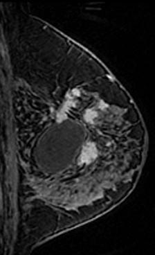
14 First Reports of Breast MRI: El Yousef et al: Radiology 1984 Stelling et al: Radiology 1985 Dash et al: AJR 1986 Contrast Enhanced Breast MRI: Kaiser et al: Radiology 1989 Heywang et al: Radiology 1989 Higher Resolution 3D Imaging: Harms et al: Radiology 1993 Orel et al: Radiology 1994 MRI Guided Bx: Schnall et al: RSNA 1993 Heywang et al: RSNA 1993 MRI of the Breast 2009 Courtesy M. Schnall U of PA 150 μm spatial resolution

15 Benign Fibroadenoma

16 Intraductal Carcinoma

17
18 Current ACR MRI Recommendations A. Screening 1. High-Risk Patients 2. Contralateral breast of patients with new malignancies 3. Patients pre-augmentation and having free injection augmentation
19
20
21 Current ACR MRI Recommendations B. Extent of Disease 4. Invasive and ductal carcinoma in situ 5. Invasion deep to fascia 6. Post lumpectomy with positive margins 7. Neoadjuvant chemotherapy Diagnostic imaging of all women with cancer prior to treatment % of women will have multifocal or multi-centric breast cancer.


22 Chemoprevention J. P. Delille et al., Radiology 235, 36 (2005).
23 Neoadjuvant Chemotherapy
24 Current ACR MRI Recommendations C. Additional Evaluation of Findings 8. Recurrence of Breast Cancer 9. Metastatic disease with unknown origin of primary 10.Lesion characterization 11.Post-operative reconstruction 12.MRI guided biopsy
 Scanned at 30s and 120s post-injection M Nishino et al, J CAT, 27(5), 771-8")
25 Breast CT 100 ml (3 ml/s) intravenous injection Iopamiron 300 (Nihon Schering, Osaka) Scanned at 30s and 120s post-injection M Nishino et al, J CAT, 27(5),


26 Invasive IDC Invasive IDC Multiple Fibroadenomas Papilloma


27 Dedicated breast CT scanner pendant geometry Slide Courtesy John Boone, Ph.D.


28 PRE CONTRAST BCT SUBTRACTION IMAGES SUBTRACTION POST CONTRAST Pt 122
29 Contrast-Enhanced Digital Mammography The advent of modern digital mammography has led to a renewed interest in breast angiography. The field is now called contrast-enhanced digital mammography. Rather than image blood vessels, the desire is to fractionally increase the contrast of breast lesions by virtue of their slightly increased uptake and retention of contrast media as compared to surrounding tissue.
30 Temporal Subtraction In temporal subtraction angiography, an initial mask image is produced and stored prior to the injection of contrast material The contrast media is then injected Finally, a series of post-contrast images are acquired Subtracted images are produced by logarithmically subtracting the initial mask image from each of the post-contrast images
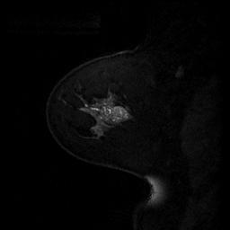
31 Temporal Subtraction Mass attenuation[cm 2 /g] ICRU-44 Breast Tissue Iodine 0.27 mm Cu Energy [kev] Photon Fluence [# photons/mm 2 ]
32 Jong and Yaffe 22 women with mammographic abnormalities underwent CE digital mammography. 6 images were obtained with contrast injected intravenously between the 1st and 2nd images. Enhancement was observed in 8 of 10 patients with cancer. In 2 cancer cases, no enhancement was observed. No enhancement was seen in seven of 12 benign cases, which were otherwise suspected as cancer. Lesion enhancement kinetics was similar to MRI. RA Jong, MJ. Yaffe, et al. Radiology 228: , 2003


33 CC 1 Min IDC IDC Papilloma Papilloma Courtesy M. Yaffe
 Scout 1 min.")


34 Patient With Benign Lesion (Fibrocystic Change) Scout 1 min. 5 min. 30 Enhancement Kinetics Lesion Mean - Tissue Mean in Subtracted Images Time (minutes) Courtesy M. Yaffe


")
35 Infiltrating Ductal Carcinoma Scout 1 min. 7 min. 3 Kinetics mg/cm Time (minutes) Courtesy M. Yaffe
36 CE Tomosynthesis Method MLO projection with 5-7 dn compression X-ray tube 49 kvp, Rh target, 0.27 mm Cu filter 9 projections per data set: 50 o arc, 6.25 o apart 1 projection each 30 sec Dose per data set ~ mammogram (~2 mgy) 68 cm 20 cm Pivot point Compression plate Breast Detector
37 CE Tomosynthesis Method 1 ml/kg of Visipaque-320 (320 mg I/ml iodixanol) - Amersham, Princeton, NJ. 60 ml saline flush. First post-contrast image obtained 90 sec after start of contrast injection. Total exam time ~ 10 minutes. 17 patients to date


38 Patient 1: Digital Mammography Right breast cm ill-defined asymmetry overlying pectoralis muscle


39 Patient 1: MRI Pre Gd T1 FS Post Gd FS Post Gd - sub 1.3 cm enhancing mass at 12:00 in right breast.


40 Contrast-Enhanced Tomosynthesis Pre-contrast Post-contrast Subtraction Spiculated mass with rim enhancement.
41 Image Registration The registration algorithm is a multi-scale, globally smooth and locally affine. Before registration, pre- and post-contrast images are sampled and averaged to preserve original geometry while reducing computational time, and improving the per pixel signal-to-noise ratio.
42 Raw files (18) Sort Pre-contrast Images (9) Post-contrast Images (9) 2294x1914 Sample, Average, Register 2294x1914 Registered post-contrast Images (9) 256x256 Sample, Mask, Normalize intensities Log (pre-contrast) Log (post-contrast) (9) 1147x957 Subtraction Images (9) Write format 1147x957 Projection Images (9) 1147x957


43 Patient 1: Motion Correction 38 yo with ductal carcinoma demonstrating strong enhancement. Registration reduced motion artifacts in axilla and inferior breast, resulting in superior visualization of the lesion and vasculature. BEFORE AFTER


44 Patient 2: Motion Correction BEFORE AFTER AFTER 43 yo with segmental enhancement in upper half of breast, concordant with MRI. In the pre-registered image, the enhancement was not discernable from motion artifacts.
45 Temporal Subtraction Advantages: Superior separation of pre- and post-contrast images High kvp pre- and post- contrast images Reduced total dose Disadvantages: Motion Artifacts
46 Dual-Energy Imaging At diagnostic energies, there are two main x-ray interactions Photoelectric effect Compton effect The relative contribution of the two effects depends upon the energy and the atomic number of the material Therefore, the attenuation coefficients of different materials have different trends as a function of energy
47 Coherent
48 Dual-Energy Radiography
49 Dual-E Example μ f t f The attenuation of the two materials is given by μ g ( μ ) t f t f +μ g tg g I = I e a o
50 Dual-E Example μ f t f We can define a quantity, T, such that μ g t g T 18 = μ f t f + μ g t g and T 40 = μ f t f + μ g t g at 18 kev at 40 kev
51 Dual-E Example μ f μ g t f t g Using data from Johns and Yaffe (1987) for fat and glandular tissue gives T 18 = 0.6t f + 1.0t g at 18 kev and T 40 = 0.2t f + 0.3t g at 40 kev
52 μ f μ g t f t g Dual-E Example If you multiply T 40 by 4, and subtract T 18, you get T sub = (4* )t f + (4* )t g = 0.2 (t f + t g ). Now, fat and glandular tissue have the same attenuation, and thus they lack contrast

53
54 Energy Subtraction In energy subtraction, contrast media is injected first Then, a series of sequential image pairs is obtained Each image pair consists of one high energy and one low energy image Images are then processed and subtracted pair-wise
55 Energy Subtraction Low High Time


 = ln(")

56 Dual-Energy Contrast- High Energy Enhanced Imaging Low Energy High Energy Low Energy SI DE ( x, y) = ln( SI ( x, y)) w ln( SI ( x, y)) H t L
57 Lewin et al. CE DSM was performed on 26 subjects with mammographic or clinical findings warranting biopsy. High-energy (44-49 kvp, + 8mm Al filtration) and lowenergy (30-33 kvp) images were obtained pair-wise, following administration of iodinated contrast. A weighted logarithmic subtraction of the images was performed to obtain images that preferentially show iodine. 13 subjects had invasive cancers, 11 of which enhanced strongly, 1 moderately and 1 weakly. 1 case of DCIS demonstrated a weak enhancement In the 12 benign cases, 2 enhanced diffusely and 2 enhanced weakly focally. JM Lewin et al., Radiology 228: , 2003.

")
58 Two-View Film Mammogram (wire on excisional biopsy scar) (cyst)


59 Lateral... Sagittal Post-contrast MRI to Medial


60 Post-Contrast Dual-Energy Digital Subtraction Mammography

61 XCounter Mammo-3T Prototype

62

63

64

65

66
67

68 DE-DBT: Patient 1 Age: 55 Diagnosis: Invasive ductal carcinoma Sign: mass in axillary tail region

69

70
71 Dual Energy Imaging Subtraction of images increases noise, and does not alter intrinsic subject contrast However, subtraction reduces background structure, increasing conspicuity of residual signals Dual energy imaging is sensitive to scatter and beam hardening Subtraction works best for large objects where the residual signals and noise are distinguishable.


72 Iodine HE Stepwedge Images Not corrected Corrected
73 Corrected Iodine SI 4 mg I/cm2 2 mg I/cm2 0.5 mg I/cm2 1 mg I/cm2 10 mg I/cm2 Not corrected 4 mg I/cm2 2 mg I/cm2 1 mg I/cm2 0.5 mg I/cm2 10 mg I/cm2 20 mg I/cm2 10 mg I/cm2 20 mg I/cm2


74 Iodine HE Stepwedge Images Radius: 70 Not corrected Corrected Proj images, angle 1, W/Cu,49 kv, 100mAs w/sn, 45kV, 100 mas Total Thickness: 25 mm iodine phantom + 40 mm stepwedge
75 Quantification of Glandularity and Iodine uptake 100% Ad 2 mg I/cm2 100% Gl 10 mg I/cm2 23 degrees Corrected
76 Energy Subtraction Advantages: Motion artifacts are rare Disadvantages: System modifications are necessary to allow rapid change of filter material and kvp Detector must be suited to rapid readout Poorer separation of tissue and contrast agent Beam hardening artifacts
77 Molecular Imaging Molecular Imaging in a broad sense implies visualizing normal and abnormal cellular functions by utilizing either biochemical or pharmacological probes. Ideally, the imaging technique should not perturb the function which is being assessed.
78 Molecular Imaging Molecular Imaging in a broad sense implies visualizing normal and abnormal cellular functions by utilizing either biochemical or pharmacological probes. Ideally, the imaging technique should not perturb the function which is being assessed.
79 Molecular imaging in intact species: methods and agents Sensitivity pm Modality Agents H R Primary uses Examples Optical FMT fluorescent proteins X gene expression, tagging superficial structures BLI luciferin X gene expression, therapeutic monitoring GFP, RFP, NIRF probes fluc rluc nm Nuclear SPECT 99m Tc, 123/5 I, 111 In X X site-selectivity, protein labeling 99m Tc-annex in V, 123 I- A85380 PET 11 C, 18 F, 124 I, 64/62/60 Cu X X site-selectivity, gene expression, drug dev mnt 11 C-RAC, 124 I-FIAU, 64 Cu-ATSM μm MRI Spectroscopy Contrast agents endogenous metabolites X X CNS, prostate, heart, breast NAA, Cr, Cho, Glx, mi, 31 P Gd, Mn, FeO X cell trafficking, enzymatic activation poly-l-lysine, dendrimers, MION μm X-ray contrast agents Iodine, Au Nanoparticles X Cancer Gold Nanoparticles (10 μm) Ultrasound contrast agents perfluorinated microbubbles H=human, R=rodent X drug-delivery, gene transfection human albumin (Optison)

80 Nanoparticles Nanoparticles are small polymeric colloidal particles with a therapeutic and/or imaging agent(s) either dispersed in polymer matrix or encapsulated in polymer. Sahoo SK. Labhasetwar V. Nanotech approaches to drug delivery and imaging Drug Discovery Today. 8: , 2003

81 Liposome-encapsulated iodine Karathanasis, et al., Radiology 250(2), 398, 2009

82 In vivo cancer targeting and imaging with semiconductor quantum dots, XH Gao, YY Cui, et al., Nature Biotech, advanced online publication 18 July 2004 Gold NP Tumors are targeted passively by virtue of the leaky tumor vasculature and actively by high affinity binding of NP-antibody conjugates to tumor antigens
83 At mammographic energies, ten 100nm gold nanoparticles/cell will result in 5% subject contrast. Alexander G. Tkachenko, J. AM. CHEM. SOC. 2003, 125,
84 Summary Contrast-enhanced digital breast tomosynthesis is possible using either temporal subtraction or energy subtraction. Early clinical results indicate that CE DBT gives clinical results which are concordant with MRI. Technical challenges still exist before CE DBT will be widely available.
Contrast-Enhanced Digital Mammography
 2015 ARRS Breast Symposium Contrast-Enhanced Digital Mammography John Lewin, M.D. Diversified Radiology of Colorado CEDM - Outline History Technique Literature Review / Cases Clinical Status Inexpensive,
Contrast Enhanced Spectral Mammography (CESM) Updates
 Updates") Contrast Enhanced Spectral Mammography (CESM) Updates Georgeta Mihai, PhD, DABR Medical Physicist, BIDMC, Boston Assistant Professor, Harvard Medical School, Boston Disclosures None Acknowledgments: Da
Contrast Enhanced Spectral Mammography (CESM) Updates Georgeta Mihai, PhD, DABR Medical Physicist, BIDMC, Boston Assistant Professor, Harvard Medical School, Boston Disclosures None Acknowledgments: Da
Pitfalls and Limitations of Breast MRI. Susan Orel Roth, MD Professor of Radiology University of Pennsylvania
 Pitfalls and Limitations of Breast MRI Susan Orel Roth, MD Professor of Radiology University of Pennsylvania Objectives Review the etiologies of false negative breast MRI examinations Discuss the limitations
8/3/2016. DBT Physics Basic to Advanced: Primer On Tomosynthesis. Tomosynthesis Pedigree
 DBT Physics Basic to Advanced: Primer On Tomosynthesis Andrew D. A. Maidment, Ph.D. University of Pennsylvania Department of Radiology Acknowledgements of Support Research support from the Komen Foundation,
Emerging Techniques in Breast Imaging: Contrast-Enhanced Mammography and Fast MRI
 Emerging Techniques in Breast Imaging: Contrast-Enhanced Mammography and Fast MRI Lilian Wang, M.D. Breast Imaging Section Department of Radiology Northwestern Medicine Overview Rationale for new imaging
Here are examples of bilateral analog mammograms from the same patient including CC and MLO projections.
 Good afternoon. It s my pleasure to be discussing Diagnostic Breast Imaging over the next half hour. I m Wei Yang, Professor of Diagnostic Radiology and Chief, the Section of Breast Imaging as well as
SenoBright Contrast Enhanced Spectral Mammography Technology. Ann-Katherine Carton Sylvie Saab-Puong Matt Suminski
 SenoBright Contrast Enhanced Spectral Mammography Technology Ann-Katherine Carton Sylvie Saab-Puong Matt Suminski White Paper October 2012 SenoBright Contrast Enhanced Spectral Mammography Technology Ann-Katherine
THE FUTURE OF MEDICAL IMAGING A. D. A. Maidment* Department of Radiology, University of Pennsylvania, 3400 Spruce Street, Philadelphia, PA 19104, USA
 Radiation Protection Dosimetry (2010), Vol. 139, No. 1 3, pp. 3 7 Advance Access publication 18 March 2010 doi:10.1093/rpd/ncq090 A. D. A. Maidment* Department of Radiology, University of Pennsylvania,
Standard Breast Imaging Modalities. Lilian Wang, M.D. Breast Imaging Section Department of Radiology Northwestern Medicine
 Standard Breast Imaging Modalities Lilian Wang, M.D. Breast Imaging Section Department of Radiology Northwestern Medicine Overview Standard breast imaging modalities Mammography Ultrasound MRI Imaging
Mammographic imaging of nonpalpable breast lesions. Malai Muttarak, MD Department of Radiology Chiang Mai University Chiang Mai, Thailand
 Mammographic imaging of nonpalpable breast lesions Malai Muttarak, MD Department of Radiology Chiang Mai University Chiang Mai, Thailand Introduction Contents Mammographic signs of nonpalpable breast cancer
Update of Digital Breast Tomosynthesis. Susan Orel Roth, MD
 Update of Digital Breast Tomosynthesis Susan Orel Roth, MD NCI estimates that : Why DBT? Approximately 20% of breast cancers are missed at mammography screening Average recall rates approximately 10%
CURRENTLY FDA APPROVED ARE FULL FIELD DIGITAL MAMMOGRAPHY SYSTEMS AND FILM SCREEN STILL BEING USED AT SOME INSTITUTIONS
 ABBY DUROJAYE,M.D CURRENTLY FDA APPROVED ARE FULL FIELD DIGITAL MAMMOGRAPHY SYSTEMS AND FILM SCREEN STILL BEING USED AT SOME INSTITUTIONS BOTH HAVE BEEN SHOWN TO BE EFFECTIVE TOOLS EARLY DETECTION OF BREAST
Updates in Mammography. Dr. Yang Faridah A. Aziz Department of Biomedical Imaging University Malaya Medical Centre
 Updates in Mammography Dr. Yang Faridah A. Aziz Department of Biomedical Imaging University Malaya Medical Centre Updates in Mammography Breast Imaging Dr. Yang Faridah A. Aziz Department of Biomedical
Successful Breast MRI Program : The ingredients
 Successful Breast MRI Program : The ingredients Dr. Smriti Hari Associate Professor Deptt. Of Radiology All India Institute of Medical Sciences New Delhi How to perform Breast MRI Breast MRI descriptors
Since its introduction in 2000, digital mammography has become
 Review Article Smith A, PhD email : Andrew.smith@hologic.com Since its introduction in 2000, digital mammography has become an accepted standard of care in breast cancer screening and has paved the way
Breast Tomosynthesis. What is breast tomosynthesis?
 Scan for mobile link. Breast Tomosynthesis Breast tomosynthesis is an advanced form of mammography, a specific type of breast imaging that uses low-dose x-rays to detect cancer early when it is most treatable.
Digital Breast Tomosynthesis from a first idea to clinical routine
 International Master Programm Biomedical Engineering Digital Breast Tomosynthesis from a first idea to clinical routine Historical background 2D imaging of 3D objects has important limitations Jörg Barkhausen
Financial Disclosures
 Financial Disclosures 3D Mammography: The Latest Developments in the Breast Imaging Arena I have no financial disclosures Dr. Katharine Lampen-Sachar Breast and Body Radiologist Radiology Associates of
Outline. Digital Breast Tomosynthesis: Update and Pearls for Implementation. Tomosynthesis Dataset: 2D/3D (Hologic Combo Acquisition)
") Outline Digital Breast Tomosynthesis (DBT) the new standard of care Digital Breast Tomosynthesis: Update and Pearls for Implementation Emily F. Conant, M.D. Professor, Chief of Breast Imaging Department
Outline Digital Breast Tomosynthesis (DBT) the new standard of care Digital Breast Tomosynthesis: Update and Pearls for Implementation Emily F. Conant, M.D. Professor, Chief of Breast Imaging Department
Armed Forces Institute of Pathology.
 Armed Forces Institute of Pathology www.radpath.com Armed Forces Institute of Pathology Breast Disease www.radpath.org Armed Forces Institute of Pathology Interpretation of Breast MRI Leonard M. Glassman
Emerging Technologies in Breast Imaging
 June 30, 2016 Emerging Technologies in Breast Imaging Jay A. Baker, M.D. Division of Breast Imaging New Technologies in Breast Imaging Full Field Digital Mammography (FFDM) X-ray/Mammography Computer-Aided
Breast Cancer Imaging
 Breast Cancer Imaging I. Policy University Health Alliance (UHA) will cover breast imaging when such services meet the medical criteria guidelines (subject to limitations and exclusions) indicated below.
Breast CT and Dosimetry
 2013 ICTP/IAEA Training Course on Radiation Protection of Patients Trieste Breast CT and Dosimetry John M. Boone, Ph.D., FAAPM, FSBI, FACR Professor and Vice Chair (Research) of Radiology Professor of
Hacia la imagenología tomográfica de mama
 Hacia la imagenología tomográfica de mama Futuro y presente Ioannis Sechopoulos, Ph.D., DABR Advanced X ray Tomographic Imaging (AXTI) Lab Department of Radiology and Nuclear Medicine Radboud University
Radiation Dosimetry in Digital Breast Tomosynthesis. March, 2015 William J. O Connel, Dr. Ph, Senior Medical Physicist
 Radiation Dosimetry in Digital Breast Tomosynthesis March, 2015 William J. O Connel, Dr. Ph, Senior Medical Physicist Imagination at work. Syllabus 1. Introduction 2. Dosimetry in Mammography 3. Dosimetry
Role of PEM in Breast Cancer Management. Judy Kalinyak, MD, PhD Chief Medical Officer Naviscan, Inc (San Diego, CA)
") Role of PEM in Breast Cancer Management Judy Kalinyak, MD, PhD Chief Medical Officer Naviscan, Inc (San Diego, CA) Role of PEM in Breast Cancer Management Introduction to Positron Emission Mammography
Role of PEM in Breast Cancer Management Judy Kalinyak, MD, PhD Chief Medical Officer Naviscan, Inc (San Diego, CA) Role of PEM in Breast Cancer Management Introduction to Positron Emission Mammography
Opportunities and Innovations in Digital Mammography John M. Sandrik, Ph.D. GE Healthcare Milwaukee, WI
 Opportunities and Innovations in Digital Mammography John M. Sandrik, Ph.D. GE Healthcare Milwaukee, WI john.sandrik@med.ge.com with many thanks to Vince Polkus, Advanced Applications Product Mgr. 1 Content
EARLY DETECTION: MAMMOGRAPHY AND SONOGRAPHY
 EARLY DETECTION: MAMMOGRAPHY AND SONOGRAPHY Elizabeth A. Rafferty, M.D. Avon Comprehensive Breast Center Massachusetts General Hospital Harvard Medical School Breast Cancer Screening Early detection of
An Introduction to Dual Energy Computed Tomography
 An Introduction to Dual Energy Computed Tomography Michael Riedel University of Texas Health Science Center at San Antonio Introduction The idea of computed tomography (CT) was first introduced in the
RADIATION PROTECTION IN DIAGNOSTIC AND INTERVENTIONAL RADIOLOGY. L19: Optimization of Protection in Mammography
 IAEA Training Material on Radiation Protection in Diagnostic and Interventional Radiology RADIATION PROTECTION IN DIAGNOSTIC AND INTERVENTIONAL RADIOLOGY L19: Optimization of Protection in Mammography
Contrast-enhanced Breast MRI RSSA 2013
 Contrast-enhanced Breast MRI RSSA 2013 Prof. dr. Maurice van den Bosch University Medical Center Utrecht, the Netherlands Index 1) Breast cancer 2) Why MRI of the breast 3) Technique 4) Interpretation
AAPM Medical Physics Tutorial Session 1. Digital Breast Tomosynthesis. CT Breast Imaging. TMIST Update. Stereotactic Breast Biopsy
 Saturday SPPH01 AAPM Medical Physics Tutorial Session 1 Saturday, Nov. 25 12:00PM - 2:00PM Room: E351 BR PH AMA PRA Category 1 Credits : 2.00 ARRT Category A+ Credits: 2.00 FDA Discussions may include
TOMOSYNTHESIS: WORTH ALL THE HYPE?
 X-Ray Associates of New Mexico, P.C. TOMOSYNTHESIS: WORTH ALL THE HYPE? MICHAEL N. LINVER, MD, FACR MAMMOGRAPHY: THE GOOD, THE PRETTY GOOD, & THE NOT SO GOOD MAMMOGRAPHY: THE GOOD, THE PRETTY GOOD, & THE
New Imaging Modalities for better Screening and Diagnosis
 New Imaging Modalities for better Screening and Diagnosis Miri Sklair-Levy, MD Department of Diagnostic Imaging Sheba Medical Center, Sackler School of Medicine, Tel Aviv University Department of Diagnostic
EARLY DETECTION: MAMMOGRAPHY AND SONOGRAPHY
 EARLY DETECTION: MAMMOGRAPHY AND SONOGRAPHY Elizabeth A. Rafferty, M.D. Avon Comprehensive Breast Center Massachusetts General Hospital Harvard Medical School Breast Cancer Screening Early detection of
Breast MRI Update. Jeffrey C. Weinreb, MD, FACR Yale University School of Medicine
 Breast MRI Update Jeffrey C. Weinreb, MD, FACR jeffrey.weinreb@yale.edu Yale University School of Medicine I disclose the following financial relationships with relevant commercial interests: Bracco Bayer
Patient Dosimetry in Mammography and Tomosynthesis:
 2013 ICTP/IAEA Training Course on Radiation Protection of Patients Trieste Patient Dosimetry in Mammography and Tomosynthesis: What to measure, why and how John M. Boone, Ph.D., FAAPM, FSBI, FACR Professor
Mammography. What is Mammography?
 Scan for mobile link. Mammography Mammography is a specific type of breast imaging that uses low-dose x-rays to detect cancer early before women experience symptoms when it is most treatable. Tell your
Contrast enhanced spectral mammography: A literature review
 Contrast enhanced spectral mammography: A literature review Poster No.: R-0172 Congress: RANZCR-AOCR 2012 Type: Authors: Keywords: DOI: Educational Exhibit S. Buzynski, D. Taylor; Perth/AU Breast, Digital
Breast Imaging Update: Old Dog New Tricks
 Breast Imaging Update: Old Dog New Tricks Claire McKay, DO M&S Imaging Assoc. San Antonio, TX cmckayhart@juno.com Goals Describe modalities available, old and new Provide understanding of pros and cons
Disclosures. Outline. Learning Objectives. Introduction. Introduction. Stereotactic Breast Biopsy vs Mammography: Image Quality and Dose.
 Disclosures Stereotactic Biopsy vs Mammography: and Dose None Vikas Patel, PhD, DABR Upstate Medical Physics 2014 Annual Meeting The American Association of Physicists in Medicine Austin, TX Learning Objectives
Breast Imaging: Multidisciplinary Approach. Madelene Lewis, MD Assistant Professor Associate Program Director Medical University of South Carolina
 Breast Imaging: Multidisciplinary Approach Madelene Lewis, MD Assistant Professor Associate Program Director Medical University of South Carolina No Disclosures Objectives Discuss a multidisciplinary breast
Participants John M. Boone, PhD, Sacramento, CA (Presenter) Patent agreement, Isotropic Imaging Corporation; Consultant, RadSite;
 Patent agreement, Isotropic Imaging Corporation; Consultant, RadSite;") Saturday SPPH01 AAPM Medical Physics Tutorial Session 1 Saturday, Nov. 25 12:00PM - 2:00PM Room: E351 BR PH AMA PRA Category 1 Credits : 2.00 ARRT Category A+ Credits: 2.25 FDA Discussions may include
Saturday SPPH01 AAPM Medical Physics Tutorial Session 1 Saturday, Nov. 25 12:00PM - 2:00PM Room: E351 BR PH AMA PRA Category 1 Credits : 2.00 ARRT Category A+ Credits: 2.25 FDA Discussions may include
Fundamentals of Breast Tomosynthesis
 Fundamentals of Breast Tomosynthesis Improving the Performance of Mammography Andrew Smith, Ph.D. This white paper is one in a series of research overviws on advanced technologies in women s healthcare.
Mammography. What is Mammography? What are some common uses of the procedure?
 Mammography What is Mammography? Mammography is a specific type of imaging that uses a low-dose x-ray system to examine breasts. A mammography exam, called a mammogram, is used to aid in the early detection
Tomosynthesis and breast imaging update. Dr Michael J Michell Consultant Radiologist King's College Hospital NHS Foundation Trust
 Tomosynthesis and breast imaging update Dr Michael J Michell Consultant Radiologist King's College Hospital NHS Foundation Trust Breast imaging new technology BREAST CANCER FLT PET shows different grades
RSNA, /radiol Appendix E1. Methods
 RSNA, 2016 10.1148/radiol.2016151097 Appendix E1 Methods US and Near-infrared Data Acquisition Four optical wavelengths (740 nm, 780 nm, 808 nm, and 830 nm) were used to sequentially deliver the light
Lesion Imaging Characteristics Mass, Favoring Benign Circumscribed Margins Intramammary Lymph Node
 Lesion Imaging Characteristics Mass, Favoring Benign Circumscribed Margins Intramammary Lymph Node Oil Cyst Mass, Intermediate Concern Microlobulated Margins Obscured Margins Mass, Favoring Malignant Indistinct
Breast Cancer. Most common cancer among women in the US. 2nd leading cause of death in women. Mortality rates though have declined
 Breast Cancer Most common cancer among women in the US 2nd leading cause of death in women Mortality rates though have declined 1 in 8 women will develop breast cancer Breast Cancer Breast cancer increases
Breast Cancer. Saima Saeed MD
 Breast Cancer Saima Saeed MD Breast Cancer Most common cancer among women in the US 2nd leading cause of death in women 1 in 8 women will develop breast cancer Incidence/mortality rates have declined Breast
Ana Sofia Preto 19/06/2013
 Ana Sofia Preto 19/06/2013 Understanding the underlying pathophysiologic processes leading to the various types of calcifications Description and illustration of the several types of calcifications, according
MRI in breast cancer: diagnosis and intervention. Dr Sue Barter Addenbrookes Hospital, Cambridge UK
 MRI in breast cancer: diagnosis and intervention Dr Sue Barter Addenbrookes Hospital, Cambridge UK Intervention will be discussed in High Risk Screening! Indications UK and Europe: Breast MRI is well established
Breast MRI: Friend or Foe?
 Breast MRI: Friend or Foe? UCSF Postgraduate Course May 18, 2013 Cheryl Ewing, MD Clinical Professor of Surgery UCSF Department of Surgery APPLEGATE HAS DOUBLE MASTECTOMY IN CANCER SCARE DIAGNOSED WITH
Accreditation Case Review: Mammography and Stereotactic Biopsy
 Accreditation Case Review: Mammography and Stereotactic Biopsy Brett T. Parkinson MD Breast Imaging Director Breast Care Services Intermountain Healthcare Chair, ACR Committee on Mammography Accreditation
Mammography. Background and Perspective. Mammography Evolution. Background and Perspective. T.R. Nelson, Ph.D. x41433
 - 2015 Background and Perspective 2005 (in US) Women Men Mammography Invasive Breast Cancer Diagnosed 211,240 1,690 Noninvasive Breast Cancer Diagnosed 58,940 Deaths from Breast Cancer 40,410 460 T.R.
I. Cancer staging problem
 Instrumentation Lab (Craig Levin) Angela Craig Peter Jin Frezghi Guillem Billie Garry Research interests (by imaging modality) I. High resolution radionuclide imaging : positron emission tomography (PET)
FACT: FACT: Breast Cancer Staging. For cancer to occur, something must damage nucleus of the cell. Stage I. Stage II 10/9/2018
 Digital Breast Tomosynthesis (DBT) Pathology Findings: Case Studies and Beyond For cancer to occur, something must damage nucleus of the cell. Advanced Health Education Center www.aheconline.com Copyright
Molecular Imaging and Breast Cancer
 Molecular Imaging and Breast Cancer Breast cancer forms in tissues of the breast usually in the ducts, tubes that carry milk to the nipple, and lobules, the glands that make milk. It occurs in both men
BREAST MRI. Elizabeth A. Rafferty, M.D. Avon Comprehensive Breast Center Massachusetts General Hospital Harvard Medical School
 BREAST MRI Elizabeth A. Rafferty, M.D. Avon Comprehensive Breast Center Massachusetts General Hospital Harvard Medical School BREAST MRI Any assessment of the breast parenchyma requires the administration
Imaging in breast cancer. Mammography and Ultrasound Donya Farrokh.MD Radiologist Mashhad University of Medical Since
 Imaging in breast cancer Mammography and Ultrasound Donya Farrokh.MD Radiologist Mashhad University of Medical Since A mammogram report is a key component of the breast cancer diagnostic process. A mammogram
2/14/2019. Advances in Breast Imaging. Outline. Jiali Wang, Ph.D., DABR Medical Physicist. February 23, 2019
 Advances in Breast Imaging SCPMG Medical Imaging Technology & Informatics Jiali Wang, Ph.D., DABR Medical Physicist February 23, 2019 Outline History of breast imaging Advances in breast imaging a) Full-Field
Role of positron emission mammography (PEM) for assessment of axillary lymph node status in patients with breast cancer
 for assessment of axillary lymph node status in patients with breast cancer") Role of positron emission mammography (PEM) for assessment of axillary lymph node status in patients with breast cancer Poster No.: C-1260 Congress: ECR 2011 Type: Scientific Paper Authors: K. M. Kulkarni,
Role of positron emission mammography (PEM) for assessment of axillary lymph node status in patients with breast cancer Poster No.: C-1260 Congress: ECR 2011 Type: Scientific Paper Authors: K. M. Kulkarni,
What s New in Breast Imaging. Jennifer A. Harvey, M.D., FACR Professor of Radiology University of Virginia
 What s New in Breast Imaging Jennifer A. Harvey, M.D., FACR Professor of Radiology University of Virginia Disclosure Hologic, Inc. Shareholder and research agreement. Volpara Solutions, Ltd. Shareholder
Contrast-Enhanced Breast Tomosynthesis: Combining the Best of Both Worlds for Better Breast-Cancer Diagnosis
 Contrast-Enhanced Breast Tomosynthesis: Combining the Best of Both Worlds for Better Breast-Cancer Diagnosis T Wu (twu2@partners.org), E Rafferty, R Moore, D Kopans, Massachusetts General Hospital, Boston,
BREAST IMAGING and NEW IMAGING MODALITIES- A Surgeons view
 BREAST IMAGING and NEW IMAGING MODALITIES- A Surgeons view DR CHANTEL THORNTON SPECIALIST BREAST CANCER SURGEON BMSc (hons) MBBS (hons) FRACS Epworth Hospital, Richmond- Agora Centre for Women s Health
POSITIONING ACR REQUIREMENTS IMAGE REVIEW CATEGORIES DEFICIENCIES IN POSITIONING ACR REQUIREMENTS 3/28/2016 NUMBER 1 REASON FOR ACR FAILURE
 CERTIFYING AGENCIES MAMMOGRAPHY CLINICAL IMAGE EVALUATION Pam Fulmer, BA RT (R)(M)(QM) FDA approved certifying states States can only certify facilities within their state borders Illinois Iowa South Carolina
Assessment of extent of disease: digital breast tomosynthesis (DBT) versus full-field digital mammography (FFDM)
 versus full-field digital mammography (FFDM)") Assessment of extent of disease: digital breast tomosynthesis (DBT) versus full-field digital mammography (FFDM) Poster No.: C-1237 Congress: ECR 2012 Type: Scientific Paper Authors: N. Seo 1, H. H. Kim
Assessment of extent of disease: digital breast tomosynthesis (DBT) versus full-field digital mammography (FFDM) Poster No.: C-1237 Congress: ECR 2012 Type: Scientific Paper Authors: N. Seo 1, H. H. Kim
FDA Executive Summary
 Meeting of the Radiological Devices Advisory Panel On October 24, 22, the panel will discuss, make recommendations, and vote on a premarket approval application supplement (P83/S) to expand the indications
Categorical Classification of Spiculated Mass on Breast MRI
 Categorical Classification of Spiculated Mass on Breast MRI Poster No.: C-1974 Congress: ECR 2013 Type: Authors: Scientific Exhibit Y. Kanda 1, S. Kanao 2, M. Kataoka 2, K. Togashi 2 ; 1 Kyoto City/JP,
Breast positioning system for full field digital mammography and digital breast tomosynthesis system
 Breast positioning system for full field digital mammography and digital breast tomosynthesis system Mari Varjonen* a, Martti Pamilo b, Pirjo Hokka b, Riina Hokkanen a, Pekka Strömmer a a Planmed Oy Asentajankatu
Policy Library Clinical Advantages of Digital Breast Tomosynthesis in Symptomatic Patients
 Policy Library Clinical Advantages of Digital Breast Tomosynthesis in Symptomatic Patients Version: 1 Approved by: Faculty of Clinical Radiology Council Date of approval: Click and type: day month and
AB MR Interpretation Overview
 AB MR Interpretation Overview Goal of AB MR interpretation is to maintain high sensitivity and specificity In order to minimize false positives and short term follow ups, it is fundamental to focus only
Molecular Imaging and Cancer
 Molecular Imaging and Cancer Cancer causes one in every four deaths in the United States, second only to heart disease. According to the U.S. Department of Health and Human Services, more than 512,000
Current Status of Supplementary Screening With Breast Ultrasound
 Current Status of Supplementary Screening With Breast Ultrasound Stephen A. Feig, M.D., FACR Fong and Jean Tsai Professor of Women s Imaging Department of Radiologic Sciences University of California,
BREAST MRI. Elizabeth A. Rafferty, M.D. Avon Comprehensive Breast Center Massachusetts General Hospital Harvard Medical School
 BREAST MRI Elizabeth A. Rafferty, M.D. Avon Comprehensive Breast Center Massachusetts General Hospital Harvard Medical School BREAST MRI Any assessment of the breast parenchyma requires the administration
ROLE OF MRI IN SCREENING, DIAGNOSIS AND MANAGEMENT OF BREAST CANCER. B.Zandi Professor of Radiology
 ROLE OF MRI IN SCREENING, DIAGNOSIS AND MANAGEMENT OF BREAST CANCER B.Zandi Professor of Radiology Introduction In the USA, Breast Cancer is : The Most Common Non-Skin Cancer The Second Leading cause of
BI-RADS and Breast MRI. Kathy Borovicka, M.D. Thursday February 15, 2018
 BI-RADS and Breast MRI Kathy Borovicka, M.D. Thursday February 15, 2018 Learning Objectives Be familiar with the Breast Imaging Reporting and Data System (BI-RADS) Understand the components of a breast
Essentials of Clinical MR, 2 nd edition. 73. Urinary Bladder and Male Pelvis
 73. Urinary Bladder and Male Pelvis Urinary bladder carcinoma is best locally staged with MRI. It is important however to note that a thickened wall (> 5 mm) is a non-specific finding seen in an underfilled
Molecular Breast Imaging: History and Recent Developments
 Molecular Breast Imaging: History and Recent Developments Associate Professor, Department of Imaging Physics The University of Texas MD Anderson Cancer Center, Houston, Texas Educational Objectives 1.
Detailed Program of the second BREAST IMAGING AND INTERVENTIONS PROGRAM am am : Clinician s requirements from breast imaging
 Detailed Program of the second BREAST IMAGING AND INTERVENTIONS PROGRAM 2012 Day one, 2 nd November BREAST IMAGING AND INTERVENTIONS PROGRAM 2012 9.00 AM 9.10 am Introduction 9.10 am - 9.30 am : Clinician
Dual Energy CT of the Liver
 34th Annual Course October 2011 Washington, DC Dual Energy CT of the Liver Vassilios Raptopoulos, MD Beth Israel Deaconess Medical Center Harvard Medical School Dual Energy CT (DECT) Different materials
Note: This copy is for your personal non-commercial use only. To order presentation-ready copies for distribution to your colleagues or clients, conta
 Note: This copy is for your personal non-commercial use only. To order presentation-ready copies for distribution to your colleagues or clients, contact us at www.rsna.org/rsnarights. Karen K. Lindfors,
Epworth Healthcare Benign Breast Disease Symposium. Sat Nov 12 th 2016
 Epworth Healthcare Benign Breast Disease Symposium Breast cancer is common Sat Nov 12 th 2016 Benign breast disease is commoner, and anxiety about breast disease commoner still Breast Care Campaign UK
Breast MRI: Friend or Foe?
 Breast : Friend or Foe? APPLEGATE HAS DOUBLE MASTECTOMY IN CANCER SCARE DIAGNOSED WITH CANCER IN ONE BREAST Comments: 0 ASSOCIATED PRESS 8/19/2008 UCSF Postgraduate Course March 19, 2009 E. Shelley Hwang
Radiologic and pathologic correlation of non-mass like breast lesions on US and MRI: Benign, high risk, versus malignant
 Radiologic and pathologic correlation of non-mass like breast lesions on US and MRI: Benign, high risk, versus malignant Poster No.: C-1161 Congress: ECR 2013 Type: Educational Exhibit Authors: J. Kwak,
Anyone can get breast cancer BREAST MRI BREAST CANCER. The incidence of getting breast cancer is 1:19 in Malaysia
 Anyone can get breast cancer BREAST MRI KATE Datin Dr Fatimah Moosa Sunway Medical Centre DATIN SERI ENDON KYLIE SIZE DOES NOT MAKE A DIFFERENCE BREAST CANCER The incidence of getting breast cancer is
Physics of MBI (~10 slides)
") Physics of MBI (~10 slides) Molecular Breast Imaging (MBI) physics and performance testing JW Hugg, BR Simrak, PD Smith, BE Patt Gamma Medica, Inc., Northridge, CA Molecular Breast Imaging (MBI) is an
Physics of MBI (~10 slides) Molecular Breast Imaging (MBI) physics and performance testing JW Hugg, BR Simrak, PD Smith, BE Patt Gamma Medica, Inc., Northridge, CA Molecular Breast Imaging (MBI) is an
Diagnostic Medical Physicist Via Christi Hospitals Wichita, Wichita, KS
 Digital Breast Tomosynthesis SWAAPM Meeting 30 Mar 2012 Jerry A. Thomas, MS, FAAPM, DABR, CHP, DABSNM Diagnostic Medical Physicist Via Christi Hospitals Wichita, Wichita, KS Talk Overview Breast Cancer
High Risk Screening: A Multimodality Approach
 High Risk Screening: A Multimodality Approach John Lewin, M.D., FACR, FSBI The Women s Imaging Center Denver, Colorado Disclosures Consultant to Hologic Previously received research funds from Hologic
Breast Cancer Diagnosis, Treatment and Follow-up
 Breast Cancer Diagnosis, Treatment and Follow-up What is breast cancer? Each of the body s organs, including the breast, is made up of many types of cells. Normally, healthy cells grow and divide to produce
DCIS of the Breast--MRI findings with mammographic correlation.
 DCIS of the Breast--MRI findings with mammographic correlation. Poster No.: C-1560 Congress: ECR 2013 Type: Educational Exhibit Authors: N. B. Ibrahim, P. Morris, S. ANANDAN; Burlington, MA/US Keywords:
Breast Cancer. What is breast cancer?
 Scan for mobile link. Breast Cancer Breast cancer is a malignant tumor in or around breast tissue. It usually begins as a lump or calcium deposit that develops from abnormal cell growth. Most breast lumps
To Shield or Not to Shield? Lincoln L. Berland, M.D.
 To Shield or Not to Shield? Lincoln L. Berland, M.D. Disclosures Consultant to: Nuance, Inc. Page 2 Breast Radiation on CT Use of chest CT has increased in women vulnerable to cancer induction by radiation.
Innovations and Applications of Tomosynthesis. Andrew D. A. Maidment, Ph.D. University of Pennsylvania Department of Radiology
 Innovations and Applications of Tomosynthesis Andrew D. A. Maidment, Ph.D. University of Pennsylvania Department of Radiology Acknowledgements of Support Grant support from the Komen Foundation, DOD, NIH,
Non-mass Enhancement on Breast MRI. Aditi A. Desai, MD Margaret Ann Mays, MD
 Non-mass Enhancement on Breast MRI Aditi A. Desai, MD Margaret Ann Mays, MD Breast MRI Important screening and diagnostic tool, given its high sensitivity for breast cancer detection Breast MRI - Indications
Contrast Enhanced Spectral Mammography (CESM) Initial UK Experience. Dr Sarah L Tennant BMedSci, BMBS, MRCP, FRCR
 Initial UK Experience. Dr Sarah L Tennant BMedSci, BMBS, MRCP, FRCR") Contrast Enhanced Spectral Mammography (CESM) Initial UK Experience Dr Sarah L Tennant BMedSci, BMBS, MRCP, FRCR Vote Now Your experience of CESM 1. No experience of CESM 44% 2. I ve seen some cases in
Contrast Enhanced Spectral Mammography (CESM) Initial UK Experience Dr Sarah L Tennant BMedSci, BMBS, MRCP, FRCR Vote Now Your experience of CESM 1. No experience of CESM 44% 2. I ve seen some cases in
Index. C Calcifications fat necrosis 1, 61 fat necrosis 4, 69 nipple/peri-areolar involvement 1, 165
 A ADH. See Atypical ductal hyperplasia (ADH) American College of Radiology (ACR), BI-RADS background parenchymal enhancement, 8, 9, 81, 82 fibroglandular tissue guidelines, 6 American Joint Committee on
Molecular Breast Imaging
 Molecular Breast Imaging Development of a Low-Dose Screening Test for Dense Breasts Conflict of Interest Royalties for technologies licensed to Gamma Medica Ideas Carrie B. Hruska, Ph.D. Department of
Contrast-Enhanced Spectral Mammography
 Contrast-Enhanced Spectral Mammography Illuminating Breast Cancer Detection SenoBright HD TM gehealthcare.com/senobright Mammography is the most reliable imaging technique for breasts, but limitations