Mechanism of Vesicular Transport
|
|
|
- Mark Booth
- 5 years ago
- Views:
Transcription
1 Mechanism of Vesicular Transport Transport vesicles play a central role in the traffic of molecules between different membrane-enclosed enclosed compartments. The selectivity of such transport is therefore a key to maintaining the functional organization of the cell. The specificity of transport of transport is based on the selective packaging of the intended cargo into vesicles that recognize and fuse only with the appropriate target membrane.
2 Experimental Approaches to understanding vesicular transport 1. Isolation of yeast mutants that are defective in protein transport and sorting 2. Biochemical i approach: Reconstitution of vesicular transport t in cell-free system / isolation of enzymes and proteins involved in protein processing and sorting 3. Studies of synaptic vesicles, which are responsible for the regulated secretion of neurotransmitters by neurons 4. Fluorescent Microscopy Studies using yeast mutants Advantageous because they are readily amenable to genetic analysis Randy Schekman and his colleagues have pioneered the isolation of yeast mutants defective in vesicular transport including mutants that are defective at various stages of protein secretion (sec mutants), mutants that are unable to transport proteins to the vacuole, and mutants that are unable to retain resident ER proteins. The isolation of such mutants led directly to the molecular cloning and analysis of the corresponding genes, thereby identifying a number of proteins involved in various steps of the secretory pathway
3 Fig Yeast mutants defective in protein trafficking and sorting
4 Other studies have been done using yeast mutants which has even defined the pathway by which secretory proteins mature. A large number of temperature sensitive mutant yeast strains were identified in which the secretion of all proteins is blocked at the higher, nonpermissive temperature (at which h cells cannot grow) but is normal at the lower permissive i temperatures, (at which cells grow normally). When transferred from the lower to the higher temperature, these so-called sec mutants accumulate secretory proteins at the point in the pathway a that is blocked. Analysis of such mutants identified five classes (A-E), corresponding to the five steps in the secretory pathway, in which secretory proteins accumulate in the cytosol, RER, small vesicles taking proteins from the ER to the Golgi complex, Golgi cisternae, or secretory vesicles. To determine the order of the steps in the pathway, researchers analyzed double sec mutants. For instance, when yeast cells contain mutations in both class B and class D functions, proteins accumulate in the RER, not in the Golgi cisternae. Since proteins accumulate at the earliest blocked step, this finding shows that class B mutations must act at an earlier point in the maturation pathway than class D mutations do, These studies confirmed that as a secretory yprotein matures it moves sequentially from the cytosol -> RER -> ER-to-Golgi transport vesicles -> Golgi cisternae -> secretory vesicles and finally is exocytosed.
5 Isolation of temperature-sensitive mutants in yeast


6 Reconstituted vesicular transport The first cell-free transport system was developed by James Rothman and his colleagues A mutant mammalian cell line that lacked functional N-acetylglucosaminetransferase at an early stage of N-glycosylation o in the Golgi. Consequently, the glycoproteins produced by this mutant lacked added N- acetylglucosamine yg (GlcNAc) units. Golgi stacks isolated from a virus-infected mutant cell line unable to catalyze the addition of GlcNAc to N-linked oligosaccharides are mixed with Golgi stackes from a normal cell line. Because the mutant cells were infected by a virus, the proteins it synthesizes can be specifically detected. Transport of these proteins to normal Golgi stacks is signaled by the addition of radiolabelled ll d GlcNAc.


7 Cargo selection, Coat proteins, and Vesicle Budding Most transport vesicles are coated with cytosolic coat proteins, thus called coated vesicle overview 1. Secretory proteins are sorted from proteins targeted for other destinations and from proteins that need to remain behind 2. The caots assemble as the secretory protein-containing vesicles bud off the donor membrane and are generally removed from the vesicle in the cytosol before they reach their target 3. At the target membrane, the vesicles dock and fuse with the membrane, emptying their lumenal cargo and inserting their membrane proteins into the target membrane
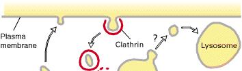
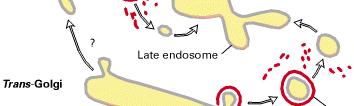
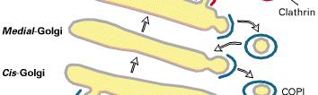
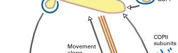
8 Three types of Coated vesicles COP-coated vesicles COPI : vesicles moving between the Golgi cisternae or retrieval vesicles that returen resident ER proteins marked by the KDEL or KKXX retrieval signals back to the ER from the ER-Golgi interamediate compartment or the cis Golgi network (Retrograde transport) COPII : carry secretory proteins from the ER to the ER-Golgi intermediate compartment or Golgi apparatus, budding from the transitional ER and carrying their cargo forward along the secretory pathway Clathrin-coated vesicles: the uptake of extracellular l molecules l from the plasma membrane by endocytosis as well as the transport of molecules from the trans Golgi network to endosomes, lysosomes or the plasma membrane
9 Different Coat Proteins Act at Specific Points in the Secretory Pathway
10 removal by retrograde flow maintains identity of ER and Golgi KDEL receptor for soluble proteins TM proteins with -KKXX interact with COP I tubulation separates membrane and soluble proteins


11 Early Secretory Pathway - Forward and Retrograde Traffic KDEL-receptors bind to KDEL-bearing proteins in the low ph environment of the Golgi and release that Cargo in the neutral ph of the ER. ph probably alters KDEL receptor conformation - regulating cargo binding and inclusion in COPI vesicles.
, adaptor proteins 1. ARF/GDP on the Golgi membrane 2.")


12 The formation of clathrin-coated vesicles Clathrin, GTP-binding proteins (ARF1, ADP-ribosylation factor1), adaptor proteins 1. ARF/GDP on the Golgi membrane 2. ARF-GEF (ARF-guanine nucleotide exchange factor) stimulated the exchange of the GDP for GTP 3. ARF/GTP initiates the budding process by recruiting adaptor proteins, which h then serve as binding sites for both transmembrane receptors and for clathrin. 4. Clathrin actually plays a structural role in vesicular budding by assembling into a basketlike lattice structure that distorts the membrane and initiates i i the bud 5. During the transport, The GTP bound to ARF1 is hydrolyzed to GDP and the ARF/GDP is released from the membrane for recycling. 6. The loss of ARF1 and the action of uncoating enzymes (e.g. Hsc70) weakens the coopertive binding of the clathrin coat complex such as by elicit conformational change of clathrin, allowing chaperone proteins in the cytoplasm to dissociate most of the coat from the vesicle membrane * clathrin-coated vesicles exit the trans Golgi for different destinations: endosomes, lysosomes, or different plasma membrane domains. Since these targets require specific cargeos, different adaptor proteins play a role in the assembly of vesicles for different destinations.
13


.")
14 CopI Made of coatamer subunits. Mediates retrieval of proteins from Golgi to ER (retrograde transport). COPI vesicles transport ER resident proteins with KKXX or RRXX signals. Uses GTP binding protein ARF (as does clathrin). CopII Mediates forward movement of vesicles from ER to Golgi (anterograde transport). Regulated by a GTP binding protein Sar1. 14
15 Vesicle fusion The fusion of a transport t vesicle with its target t involves two types of events 1. The transport vesicle must recognize the correct target membrane 2. The vesicle and target membranes must fuse, delivering the contents to the target organelle. SNARE hypothesis by Rothman Vesicle fusion is mediated by interactions between specific pairs of transmembrane proteins called SNAREs on the vesicle and target membrane (v- SNARE and t-snare respectively) This hypothesis was supported by the identification of SNAREs that were present on synaptic vesicles and by the finding of yeast sec mutants that appeared to encode SNAREs required for a variety of vesicle transport events. Basically, SNAREs are required for vesicle fusion with a target membrane and that SNARE-SNARE pairing provides the energy to bring the two bilayers sufficiently close to destabilize them and result in fusion. Docking, tethering and fusion to specific target membranes, however, require much more additional i proteins; members of the Rab family of small GTPbinding proteins play key roles in this docking. More than 60 different Rab proteins have been identified and shown to function in specific vesicle transport t processes (table 10.1). 1) They function in many steps of vesicle trafficking, including interating with SNAREsto regulate and facilitate the foramtion of SNARE/SNARE complex


16 Individual Rab or combinations of Rab proteins mark different organelles and transport vesicles, so their localization on the correct membrane is key to establishing the specificity of vesicular transport The Rab proteins are carried through the cytosol in their GDP-bound form by GDPdissociation inhibitor (GDIs). At a membrane, they are removed from GDIs by GDI-displacement factors. Specific guaninenucleotide exchange factors then convert Rab/GDP to the active Rab/GTP state. Individual guanine nucleotide exchange factors are localized to specific membranes and act on specific members of the Rab facmily, so they are responsible for formation of active Rab/GDP complexes at the correct membrane sites. In the absence of the appropriate exchange factor, Rab proteins remain as GDP-bound form and are removed from the membrane by a GDI and carried to another membrane


17 Rab/GTP on the transport vesicle and on the target membrane interacts with effector proteins and SNAREs to assemble a prefusion complex When the transport vesicle encounters this target membrane, the effector proteins link the membranes by protein-protein interactions. This tethering of the vesicle to the target membrane stimulates Rab/GTP hydrolysis and allows the contact between v- & t-snares. All SNAREs have along central coil-coil domain and this domain binds strongly to other coil-coil domains and, in effect, zips the SNAREs together, brining the two membrane into nearly direct contact. The simplest hypothesis h is that this creates instability in the lipid bilayers and they fuse. Following membrane fusion, the NSF/SNAP complex disassembles the SNARE complex, allowing the SNAREs to be reused for subsequent rounds of vesicle transport. As the energy of SNARE-SNARE interaction drives the fusion of the membrane, energy from hydrolysis of ATP is required to separate the SNAREs.


18 Specific types of jusion may involve specialized sites on the plasma membrane. One of these is exosytosis, the fusion of a transport vesicle with the plasma membrane, resulting in secretion of the vesicle contents. Many types of exocytosis occur at specific protein complexes, called exocysts, on the plasma membrane. This eight protien complex was first discovered to be required for secretion in the yeast, but is also plays an important role in secretion in polarized mammalian cells. The structure of exocysts is not well understood but their assembly appears to require sequential interactions among eight exocyst proteins localized on both the transport vesicle and the target membrane site. Interaction of these proteins results in efficient targeting of the transport vesicle to a specific location on the plasma membrane. Several small GTP-binding proteins are also associated with exocysts and these are involved in vesicle docking and fusion but others may yplay a role in localizing exocysts ot apical or basolateral membranes or to axons or dendrites
19 Lysosomes Membrane-enclosed organelles that contain an array of enzymes capable of breaking down all types of biological polymers Lysosomes function as the digestive system of the cell, serving both to degrade material taken up from outside the cell and to digest obsolete components of the cell itself. In their simplest form lysosomes are visualized as dense spherical vacuoles and display considerable variation in size and shape as a result of differences in the materials that have benn taken up for digestion Lysosomal acid hydrolases Lysosomes contain about 50 different degradative enzymes that can hydrolyze proteins, DNA, RNA, polysaccharides, and lipids. Mutations in the genesthat endoce these enzymes are responsible for more than 30 different hyman genetic diseases, which are called lysosomal storage diseases because undegraded d d material accumulates within thelysosomes of affected individuals. Most of theses diseases result from deficiencies in single lysosomal enzyme (Gaucher disease results from a mutation in the gene that encodes a lysosomal enzyme required for the breakdown of glycolipids. I cell disease is caused by a deficiency in the enzyme that catalyzes the first step in the tagging of lysosomal enzymes with mannos-6-phosphate in the golgi apparatus. The result is a general failure of lysosomal enzymes to be incorporated into lysosomes
 that is maintained within")


20 Most lysosomal enzymes are acid hydrolases, which are active at the acidic ph (~5) that is maintained within lysosomes but not at the neutral ph characteristic of the rest of the cytoplasm: protection against uncontrolled digestion of the contents of the cytosol even if lysosomal membrane were broken down. To maintain their acidic internal ph, lysosomes concentrate H+ ions using a proton pump in the lysosomal membrane (a hundred fold higher H+ inside the lysosome)
21 Endocytosis and lysosome formation One of the major functions of lysosomes is the digestion of material taken up form ouside the cell by endocytosis (chapter 13). In particular, lysosomes are formed when transport vesicles from the trans Golgi network fuse with endosomes, which contain molecules taken up by endocytosis at the plasma membrane. The formation of endosomes and lysosomes thus represents an intersection between the secretory pathway and the endocytic pathway. Materials from outside the cell is taken up in clathrin-coated endocytic vesicles, which bud from the plasma membrane and then fuse with early endosomes. Membrane components are then recycled to the plasma membrane (chap 13) and the early endosomes gradually mature into late endosomes, which are the precursors to lysosomes. One of the important changes during endosome maturation is the lowering of the internal ph Acid hydrolases targeted to lysosomes by mannose-6-phosphate are recognized by mannose-6-p receptor in the trans Golgi network and packaged into clathrin-coated t vesicles. Following fusion of the vesicles with late endosomes, the acidic ph causes the hydrolases to dissociate from the receptor. The hydrolases are thus released into the lumen of the endosome, while the receptores remain in the membrane and are eventually recycled to the Golgi. Late endosomes then mature into lysosomes as they acquire a full complement of acid hydrolases
22
23 The mannose 6-phosphate (M6P) pathway Sorting of lumenal proteins can occur by binding transmembrane receptors. Lysosomal enzymes modified df dwith M6P are bound by the lumenal domain of MP6R. MP6R-lysosomal l enzyme complexes are recruited into clathrin/ap1 coated pits. Vesicles deliver the MP6R-lysosomal enzyme complexes to the late endosome. MP6R recycles to the golgi. Lysosomal enzymes are delivered to lysosomes.
24
Molecular Cell Biology - Problem Drill 17: Intracellular Vesicular Traffic
 Molecular Cell Biology - Problem Drill 17: Intracellular Vesicular Traffic Question No. 1 of 10 1. Which of the following statements about clathrin-coated vesicles is correct? Question #1 (A) There are
Vesicle Transport. Vesicle pathway: many compartments, interconnected by trafficking routes 3/17/14
 Vesicle Transport Vesicle Formation Curvature (Self Assembly of Coat complex) Sorting (Sorting Complex formation) Regulation (Sar1/Arf1 GTPases) Fission () Membrane Fusion SNARE combinations Tethers Regulation
endomembrane system internal membranes origins transport of proteins chapter 15 endomembrane system
 endo system chapter 15 internal s endo system functions as a coordinated unit divide cytoplasm into distinct compartments controls exocytosis and endocytosis movement of molecules which cannot pass through
Homework Hanson section MCB Course, Fall 2014
 Homework Hanson section MCB Course, Fall 2014 (1) Antitrypsin, which inhibits certain proteases, is normally secreted into the bloodstream by liver cells. Antitrypsin is absent from the bloodstream of
Chapter 1: Vesicular traffic. Biochimica cellulare parte B 2017/18
 Chapter 1: Vesicular traffic Biochimica cellulare parte B 2017/18 Major Protein-sorting pathways in eukaryotic cells Secretory and endocytic pathways Unifying principle governs all protein trafficking
Intracellular Vesicular Traffic Chapter 13, Alberts et al.
 Intracellular Vesicular Traffic Chapter 13, Alberts et al. The endocytic and biosynthetic-secretory pathways The intracellular compartments of the eucaryotic ell involved in the biosynthetic-secretory
Protein Trafficking in the Secretory and Endocytic Pathways
 Protein Trafficking in the Secretory and Endocytic Pathways The compartmentalization of eukaryotic cells has considerable functional advantages for the cell, but requires elaborate mechanisms to ensure
Chapter 13: Vesicular Traffic
 Chapter 13: Vesicular Traffic Know the terminology: ER, Golgi, vesicle, clathrin, COP-I, COP-II, BiP, glycosylation, KDEL, microtubule, SNAREs, dynamin, mannose-6-phosphate, M6P receptor, endocytosis,
Intracellular vesicular traffic. B. Balen
 Intracellular vesicular traffic B. Balen Three types of transport in eukaryotic cells Figure 12-6 Molecular Biology of the Cell ( Garland Science 2008) Endoplasmic reticulum in all eucaryotic cells Endoplasmic
Molecular Trafficking
 SCBM 251 Molecular Trafficking Assoc. Prof. Rutaiwan Tohtong Department of Biochemistry Faculty of Science rutaiwan.toh@mahidol.ac.th Lecture outline 1. What is molecular trafficking? Why is it important?
In the previous chapter we explored how proteins are targeted
 17 VESICULAR TRAFFIC, SECRETION, AND ENDOCYTOSIS Electron micrograph of clathrin cages, like those that surround clathrin-coated transport vesicles, formed by the in vitro polymerization of clathrin heavy
Summary of Endomembrane-system
 Summary of Endomembrane-system 1. Endomembrane System: The structural and functional relationship organelles including ER,Golgi complex, lysosome, endosomes, secretory vesicles. 2. Membrane-bound structures
CELL BIOLOGY - CLUTCH CH INTRACELLULAR PROTEIN TRANSPORT.
 !! www.clutchprep.com CONCEPT: MEMBRANE ENCLOSED ORGANELLES Table of eukaryotic organelles and their functions Organelle Function % volume of cell Cytosol Aqueous fluid where metabolic pathways and chemical
Lecture Readings. Vesicular Trafficking, Secretory Pathway, HIV Assembly and Exit from Cell
 October 26, 2006 1 Vesicular Trafficking, Secretory Pathway, HIV Assembly and Exit from Cell 1. Secretory pathway a. Formation of coated vesicles b. SNAREs and vesicle targeting 2. Membrane fusion a. SNAREs
1. endoplasmic reticulum This is the location where N-linked oligosaccharide is initially synthesized and attached to glycoproteins.
 Biology 4410 Name Spring 2006 Exam 2 A. Multiple Choice, 2 pt each Pick the best choice from the list of choices, and write it in the space provided. Some choices may be used more than once, and other
Localization and Retention of Glycosyltransferases And the Role of Vesicle Trafficking in Glycosylation
 Localization and Retention of Glycosyltransferases And the Role of Vesicle Trafficking in Glycosylation Richard Steet, Ph.D. 3/8/2011 glycosylation is a non-template derived phenomenon - the presence of
Advanced Cell Biology. Lecture 33
 Advanced Cell Biology. Lecture 33 Alexey Shipunov Minot State University April 22, 2013 Shipunov (MSU) Advanced Cell Biology. Lecture 33 April 22, 2013 1 / 38 Outline Questions and answers Intracellular
Localization and Retention of Glycosyltransferases And the Role of Vesicle Trafficking in Glycosylation
 Localization and Retention of Glycosyltransferases And the Role of Vesicle Trafficking in Glycosylation Richard Steet, Ph.D. 2/21/17 glycosylation is a non-template derived phenomenon - the presence of
MOLECULAR CELL BIOLOGY
 1 Lodish Berk Kaiser Krieger scott Bretscher Ploegh Matsudaira MOLECULAR CELL BIOLOGY SEVENTH EDITION CHAPTER 22 NERVE CELLS Copyright 2013 by W. H. Freeman and Company Figure 22.1 Typical morphology of
Introduction and protein sorting
 Introduction and protein sorting Membrane proteins Major components of cells Nucleic acids Carbohydrates Proteins Lipids (50% of mass of plasma membranes, 30% of mitochondrial membranes, 80% of myelin
Chapter 17: Vesicular traffic, secretion, and endocytosis
 Chapter 17: Vesicular traffic, secretion, and endocytosis SEM of the formation of clathrin-coated vesicles on the cytosolic face of the plasma membrane Outline: 1. Techniques for studying the secretory
BIOL 4374/BCHS 4313 Cell Biology Exam #2 March 22, 2001
 BIOL 4374/BCHS 4313 Cell Biology Exam #2 March 22, 2001 SS# Name This exam is worth a total of 100 points. The number of points each question is worth is shown in parentheses. Good luck! 1. (2) In the
PROTEIN TRAFFICKING. Dr. SARRAY Sameh, Ph.D
 PROTEIN TRAFFICKING Dr. SARRAY Sameh, Ph.D Overview Proteins are synthesized either on free ribosomes or on ribosomes bound to endoplasmic reticulum (RER). The synthesis of nuclear, mitochondrial and peroxisomal
Zool 3200: Cell Biology Exam 4 Part I 2/3/15
 Name: Key Trask Zool 3200: Cell Biology Exam 4 Part I 2/3/15 Answer each of the following questions in the space provided, explaining your answers when asked to do so; circle the correct answer or answers
Lysosomes and endocytic pathways 9/27/2012 Phyllis Hanson
 Lysosomes and endocytic pathways 9/27/2012 Phyllis Hanson General principles Properties of lysosomes Delivery of enzymes to lysosomes Endocytic uptake clathrin, others Endocytic pathways recycling vs.
1. This is the location where N-linked oligosaccharide is initially synthesized and attached to glycoproteins.
 Biology 4410 Name Spring 2006 Exam 2 A. Multiple Choice, 2 pt each Pick the best choice from the list of choices, and write it in the space provided. Some choices may be used more than once, and other
Molecular Cell Biology Problem Drill 16: Intracellular Compartment and Protein Sorting
 Molecular Cell Biology Problem Drill 16: Intracellular Compartment and Protein Sorting Question No. 1 of 10 Question 1. Which of the following statements about the nucleus is correct? Question #01 A. The
Practice Exam 2 MCBII
 1. Which feature is true for signal sequences and for stop transfer transmembrane domains (4 pts)? A. They are both 20 hydrophobic amino acids long. B. They are both found at the N-terminus of the protein.
Subcellular biochemistry
 Department of Medical Biochemistry Semmelweis University Subcellular biochemistry February-March 2017 Subcellular biochemistry (biochemical aspects of cell biology) Miklós Csala Semmelweis University Dept.
Intracellular Compartments and Protein Sorting
 Intracellular Compartments and Protein Sorting Intracellular Compartments A eukaryotic cell is elaborately subdivided into functionally distinct, membrane-enclosed compartments. Each compartment, or organelle,
Essential Cell Biology
 Alberts Bray Hopkin Johnson Lewis Raff Roberts Walter Essential Cell Biology FOURTH EDITION Chapter 15 Intracellular Compartments and Protein Transport Copyright Garland Science 2014 CHAPTER CONTENTS MEMBRANE-ENCLOSED
Renáta Schipp Gergely Berta Department of Medical Biology
 The cell III. Renáta Schipp Gergely Berta Department of Medical Biology Size and Biology Biology is a visually rich subject many of the biological events and structures are smaller than the unaided human
17/01/2017. Protein trafficking between cell compartments. Lecture 3: The cytosol. The mitochondrion - the power plant of the cell
 ell biology 2017 version 13/1 2017 ote endosome vs lysosome handout Lecture 3: Text book Alberts et al.: hapter 12-14 (Topics covered by the lecture) A lot of reading! Focus on principles ell Biology interactive
Lecture 6 - Intracellular compartments and transport I
 01.25.10 Lecture 6 - Intracellular compartments and transport I Intracellular transport and compartments 1. Protein sorting: How proteins get to their appropriate destinations within the cell 2. Vesicular
Molecular Cell Biology 5068 In Class Exam 1 October 3, 2013
 Molecular Cell Biology 5068 In Class Exam 1 October 3, 2013 Exam Number: Please print your name: Instructions: Please write only on these pages, in the spaces allotted and not on the back. Write your number
Renata Schipp Medical Biology Department
 Renata Schipp Medical Biology Department Deffinition of cell The cell is the smallest structural and functional unit of all known living organisms The cell was discovered by Robert Hooke in 1665 and also
Intracellular Vesicle Trafficking
 Intracellular Vesicle Trafficking Chi-Kuang Yao (IBC, Academia Sinica) 11-6-2017 ckyao@gate.sinica.edu.tw 1 Compartmentalization makes difference between bacteria and yeast 1. More compartments with specific
Cells: The Living Units
 Chapter 3 Part B Cells: The Living Units Annie Leibovitz/Contact Press Images PowerPoint Lecture Slides prepared by Karen Dunbar Kareiva Ivy Tech Community College 3.4 Active Membrane Transport Two major
The Cell Organelles. Eukaryotic cell. The plasma membrane separates the cell from the environment. Plasma membrane: a cell s boundary
 Eukaryotic cell The Cell Organelles Enclosed by plasma membrane Subdivided into membrane bound compartments - organelles One of the organelles is membrane bound nucleus Cytoplasm contains supporting matrix
Molecular Cell Biology 5068 In class Exam 1 October 2, Please print your name: Instructions:
 Molecular Cell Biology 5068 In class Exam 1 October 2, 2012 Exam Number: Please print your name: Instructions: Please write only on these pages, in the spaces allotted and not on the back. Write your number
Module 3 Lecture 7 Endocytosis and Exocytosis
 Module 3 Lecture 7 Endocytosis and Exocytosis Endocytosis: Endocytosis is the process by which cells absorb larger molecules and particles from the surrounding by engulfing them. It is used by most of
AP Biology
 Tour of the Cell (1) 2007-2008 Types of cells Prokaryote bacteria cells - no organelles - organelles Eukaryote animal cells Eukaryote plant cells Cell Size Why organelles? Specialized structures - specialized
Legionella pneumophila: an intracellular pathogen of phagocytes Prof. Craig Roy
 an intracellular pathogen of phagocytes Section of Microbial Pathogenesis, Yale University School of Medicine 1 Legionella pneumophila Gram-negative bacterium Facultative intracellular pathogen Protozoa
Lysosomes, Peroxisomes and Centrioles. Hüseyin Çağsın
 Lysosomes, Peroxisomes and Centrioles Hüseyin Çağsın Lysosomes Outline Endosomes Molecule transport to the lysosomes Endocytosis Exocytosis Autophagy Vacuoles Peroxisomes Centrioles Lysosomes Lysosomes
/searchlist/6850.html Tour of the Cell 1
 http://www.studiodaily.com/main /searchlist/6850.html Tour of the Cell 1 2011-2012 Cytology: science/study of cells To view cells: Light microscopy resolving power: measure of clarity Electron microscopy
Chapt. 10 Cell Biology and Biochemistry. The cell: Student Learning Outcomes: Describe basic features of typical human cell
 Chapt. 10 Cell Biology and Biochemistry Cell Chapt. 10 Cell Biology and Biochemistry The cell: Lipid bilayer membrane Student Learning Outcomes: Describe basic features of typical human cell Integral transport
The endoplasmic reticulum is a network of folded membranes that form channels through the cytoplasm and sacs called cisternae.
 Endoplasmic reticulum (ER) The endoplasmic reticulum is a network of folded membranes that form channels through the cytoplasm and sacs called cisternae. Cisternae serve as channels for the transport of
October 26, Lecture Readings. Vesicular Trafficking, Secretory Pathway, HIV Assembly and Exit from Cell
 October 26, 2006 Vesicular Trafficking, Secretory Pathway, HIV Assembly and Exit from Cell 1. Secretory pathway a. Formation of coated vesicles b. SNAREs and vesicle targeting 2. Membrane fusion a. SNAREs
lysosomes Ingested materials Defective cell components Degrades macromolecules of all types:
 lysosomes Digests Ingested materials Defective cell components Degrades macromolecules of all types: Proteins Nucleic acids Carbohydrates Lipids Single membrane bound vesicle, contains up to 50 digestive
2013 John Wiley & Sons, Inc. All rights reserved. PROTEIN SORTING. Lecture 10 BIOL 266/ Biology Department Concordia University. Dr. S.
 PROTEIN SORTING Lecture 10 BIOL 266/4 2014-15 Dr. S. Azam Biology Department Concordia University Introduction Membranes divide the cytoplasm of eukaryotic cells into distinct compartments. The endomembrane
Molecular Cell Biology 5068 In Class Exam 1 September 29, Please print your name:
 Molecular Cell Biology 5068 In Class Exam 1 September 29, 2015 Exam Number: Please print your name: Instructions: Please write only on these pages, in the spaces allotted and not on the back. Write your
Cytosol the fluid Cytoplasm cell interior, everything outside the nucleus but within the cell membrane, includes the organelles, cytosol, and
 Cell Organelles Plasma Membrane comprised of a phospholipid bilayer and embedded proteins Outer surface has oligosaccharides separates the cells s contents from its surroundings Cytosol the fluid Cytoplasm
Protein sorting (endoplasmic reticulum) Dr. Diala Abu-Hsasan School of Medicine
 Dr. Diala Abu-Hsasan School of Medicine") Protein sorting (endoplasmic reticulum) Dr. Diala Abu-Hsasan School of Medicine dr.abuhassand@gmail.com An overview of cellular components Endoplasmic reticulum (ER) It is a network of membrane-enclosed
Protein sorting (endoplasmic reticulum) Dr. Diala Abu-Hsasan School of Medicine dr.abuhassand@gmail.com An overview of cellular components Endoplasmic reticulum (ER) It is a network of membrane-enclosed
I. Fluid Mosaic Model A. Biological membranes are lipid bilayers with associated proteins
 Lecture 6: Membranes and Cell Transport Biological Membranes I. Fluid Mosaic Model A. Biological membranes are lipid bilayers with associated proteins 1. Characteristics a. Phospholipids form bilayers
Lipids and Membranes
 Lipids and Membranes Presented by Dr. Mohammad Saadeh The requirements for the Pharmaceutical Biochemistry I Philadelphia University Faculty of pharmacy Membrane transport D. Endocytosis and Exocytosis
5.6 Diffusion, Membranes, and Metabolism
 5.6 Diffusion, Membranes, and Metabolism Concentration of a substance Number of atoms or molecules in a given volume Concentration gradient of a substance A difference in concentration between two regions
MCB130 Midterm. GSI s Name:
 1. Peroxisomes are small, membrane-enclosed organelles that function in the degradation of fatty acids and in the degradation of H 2 O 2. Peroxisomes are not part of the secretory pathway and peroxisomal
Cellular control of cholesterol. Peter Takizawa Department of Cell Biology
 Cellular control of cholesterol Peter Takizawa Department of Cell Biology Brief overview of cholesterol s biological role Regulation of cholesterol synthesis Dietary and cellular uptake of cholesterol
The contribution of proteins and lipids to COPI vesicle formation and consumption. Fredrik Kartberg
 The contribution of proteins and lipids to COPI vesicle formation and consumption Fredrik Kartberg Institute of Biomedicine Department of Medical Genetics 2008 A doctoral thesis at a Swedish University
The Cell. Copyright 2003 Pearson Education, Inc. publishing as Benjamin Cummings
 The Cell Cell Theory The cell is the basic structural and functional unit of life The organism activity depends on individual and collective activity of cells Biochemical activities of cells are dictated
Overview of clathrin-mediated endocytosis
 Overview of clathrin-mediated endocytosis Accessory and adaptor proteins promote clathrin nucleation on the plasma membrane and some help deform membrane. Clathrin assembly into lattices stabilize the
Endocytosis and Intracellular Trafficking of Notch and Its Ligands
 CHA P T E R F IVE Endocytosis and Intracellular Trafficking of Notch and Its Ligands Shinya Yamamoto, *,1 Wu-Lin Charng, *,1 and Hugo J. Bellen *,,, Contents 1. Notch Signaling and its Regulation by Endocytosis
7.06 Spring of PROBLEM SET #6
 7.6 Spring 23 1 of 6 7.6 PROBLEM SET #6 1. You are studying a mouse model of hypercholesterolemia, a disease characterized by high levels of cholesterol in the blood. In normal cells, LDL particles in
Zool 3200: Cell Biology Exam 4 Part I 2/3/15
 Name: Trask Zool 3200: Cell Biology Exam 4 Part I 2/3/15 Answer each of the following questions in the space provided, explaining your answers when asked to do so; circle the correct answer or answers
MOLECULAR CELL BIOLOGY
 1 Lodish Berk Kaiser Krieger scott Bretscher Ploegh Matsudaira MOLECULAR CELL BIOLOGY SEVENTH EDITION CHAPTER 13 Moving Proteins into Membranes and Organelles Copyright 2013 by W. H. Freeman and Company
A Tour of the Cell. Chapter 6. Biology Eighth Edition Neil Campbell and Jane Reece. PowerPoint Lecture Presentations for
 Chapter 6 A Tour of the Cell PowerPoint Lecture Presentations for Biology Eighth Edition Neil Campbell and Jane Reece Lectures by Chris Romero, updated by Erin Barley with contributions from Joan Sharp
A Tour of the Cell. Ch. 7
 A Tour of the Cell Ch. 7 Cell Theory O All organisms are composed of one or more cells. O The cell is the basic unit of structure and organization of organisms. O All cells come from preexisting cells.
Posttranslational Modification and Targeting of Proteins
 Posttranslational Modification and Targeting of Proteins Graduate Biochemistry Term 2/2016 Assist. Prof. Dr. Panida Khunkaewla School of Chemistry, Institute of Science Suranaree University of Technology
PHSI3009 Frontiers in Cellular Physiology 2017
 Overview of PHSI3009 L2 Cell membrane and Principles of cell communication L3 Signalling via G protein-coupled receptor L4 Calcium Signalling L5 Signalling via Growth Factors L6 Signalling via small G-protein
Chapter 7: Inside the Cell
 Chapter 7: Inside the Cell 7.1 Bacterial and Archael Cell Structures and Their Functions - Eukaryotic cells have a membrane-bound compartment called a nucleus, while prokaryotic cells do not. - Morphology
Don t Freak Out. Test on cell organelle on Friday!
 Cell Structure 1 Don t Freak Out Test on cell organelle on Friday! This test should be a buffer test and help raise your overall test score. All information will come from this week! 2 Cells Provide Compartments
A Tour of the Cell Lecture 2, Part 1 Fall 2008
 Cell Theory 1 A Tour of the Cell Lecture 2, Part 1 Fall 2008 Cells are the basic unit of structure and function The lowest level of structure that can perform all activities required for life Reproduction
Delve AP Biology Lecture 4: 10/9/11 Melissa Ko and Anne Huang
 Today s Agenda: I. Review of organelles II. More important organelles III. Plasma membrane structure IV. Diffusion and transport Delve AP Biology Lecture 4: 10/9/11 Melissa Ko and Anne Huang I. Review
Organization of ATPases
 The Primary Active Transporter II: The ATPase Objectives: Organization P type with NPA domains Proton pumps of the rotary V type ATPase 1 Organization of P type, solute transport, found in plasma membranes
Cells. 1. Smallest living structures. 2. Basic structural and functional units of the body. 3. Derived from pre-existing cells. 4. Homeostasis.
 Cells The Cell The human body has about 75 trillion cells All tissues and organs are made up of cells Smallest functional unit of life Cytology Histology Cytology Epithelial cells Fibroblasts Erythrocytes
CELL PART OF THE DAY. Chapter 7: Cell Structure and Function
 CELL PART OF THE DAY Chapter 7: Cell Structure and Function Cell Membrane Cell membranes are composed of two phospholipid layers. Cell membrane is flexible, not rigid The cell membrane has two major functions.
Cellular compartments
 Cellular compartments 1. Cellular compartments and their function 2. Evolution of cellular compartments 3. How to make a 3D model of cellular compartment 4. Cell organelles in the fluorescent microscope
Name: Multiple choice questions. Pick the BEST answer (2 pts ea)
") Exam 1 202 Oct. 5, 1999 Multiple choice questions. Pick the BEST answer (2 pts ea) 1. The lipids of a red blood cell membrane are all a. phospholipids b. amphipathic c. glycolipids d. unsaturated 2. The
Exam 1 202 Oct. 5, 1999 Multiple choice questions. Pick the BEST answer (2 pts ea) 1. The lipids of a red blood cell membrane are all a. phospholipids b. amphipathic c. glycolipids d. unsaturated 2. The
Cell Overview. Hanan Jafar BDS.MSc.PhD
 Cell Overview Hanan Jafar BDS.MSc.PhD THE CELL is made of: 1- Nucleus 2- Cell Membrane 3- Cytoplasm THE CELL Formed of: 1. Nuclear envelope 2. Chromatin 3. Nucleolus 4. Nucleoplasm (nuclear matrix) NUCLEUS
AP Biology Book Notes Chapter 4: Cells v Cell theory implications Ø Studying cell biology is in some sense the same as studying life Ø Life is
 AP Biology Book Notes Chapter 4: Cells v Cell theory implications Ø Studying cell biology is in some sense the same as studying life Ø Life is continuous v Small cell size is becoming more necessary as
Types of ER. Chapter 6 The Cell: Endomembrane System Endoplasmic Reticulum, Golgi Apparatus, Lysosomes, Peroxisomes, Vacuoles, Vesicles.
 WH Chapter 6 The Cell: Endomembrane System Endoplasmic Reticulum, Golgi Apparatus, Lysosomes, Peroxisomes, Vacuoles, Vesicles Types of ER Overview Play key role in synthesis (& hydrolysis) of macromolecules
Section 6. Junaid Malek, M.D.
 Section 6 Junaid Malek, M.D. The Golgi and gp160 gp160 transported from ER to the Golgi in coated vesicles These coated vesicles fuse to the cis portion of the Golgi and deposit their cargo in the cisternae
Cell morphology. Cell organelles structure and function. Chapter 1: UNIT 1. Dr. Charushila Rukadikar
 UNIT 1 Cell morphology Cell organelles structure and function Chapter 1: Dr. Charushila Rukadikar Assistant Professor Department Of Physiology ZMCH, Dahod Physiology The science that is concerned with
The Cell. Biology 105 Lecture 4 Reading: Chapter 3 (pages 47 62)
") The Cell Biology 105 Lecture 4 Reading: Chapter 3 (pages 47 62) Outline I. Prokaryotic vs. Eukaryotic II. Eukaryotic A. Plasma membrane transport across B. Main features of animal cells and their functions
The Cell Biology 105 Lecture 4 Reading: Chapter 3 (pages 47 62) Outline I. Prokaryotic vs. Eukaryotic II. Eukaryotic A. Plasma membrane transport across B. Main features of animal cells and their functions
The Molecular Mechanism of Intracellular Membrane Fusion. Richard H. Scheller
 The Molecular Mechanism of Intracellular Membrane Fusion Richard H. Scheller The human brain contains approximately 10 15 connections between nerve cells (Figure 2). The specific formation and modulation
A. Major parts 1. Nucleus 2. Cytoplasm a. Contain organelles (see below) 3. Plasma membrane (To be discussed in Cellular Transport Lecture)
 3. Plasma membrane (To be discussed in Cellular Transport Lecture)") Lecture 5: Cellular Biology I. Cell Theory Concepts: 1. Cells are the functional and structural units of living organisms 2. The activity of an organism is dependent on both the individual and collective
Lecture 5: Cellular Biology I. Cell Theory Concepts: 1. Cells are the functional and structural units of living organisms 2. The activity of an organism is dependent on both the individual and collective
Signal Transduction Cascades
 Signal Transduction Cascades Contents of this page: Kinases & phosphatases Protein Kinase A (camp-dependent protein kinase) G-protein signal cascade Structure of G-proteins Small GTP-binding proteins,
Cell Structure & Function. Source:
 Cell Structure & Function Source: http://koning.ecsu.ctstateu.edu/cell/cell.html Definition of Cell A cell is the smallest unit that is capable of performing life functions. http://web.jjay.cuny.edu/~acarpi/nsc/images/cell.gif
Antigen presenting cells
 Antigen recognition by T and B cells - T and B cells exhibit fundamental differences in antigen recognition - B cells recognize antigen free in solution (native antigen). - T cells recognize antigen after
Main differences between plant and animal cells: Plant cells have: cell walls, a large central vacuole, plastids and turgor pressure.
 Main differences between plant and animal cells: Plant cells have: cell walls, a large central vacuole, plastids and turgor pressure. Animal cells have a lysosome (related to vacuole) and centrioles (function
10/13/11. Cell Theory. Cell Structure
 Cell Structure Grade 12 Biology Cell Theory All organisms are composed of one or more cells. Cells are the smallest living units of all living organisms. Cells arise only by division of a previously existing
Cell wall components:
 Main differences between plant and animal cells: Plant cells have: cell walls, a large central vacuole, plastids and turgor pressure. The Cell Wall The primary cell wall is capable of rapid expansion during
Chapter 2: Exocytosis and endocytosis. Biochimica cellulare parte B 2016/17
 Chapter 2: Exocytosis and endocytosis Biochimica cellulare parte B 2016/17 Exocytosis and endocytosis Transport from the trans-golgi network to the cell exterior: exocytosis. All eukaryotic cells continuously
Cell Quality Control. Peter Takizawa Department of Cell Biology
 Cell Quality Control Peter Takizawa Department of Cell Biology Cellular quality control reduces production of defective proteins. Cells have many quality control systems to ensure that cell does not build
Eukaryotic Cell Structure
 5 Eukaryotic Cell Structure 1 5.1 A typical eukaryotic cell 1. Compare and contrast eukaryotic, bacterial, and archaeal cells in terms of their use of membranes, size, morphological diversity, and organelles.
Zool 3200: Cell Biology Exam 4 Part II 2/3/15
 Name:Key Trask Zool 3200: Cell Biology Exam 4 Part II 2/3/15 Answer each of the following questions in the space provided, explaining your answers when asked to do so; circle the correct answer or answers
Cell Membranes and Signaling
 5 Cell Membranes and Signaling Concept 5.1 Biological Membranes Have a Common Structure and Are Fluid A membrane s structure and functions are determined by its constituents: lipids, proteins, and carbohydrates.
AP Biology. Overview. Endoplasmic Reticulum (ER) Types of ER. Smooth ER function. Rough ER function
 Types of ER. Smooth ER function. Rough ER function") The Cell: Endomembrane System Endoplasmic Reticulum,, Lysosomes, Peroxisomes, Vacuoles, Vesicles Overview Play key role in synthesis (& hydrolysis) of macromolecules in cell Various players modify macromolecules
The Cell: Endomembrane System Endoplasmic Reticulum,, Lysosomes, Peroxisomes, Vacuoles, Vesicles Overview Play key role in synthesis (& hydrolysis) of macromolecules in cell Various players modify macromolecules
SHORT ANSWER. Write the word or phrase that best completes each statement or answers the question.
 SHORT ANSWER. Write the word or phrase that best completes each statement or answers the question. Figure 2.1 Using Figure 2.1, match the following: 1) Rough endoplasmic reticulum 1) 2) Nucleolus 2) 3)
4 A Tour of the Cell CAMPBELL BIOLOGY IN FOCUS. Urry Cain Wasserman Minorsky Jackson Reece
 CAMPBELL BIOLOGY IN FOCUS Urry Cain Wasserman Minorsky Jackson Reece 4 A Tour of the Cell Lecture Presentations by Kathleen Fitzpatrick and Nicole Tunbridge Overview: The Fundamental Units of Life All