Maria Immaculata iwo/sf ITB
|
|
|
- Lee Casey
- 6 years ago
- Views:
Transcription
1 1

2 3.1 Cellular Organization three main part of a human cell Describe the structure, function and the role of the plasma membrane, nucleus, endoplasmic reticulum and the Golgi apparatus. Describe the structure and the function of lysosomes and the role of these organelles in the breakdown of mlecules Describe the structure and the function of mitochondria and their role in produicng ATP 3.2 Crossing the Plasma membrane Describe how substances move across the plasma membrane, and distinguish between passive and active transport The Cell Cycle 2 Describe the phases of the cell cycle
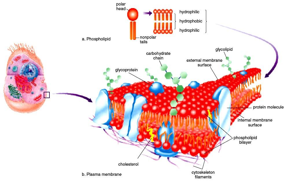
3 3.1 Cellular Organization Three main part of every human cell: a plasma membrane, a nucleus, cytoplasm The plasma membrane, - surrounds the cell and keeps it intact, regulates what enters and exits a cell. - is a phospholipid bilayer that is said to be semipermeable because it allows certain molecules but not others to enter the cell. - Proteins present in the plasma membrane play important 3 roles in allowing substances to enter the cell.
4 The nucleus - is a large, centrally located structure that can often be seen with a light microscope. - contains the chromosomes - is the control center of the cell. - It controls the metabolic functioning and structural characteristics of the cell. The nucleolus is a region inside the nucleus. - is of primary importance because it stores the genetic information that determines the characteristics of the body s cells and their metabolic functioning. - Every cell contains a copy of genetic information, but each cell type has certain genes turned on, and others turned off. 4
5 The cytoplasm - is the portion of the cell between the nucleus and the plasma membrane. - The matrix of the cytoplasm is a semi-fluid medium that contains water and various types of molecules suspended or dissolved in the medium. The presence of proteins accounts for the semi-fluid nature of the matrix. - contains various organelles 5
6 Organelles - are small, usually membranous structures that are best seen with an electron microscope. - Each type of organelle has a specific function. For example, one type of organelle transports substances, and another type produces ATP for the cell. Because organelles are composed of membrane, keeping the various cellular activities separated from one another. Just as the rooms in your house have particular pieces of furniture that serve a particular purpose, organelles have a structure that suits their function. 6
7 Cytoskeleton Cells also have a cytoskeleton, a network of interconnected filaments and microtubules in the cytoplasm. The name cytoskeleton is convenient in that it allows us to compare the cytoskeleton to our bones and muscles. Bones and muscles give us structure and produce movement. Similarly, the elements of the cytoskeleton maintain cell shape and allow the cell and its contents to move. Some cells move by using cilia and flagella, which are made up of microtubules. 7
8 8
9 9
10 10
11 11 Ima Mariaculata Maria Immaculataiwo/SF iwo/sfitb ITB

12 Figure 3.1 A generalized cell, with a blowup of the cytoskeleton. Cilia Peroxisome Cytoplasm Vesicle formation Nuclear pore chromatin Nucleus Nucleolus Nuclear envelope Vesicle Rough ER Ribosomes Centrioles Golgi apparatus Lysosome Plasma membrane Smooth ER mitochondrion Plasma microtubule membrane Intermediate Actin filament filament 12
13 Figure 3.2 Fluid-mosaic model of the plasma membrane. a. In the phospholipid bilayer, the polar (hydrophilic) heads project outward and the nonpolar (hydrophobic) tails project inward. b. Proteins are embedded in the membrane. Glycoproteins have 13 attached
14 14
15 Ribosomes Ribosomes are composed of two subunits, one large and one small. Each subunit has its own mix of proteins and rrna. Protein synthesis occurs at the ribosomes. Ribosomes are found free within the cytoplasm either singly or in groups called polyribosomes. Ribosomes are often attached to the endoplasmic reticulum 15
16 Endomembrane System The endomembrane system consists of - the nuclear envelope, the endoplasmic reticulum, he Golgi apparatus, lysosomes, vesicles (tiny membranous sacs) These components of the cell work together to produce and secrete a product. 16

17 Figure 3.5 The endomembrane system. Vesicles from the ER bring proteins and lipids to the Golgi apparatus where they are modified and repackaged into vesicles. Secretion occurs when vesicles fuse with the plasma membrane. Lysosomes made at the Golgi apparatus digest macromolecules after fusing with incoming 17
18 The Endoplasmic Reticulum The ER, a complicated system of membranous channels and saccules (flattened vesicles), is physically continuous with the outer membrane of the nuclear envelope. Rough ER - is studded with ribosomes on the side of the membrane that faces the cytoplasm. - Here proteins are synthesized and enter the ER interior where processing and modification begin. - Some of these proteins are incorporated into membrane, and some are for export. Smooth ER, which is continuous with rough ER, does not have attached ribosomes. - synthesizes the phospholipids that occur in membranes and has various other functions, depending on the particular cell. - In the testes, it produces testosterone, - in the liver it helps detoxify drugs. 18

19 Figure 3.4 Rough endoplasmic reticulum is studded with ribosomes where protein synthesis occurs. Nuclear envelope Ribosome Rough ER Smooth ER Smooth endoplasmic reticulum, which has no attached ribosomes, produces lipids and often has other functions as well in particular cells. ER also forms vesicles in which large molecules are transported to other parts of the cell. 19
20 The Golgi Apparatus The Golgi apparatus is named for Camillo Golgi, who discovered its presence in cells in The Golgi apparatus receives protein and/or lipid-filled vesicles that bud from the ER. The Golgi apparatus contains enzymes that modify proteins and lipids. For example, it can add a chain of sugars to proteins and lipids, thereby making them glycoproteins and glycolipids, which are molecules found in the plasma membrane. 20
21 Lysosomes Lysosomes, membranous sacs produced by the Golgi apparatus, contain hydrolytic digestive enzymes. Sometimes macromolecules are brought into a cell by vesicle formation at the plasma membrane (Fig. 3.5). When a lysosome fuses with such a vesicle, its contents are digested by lysosomal enzymes into simpler subunits that then enter the cytoplasm. Even parts of a cell are digested by its own lysosomes (called autodigestion). Normal cell rejuvenation most likely takes place in this manner, but autodigestion is also important during development. 21
22 Lysosomes For example, The fingers of a human embryo are at first webbed, but they are freed from one another as a result of lysosomal action. Occasionally, a child is born with Tay-Sachs disease, a metabolic disorder involving a missing or inactive lysosomal enzyme. In these cases, the lysosomes fill to capacity with macromolecules that cannot be broken down. The cells become so full of these lysosomes that the child dies. Someday soon, it may be possible to provide the missing enzyme for these children. 22
23 Mitochondria Although the size and shape of mitochondria (sing., mitochondrion) can vary, all are bounded by a double membrane. The inner membrane is folded to form little shelves called cristae, which project into the matrix, an inner space filled with a gel-like fluid Mitochondria are the site of ATP (adenosine triphosphate) production involving complex metabolic pathways. ATP molecules are the common carrier of energy in cells. 23

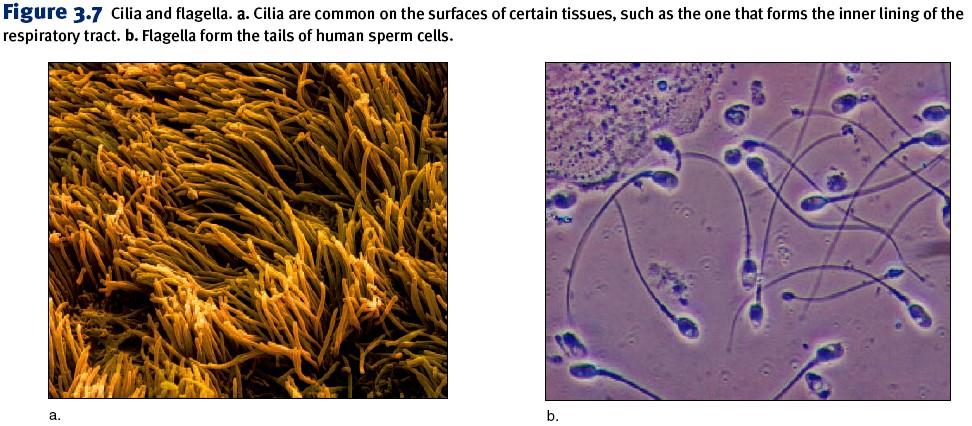
24 The chemical transformation that involves mitochondria is as follows: In the process, mitochondria use up oxygen and give off carbon dioxide and water. Because oxygen is involved, we say that mitochondria carry on cellular respiration. 24
25 The matrix of a mitochondrion contains enzymes for breaking down glucose products. ATP production then occurs at the cristae. Every cell uses a certain amount of ATP energy to synthesize molecules, but many cells use ATP to carry out their specialized functions, For example, - muscle cells use ATP for muscle contraction, which produces movement - nerve cells use it for the conduction of nerve impulses, which make us aware of our environment. 25

26 cristae 26
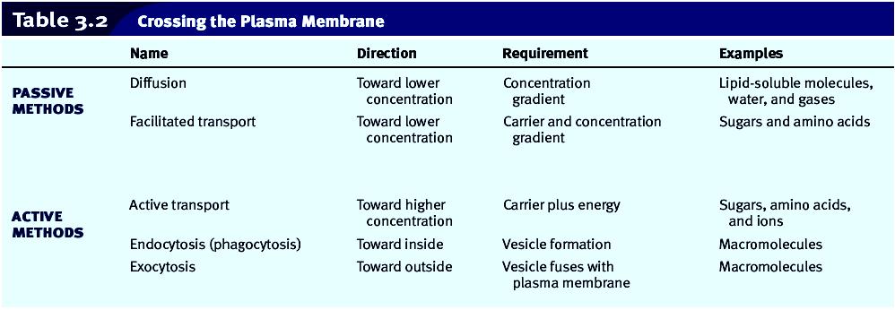
27 The Cytoskeleton Several types of filamentous protein structures form a cytoskeleton that helps maintain the cell s shape and either anchors the organelles or assists their movement as appropriate. The cytoskeleton includes microtubules, intermediate filaments, actin filaments 27
28 Microtubules are hollow cylinders whose wall is made up of 13 logitudinal rows of the globular protein tubulin. Microtubule assembly is regulated by the centrosome which lies near the nucleus. Microtubules radiate from the centrosome, helping to maintain the shape of the cell and acting as tracks along which organelles move. During cell division, microtubules form spindle fibers, which assist the movement of chromosomes. 28
29 Intermediate filaments differ in structure and function. Actin filaments are long, extremely thin fibers that usually occur in bundles or other groupings. Actin filaments have been isolated from various types of cells, especially those in which movement occurs. Microvilli, which project from certain cells and can shorten and extend, contain actin filaments. Actin filaments, like microtubules, can assemble and disassemble. 29
30 Centrioles are short cylinders with nine outer microtubule triplets and no center microtubules Each cell has a pair of centrioles in the centrosome near the nucleus. The members of each pair of centrioles are at right angles to one another. Before a cell divides, the centrioles duplicate, and the members of the new pair are also at right angles to one another. During cell division, the pairs of centrioles separate so that each daughter cell gets one centrosome. Centrioles may be involved in the formation of the spindle that functions during cell division. Centrioles also give rise to basal bodies that direct the formation of cilia and flagella. 30
31 Cilia and Flagella Cilia and flagella (sing., cilium, flagellum) are projections of cells that can move either in an undulating fashion, like a whip, or stiffly, like an oar. Cilia are shorter than flagella Cells that have these organelles are capable of self movement or moving material along the surface of the cell. For example, sperm cells, carrying genetic material to the egg, move by means of flagella. The cells that line our respiratory tract are ciliated. These cilia sweep debris trapped within mucus back up the throat, and this action helps keep the lungs clean. 31
32 Each cilium and flagellum has a basal body at its base, which lies in the cytoplasm. Basal bodies, like centrioles, have (9+0 pattern): nine outer microtubule triplets and no center microtubules They are believed to organize the structure of cilia and flagella even though cilia and flagella have a 9+ 2 pattern of microtubules. In cilia and flagella, nine microtubule doublets surround two central microtubules. This arrangement is believed to be necessary to their ability to move. 32
33 33
34 3.2 Crossing the Plasma Membrane The plasma membrane keeps a cell intact. It allows only certain molecules and ions to enter and exit the cytoplasm freely; therefore, the plasma membrane is said to be selectively permeable. Both passive and active methods are used to cross the plasma membrane 34
35 Diffusion Diffusion is the random movement of molecules from the area of higher concentration to the area of lower concentration until they are equally distributed. To illustrate diffusion, imagine putting a tablet of dye into water. The water eventually takes on the color of the dye as the dye molecules diffuse. The chemical and physical properties of the plasma membrane allow only a few types of molecules to enter and exit a cell simply by diffusion. Lipid-soluble molecules such as alcohols can diffuse through the membrane because lipids are the membrane s main structural components. 35
36 Diffusion Gases can also diffuse through the lipid bilayer; this is the mechanism by which oxygen enters cells and carbon dioxide exits cells. After inhalation (breathing in), the concentration of oxygen in the alveoli is higher than that in the blood; therefore, oxygen diffuses into the blood. When molecules simply diffuse from higher to lower concentration across plasma membranes, no cellular energy is involved. 36
37 Osmosis Osmosis is the diffusion of water across a plasma membrane. It occurs whenever an unequal concentration of water exists on either side of a selectively permeable membrane. Normally, body fluids are isotonic to cells, that is, there is an equal concentration of solutes (substances) and solvent (water) on both sides of the plasma membrane, and cells maintain their usual size and shape. Intravenous solutions medically administered usually have this tonicity. Tonicity is the degree to which a solution s concentration of solute versus water causes water to 37 move into or out of cells.
38 Hypotonic & hypertonic solutions Solutions (solute plus solvent) that cause cells to swell or even to burst due to an intake of water are said to be hypotonic solutions. If red blood cells are placed in a hypotonic solution, which has a higher concentration of water (lower concentration of solute) than do the cells, water enters the cells and they swell to bursting (Fig. 3.8b). The term lysis refers to disrupted cells; hemolysis, then, is disrupted red blood cells. Solutions that cause cells to shrink or to shrivel due to a loss of water are said to be hypertonic solutions. If red blood cells are placed in a hypertonic solution, whichhas a lower concentration of water (higher concentration of solute) than do the cells, water leaves the cells and they shrink (Fig. 3.8c). 38
39 Solutions that cause cells to shrink or to shrivel due to a loss of water are said to be hypertonic solutions. If red blood cells are placed in a hypertonic solution, which has a lower concentration of water (higher concentration of solute) than do the cells, water eaves the cells and they shrink (Fig. 3.8c). The term crenation refers to red blood cells in this condition. These changes have occurred due to osmotic pressure. Osmotic pressure is the force exerted on a selectively permeable membrane because water has moved from the area of higher concentration of water to the area of lower concentration (higher concentration of solute). 39
40 40
41 Filtration Because capillary walls are only one cell thick, small molecules (e.g., water or small solutes) tend to passively diffuse across these walls, from areas of higher concentration to those of lower concentration. However, blood pressure aids matters by pushing water and dissolved solutes out of the capillary. This process is called filtration. Filtration is easily observed in the laboratory when a solution is poured past filter paper into a flask. Large substances stay behind, but small molecules and water pass through. Filtrasi memerlukan tekanan! 41
42 Filtration of water and substances in the region of capillaries is largely responsible for the formation of tissue fluid, the fluid that surrounds the cells. Filtration is also at work in the kidneys when water and small molecules move from the blood to the inside of the kidney tubules. 42
43 Transport by Carriers Most solutes do not simply diffuse across a plasma membrane; rather, they are transported by means of protein carriers within the membrane. During facilitated transport, a molecule (e.g., an amino acid or glucose) is transported across the plasma membrane from the side of higher concentration to the side of lower concentration. The cell does not need to expend energy for this type of transport because the molecules are moving down their concentration gradient. During active transport, a molecule is moving contrary to the normal direction that is, from lower to higher concentration (Fig. 3.9). 43
44 For example, - iodine collects in the cells of the thyroid gland; - sugar is completely absorbed from the gut by cells that line the digestive tract; - and sodium (Na) is sometimes almost completely withdrawn from urine by cells lining kidney tubules. Active transport requires a protein carrier and the use of cellular energy obtained from the breakdown of ATP. When ATP is broken down, energy is released, and in this case the energy is used by a carrier to carry out active transport. Therefore, it is not surprising that cells involved in active transport have a large number of mitochondria near the plasma membrane at which active transport is occurring. 44
45 Proteins involved in active transport often are called pumps because just as a water pump uses energy to move water against the force of gravity, proteins use energy to move substances against their concentration gradients. One type of pump that is active in all cells but is especially associated with nerve and muscle cells moves sodium ions (Na) to the outside of the cell and potassium ions (K) to the inside of the cell. The passage of salt (NaCl) across a plasma membrane is of primary importance in cells. First, sodium ions are pumped across a membrane; then, chloride ions simply diffuse through channels that allow their passage. Chloride ion channels malfunction in persons with cystic fibrosis, and this leads to the symptoms of this inherited (genetic) disorder. 45
46 Endocytosis and Exocytosis During endocytosis, commonly called phagocytosis, a portion of the plasma membrane invaginates to envelop a substance, and then the membrane pinches off to form an intracellular vesicle (see Fig. 3.1, top). Digestion may be required before molecules can cross a vesicle membrane to enter the cytoplasm. During exocytosis, a vesicle fuses with the plasma membrane as secretion occurs (see Fig. 3.1, bottom). This is the way insulin leaves insulin-secreting cells, for instance. Table 3.2 summarizes the various ways molecules cross the plasma membrane. 46

47 Figure 3.9 Active transport through a plasma membrane. Active transport allows a molecule to cross the membrane from lower concentration to higher concentration. ➀ Molecule enters carrier. ➁ Breakdown of ATP induces a change in shape that ➂ drives the molecule across the membrane. 47
48 48
49 Dehydration and Water Intoxication Dehydration is due to a loss of water. The solute concentration in extracellular fluid increases that is, tissue fluid becomes hypertonic to cells, and water leaves the cells. Common causes of dehydration are excessive sweating, perhaps during exercise, without any replacement of the water lost. Dehydration can also be a side effect of any illness that causes prolonged vomiting or diarrhea. The signs of moderate dehydration are a dry mouth, sunken eyes, and skin that will not bounce back after light pinching. 49
50 Dehydration and Water Intoxication If dehydration becomes severe, the pulse and breathing rate are rapid, the hands and feet are cold, the lips are blue. Although dehydration leads to weight loss, it is never a good idea to dehydrate on purpose for this reason. To cure dehydration, intake of a low-sodium solution is needed because water intake alone could lead to water intoxication. 50
51 Water intoxication is due to a gain in water. The solute concentration in extracellular fluid decreases that is, tissue fluid becomes hypotonic to the cells, and water enters the cells. One cause can be the intake of too much water during a marathon race. Marathoners who collapse and have nausea and vomiting after a race are probably not suffering from a heart attack, but they may be suffering from water intoxication, which can lead to pulmonary edema and swelling in the brain. The cure, an intravenous solution containing high amounts of sodium, is the opposite of that for dehydration. Therefore, it is important that physicians be able to diagnose water intoxication in athletes who have had an opportunity to drink fluids for the past several hours. 51
52 Figure 3A Dehydration versus water intoxication. a. If extracellular fluid loses much water, cells lose water by osmosis, and become dehydrated. 52
53 Figure 3A Dehydration versus water intoxication. b. If extracellular fluid gains water, cells gain water by osmosis, and water intoxication occurs. 53
54 3.3 The Cell Cycle 54
 and a DNA synthesis (S) phase.")
55 Figure 3.10 The cell cycle consists of interphase, during which cellular components duplicate, a mitotic stage, during which the cell divides. Interphase consists of two socalled growth phases (G1 and G2) and a DNA synthesis (S) phase. The mitotic stage consists of the phases noted plus cytokinesis. 55
56 During G1, a cell doubles its organelles (such as mitochondria and ribosomes) and accumulates materials that will be used for DNA synthesis. S Phase Following G1, the cell enters the S (for synthesis ) phase. During the S phase, DNA replication occurs. At the beginning of the S phase, each chromosome is composed of one DNA double helix, which is equal to a chromatid. At the end of this phase, each chromosome has two identical DNA double helix molecules, and therefore is composed of two sister chromatids. Another way of expressing these events is to say that DNA replication has resulted in duplicated chromosomes. 56
57 G2 Phase During this phase, the cell synthesizes proteins that will assist cell division, such as the protein found in microtubules. The role of microtubules in cell division is described later in this section. Also, chromatin condenses, and the chromosomes become visible. Mitotic Stage Following interphase, the cell enters the M (for mitotic) stage. This cell division stage includes mitosis (division of the nucleus) and cytokinesis (division of the cytoplasm). During mitosis, daughter chromosomes are distributed to two daughter nuclei. When cytokinesis is complete, two daughter cellsare present. 57
58 Events During Interphase Two significant events during interphase are replication of DNA protein synthesis. 58
59 Replication of DNA During replication, an exact copy of a DNA helix is produced. The double-stranded structure of DNA aids replication because each strand serves as a template for the formation of a complementary strand. A template is most often a mold used to produce a shape opposite to itself. In this case, each old (parental) strand is a template for each new (daughter) strand. 59
60 Replication stages 1. Before replication begins, the two strands that make up parental DNA are hydrogen-bonded to one another. 2. During replication, the old (parental) DNA strands unwind and upzip (i.e., the weak hydrogen bonds between the two strands break). 3. New complementary nucleotides, always present in the nucleus, pair with the nucleotides in the old strands. A pairs with T and C pairs with G. The enzyme DNA polymerase joins the new nucleotides forming new (daughter) complementary strands. 4. When replication is complete, the two double helix molecules are identical. 60
61 Each strand of a double helix is equal to a chromatid, which means that at the completion of replication each chromosome is composed of two sister chromatids. They are called sister chromatids because they are identical. The chromosome is called a duplicated chromosome. Cancer, which is characterized by rapidly dividing cells, is treated with chemotherapeutic drugs that stop replication and therefore cell division. Some chemotherapeutic drugs are analogs that have a similar, but not identical, structure to the four nucleotides in DNA. When these are mistakenly used by the cancer cells to synthesize DNA, replication stops, and the cells die off. 61

62 Figure 3.11 Overview of DNA replication. During replication, an old strand serves as a template for a new strand. The new double helix is composed of an old (parental) strand and a new (daughter) strand. 62

63 Figure 3.12 Ladder configuration and DNA replication. Use of the ladder configuration better illustrates how complementary nucleotides available in the cell pair with those of each old strand before they are joined together to form a daughter strand. 63
64 Protein Synthesis DNA not only serves as a template for its own replication, but is also a template for RNA formation. Protein synthesis requires two steps, called - transcription - translation. During transcription, an mrna molecule is produced, and during translation, this mrna specifies the order of amino acids in a particular polypeptide (Fig. 3.13). A gene (i.e., DNA) contains coded information for the sequence of amino acids in a particular polypeptide. The code is a triplet code: Every three bases in DNA (and therefore in mrna) stands for a particular amino acid. 64

65 Figure 3.13 Protein synthesis. The two steps required for protein synthesis are transcription, which occurs in the nucleus, and translation, which occurs in the cytoplasm 65 at the ribosomes.
66 Events During the Mitotic Stage The mitotic stage of the cell cycle consists of mitosis and cytokinesis. By the end of interphase (Fig. 3.14, upper left), the centrioles have doubled and the chromosomes are becoming visible. Each chromosome is duplicated it is composed of two chromatids held together at a centromere. The mitosis process is divided into four phases: prophase, metaphase, anaphase, and telophase (Fig.3.14). The parental cell is the cell that divides, and the daughter cells are the cells that result. 66
67 67
68 Prophase The two pairs of centrioles outside the nucleus begin moving away from each other toward opposite ends of the nucleus. Spindle fibers appear between the separating centriole pairs, the nuclear envelope begins to fragment, and the nucleolus begins to disappear. The chromosomes are now fully visible. Spindle fibers attach to the centromeres as the chromosomes continue to shorten and thicken. During prophase, chromosomes are randomly placed in the nucleus. Structure of the Spindle At the end of prophase, a cell has a fully formed spindle. A spindle has poles, asters, and fibers. The asters are arrays of short microtubules that radiate from the poles, and the fibers are bundles of microtubules that stretch between the poles. Centrioles are located in centrosomes, which are believed to organize the spindle. 68
69 Metaphase During metaphase, the nuclear envelope is fragmented, and the spindle occupies the region formerly occupied by the nucleus. The chromosomes are now at the equator (center) of the spindle. Metaphase is characterized by a fully formed spindle, and the chromosomes, each with two sister chromatids, are aligned at the equator (Fig. 3.15). 69
70 Anaphase At the start of anaphase, the sister chromatids separate. Once separated, the chromatids are called chromosomes. Separation of the sister chromatids ensures that each cell receives a copy of each type of chromosome and thereby has a full complement of genes. During anaphase, the daughter chromosomes move to the poles of the spindle. Anaphase is characterized by the movement of chromosomes toward each pole. Function of the Spindle The spindle brings about chromosome movement. Two types of spindle fibers are involved in the movement of chromosomes during anaphase. Spindle fibers, as stated earlier, are composed of microtubules. Microtubules can assemble and disassemble by the addition or subtraction of tubulin (protein) subunits. This is what enables spindle fibers to lengthen and shorten, and it ultimately causes the movement of the chromosomes. 70
71 Telophase and Cytokinesis Telophase begins when the chromosomes arrive at the poles. During telophase, the chromosomes become indistinct chromatin again. The spindle disappears as nucleoli appear, and nuclear envelope components reassemble in each cell. Telophase is characterized by the presence of two daughter nuclei. Cytokinesis is division of the cytoplasm and organelles. In human cells, a slight indentation called a cleavage furrow passes around the circumference of the cell. Actin filaments form a contractile ring, and as the ring gets smaller and smaller, the cleavage furrow pinches the cell in half. As a result, each cell becomes enclosed by its own plasma membrane. 71
72 Importance of Mitosis Because of mitosis, each cell in our body is genetically identical, meaning that it has the same number and kinds of chromosomes. Mitosis is important to the growth and repair of multicellular organisms. When a baby develops in the mother s womb, mitosis occurs as a component of growth. As a wound heals, mitosis occurs, and the damage is repaired. 72
Ch. 3 CELLS AND TISSUES. Copyright 2010 Pearson Education, Inc.
 Ch. 3 CELLS AND TISSUES Generalized Cell All cells: Human cells have three basic parts: Plasma membrane flexible outer boundary Cytoplasm intracellular fluid containing organelles Nucleus control center
Cells. Unit 3 Cell Structure and Function. Cells. Plasma Membrane
 Unit 3 Cell Structure and Function Cells Cell theory The cell is the basic unit of life The cells of all living things exhibit the seven characteristics of life All living things are made of cells Cells
Chapter 3: Cytology. Cytology is the study of cells. Cells are the basic units of life. We are made up of trillions of cells.
 PLEASE NOTE THAT THE ITEMS IN THE TEXT THAT ARE HIGHLIGHTED IN YELLOW ARE THOSE THAT ARE TOUCHED ON IN THE READING ASSIGNMENT (PAGES 90-99) AND IN THE LECTURE. ESPECIALLY KNOW THIS MATERIAL FOR THE FIRST
Chapter 3: Cells 3-1
 Chapter 3: Cells 3-1 Introduction: A. Human body consists of 75 trillion cells B. About 260 types of cells that vary in shape & size yet have much in common B. Differences in cell shape make different
Chapter 3: Cells. I. Overview
 Chapter 3: Cells I. Overview A. Characteristics 1. Basic structural/functional unit 2. Diameter is too small to see by the naked eye 3. Can be over 3 feet long 4. Trillions of cells in over 200 basic types
The Cell. Biology 105 Lecture 4 Reading: Chapter 3 (pages 47 62)
") The Cell Biology 105 Lecture 4 Reading: Chapter 3 (pages 47 62) Outline I. Prokaryotic vs. Eukaryotic II. Eukaryotic A. Plasma membrane transport across B. Main features of animal cells and their functions
The Cell Biology 105 Lecture 4 Reading: Chapter 3 (pages 47 62) Outline I. Prokaryotic vs. Eukaryotic II. Eukaryotic A. Plasma membrane transport across B. Main features of animal cells and their functions
Biology 218 Human Anatomy
 Chapter 2 Adapted from Tortora 10 th ed LECTURE OUTLINE A. A Generalized Cell (p. 25) 1. A human cell consists of three major parts (see Table 2.2 on p. 42): a. Plasma membrane b. Cytoplasm which includes
Chaffey College: Anatomy and Physiology Chapter 3: Cells - The Living Units
 Cell Theory Chaffey College: Anatomy and Physiology Chapter 3: Cells - The Living Units The cell is the basic structural and functional unit of life Organismal activity depends on individual and collective
Biology 12 Cell Structure and Function. Typical Animal Cell
 Biology 12 Cell Structure and Function Typical Animal Cell Vacuoles: storage of materials and water Golgi body: a series of stacked disk shaped sacs. Repackaging centre stores, modifies, and packages proteins
1.3 - Cells. Chapter 3 - Cells
 1.3 - Cells Chapter 3 - Cells Cells Cytology = the study of cells All animal cells have 3 main parts: Nucleus Cell Membrane Cell membrane is semipermeable Cytoplasm (cytosol): where remaining organelles
Structures in Cells. Lecture 5, EH1008: Biology for Public Health, Biomolecules.
 Structures in Cells Lecture 5, EH1008: Biology for Public Health, Biomolecules Limian.zheng@ucc.ie 1 Cytoplasm Nucleus Centrioles Cytoskeleton Cilia Microvilli 2 Cytoplasm Cellular material outside nucleus
Cell Category? Prokaryote
 CELLS Cell Category? Prokaryote Prokaryote Eukaryote Cell Category? Cell Type? Cell Category? Cell Type? Endosymbiosis eukaryotic cells were formed from simpler prokaryotes Endo within Symbiosis together
Organelles. copyright cmassengale 1
 Organelles copyright cmassengale 1 Organelles Very small (Microscopic) Perform various functions for a cell Found in the cytoplasm May or may not be membrane-bound 2 Animal Cell Organelles Nucleolus Nucleus
Structures in Cells. Cytoplasm. Lecture 5, EH1008: Biology for Public Health, Biomolecules
 Structures in Cells Lecture 5, EH1008: Biology for Public Health, Biomolecules Limian.zheng@ucc.ie 1 Cytoplasm Nucleus Centrioles Cytoskeleton Cilia Microvilli 2 Cytoplasm Cellular material outside nucleus
Title: Sep 10 7:59 PM (1 of 36) Ch 3 Cell Organelles and Transport
 Ch 3 Cell Organelles and Transport") Title: Sep 10 7:59 PM (1 of 36) Ch 3 Cell Organelles and Transport Title: Sep 10 8:02 PM (2 of 36) Cell organelles Nucleus: contains DNA Title: Sep 10 8:03 PM (3 of 36) Nuclear envelope double membrane
Title: Sep 10 7:59 PM (1 of 36) Ch 3 Cell Organelles and Transport Title: Sep 10 8:02 PM (2 of 36) Cell organelles Nucleus: contains DNA Title: Sep 10 8:03 PM (3 of 36) Nuclear envelope double membrane
The Cell. BIOLOGY OF HUMANS Concepts, Applications, and Issues. Judith Goodenough Betty McGuire
 BIOLOGY OF HUMANS Concepts, Applications, and Issues Fifth Edition Judith Goodenough Betty McGuire 3 The Cell Lecture Presentation Anne Gasc Hawaii Pacific University and University of Hawaii Honolulu
Overview of the Cellular Basis of Life. Copyright 2009 Pearson Education, Inc., publishing as Benjamin Cummings
 Overview of the Cellular Basis of Life Cells and Tissues Cells: Carry out all chemical activities needed to sustain life Cells are the building blocks of all living things Tissues Cells vary in length,
Cell Structure and Function. Biology 12 Unit 1 Cell Structure and Function Inquiry into Life pages and 68-69
 Cell Structure and Function Biology 12 Unit 1 Cell Structure and Function Inquiry into Life pages 45 59 and 68-69 Assignments for this Unit Pick up the notes/worksheet for this unit and the project There
Cell Structure and Function
 C h a p t e r 3 Cell Structure and Function PowerPoint Lecture Slides prepared by Jason LaPres Lone Star College - North Harris 3-1 The study of cells provides the foundation for understanding human physiology
SHORT ANSWER. Write the word or phrase that best completes each statement or answers the question.
 SHORT ANSWER. Write the word or phrase that best completes each statement or answers the question. Figure 2.1 Using Figure 2.1, match the following: 1) Rough endoplasmic reticulum 1) 2) Nucleolus 2) 3)
6 functions of membrane proteins integral & peripheral proteins Membrane Junctions
 Cells Cells are the structural units of all living organisms ranging from unicellular to multicellular organisms. Biochemical activities of cells are dictated by cell shape and specific subcellular structures.
Principles of Anatomy and Physiology
 Principles of Anatomy and Physiology 14 th Edition CHAPTER 3 The Cellular Level of Organization Introduction The purpose of the chapter is to: 1. Introduce the parts of a cell 2. Discuss the importance
Cell Theory. Passive Transport
 Cell Theory 4 basic concepts of cell theory are: Cells are the units of structure (building blocks) of all animals and plants. Cells are the smallest unit of function in all animals and plants. Cells originate
Anatomy Chapter 2 - Cells
 Cells Cells are the basic living structural, functional unit of the body Cytology is the branch of science that studies cells The human body has 100 trillion cells 200 different cell types with a variety
Chapter 3 Review Assignment
 Class: Date: Chapter 3 Review Assignment Multiple Choice 40 MC = 40 Marks Identify the choice that best completes the statement or answers the question. 1. Which of the following organelles produces transport
Cells. Prokaryotic vs. Eukaryotic Euakryotic cells are generally one to one hundred times bigger than prokaryotic cells
 Cell Theory Cells 1. All living things are composed of one or more cell 2. Cell is the basic unit of life 3. All cells come from the division of pre-existing cells Cells are divided into 2 categories:
3UNIT. Photosynthesis and. Cellular Respiration. Unit PreQuiz? General Outcomes. Unit 3 Contents. Focussing Questions
 3UNIT Photosynthesis and Cellular Respiration General Outcomes In this unit, you will relate photosynthesis to the storage of energy in organic compounds explain the role of cellular respiration in releasing
The Study of Cells The diversity of the cells of the body The following figure shows the proportion of cell size of the variety of cells in the body
 Adapted from Martini Human Anatomy 7th ed. Chapter 2 Foundations: The Cell Introduction There are trillions of cells in the body Cells are the structural building blocks of all plants and animals Cells
Cell Structure and Function
 Cell Structure and Function Agre and cells in the news Cells Smallest living unit Most are microscopic Discovery of Cells Robert Hooke (mid-1600s) Observed sliver of cork Saw row of empty boxes Coined
(impermeable; freely permeable; selectively permeable)
") BIOL 2457 CHAPTER 3 Part 1 SI 1 1. A is the basic structure of life. 2. The gelatinous inside of the cell is called the. 3. Name the structure that increases the cell s surface area? 4. Name the structure
BIOL 2457 CHAPTER 3 Part 1 SI 1 1. A is the basic structure of life. 2. The gelatinous inside of the cell is called the. 3. Name the structure that increases the cell s surface area? 4. Name the structure
BIOLOGY 12 - CELL STRUCTURE & FUNCTION: Chapter Notes THE CELL THEORY
 BIOLOGY 12 - CELL STRUCTURE & FUNCTION: Chapter Notes THE CELL THEORY 1. All living organisms are made up of one or more cells 2. The cell is the basic unit of life 3. All cells come from the division
Cell. ~ 75 trillion make up adult Living cells ~ 60% water Carry materials for exchange
 Chapter 3 Cells 1 Cell ~ 75 trillion make up adult Living cells ~ 60% water Carry materials for exchange 2 Cell Shapes Shape and size vary Ex: Nerve cells can be 3ft long! Epithelial often flat and thin
Cytosol the fluid Cytoplasm cell interior, everything outside the nucleus but within the cell membrane, includes the organelles, cytosol, and
 Cell Organelles Plasma Membrane comprised of a phospholipid bilayer and embedded proteins Outer surface has oligosaccharides separates the cells s contents from its surroundings Cytosol the fluid Cytoplasm
Modern Cell Theory. Plasma Membrane. Generalized Cell Structures. Cellular Form and Function. Three principle parts of a cell
 Cellular Form and Function Concepts of cellular structure Cell surface Membrane transport Cytoplasm Modern Cell Theory All living organisms are composed of cells. the simplest structural and functional
CELL MEMBRANES. CELL MEMBRANE- Structure and Function
 BIOLOGY 12 CELL MEMBRANES NAME: INTRODUCTION 1. The cell membrane the passage of molecules into and out of the cell. 2. Some types of molecules, particularly molecules, pass freely across the cell membrane
Cells and Tissues 3PART A. PowerPoint Lecture Slide Presentation by Patty Bostwick-Taylor, Florence-Darlington Technical College
 PowerPoint Lecture Slide Presentation by Patty Bostwick-Taylor, Florence-Darlington Technical College Cells and Tissues 3PART A Cells and Tissues Carry out all chemical activities needed to sustain life
Cell Structure and Function Practice Exam - KEY
 Biology 12 Name: Cell Structure and Function Practice Exam - KEY Cell parts and Function 1. Identify each part of the cell indicated and give one role for each structure in the secretion and/or synthesis
CELL PARTS TYPICAL ANIMAL CELL
 AP BIOLOGY CText Reference, Campbell v.8, Chapter 6 ACTIVITY1.12 NAME DATE HOUR CELL PARTS TYPICAL ANIMAL CELL ENDOMEMBRANE SYSTEM TYPICAL PLANT CELL QUESTIONS: 1. Write the name of the cell part in the
Notes Chapter 7 Cell Structure and Function Hooke looked at cork under a simple microscope and found tiny chambers he named cells.
 Notes Chapter 7 Cell Structure and Function 7.1 Cell discovery and Theory 1665 Hooke looked at cork under a simple microscope and found tiny chambers he named cells. Cells are the basic structural and
Cells. I. Introduction to the Cell. II. Composite Cell
 I. Introduction to the Cell Cells A. 75 trillion cells in the human adult. B. Size 1. Large enough for organelles 2. Limited by the cell s surface to volume ratio I. Introduction to the Cell C. Shape 1.
Cells. Introduction: Composite Cell: Bi100 Chapter 3
 Bi100 Chapter 3 Cells Introduction: A. The human body consists of almost 100 trillion cells that vary considerably in shape and size yet have much in common. B. Differences in cell shape and composition
Basrah University Pharmacy College. Human Biology. Cells: The Basic Units of Life Dr. Rawaa Salim Hameed
 Basrah University Pharmacy College Human Biology Cells: The Basic Units of Life Cells: The Basic Units of Life All organisms are composed of one (e.g. bacteria) or more cells (e.g. human body contains
Chapter 7. (7-1 and 7-2) A Tour of the Cell
 A Tour of the Cell") Chapter 7 (7-1 and 7-2) A Tour of the Cell Microscopes as Windows to the World of Cells Cells were first described in 1665 by Robert Hooke. By the mid-1800s, the accumulation of scientific evidence led
Chapter 7 (7-1 and 7-2) A Tour of the Cell Microscopes as Windows to the World of Cells Cells were first described in 1665 by Robert Hooke. By the mid-1800s, the accumulation of scientific evidence led
LIFE IS CELLULAR. Cell Theory. Cells Are Small. Prokaryotic Cell 10/4/15. Chapter 7 Cell Structure and Function
 Chapter 7 Cell Structure and Function The cell basic unit of life, all living things are made of a cell (unicellular) or more than one cell (multicellular). LIFE IS CELLULAR The invention of the microscope
Chapter 4 Organization of the Cell
 Chapter 4 Organization of the Cell Cell basic unit of life o Small o Self-sufficient o Self-replicating Cell Theory organisms are composed of cells and all cells come from the division of other cells Cells
Cell Biology. a review! Cell Theory & Cell Structures
 Cell Biology Cell Theory & a review! Cell Structures Cell Theory refers to the idea that cells are the basic unit of structure and function of all living things. Cells are either prokaryotic or eukaryotic
A. Major parts 1. Nucleus 2. Cytoplasm a. Contain organelles (see below) 3. Plasma membrane (To be discussed in Cellular Transport Lecture)
 3. Plasma membrane (To be discussed in Cellular Transport Lecture)") Lecture 5: Cellular Biology I. Cell Theory Concepts: 1. Cells are the functional and structural units of living organisms 2. The activity of an organism is dependent on both the individual and collective
Lecture 5: Cellular Biology I. Cell Theory Concepts: 1. Cells are the functional and structural units of living organisms 2. The activity of an organism is dependent on both the individual and collective
History of the Cell. History of the Cell 10/24/2013. Unit 3: Cellular Structure and Function. Robert Hooke (1665) Robert Hooke (1665)
 Robert Hooke (1665)") Unit 3: Cellular Structure and Function Mr. Hulse BVHS 2013-2014 Unit 3: Learning Targets 1-9 History of the Cell Robert Hooke (1665) 1 st person to see a cell Observed a piece of cork using a microscope
Unit 3: Cellular Structure and Function Mr. Hulse BVHS 2013-2014 Unit 3: Learning Targets 1-9 History of the Cell Robert Hooke (1665) 1 st person to see a cell Observed a piece of cork using a microscope
ORGANELLES OF THE ENDOMEMBRANE SYSTEM
 Membranes compartmentalize the interior of the cell and facilitate a variety of metabolic activities. Chloroplasts and a rigid cell wall are what distinguish a plant cell from an animal cell. A typical
Plasma Membrane. comprised of a phospholipid bilayer and embedded proteins separates the cells s contents from its surroundings
 Cell Organelles Plasma Membrane comprised of a phospholipid bilayer and embedded proteins separates the cells s contents from its surroundings Cytosol the fluid Cytoplasm cell interior, everything outside
CELLS.
 CELLS http://www.aimediaserver.com/studiodaily/harvard/harvard.swf INTERESTING FACTS The longest cells in the human body are the motor neurons. They can be up to 1.37 meters long and go from the spinal
MULTIPLE CHOICE. Choose the one alternative that best completes the statement or answers the question.
 MULTIPLE CHOICE. Choose the one alternative that best completes the statement or answers the question. Figure 2.1 Use the diagram above to answer the following questions. 1) Which letter indicates the
Cell and Cell Membrane Structure and Function
 Lesson 3 Cell and Cell Membrane Structure and Function Introduction to Life Processes - SCI 102 1 The Cell Theory Three principles comprise the cell theory 1) Every living organism is made up of one or
Mitosis. AND Cell DiVISION
 Mitosis AND Cell DiVISION Cell Division Characteristic of living things: ability to reproduce their own kind. Cell division purpose: When unicellular organisms such as amoeba divide to form offspring reproduction
Mitosis and Cellular Division. EQ: How do the cells in our body divide?
 Mitosis and Cellular Division EQ: How do the cells in our body divide? Cell division is the process by which cellular material is divided between two new daughter cells. 1 Mother Cell 2 Daughter cells.
Cell Transport Unit Test
 Cell Transport Unit Test ~Please DO NOT write on the test~ I CAN describe the parts of cells. 1. The ideas that all living things are composed of cells come from other cells defines: a. Central dogma b.
Human Anatomy & Physiology
 PowerPoint Lecture Slides prepared by Barbara Heard, Atlantic Cape Community College Ninth Edition Human Anatomy & Physiology C H A P T E R 3 Annie Leibovitz/Contact Press Images 2013 Pearson Education,
Chapter 3 Cell Structures & Functions
 Biology 12 Name: Cell Biology Per: Date: Chapter 3 Cell Structures & Functions Complete using BC Biology 12, pages 62-107 Diagnostic Questions (mark using the answer key on page 527) 1. 2. 3. 4. 9. What
Cytology. Cell Anatomy. Cell Theory - A good place to start! The generalized cell contains: Cell membrane Cytoplasm. Nucleus. Cytosol Organelles
 Cytology The key to every biological problem must finally be sought in the cell, for every living organism is, or at some time has been, a cell. E.B. Wilson, 1925 Cell Theory - A good place to start! Cell
Objectives. By the end of the lesson you should be able to: State the 2 types of cells Relate the structure to function for all the organelles
 Biology 11 THE Cell Objectives By the end of the lesson you should be able to: State the 2 types of cells Relate the structure to function for all the organelles Types of Cells There are two types of cells:
First discovered in 1665 since then every organism observed with microscopes shows cells
 The Cell Cell theory (1838): 1. All organisms are composed of one or more cells, and the life processes of metabolism and heredity occur within these cells. 2. Cells are the smallest living things, the
Chapter 8 The Cell Cycle
 What molecule stores your genetic information or determines everything about you? DNA a nucleic acid How are DNA molecules arranged in the nucleus? As you can see DNA is: Chapter 8 The Cell Cycle 1. Arranged
Cell Cell
 Go to cellsalive.com. Select Interactive Cell Models: Plant and Animal. Fill in the information on Plant and Animal Organelles, then Click on Start the Animation Select Plant or Animal Cell below the box.
basic unit structure and function
 Chapter 3 Cells Introduction The cell is the basic unit of structure and function in living things. Cells vary in their shape, size, and arrangements, but all cells have similar components with a particular
Modern Cell Theory. Plasma Membrane. Generalized Cell Structures. Cellular Form and Function. Three principle parts of a cell
 Cellular Form and Function Concepts of cellular structure Cell surface Membrane transport Cytoplasm Modern Cell Theory All living organisms are composed of cells. the simplest structural and functional
The Cell Organelles. Eukaryotic cell. The plasma membrane separates the cell from the environment. Plasma membrane: a cell s boundary
 Eukaryotic cell The Cell Organelles Enclosed by plasma membrane Subdivided into membrane bound compartments - organelles One of the organelles is membrane bound nucleus Cytoplasm contains supporting matrix
BIOLOGY 111. CHAPTER 3: The Cell: The Fundamental Unit of Life
 BIOLOGY 111 CHAPTER 3: The Cell: The Fundamental Unit of Life The Cell: The Fundamental Unit of Life Learning Outcomes 3.1 Explain the similarities and differences between prokaryotic and eukaryotic cells
Study Guide for Biology Chapter 5
 Class: Date: Study Guide for Biology Chapter 5 Multiple Choice Identify the choice that best completes the statement or answers the question. 1. Which of the following led to the discovery of cells? a.
Cells and Tissues. Lesson 2.1: Molecules of Life Lesson 2.2: Cells Lesson 2.3: Tissues
 2 Cells and Tissues Lesson 2.1: Molecules of Life Lesson 2.2: Cells Lesson 2.3: Tissues Chapter 2: Cells and Tissues Lesson 2.1 Molecules of Life Molecules of Life carbohydrates proteins lipids nucleic
Goals. Cells. Cells: The Living Units. By the end of this lecture you should be able to describe.
 C H A P T E R 2 Cells: The Living Units Goals By the end of this lecture you should be able to describe. Similarities and differences between cells Why cells look and function differently The function
Structure of a Generalized Cell
 A Quick Tour Through A Cell BIO130 Lab 2 Exercise 4 The Cell: Anatomy Structure of a Generalized Cell -plasma membrane -cytoplasm: cytosol organelles -nucleus Play TourOfAnimalCell.mpg Plasma membrane
Hole s Human Anatomy and Physiology Tenth Edition. Chapter 3
 PowerPoint Lecture Outlines to accompany Hole s Human Anatomy and Physiology Tenth Edition Shier w Butler w Lewis Chapter 3 Copyright The McGraw-Hill Companies, Inc. Permission required for reproduction
The Cell and Cellular transport
 Cell theory (1838): The Cell 1. All organisms are composed of one or more cells, and the life processes of metabolism and heredity occur within these cells. 2. Cells are the smallest living things, the
A&P 1 Cellular Anatomy, Division & Mitosis - Pre-Lab Exercises
 A&P 1 Cellular Anatomy, Division & Mitosis - Pre-Lab Exercises Have someone in your group read the following out loud, while the others read along: In this "Pre-lab Guide", we will be going over some of
Cell Structure and Function Cell Structure and function
 Cell Structure and Cell Structure and function Dr Badri Paudel www.badripaudel.com Smallest living unit Most are microscopic Cells Discovery of Cells Robert Hooke (mid-1600s) Observed sliver of cork Saw
Lesson 1. Cell Theory - Statements - Exceptions. Categorizing Cells - Prokaryotes vs Eukaryotes
 Lesson 1 Cell Theory - Statements - Exceptions Categorizing Cells - Prokaryotes vs Eukaryotes The Cell Theory The discovery of cells and their structure is linked to the development of the magnifying lenses,
Unduplicated. Chromosomes. Telophase
 10-2 Cell Division The Cell Cycle Interphase Mitosis Prophase Cytokinesis G 1 S G 2 Chromatin in Parent Nucleus & Daughter Cells Chromatin Daughter Nuclei Telophase Mitotic Anaphase Metaphase Use what
Cell Structure & Interactions
 Cells Structures & Interactions Overview 1830s-Botanist Matthias Schleiden and zoologist Theodor Schwann were studying tissues and proposed the unified cell theory All living things are composed of one
Genetics and Cellular Function
 Genetics and Cellular Function DNA replication and the cell cycle Mitosis Mitosis Mitosis: division of cells that results in daughter cells with the same the genetic information that the original cell
Why do cells divide? Cells divide in order to make more cells they multiply in order to create a larger surface to volume ratio!!!
 Why do cells divide? Cells divide in order to make more cells they multiply in order to create a larger surface to volume ratio!!! Chromosomes Are made of chromatin: a mass of genetic material composed
A Tour of the Cell. reference: Chapter 6. Reference: Chapter 2
 A Tour of the Cell reference: Chapter 6 Reference: Chapter 2 Monkey Fibroblast Cells stained with fluorescent dyes to show the nucleus (blue) and cytoskeleton (yellow and red fibers), image courtesy of
10/13/11. Cell Theory. Cell Structure
 Cell Structure Grade 12 Biology Cell Theory All organisms are composed of one or more cells. Cells are the smallest living units of all living organisms. Cells arise only by division of a previously existing
Unit 2: More on Matter & Energy in Ecosystems. Macromolecules to Organelles to Cells
 IN: Unit 2: More on Matter & Energy in Ecosystems Macromolecules to Organelles to Cells Where are cells on the biological scale? Sub-Atomic Particles Atoms Molecules Macromolecules (proteins, lipids, nucleic
Human height. Length of some nerve and muscle cells. Chicken egg. Frog egg. Most plant and animal cells Nucleus Most bacteria Mitochondrion
 10 m 1 m 0.1 m 1 cm Human height Length of some nerve and muscle cells Chicken egg Unaided eye 1 mm Frog egg 100 µm 10 µm 1 µm 100 nm 10 nm Most plant and animal cells Nucleus Most bacteria Mitochondrion
8/7/18. UNIT 2: Cells Chapter 3: Cell Structure and Function. I. Cell Theory (3.1) A. Early studies led to the development of the cell theory
 A. Early studies led to the development of the cell theory") 8/7/18 UNIT 2: Cells Chapter 3: Cell Structure and Function I. Cell Theory (3.1) A. Early studies led to the development of the cell theory 1. Discovery of Cells a. Robert Hooke (1665)-Used compound microscope
8/7/18 UNIT 2: Cells Chapter 3: Cell Structure and Function I. Cell Theory (3.1) A. Early studies led to the development of the cell theory 1. Discovery of Cells a. Robert Hooke (1665)-Used compound microscope
Chapter 3 Part 2! Pages (10 th and 11 th eds.)! The Cellular Level of Organization! Cellular Organelles and Protein Synthesis!
! The Cellular Level of Organization! Cellular Organelles and Protein Synthesis!") Chapter 3 Part 2! Pages 65 89 (10 th and 11 th eds.)! The Cellular Level of Organization! Cellular Organelles and Protein Synthesis! The Cell Theory! Living organisms are composed of one or more cells.!
Chapter 3 Part 2! Pages 65 89 (10 th and 11 th eds.)! The Cellular Level of Organization! Cellular Organelles and Protein Synthesis! The Cell Theory! Living organisms are composed of one or more cells.!
Biology is the only subject in which multiplication is the same thing as division
 Biology is the only subject in which multiplication is the same thing as division The Cell Cycle: Cell Growth, Cell Division 2007-2008 2007-2008 Getting from there to here Going from egg to baby. the original
A TOUR OF THE CELL 10/1/2012
 A TOUR OF THE CELL Chapter 6 KEY CONCEPTS: Eukaryotic cells have internal membranes that compartmentalize their functions The eukaryotic cell s genetic instructions are housed in the nucleus and carried
First to View Cells. copyright cmassengale
 CELL THEORY All living things are made of cells Cells are the basic unit of structure and function in an organism (basic unit of life) Cells come from the reproduction of existing cells (cell division)
Cytoskeleton. Provide shape and support for the cell. Other functions of the cytoskeleton. Nucleolus. Nucleus
 Chapter 4: Cell Structure and Function Cytoskeleton The cytoskeleton is a network of fibers that organizes structures and activities in the cell. Microtubules (the largest) Intermediate fibers Microfilaments
1. or is the study of cellular structure and function. 2. What is the purpose and characteristics of the plasma membrane?
 Chapter 3 Reading Guide The Cellular Level of Organization Name 1. or is the study of cellular structure and function. Section 3.1 Parts of a Cell 2. What is the purpose and characteristics of the plasma
Basic Structure of a Cell. copyright cmassengale
 Basic Structure of a Cell 1 Review Facts About Living Things 2 What Are the Main Characteristics of organisms? 1. Made of CELLS 2. Require ENERGY (food) 3. REPRODUCE (species) 4. Maintain HOMEOSTASIS 5.
Fig. 1. Simple columnar epithelial cells lining the small intestine.
 Mitosis Prelab Reading Fig. 1. Simple columnar epithelial cells lining the small intestine. The tall cells pictured in Fig. 1 form the lining of the small intestine in humans and other animals. These cells
Organelles of the Cell & How They Work Together. Packet #7
 Organelles of the Cell & How They Work Together Packet #7 Introduction Introduction Organization of cells is basically similar in all cells. Additionally, most cells are tiny Ranging from 1 1000 cubic
CELL CYCLE INTRODUCTION PART I ANIMAL CELL CYCLE INTERPHASE
 CELL CYCLE INTRODUCTION The nuclei in cells of eukaryotic organisms contain chromosomes with clusters of genes, discrete units of hereditary information consisting of double-stranded DNA. Structural proteins
4/12/17. Cells. Cell Structure. Ch. 2 Cell Structure and Func.on. Range of Cell Sizes BIOL 100
 Ch. 2 Cell Structure and Func.on BIOL 100 Cells Fundamental units of life Cell theory All living things are composed of one or more cells. The cell is the most basic unit of life. All cells come from pre-existing
Plasma Membrane Structure and Function
 Plasma Membrane Structure and Function The plasma membrane separates the internal environment of the cell from its surroundings. The plasma membrane is a phospholipid bilayer with embedded proteins. The
CHAPTER 3 1/21/2016. Typical Bacteria Cell. The Cell
 CHAPTER 3 The Cell Chapter 3 Learning Objectives Compare and contrast the features of prokaryotic and eukaryotic cells. Explain why surface area-to-volume ratios constrain cell size. Contrast light microscopy
Biology is the only subject in which multiplication is the same thing as division
 Biology is the only subject in which multiplication is the same thing as division 2007-2008 The Cell Cycle: Cell Growth, Cell Division 2007-2008 Getting from there to here Going from egg to baby. the original
Cell Structure and Function
 PowerPoint Lecture Slides prepared by Meg Flemming Austin Community College C H A P T E R 3 Cell Structure and Function Chapter 3 Learning Outcomes 3-1 3-2 3-3 3-4 3-5 List the main points of the cell