JBC Papers in Press. Published on September 18, 2002 as Manuscript M
|
|
|
- Claude Gibbs
- 5 years ago
- Views:
Transcription
1 JBC Papers in Press. Published on September 18, 2002 as Manuscript M Determinants for Membrane Association and Permeabilization of the Coxsackievirus 2B Protein and the Identification of the Golgi Complex as the Target Organelle Arjan S. de Jong 1, Els Wessels 1, Henri B.P.M. Dijkman 2, Jochem M.D. Galama 1, Willem J.G. Melchers 1, Peter H.G.M. Willems 3 and Frank J.M. van Kuppeveld 1* Departments of Medical Microbiology 1 and Biochemistry 3, Nijmegen Center for Molecular Life Sciences, University Medical Center Nijmegen, Department of Pathology 2, University Medical Center Nijmegen, PO Box 9101, 6500 HB Nijmegen, The Netherlands Running title: Membrane association of coxsackievirus protein 2B * Corresponding author Mailing address: Department of Medical Microbiology Nijmegen Center for Molecular Life Sciences University Medical Center Nijmegen PO Box HB Nijmegen The Netherlands. Phone:(31) Fax:(31) f.vankuppeveld@ncmls.kun.nl. Copyright 2002 by The American Society for Biochemistry and Molecular Biology, Inc.
2 2 ABSTRACT The 2B protein of enterovirus is responsible for the alterations in the permeability of secretory membranes and the plasma membrane in infected cells. The structural requirements for the membrane association and the subcellular localization of this essential virus protein, however, have not been defined. Here, we provide evidence that the 2B protein is an integral membrane protein in vivo that is predominantly localized at the Golgi complex upon individual expression. Addition of organelle-specific targeting signals to the 2B protein revealed that the Golgi localization is an absolute prerequisite for the ability of the protein to modify plasma membrane permeability. Expression of deletion mutants and heterologous proteins containing specific domains of the 2B protein demonstrated that each of the two hydrophobic regions could mediate membrane binding individually. However, the presence of both hydrophobic regions was required for the correct membrane association, efficient Golgi targeting, and the membrane-permeabilizing activity of the 2B protein, suggesting that the two hydrophobic regions are cooperatively involved in the formation of a membrane-integral complex. The formation of membrane-integral pores by the 2B protein in the Golgi complex, and the possible mechanism by which a Golgi-localized virus protein modifies plasma membrane permeability are discussed.
3 3 INTRODUCTION Enteroviruses (e.g. poliovirus (PV), coxsackievirus, ECHOvirus) belong to the family of picornaviridea, a group of nonenveloped, cytolytic viruses that have a positive stranded RNA genome of 7.5 kb. The enterovirus genome contains one large open reading frame that is translated into a single, 220 kda polyprotein. Processing of the polyprotein by virus-encoded proteases yields the structural P1 region proteins that encapsidate the viral RNA, and the non-structural P2 and P3 region proteins that are involved in replication of the viral RNA. Processing of the P2 and P3 regions yields the 2A pro, 2B, 2C, 3A, 3B, 3C pro and 3D pol proteins and the more stable cleavage intermediates 2BC, 3AB and 3CD pro, which have functions distinct from their cleavage products. Although multiple functions have been attributed to the mature viral proteins and the cleavage intermediates, the exact function of most of the viral proteins in the replication cycle is still largely unknown. Host cell membranes are subject to a number of profound alterations upon enterovirus infection. Enteroviruses gradually modify host cell membrane permeability and rearrange intracellular membranes during infection. The modification of the plasma membrane permeability is most likely important for the lysis of the cell and the release of virus progeny. Modifications of secretory pathway membranes are connected to viral functions such as genome replication. The modification of host cell membrane permeability is such that initially calcium is released from intracellular stores (1). Ionic gradients maintained by the plasma membrane are also disrupted (2). Later in infection, also small compounds like hygromycin B, a small non-permeative translation inhibitor, can efficiently enter the cell. The 2B protein has been identified as the viral protein that is responsible for the alterations in host cell membrane permeability that take place in enterovirus-infected cells. Individual expression of the 2B protein was shown to be sufficient for the release of calcium from intracellular stores and the increase in plasma membrane permeability to both calcium and hygromycin B (1;3-6). Another important membrane modification that can be observed in enterovirus-infected cells is the massive proliferation and accumulation of membrane vesicles in the cytoplasm. These vesicles are derived from the secretory pathway (7) and were shown to be the site at which viral RNA replication takes place (8;9). Individual expression of the viral proteins has shown that the 2BC protein is responsible for the induction of the vesicle formation (9-12), possibly together with the 3A protein (13). The virus-induced inhibition of protein secretion is another important modification, affecting trafficking
4 4 of host cell membranes. This inhibition may be advantageous for the virus by preventing anti-viral host cell responses like MHC expression and secretion of interferon and interleukines (14;15). Individual expression of the enterovirus non-structural proteins has shown that both the 2B protein and the 3A protein are endowed with the ability to inhibit protein secretion (1;3). The 3A protein was shown to inhibit ER-to-Golgi transport (16). The step blocked by the 2B protein is still unknown. The mechanism by which the enterovirus 2B protein exerts its effects on membranes is as yet unknown. Moreover, it remains to be established whether the different activities of the 2B protein (i.e, membrane permeabilization, membrane rearrangement, and secretion inhibition) represent different functions or whether these activities are the result of a general membrane-disturbing activity of the 2B protein. Our aim is to gain more insight into the structure and function of the coxsackievirus 2B protein. The coxsackievirus 2B protein is a small protein of 99 amino acids that, in the infected cell, is present at the virus-induced, secretory pathway derived membrane vesicles at which the viral replication takes place (17). The protein contains two hydrophobic regions (figure 1), of which one is predicted to form a cationic amphipathic α-helix with characteristics typical for the group of the so-called membrane-lytic peptides (18). Mutations in the amphipathic α-helix or the second hydrophobic region were shown to have deleterious effects on viral RNA replication and virus growth, indicating that the integrity of these regions is essential for an early function in the viral life cycle (18;19). Mutations in these domains were also found to interfere with the ability of the 2B protein to increase membrane permeability and to inhibit protein secretion (6). This strongly suggests that the membrane modifying activities of the 2B protein are in some way required for the accumulation of the membrane vesicles at which viral RNA replication takes place. At present, the determinants for the membrane interaction and the subcellular localization of the 2B protein have not been defined. In this study, we have investigated the mode of membrane association in vivo and the subcellular localization of the 2B protein. Furthermore, by testing 2B deletion mutants, we have analyzed the importance of specific domains for the mode of membrane interaction, the subcellular localization, and the ability to increase membrane permeability. Moreover, the correlation between the subcellular localization and the membrane-active character of the 2B protein was determined.
5 5 EXPERIMENTAL PROCEDURES Cells and media. Buffalo green monkey (BGM) cells were grown in minimal essential medium (MEM) (Gibco), supplemented with 10% fetal bovine serum (FBS), 100 units penicillin per ml and 100 mg streptomycin per ml. Cells were grown at 37 C in a 5% CO 2 incubator. Antibodies and conjugates. Rabbit polyclonal anti-egfp antiserum was a kind gift from Dr. J. Fransen, Department of Cell Biology and Histology, UMC Nijmegen, The Netherlands. Rabbit polyclonal anti-2b antiserum was obtained by immunizing SPF rabbits with a recombinant MBP-2B fusion protein produced in E. Coli. Western blot analysis showed that the immune sera specifically recognized the MBP-2B and 2B-EGFP fusion proteins, while no reactivity of the pre-immune sera was detected (data not shown). Mouse monoclonal anti-c-myc (clone 9E10) and mouse monoclonal anti- Golgi 58K protein (clone 58K-9) antisera were obtained from Sigma-Aldrich. Rabbit polyclonal anticalreticulin was obtained from Affinity Bioreagents, Inc. FITC-conjugated goat-anti-rabbit polyclonal antibody, Texas Red-conjugated goat-anti-mouse polyclonal antibody and Texas Red-conjugated goat-anti-rabbit polyclonal antibody were obtained from Jackson ImmunoResearch Laboratories. Peroxidase-conjugated goat anti-rabbit immunoglobulin was obtained form Dako Diagnostika. Plasmids. The coding sequences of the wild-type 2B protein or 2B deletion mutants were amplified by PCR and cloned in the pegfp fusion vectors described below (Clontech). The plasmid pcb3/t7 (20), which contains the full-length cdna of CBV3, was used as a template in the PCR reactions for the amplification of the sequence encoding wild-type 2B, 2B( 59C), 2B( 14C) and 2B( 20N) proteins, lacking aa 41-99, aa and aa 1-20, respectively. The pcb3/t7-2b HR1 and pcb3/t7-2b HR2 deletion constructs (21) were used as templates in the PCR reactions for the amplification of the sequences encoding the 2B( HR1) and 2B( HR2) proteins, lacking the amphipathic α-helix (aa 34-56) or the second hydrophobic region (aa 64-80), respectively. 2B-EGFP and EGFP-2B fusion proteins. For the construction of the p2b-egfp plasmid, the 2B coding sequence was amplified using a forward primer that introduced a SaII restriction site (restriction sites are in italics) and a start codon (underlined) preceded by a Kozak sequence (p115-16; 5 -ctcctgtggctggctagcgtcgacgccaccatgggagtgaaggactatgtggaa-3 ), and a reverse primer that
6 6 introduced a BamHI restriction site (p115-20; 5 -ccagctggatccttggcgttcagccatagg-3 ). The PCR product was cloned into pegfp-n3 digested with SalI and BamHI to yield p2b-egfp. For the construction of the pegfp-2b plasmid, the 2B coding sequence was amplified by PCR using primer p and a reverse primer that introduced a stop codon (underlined) and a SmaI restriction site (p115-7; 5 - aagccacccgggctattggcgttcagccatagg-3 ). The PCR product was cloned into pegfp-c1 digested with SmaI and SalI to yield pegfp-2b. 2B-EGFP deletion mutants. For the construction of the p2b( 59C)-EGFP plasmid, the coding sequence of the N-terminal 40 amino acids (aa) of the 2B protein was amplified using p115-16, and a reverse primer that introduced an EcoCRI restriction site (p169-3; 5 - taaggcttttagagagagctctaagatggagtc-3 ). The PCR product was cloned into pegfp-n2 digested with NheI and SmaI to yield p2b( 59C)-EGFP. For the construction of the 2B( 14C)-EGFP plasmid, the coding sequence of aa 1-85 of the 2B protein was amplified using forward primer p and a reverse primer that introduced a BamHI restriction site (p115-29; 5 -ggggggggggaccggtgccccggggaccggcggatcctgtttgagccaccgccacgg-3 ) The PCR product was cloned into pegfp-n3 digested with SalI and BamHI to yield p2b( 14C)-EGFP. For the construction of the p2b( 20N)-EGFP plasmid, the coding sequence of aa of the 2B protein was amplified using a forward primer that introduced a SalI restriction site and a start codon preceded by a Kozak sequence (p115-30; 5 -ggggggggggtcgacgccatcatggtatgtgagcaagtcaacctc-3 ) and a reverse primer p The PCR product was cloned into pegfp-n3 digested with SalI and BamHI to yield p2b( 20N)-EGFP. For the construction of the p2b( HR1)-EGFP and p2b( HR2)-EGFP plasmids, the coding sequences of proteins 2B( HR1) and 2B( HR2) were amplified using primers p and p The PCR products were cloned into p2b-egfp digested with SalI and BamHI to yield p2b( HR1)- EGFP and p2b( HR2)-EGFP, respectively. For the construction of the plasmids phr1-egfp and phr2-egfp, the coding sequences of the regions encompassing these regions (aa and aa 56-85, respectively) were amplified using primers that introduced a SalI restriction site and a start codon (underlined) preceded by a Kozak sequence at the 5 end and a BamHI restriction site at the 3 end. The HR1 coding sequence was amplified using p (5 -aacctcgtcgacgccaccatgtcactagtgggtcaagactcc-3 ) and p (5 - agtcacggattcgtcatcgtggttcctcaccac-3 ), the HR2 coding region was amplified using p (5 -
7 7 tcagccgtcgacgccaccatggtggtgaggaaccacgat-3 ) and p (5 -atattgggatccctgtttgagccaccgccacgg- 3 ). The region encompassing both HR1 and HR2 (aa 30-85) was amplified using p and p The phr1-egfp, phr2-egfp and p(hr1+hr2)-egfp plasmids were made by cloning the PCR products digested with SalI and BamHI into the pegfp-n3 vector, digested with the same enzymes. All sequences that were amplified by PCR were checked by sequence analysis. 2B-myc constructs. Plasmids p2b-myc, p2b( 20N)-myc, p2b( 14C)-myc, p2b( HR1)-myc, and p2b( HR2)-myc, were constructed by deleting the EGFP sequence (using BamHI and NotI) from the corresponding 2B-EGFP plasmids, and replacing it with a PCR product encoding the C-myc tag (aa EQKLISEEDL), followed by a stop codon. ER-targeted 2B construct. For the construction of the plasmids p2b-egfp-kkaa and p2b- EGFP-AAAA, the EGFP coding region was amplified using a forward primer containing a BamHI restriction site (p318-17; 5 -ggggggggattcatcgccaccgtcagcaagggcgaggag-3 ) and a reverse primer containing a stop codon, a NotI restriction site and a sequence encoding either the AAAAAAKKAA or the AAAAAAAAAA tag (p382-3; 5 - ggggggggcggccgcttacgccgctttcttagctgcggctgccgcgcccttgtacagctcgtccatgcc-3 and p382-4; 5 - ggggggggcggccgcttacgccgcagcagcagctgcggctgccgcgcccttgtacagctcgtccatgcc-3, respectively). The PCR product was cloned into p2b-egfp digested with BamHI and NotI to yield p2b-egfp-kkaa and 2B-EGFP-AAAA, respectively. Golgi-targeted 2B construct. For the construction of Golgi-targeted 2B-EGFP, a construct was generated that contained an additional SmaI restriction site between the 2B and the EGFP coding regions. The coding sequence of the 2B protein was amplified using p and a reverse primer that introduced a SmaI restriction site to the 5 end of the BamHI restriction site (p318-25; 5 - gggggggggggatcccccgggttggcgttcagccatagg-3 ). The PCR product was cloned into p2b-egfp digested with SalI and BamHI to yield p2b-egfp(+smai). The coding sequence of the N-terminal 103 amino acids of beta 1,2 N-acetylglucosaminyl-transferase I (NGAT-I) was amplified from pnagt-i- GFP (a kind gift from Dr. Graham Warren, Cell Biology Laboratory, Imperial Cancer Research Fund, London, UK) using a forward primer that introduced a EcoRV restriction site (p318-14: 5 - gggggggatatcctgaagaagcagtctgcagg-3 ) and a reverse primer that introduced a BamHI restriction site (p381-13: 5 -ggggggggatcccgccggcgcgggggtcacagg-3 ). The PCR product was cloned into p2b- EGFP(+SmaI) that was digested with SmaI and BamHI to yield p2b-golgi-egfp
8 8 EGFP-Golgi. The plasmid pegfp-golgi was constructed by replacing the ECFP coding region of pecfp-golgi (Clontech) by the EGFP coding region of pegfp-n1 using BamHI and NotI restriction sites. In this construct, EGFP is fused to the N-terminal 81 amino acids of human beta 1,4- galactosyltransferase, which targets the EGFP protein to the trans-medial region of the Golgi apparatus. Transfections. BGM cell monolayers were grown to 70% confluency in 6-well plates or on coverslips in 24-well plates and transfected with 5 µg of plasmid DNA per well of a 6-well plate or with 1 µg of plasmid DNA per well of a 24-well plate. Transfections were carried out using FuGENE 6 reagent (Roche) according to the instructions of the manufacturer. For each transfection reaction, the Fugene reagent was mixed with serum-free medium and incubated for 5 min at room temperature. This mixture was added drop-wise to the plasmid DNA preparation and incubated for 15 min at room temperature. The DNA/FuGENE mixture was added drop-wise to the cells. Cells were grown at 37 C until further analysis. Membrane association. BGM cells grown in 6-well plates were transfected with plasmids encoding the proteins EGFP, wild-type 2B-EGFP or the 2B-EGFP deletion mutants. At 40 h posttransfection, cells were washed twice with ice cold TES (20 mm Tris (ph 7.4), 1 mm EDTA, 100 mm NaCl). Samples were kept on ice during the entire procedure. Cells were scraped in 0.5 ml ice-cold 1:10 TES, collected by centrifugation for 10 min at 4,500 g and resuspended in 250 µl 1:10 TES. Cells were incubated for 15 min and broken by 30 strokes in a Dounce homogenizer. Nuclei and cell debris were removed by centrifugation for 10 min at 4,500 g. Membrane and cytoplasmic fractions were separated by centrifugation for 1 hour at 150,000 g at 4 C. Supernatants were removed and stored at 80 C until further analysis. Pellet fractions were either resuspended in one supernatant volume of buffer (50 mm Tris (ph 7.4), 150 mm NaCl, 1 mm EDTA, 0.1 M phenylmethylsulfonyl fluoride, 1% Nonidet P- 40, 0.05% SDS) and stored at 80 C until further analysis, or resuspended in 200 µl of either PBS, 0.1 M Na 2 CO 3 (ph 11.5), 0.5 M EDTA, 1 M NaCl, 4 M urea or 1% triton X-100. Resuspended pellet fractions were incubated on ice for 1 hour and centrifuged for 1 hour at 150,000 g and 4 C. The supernatant fractions and the pellet fractions, which were resuspended in 200 µl of lysis buffer, were analyzed by Western blot.
9 9 Western blot analysis. Supernatant and pellet fractions of each sample were run on a 12.5% SDSpolyacrylamide gel and transferred to a nitro-cellulose membrane (BioRad). EGFP fusion proteins were stained with the anti-egfp polyclonal antiserum (dilution 1:10,000) and peroxidase-conjugated goat-anti-rabbit immunoglobulins (dilution 1:1,000) and visualised using Lumi-Light plus Western Blotting Substrate (Roche) according to the instructions of the manufacturer. Immunofluorescence and Confocal Laser Scanning Microscopy. At 24 h posttransfection BGM cells grown on coverslips were fixed with 4% paraformaldehyde in PBS (ph 7.4). Cells were permeabilized using PBS/0.1% Triton X-100. Antibodies and conjugates were diluted with PBS/0.1 % Triton X-100/2 % normal goat serum (NGS). Primary antibodies were diluted 1:200 (anti-2b), 1:200 (anti-c-myc), 1:75 (anti-golgi 58K), or 1:150 (anti-calreticulin). Conjugates were diluted 1:75. Incubations with the primary antibody were carried out overnight at 4 C and incubations with the secondary antibody were carried out for 1 hour at 4 C. Cells were washed with PBS/0.1 % Triton X- 100 between incubation steps and mounted in mowiol (Sigma-Aldrich). Cells were analyzed by confocal laser scanning microscopy (CLSM) (Leica TCS NT, Leica Lasertechnik GmbH, Heidelberg, Germany). Hygromycin B assay. BGM cells grown in 6-well plates were transfected in duplicate with plasmids encoding the proteins EGFP, wild-type 2B-EGFP or the 2B-EGFP deletion mutants. At 40 h posttransfection, one of the duplicate wells was starved of methionine for 15 min in the presence of 500 µg/ml hygromycin B, whereas the other well was starved of methionine for 15 min in the absence of hygromycin B. The cells were then pulse-labeled for 1 hour with [ 35 S-]methionine (50 µci/well) in the presence or absence of hygromycin B, washed with ice-cold PBS and lysed in 1 ml of lysisbuffer (50 mm Tris (ph 7.4), 150 mm NaCl, 1 mm EDTA, 0.1 M phenylmethylsulfonyl fluoride, 1% Nonidet P- 40, 0.05% SDS). Anti-EGFP rabbit polyclonal antiserum (1:1000) was added to the cell lysate and the lysate/antiserum solution was incubated at 4 C for 18 h. To collect the antibody-protein complexes, the samples were incubated for 2 h with Protein A Sepharose (Amersham Pharmacia Biotech), washed two times with dilution buffer (0.01 M Tris (ph 8.0), 0.14 M NaCl, 0.1% BSA, 0.1% Triton X- 100), once with TSA (0.01 M Tris (ph 8.0), 0.14 M NaCl), once with 0.05 M Tris (ph 6.8) and
10 10 precipitated. Samples were resuspended in 35 µl of SDS-sample buffer, boiled for 5 min and analyzed by SDS-PAGE. The amount of radiolabeled anti-egfp precipitated protein was quantified using a PhosphorImager (Bio-Rad Multi-Analyst version 1.0.1) and the ratios of the amount of protein synthesized in the presence and absence of hygromycin B was determined. RESULTS Construction and characterisation of N- and C-terminal fusions of EGFP to the 2B protein. Tagged 2B proteins were used because (i) an anti-2b antiserum might not recognise all 2B deletion mutants due to possible loss of epitopes, and (ii) the deletion mutants may become too small (<10 kda) to be efficiently detected. Fusion proteins of wild-type and mutant 2B to EGFP were used because they may be used in subcellular localization studies in living cells and the resulting 36 kda fusion protein is efficiently recognized and detectable by an anti-egfp serum. The fusion of a fluorescent protein at the amino-terminus or carboxy-terminus of a protein may affect its biological properties. Therefore, the possible effects of the EGFP fusion on the membraneactive character and the subcellular localization of the 2B protein were studied. The ability of 2B-EGFP and EGFP-2B to increase plasma membrane permeability of transfected cells was studied by analysing the entry of hygromycin B, a small inhibitor of translation that under physiological conditions poorly passes the plasma membrane. Cells were transfected in duplicate wells with the indicated constructs and pulse labeled in the absence or presence of hygromycin B. The radiolabeled EGFP fusion proteins were immunoprecipitated and analyzed by SDS-PAGE to measure the entry of hygromycin B (figure 2A). In EGFP expressing cells, the amount of protein synthesis was similar in the absence or presence of hygromycin B, indicating that the EGFP protein does not increase plasma membrane permeability to hygromycin B. Expression of the 2B-EGFP protein resulted in the increased entry of hygromycin B, as was reflected by the almost complete inhibition of protein synthesis in the presence of hygromycin B. The increase in plasma membrane permeability induced by the 2B-EGFP protein was similar to that induced by the untagged 2B protein. The EGFP-2B protein showed a reduced ability to increase plasma membrane permeability compared to the untagged 2B protein.
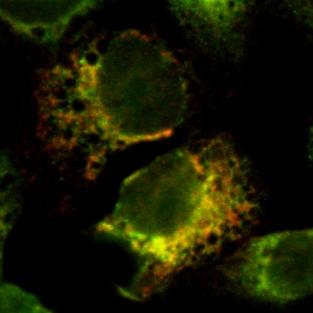
11 11 The subcellular localization of the 2B-EGFP and EGFP-2B fusion proteins was compared to that of the untagged 2B protein, stained with the anti-2b antiserum (figure 2B). The 2B-EGFP fusion protein was predominantly present in the juxtanuclear Golgi region, similar to the untagged 2B protein. In contrast, a small portion of the 2B-EGFP was observed in a reticular, ER-like pattern. The subcellular localization of the EGFP-2B protein differed substantially from that of the untagged 2B protein and showed resemblance to the ER. Based on these results, it was decided to use the 2B-EGFP protein in the subsequent experiments. In vivo membrane association of 2B-EGFP. The membrane association properties of the 2B protein were studied using an in vivo system, in which cytosolic and membrane fractions of EGFP or 2B-EGFP expressing cells were collected and analyzed on western blot. As expected, the cytosolic EGFP protein was found exclusively in the supernatant fraction (Figure 3A). The 2B-EGFP fusion protein was found exclusively in the pellet fraction, indicating that the 2B protein is associated with membranes. The mode of membrane association was further investigated by extracting the membrane fractions with buffers that discriminate between peripheral membrane proteins (which are attached to membranes by ionic interactions with membrane integral proteins or phospholipid head groups) and integral membrane proteins (which are embedded in the phospholipid bilayer). Figure 3B shows the supernatant and pellet fractions of the membrane fractions following extraction with the indicated buffers. The 2B-EGFP protein remained attached to membranes upon extraction with PBS (control) and buffers that extract peripheral membrane proteins either by increasing the ionic strength of the buffer (1M NaCl), creating mild chaotropic salt conditions (4 M urea), or chelating divalent cations (0.5 M EDTA). Moreover, the protein was still detected in the pellet fraction when membranes were extracted using alkaline conditions (0.1 M Na 2 CO 3 ph 11.5), one of the most potent methods to discriminate between peripheral and membrane-integral proteins (22). In addition, alkaline conditions convert membranes to sheets (but do not solubilize membranes) and release soluble proteins that are trapped inside membranous vesicles. The resistance of 2B-EGFP to alkaline extraction therefore argues against a peripheral association of the 2B-EGFP protein at the lumenal side of endo-membranes. The 2B-EGFP protein was found in the supernatant fraction only upon solubilization of membranes using 1% Triton X-100, a non-ionic detergent that releases integral membrane proteins. Taken together, these findings
12 12 provide evidence that the 2B protein is embedded in the phospholipid bilayer, rather than that it is peripherally associated with membranes. In vivo membrane association of 2B-EGFP deletion mutants. Deletion mutants of the 2B protein were designed to identify the regions that are important for its mode of membrane association. Wild-type 2B (99 aa) and deletion mutants of 2B are depicted in figure 3C. Protein 2B( 59C) contains only the N-terminal 40 amino acids of 2B, protein 2B( 14C) lacks the C-terminal 14 amino acid residues and protein 2B( 20N) has a deletion of the N-terminal 20 amino acids. In addition, deletion mutants were generated that lacked either the amphipathic α-helix (aa 34-56), which will further be referred to as the first hydrophobic region HR1, or the second hydrophobic region (aa 64-80), which will further be referred to as HR2. Cytosolic and membrane fractions of cells transfected with 2B-EGFP deletion mutants were prepared and analyzed as described above for the wild-type 2B-EGFP protein. Figure 3C shows that protein 2B( 59C)-EGFP is predominantly present in the cytosolic fraction, indicating that the N- terminal 40 amino acids of the 2B protein are not sufficient to mediate membrane association. The 2B( 14C)-EGFP and 2B( 20N)-EGFP proteins were found predominantly in the pellet fraction, demonstrating that the N- and the C-terminus of the protein do not contribute to the membrane association of the 2B protein. Remarkably, also the 2B( HR1)-EGFP and 2B( HR2)-EGFP proteins were found predominantly in the membrane fraction, indicating that the presence of either one of the hydrophobic regions of the 2B protein was sufficient to mediate membrane binding. The membrane associated 2B-EGFP deletion mutants were further investigated to determine their mode of membrane association. Membrane fractions were extracted using PBS, 0.1 M Na 2 CO 3, ph 11.5, and 1% Triton X-100 (figure 3D). The 2B( 14C)-EGFP and 2B( 20N)-EGFP proteins behaved like the wild-type 2B protein and were found predominantly in the membrane fraction after the extraction with 0.1 M Na 2 CO 3, suggesting that the N- and C-terminus of the 2B protein have little influence on its mode of membrane association. Both protein 2B( HR1)-EGFP and protein 2B( HR2)- EGFP behaved differently upon Na 2 CO 3 extraction. Substantial amounts of these proteins were found in the supernatant fraction upon Na 2 CO 3 extraction, whereas both proteins were almost exclusively detected in the pellet fraction upon PBS extraction. These results indicate that both hydrophobic regions are required for the correct membrane association of the 2B protein. When the membranes
13 13 were solubilized using 1% Triton X-100, the wild-type 2B-EGFP protein as well as 2B( 14C)-EGFP, 2B( 20N)-EGFP and 2B( HR2)-EGFP were predominantly present in the supernatant fraction. In contrast, the majority of the 2B( HR1)-EGFP protein remained present in the pellet fraction upon Triton X-100 extraction, indicating that 2B( HR1)-EGFP was pelleted in a membrane-independent manner upon solubilization of membranes. This result suggests that the 2B( HR1)-EGFP protein is prone to form aggregates in the absence of membranes (due to the Triton X-100 extraction). It cannot be excluded that the 2B( HR1)-EGFP protein also (partially) formed aggregates in the Na 2 CO 3 extraction experiment. It is therefore difficult to draw conclusions regarding the membrane association properties of 2B( HR1), other than that it behaves differently from the wild-type 2B protein, suggesting that this region is an important determinant for the membrane association properties of the 2B protein. In the subcellular localization experiments documented below, however, evidence will be provided that the 2B( HR1)-EGFP protein is present as a membrane associated protein in vivo. The results of the in vivo membrane association experiments are summarized in Table 1. Subcellular localization of protein 2B deletion mutants. The importance of specific domains of the 2B protein for its subcellular localization was investigated in living cells expressing 2B-EGFP deletion mutants (figure 4). The wild-type 2B-EGFP protein was predominantly present in the juxtanuclear Golgi region and was additionally found in an ER-like reticular pattern, similar to what was observed in fixed cells (figure 2B). The 2B( 59C)-EGFP was present throughout the cell, staining both the cytoplasm and the nucleus, consistent with the observation that the protein is not membrane-associated (figure 3C). Localization of the 2B( 14C)- EGFP and 2B( 20N)-EGFP was essentially the same as that of the wild-type 2B-EGFP protein (data not shown). The 2B( HR1)-EGFP and 2B( HR2)-EGFP proteins showed an altered localization compared to the wild-type 2B-EGFP protein. The 2B( HR1)-EGFP protein was present in an ER-like network and staining of the juxtanuclear Golgi region was virtually absent. The 2B( HR2)-EGFP protein was observed in the juxtanuclear Golgi region, however, staining of the ER-like network was more apparent than for wild-type 2B-EGFP. A more detailed examination of the subcellular localization was undertaken in fixed cells by investigating colocalization of the 2B protein with markers for the Golgi and the ER. In these experiments, the 2B deletion mutants were analyzed as myc-tagged proteins. This procedure was
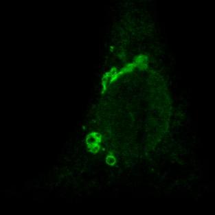
14 14 followed because the subcellular localization of the myc-tagged 2B protein better resembled that of the untagged 2B protein (compare figures 5A and 2B). Colocalization experiments with the Golgi complex were performed using the EGFP-Golgi fusion protein, in which EGFP is targeted to the trans-medial region of the Golgi complex by fusion to the N-terminal 81 amino acids of human beta 1,4- galactosyltransferase. Colocalization experiments with the ER were performed using the anticalreticulin antiserum. The 2B-myc, 2B( 14C)-myc and 2B( 20N)-myc proteins colocalized with the EGFP-Golgi marker and were virtually absent outside of the Golgi region (figure 5A, 5B and 5C, respectively), indicating that the N- and the C-terminus of the 2B protein have little or no influence on its subcellular localization. Interestingly, 2B( HR1)-myc staining was completely different from the wild-type 2B-myc protein. The 2B( HR1)-myc protein did not colocalize with EGFP-Golgi (figure 5D), but was exclusively present in a network-like pattern that was identical to the anti-calreticulin stained ER (figure 5F). Also the 2B( HR1)-myc staining differed from that of the wild-type 2B-myc protein. The 2B( HR2)-myc protein was present in the Golgi complex (figure 5E), but also a substantial part was observed in a network-like pattern that was identified as the ER (figure 5G). To further investigate the role of HR1 and HR2 in the Golgi localization, the ability of these domains to provide Golgi localization in the absence of other determinants of the 2B protein was analyzed. Initially, myc-tagged HR1 and HR2 constructs were generated. However, it was found that HR1-myc and HR2-myc proteins were too small to be preserved during fixation (data not shown). Therefore, heterologous proteins were constructed in which regions of the 2B protein encompassing either HR1 or HR2, or both HR1 and HR2, were fused to the N-terminus of the EGFP protein. The Golgi complex was stained using the anti-golgi 58K marker. Colocalization studies of these proteins with this endogenously expressed Golgi marker showed that the presence of HR1 could indeed partially target the EGFP protein to the juxtanuclear Golgi region (figure 6A), indicating that HR1 acts as a Golgi targeting signal in the absence of other domains of the 2B protein. The HR2-EGFP protein was present in a reticular network that showed close resemblance to the ER (figure 6B). Its absence from the Golgi complex suggests that HR2 does not contain any Golgi targeting signals. In contrast to the HR1-EGFP protein, which was partially localized in the ER, the (HR1+HR2)-EGFP fusion protein was predominantly present in the Golgi complex (figure 6C), giving support for an additional role of HR2 in Golgi localization. Taken together, these findings indicate that, although the amphipathic α- helix is the Golgi targeting signal, the second hydrophobic region is an important additional
15 15 determinant for the Golgi localization of both the 2B protein and the heterologous EGFP protein. The results of the subcellular localization experiments are summarized in Table 1. Membrane-permeabilizing activity of 2B-EGFP deletion mutants. In the experiments described above, we have shown that the 2B protein is an integral membrane protein that is predominantly localized at the Golgi complex and that these properties are largely dependent on the presence of both hydrophobic regions of the protein. To investigate the importance of different regions of the 2B protein for its membrane-active character, we also tested the 2B-EGFP deletion mutants for their ability to increase membrane permeability to hygromycin B. First, a time-course experiment was performed to define the time point at which plasma membrane permeability induced by the 2B-EGFP protein was maximal (figure 7A). From 24 h posttransfection to 40 h posttransfection, an increased influx of hygromycin B was observed, resulting in almost complete inhibition of protein synthesis in its presence at 40 h posttransfection. When cells were analyzed at 48 h posttransfection no increase in plasma membrane permeability to hygromycin B was observed. Therefore, all subsequent experiments were performed at 40 h posttransfection. Figure 7B shows the results of the hygromycin B assay for the 2B-EGFP deletion mutants. All mutants were tested in three independent assays, the amount of protein synthesized was determined by phosphor-imaging and the ratio of protein synthesis in the presence and absence of hygromycin B was calculated. Cells expressing wild-type protein 2B-EGFP showed an increased entry of hygromycin B, reflected by the reduction in protein synthesis to 10% of protein synthesis in cells expressing the wild-type 2B-EGFP protein in the absence of hygromycin B. Cells expressing protein 2B( 59C)-EGFP showed no influx of hygromycin B, indicating that the N-terminal 40 amino acids of the 2B protein are not sufficient to increase plasma membrane permeability. Hygromycin B entry in cells expressing the 2B( 14C)-EGFP protein was approximately equal to that of cells expressing the wild-type 2B protein. In cells expressing 2B( 20N)-EGFP, protein synthesis in the presence of hygromycin B was approximately 45% of that in the absence of hygromycin B, reflecting that plasma membrane permeability was reduced compared to that of cells expressing the wild-type 2B-EGFP protein. Cells expressing either the 2B( HR1)-EGFP or 2B( HR2)-EGFP fusion protein showed no entry of hygromycin B, indicating that the ability of these proteins to increase plasma membrane permeability was completely disrupted. Thus, the combined presence of both hydrophobic regions is not only
16 16 required for the membrane-integral character of the 2B protein, but also for its membrane-active function, suggesting that the membrane integral topology is essential for its ability to modify membrane permeability. The results of the membrane-permeabilization experiments are summarized in Table 1. Functional characterization of organelle-targeted 2B proteins. The 2B protein is predominantly present in the Golgi complex of transfected cells. We investigated the importance of the Golgi localization for the ability to increase plasma membrane permeability. For this purpose, the 2B-EGFP protein was targeted exclusively to the ER or to the Golgi complex by fusion of specific targeting signals. It was shown previously that membrane proteins acquired ER localization by the addition of a tag sequence containing the KKAA dilysine motif (AAAAAAKKAA), which acts as an ER retention signal in mammalian cells (23). The fusion of the KKAA motif to the 2B-EGFP protein indeed abolished the Golgi localization and resulted in a typical ER localization pattern (figure 8A). The presence of a control tag of 10 alanine residues, containing no additional localization signals, did not affect the subcellular localization of the 2B-EGFP protein (figure 8A). To confer complete Golgi localization to the 2B-EGFP protein, a construct was generated in which the 2B protein was fused at the N-terminus of the NAGT-I-GFP construct (which contains the targeting information of the beta 1,2 N-acetylglucosaminyl-transferase I, a type I med-trans Golgi membrane protein) (24). The resulting 2B-Golgi-EGFP protein was exclusively present in the Golgi complex (figure 8A). To analyze the importance of the subcellular localization of the 2B protein for its membraneactive function, the entry of hygromycin B was analyzed in cells expressing the targeted 2B proteins. Figure 8B shows that the ER-targeted 2B-EGFP-KKAA protein was severely impaired in its ability to increase plasma membrane permeability to hygromycin B. The 2B-EGFP-AAAA protein behaved like the untagged protein, indicating that the decrease in hygromycin B entry was not the result of the fusion of a tag to the C-terminus of 2B-EGFP. Rather, the altered subcellular localization of the protein was responsible for this result, suggesting that the Golgi localization of the 2B protein is important for its effect on plasma membrane permeability. This finding was further substantiated by the observation that the 2B-Golgi-EGFP protein increases plasma membrane permeability to hygromycin B to the same extend as the untagged 2B-EGFP protein. These data indicate that the Golgi localization is a critical determinant for the ability of the 2B protein to modify permeability of the plasma membrane.
17 17 DISCUSSION The enterovirus 2B protein is responsible for the alterations in the permeability of secretory membranes and the plasma membrane that are observed in infected cells (1;3;5;6). The molecular mechanism by which the 2B protein disturbs host cell membranes is largely unknown. In this study, we have investigated the structural and functional requirements for the in vivo membrane association and the membrane-active function of this important virus protein. The membrane association of the 2B protein was resistant against extraction with buffers that release peripherally associated membrane proteins, indicating that one or more domains of the protein are embedded in the phospholipid bilayer rather than that the protein associates with membranes through electrostatic interactions with membrane proteins or polar lipid head groups. Expression of deletion mutants and heterologous proteins showed that each of the hydrophobic regions HR1 and HR2 alone could mediate membrane binding. However, in contrast to the wild-type 2B protein, a substantial amount of the 2B proteins lacking either HR1 or HR2 was released upon Na 2 CO 3 extraction, indicating that both hydrophobic regions are required for the correct membrane association. Moreover, the presence of both HR1 and HR2 was absolutely required for the membrane-active function of the 2B protein, consistent with previous reports (6;11). Together, these findings suggest that the two hydrophobic regions are cooperatively involved in the formation of a membrane-integral complex, and that the formation of this complex is required for the membrane association and membrane-active function of the 2B protein. These findings provide important new insights into the molecular architecture of the 2B protein in the membrane. The first hydrophobic region of the 2B protein has been predicted to form a cationic amphipathic α-helix that shows similarities with the group of lytic polypeptides (18). Amphipathic α- helical lytic polypeptides may form multimers that build membrane-integral pores by exposing their hydrophobic sides to the lipid bilayer, while their hydrophilic faces form an aqueous interior (25-27). The 2B protein has been shown to form homo-multimers by yeast two-hybrid analysis (28), mammalian two-hybrid analysis (21), and in living mammalian cells using FRET microscopy (29). The behavior of the 2B protein as an integral membrane protein and the observation that each of the hydrophobic regions HR1 and HR2 can independently mediate membrane binding lend support to the idea that multimers of protein 2B build membrane-integral pores and that each of these hydrophobic
18 18 regions is involved in the formation of these pore complexes. Figure 9 shows a model of a multimeric pore complex formed by 2B proteins. According to this model, HR1 and HR2 interact with each other to form a helix-loop-helix hairpin motif that traverses the lipid bilayer. The hydrophobic regions are separated by a hydrophilic loop sequence (RNHDD in the CVB3 2B protein). Presumably both the N- and C-terminus of the 2B protein reside at the cytosolic side of the membrane. This suggestion is supported by the observation that a cytosolic ER retention signal (KKAA) (23;30) at the C-terminus of the 2B protein is functional in conferring ER retention to the 2B protein. Furthermore, the lack of glycosylation at NxS/T glycosylation signals introduced at different positions in the region upstream HR1 argues that the N-terminus is localized at the cytosolic side of the membrane (unpublished results). Finally, localization of the N- and C-terminus at the cytosolic side of the membrane is consistent with the need of proteolytic liberation of the 2B protein from the viral polyprotein by the viral protein 3C pro, a cytosolic protease. Both the membrane association and the membrane-active function were independent of the C-terminal 15 amino acids. Although dispensable for the membrane association, the N-terminal 20 amino acids were found to be of importance for the membrane-active function of the 2B protein, indicating that the molecular structure and function of the 2B protein depends on more than just the presence of HR1 and HR2, and therefore requires further investigation. Upon expression in the absence of other viral proteins, the CVB3 2B protein was localising in the Golgi complex, consistent with the Golgi localization described for the 2B protein of the closely related PV (31). Retention of the 2B protein in the ER by fusion of an ER retention signal severely inhibited the ability to increase plasma membrane permeability. This activity, however, was unaffected when the 2B protein was exclusively targeted to the Golgi complex by fusion of a Golgi targeting signal. This strongly suggests that the Golgi complex is the target organelle of the 2B protein to exert its effect on the plasma membrane. It is remarkable that a virus protein that is localized at the Golgi complex can modify plasma membrane permeability. The mechanism underlying this activity is as yet unknown. We predict that this activity is somehow linked to the ability of the 2B protein to increase Golgi membrane permeability. Previously, we have shown that expression of the 2B protein resulted in a decrease in the amount of calcium that could be released from thapsigargin-sensitive stores (1). Because at that time only the ER was recognized as a thapsigargin-sensitive store (32), we concluded that the 2B protein modified ER membrane permeability. Recently, however, also the Golgi complex was identified as a thapsigargin-sensitive intracellular calcium store (33). This finding, together with
19 19 the identification of the Golgi complex as the main target organelle of the 2B protein, suggests that the 2B protein causes an increased Golgi membrane permeability and a general disruption of the ionic gradients maintained by the Golgi membrane. Alterations in ionic gradients maintained by membranes of the Golgi complex have been demonstrated to result in functional disturbances of the Golgi complex. Disruption of cationic gradients by monensin, a H + /Na + ionophore that mediates cation diffusion over Golgi membranes, results in the inhibition of complex glycosylation in the Golgi complex (reflected by the inability of secretory proteins to acquire resistance to endo H treatment) and inhibition of protein secretion (34). Analogous to the effects of monensin, expression of the 2B protein also results in the inhibition of complex glycosylation (as demonstrated by the endo H sensitivity of glycoproteins in 2B-expressing cells) and inhibition of protein secretion (3). We propose that the disruption of the ionic content of the Golgi complex by 2B is responsible for this effect. How the disturbance of the Golgi milieu leads to an increase in plasma membrane permeability remains to be established. Possibly, the 2B-induced defect in transport of membrane proteins or lipids beyond the Golgi complex result in destabilization and permeabilization of the plasma membrane. However, it cannot be excluded that the effect on the plasma membrane is caused by the disturbance of another Golgi function(s). Having identified the Golgi complex as the main target organelle of the 2B protein, the question arises what determines the Golgi localization of the 2B protein. In this study, the amphipathic α-helix was identified as a Golgi targeting signal that could confer partial Golgi localization to the heterologous EGFP protein in the absence of other domains of the 2B protein. However, the presence of both the amphipathic α-helix and the second hydrophobic domain was found to be required for efficient Golgi targeting. The Golgi localization of the 2B protein might depend on the length of the transmembrane domain(s), as suggested by the bilayer-thickness model (35-40). HR1 and HR2 are predicted to consist of 20 aa and 18 aa respectively, which is similar to the TMD of some Golgi resident proteins, but shorter than the TMD of plasma membrane proteins (35;36;41). Alternatively, the kin-recognition model suggests that Golgi residents form large, oligomeric complexes as they reach the correct Golgi compartment that become too large to be transported (42;43). It is unlikely that homo-multimerisation reactions of the 2B protein (21;28;29) are involved in determining its Golgi localization, as 2B deletion mutants that lacked either HR1 or HR2 were not transported beyond the Golgi complex, although they were unable to form homo-multimers (28). Another possible explanation
20 20 is that the 2B protein is not transported further along the secretory pathway because of the lack of specific signals, as was suggested for some trans-golgi network markers (35). The possibility that trafficking of the 2B protein out of the Golgi complex is blocked as a consequence of the inhibition of protein secretion that is induced by the 2B protein itself (3) is unlikely, as (6)2B deletion mutants lacking either HR1 or HR2 are not transported beyond the Golgi complex, although their function was severely impaired. How do our findings relate to the situation in virus-infected cells, where the 2B protein is present at the virus-induced membrane vesicles that build the viral RNA replication complex? Initially, these membrane vesicles are derived from the ER (9). Later in infection, however, the Golgi complex has disappeared (12;31;44) and Golgi membranes contribute to the vesicle population (7;44), suggesting that the Golgi complex is gradually used up to produce the membrane vesicles that build the viral replication complex. Therefore, our findings that identified Golgi membranes as the main target of the 2B protein are in agreement with the localization of the 2B and 2BC proteins at the ERand Golgi-derived membrane vesicles in infected cells. The exact function of the 2B protein in the early steps in viral RNA replication remains to be established. The viability of viruses that carry mutations in the 2B protein closely correlates with the ability of the 2B protein to increase membrane permeability and inhibit protein secretion (1;6), suggesting that the destabilization and/or permeabilization of secretory pathway membranes by the 2B protein is somehow required for the accumulation of membrane vesicles or the formation of the membranous replication complex. Additional studies are needed to gain better insight in the membrane-active functions of the 2B and 2BC proteins, and how these functions might aid in the processes that are required to facilitate the formation of membrane-associated replication complexes. ACKNOWLEDGEMENTS We are grateful to Dr. J. Fransen (Department of Cell Biology and Histology, UMC Nijmegen, The Netherlands) for the kind gift of the anti-egfp antiserum. This work was partly supported by grants from the European Communities (INTAS 2012).
21 21 REFERENCES 1. van Kuppeveld, F.J.M., Hoenderop, J.G.J., Smeets, R.L.L., Willems, P.H.G.M., Dijkman, H.B.P.M., Galama, J.M.D., and Melchers, W.J.G. (1997) EMBO J. 16, Carrasco, L. (1995) Adv.Virus Res. 45, Doedens, J.R. and Kirkegaard, K. (1995) EMBO J. 14, Aldabe, R., Barco, A., and Carrasco, L. (1996) J.Biol.Chem. 271, Aldabe, R., Irurzun, A., and Carrasco, L. (1997) J.Virol. 71, van Kuppeveld, F.J.M., Melchers, W.J.G., Kirkegaard, K., and Doedens, J.R. (1997) Virology 227, Schlegel, A., Giddings, T.H., Ladinsky, M.S., and Kirkegaard, K. (1996) J.Virol. 70, Bienz, K., Egger, D., and Pfister, T. (1994) Arch.Virol.Suppl. 9, Rust, R.C., Landmann, L., Gosert, R., Tang, B.L., Hong, W., Hauri, H.P., Egger, D., and Bienz, K. (2001) J.Virol. 75, Aldabe, R. and Carrasco, L. (1995) Biochem.Biophys.Res.Commun. 206, Barco, A. and Carrasco, L. (1995) EMBO J. 14, Cho, M.W., Teterina, N., Egger, D., Bienz, K., and Ehrenfeld, E. (1994) Virology 202, Suhy, D.A., Giddings, T.H., and Kirkegaard, K. (2000) J.Virol. 74, Deitz, S.B., Dodd, D.A., Cooper, S., Parham, P., and Kirkegaard, K. (2000) Proc.Natl.Acad.Sci.U.S.A. 97, Dodd, D.A., Giddings, T.-H.J., and Kirkegaard, K. (2001) J.Virol. 75, Doedens, J.R., Giddings, T.H., and Kirkegaard, K. (1997) J.Virol. 71,
22 Bienz, K., Egger, D., and Pasamontes, L. (1987) Virology 160, van Kuppeveld, F.J.M., Galama, J.M.D., Zoll, J., van den Hurk, P.J.J.C., and Melchers, W.J.G. (1996) J.Virol. 70, van Kuppeveld, F.J.M., Galama, J.M.D., Zoll, J., and Melchers, W.J.G. (1995) J.Virol. 69, Klump, W.M., Bergmann, I., Muller, B.C., Ameis, D., and Kandolf, R. (1990) J.Virol. 64, De Jong, A.S., Schrama, I.W.J., Willems, P.H.G.M., Galama, J.M.D., Melchers, W.J.G., and van Kuppeveld, F.J.M. (2002) J.Gen.Virol. 83, Fujiki, Y., Hubbard, A.L., Fowler, S., and Lazarow, P.B. (1982) J.Cell Biol. 93, Andersson, H., Kappeler, F., and Hauri, H.P. (1999) J.Biol.Chem. 274, Shima, D.T., Haldar, K., Pepperkok, R., Watson, R., and Warren, G. (1997) J.Cell Biol. 137, Shai, Y. (1995) Trends.Biochem.Sci. 20, Shai, Y. (1999) Biochim.Biophys.Acta 1462, Segrest, J.P., De-Loof, H., Dohlman, J.G., Brouillette, C.G., and Anantharamaiah, G.M. (1990) Proteins 8, Cuconati, A., Xiang, W., Lahser, F., Pfister, T., and Wimmer, E. (1998) J.Virol. 72, van Kuppeveld, F. J. M.,Melchers, W.J.G., Willems, P.H.G.M., and Gadella, T.W.J. Jr (2002) J.Virol. 76, Hauri, H.P., Kappeler, F., Andersson, H., and Appenzeller, C. (2000) J.Cell Sci. 113, Sandoval, I.V. and Carrasco, L. (1997) J.Virol. 71, Lytton, J., Westlin, M., and Auley, M.R. (1991) J.Biol.Chem. 266,
23 Pinton, P., Pozzan, T., and Rizzuto, R. (1998) EMBO J. 17, Dinter, A. and Berger, E.G. (1998) Histochem.Cell Biol. 109, Munro, S. (1998) Trends.Cell Biol. 8, Bretscher, M.S. and Munro, S. (1993) Science 261, Masibay, A.S., Balaji, P.V., Boeggeman, E.E., and Qasba, P.K. (1993) J.Biol.Chem. 268, Breton, C., Mucha, J., and Jeanneau, C. (2001) Biochimie 83, Opat, A.S., van-vliet, C., and Gleeson, P.A. (2001) Biochimie 83, Munro, S. (1995) EMBO J. 14, Watson, R.T. and Pessin, J.E. (2001) Am.J.Physiol.Cell Physiol. 281, C215-C Nilsson, T., Slusarewicz, P., Hoe, M.H., and Warren, G. (1993) FEBS Lett. 330, Nilsson, T., Rabouille, C., Hui, N., Watson, R., and Warren, G. (1996) J.Cell Sci. 109 Part 7, Bolten, R., Egger, D., Gosert, R., Schaub, G., Landmann, L., and Bienz, K. (1998) J.Virol. 72, Kyte, J. and Doolittle, R.F. (1982) J Mol.Biol. 157,
24 24 LEGENDS TO THE FIGURES Figure 1. The 2B protein contains two hydrophobic regions, HR1 and HR2. (A) Hydropathy plot of the 2B protein of CVB3, according to Kyte and Doolittle (45), using a window size of 9 residues. (B) Amino acid sequence of the two hydrophobic regions shown in the hydropathy plot. (C) Helical wheel diagram of the putative amphipathic α-helix formed by HR1. The hydrophobic residues (boxed) are positioned at one face of the α-helix, the charged and polar residues are positioned at the other face of the α-helix. Figure 2. Characterisation of fusion proteins of EGFP and the 2B protein. (A) Effect of the 2B fusion proteins on plasma membrane permeability. BGM cells were transfected with EGFP, 2B-EGFP or EGFP-2B, or co-transfected with EGFP and 2B. At 48 h posttransfection, duplicate wells were pulse-labeled in the absence or the presence of 500 µg/ml of the translation inhibitor hygromycin B (HB), and then subjected to immunoprecipitation with anti-egfp. Increased plasma membrane permeability to hygromycin B resulted in the reduction of protein synthesis in cells incubated in the presence of hygromycin B compared to cells incubated in the absence of hygromycin B. The 2B-EGFP protein increases plasma membrane permeability to similar levels as the untagged 2B protein. The ability of the EGFP-2B protein to increase plasma membrane permeability is severely impaired. (B) Subcellular localization of the 2B fusion proteins. Cells were transfected with the indicated constructs, fixed at 24 h posttransfection and analyzed using CLSM. The 2B-EGFP protein is predominantly present in the juxtanuclear Golgi region, similar to the untagged anti-2b-stained 2B protein. Additional staining of an ER-like network is more apparent for the 2B-EGFP protein than for the untagged 2B protein. Subcellular localization of the EGFP-2B protein is different and closely resembles the ER. (bar = 10µm) Figure 3. Membrane association of the wild-type and mutant 2B-EGFP proteins. Cells were transfected with the indicated constructs. At 40 h posttransfection, cells were harvested and broken in a Dounce homogenizer. Cytosolic (supernatant, s) and membrane (pellet, p) fractions were prepared and analyzed by western blot using the anti-egfp polyclonal antiserum. (A) Analysis of cytosolic and membrane fractions of cells expressing either EGFP or 2B-EGFP. The EGFP protein is present in the
25 25 cytosolic fraction, the 2B-EGFP protein in the membrane fraction. (B) Mode of membrane association of the 2B-EGFP protein. Membrane fractions were extracted with the indicated buffers. The 2B-EGFP protein is only released using Triton X-100 (TX-100). (C) Schematic representation and membrane association characteristics of the 2B-EGFP protein and 2B-EGFP deletion mutants. Lines indicate the deleted regions of the protein. All proteins, except 2B( 59C)-EGFP, are present in the membrane fractions. (D) Mode of membrane association of 2B-EGFP deletion mutants. The 2B( HR1)-EGFP and 2B( HR2)-EGFP proteins behave differently from the wild-type 2B-EGFP protein and are partially present in the supernant fractions upon Na 2 CO 3 extractions. All proteins, except the 2B( HR1)-EGFP protein, are predominantly present in the supernatant fraction upon TX-100 extraction. For each panel, one of three representative experiments is shown. Figure 4. Subcellular localization of the wild-type and mutant 2B-EGFP proteins in living cells. Cells were transfected with the indicated constructs. At 24 h posttransfection, living cells were analyzed by CLSM. Wild-type 2B-EGFP is predominantly present in the juxtanuclear Golgi region and additionally in a reticular, ER-like pattern. 2B( 59C)-EGFP is present in the cytoplasm and the nucleus, 2B( HR1)-EGFP in a reticular pattern, and 2B( HR2)-EGFP in the Golgi region and in a reticular pattern. (bar = 10 µm) Figure 5. Subcellular localization of the wild-type and mutant 2B-EGFP proteins. (A-E) Cells were cotransfected with EGFP-Golgi and the indicated 2B-myc constructs, fixed at 24 h were transfected with the indicated constructs, fixed at 24 h posttransfection, stained using the anti- posttransfection and stained with the anti-c-myc antiserum. (F-G) Cells were transfected with the indicated 2B-myc constructs, fixed at 24 h posttransfection and stained with the anti-c-myc and the anti-calreticulin (ER-marker) antisera. Cells were analyzed by CLSM. The wild-type 2B-myc protein (A), the 2B( 14C)-myc protein (B), and the 2B( 20N)-myc protein (C) show clear colocalization with EGFP-Golgi. The 2B( HR1)-myc protein does not colocalize with the Golgi marker (D), but is present in the ER (F). The 2B( HR2)-myc protein partially colocalizes with the Golgi marker (E) and with the ER marker (G). (bar = 10µm) Figure 6. Subcellular localization of the HR1-EGFP, HR2-EGFP and (HR1+HR2)-EGFP. Cells
26 26 Golgi 58K antiserum and analyzed using CLSM. (A) HR1-EGFP is partially present in the Golgi complex. (B) HR2-EGFP is absent from the Golgi comlpex, and stains a reticular, ER-like pattern. (C) (HR1+HR2)-EGFP is predominantly present in the Golgi complex. (bar = 10 µm) Figure 7. Modification of plasma membrane permeability by the wild-type and mutant 2B-EGFP proteins. (A) Time course experiment of plasma membrane permeability to hygromycin B in 2B-EGFP expressing cells. The experimental setup is described in the legend of Figure 1. The 2B-induced entry of hygromycin B was maximal at 40 h posttransfection. (B) Analysis of plasma membrane permeability in cells expressing 2B-EGFP and deletion mutants of 2B-EGFP at 40 h posttransfection. The amount of anti-egfp precipitated fusion protein was quantified by means of phosphor-imaging. Plasma membrane permeability is depicted as the ratio of the amount of radiolabeled protein synthesized in the presence and absence of hygromycin B. Values represent means ± standard errors of measurements of three independent experiments. Deletion of the N-terminal 20 aa reduced the plasma membrane permeabilizing activity of protein 2B. Deletion of either HR1 or HR2, or HR1 and HR2 completely abolished the 2B-induced increase in plasma membrane permeability. Figure 8. Functional characterization of organelle-targeted 2B-EGFP proteins. The 2B-EGFP- KKAA construct encodes the wild-type 2B-EGFP protein carrying the AAAAAAKKAA sequence (containing the KKAA ER retention signal) at its C-terminus. The control 2B-EGFP-AAAA construct contains the AAAAAAAAAA sequence (lacking targeting information) at its C-terminus. The 2B-Golgi- EGFP construct contains the N-terminal 103 aa of NGAT-I between the 2B and EGFP coding sequences. (A) Subcellular localization of the targeted 2B-EGFP proteins. Cells were transfected with the indicated constructs, fixed at 24 h posttransfection, and analyzed by CLSM. The 2B-EGFP protein is predominantly present in the juxtanuclear Golgi region and additionally in a reticular pattern. The 2B-EGFP-KKAA protein is exclusively present in the ER, while the subcellular localization of 2B-EGFP and 2B-EGFP-AAAA are identical. The 2B-Golgi-EGFP is exclusively present in the Golgi comlpex. (B) Analysis of plasma membrane permeability to hygromycin B of cells expressing the targeted 2B- EGFP proteins. The experimental setup is described in the legend of Figure 1. Values represent means ± standard errors of measurements of three independent experiments. The 2B-EGFP-AAAA and 2B-Golgi-EGFP proteins increased plasma membrane permeability to a level that is similar to that
27 27 of the untagged 2B-EGFP protein. The 2B-EGFP-KKAA protein is severely impaired in its ability to increase plasma membrane permeability. Figure 9. Pore forming model for the membrane-active 2B protein. A multimeric complex of four 2B monomers is shown in a helix-loop-helix conformation. Of each monomer, the hydrophobic regions span the lipid bilayer and are spaced by a short loop sequence. The N- and C-terminus of the 2B monomers are facing the same side of the membrane. The hydrophilic faces of the amphipatic α-helix of the monomers are facing each other, thereby creating an aqueous interior or pore. This structural model is consistent with the ability of the 2B protein to modify membrane permeability and the observation that both hydrophobic regions are involved in the function and the topological architecture of the 2B protein.
28 28 Table 1. membrane association resistance to Na 2 CO 3 extraction a subcellular localization membrane-active character b 2B(wt) Golgi B( 59C) n.t. cytosol + nucleus 2B( 15C) Golgi +++ 2B( 20N) Golgi ++ 2B( HR1) + ++ ER 2B( HR2) Golgi + ER a The resistance to Na 2 CO 3 extraction is scored ++++ when % of the protein was present in the pellet fraction upon extraction, +++ when 60-80% was present in the pellet fraction, ++ when 40-60% was present in the pellet fraction, + when 20-40% was present in the pellet fraction, and - when less than 20% was present in the pellet fraction. b The membrane-active character is scored ++++ when protein synthesis in the presence of hygromycin B was reduced by % compared to protein synthesis in the absence of hygromycin B, +++ when protein synthesis was reduced by 60-80%, ++ when protein synthesis was reduced by 40-60%, + when protein synthesis was reduced by 20-40%, and when protein synthesis was reduced by less than 20%. n.t. = not tested
29 Figure 1 A hydropathy amino acid B C aa 37-54: SILEKSLKALVKIISALV aa 63-80: LITVTATLALIGCTSSPW + S + K K S K + - E A V A aa V I L I I S L L L


30 Figure 2 A EGFP 2B-EGFP EGFP-2B HB EGFP + 2B + B 2B-EGFP EGFP-2B 2B

 HR1 D 2B EGFP 2B(D14C)")
31 Figure 3 A EGFP 2B-EGFP S P S P B PBS NaCl urea Na CO 2 3 EDTA TX100 control (1M) (4M) ph M (1%) S P S P S P S P S P S P C 2B (wt) 2B (D59C) 2B (D14C) 2B (D20N) 2B (DHR1) a-helix HR1 HR HR1 HR2 HR1 HR2 HR2 S P 2B (DHR2) HR1 D 2B EGFP 2B(D14C) EGFP 2B(D20N) EGFP 2B(DHR1) EGFP 2B(DHR2) EGFP S P S P S P S P S P PBS Na CO 2 3 TX-100


32 Figure 4 2B-EGFP 2B(D59C)-EGFP 2B(DHR1)-EGFP 2B(DHR2)-EGFP


-myc")

-myc")
-myc")

33 Figure 5 A B C 2B-myc EGFP-Golgi merged 2B(D14C)-myc EGFP-Golgi merged 2B(D20N)-myc EGFP-Golgi merged D 2B(DHR1)-myc EGFP-Golgi merged E F 2B(DHR2)-myc EGFP-Golgi merged 2B(DHR1)-myc calreticulin merged G 2B(DHR2)-myc calreticulin merged


34 Figure 6 A p58 HR1-EGFP merged B p58 HR2-EGFP merged C p58 (HR1+HR2)-EGFP merged
35 De Jong et.al., Figure 7 A 24 hours 32 hours 40 hours 48 hours HB B HB - + ratio protein synthesis +/- HB 2B-EGFP 2B(D59C)-EGFP 2B(1-40)-EGFP 2B(D14C)-EGFP 2B(1-85)-EGFP 2B(21-99)-EGFP 2B(D20N)-EGFP 2B(DHR1)-EGFP 2B(dHR1)-EGFP 2B(DHR2)-EGFP 2B(dHR2)-EGFP EGFP 0,00 0,50 1,00


36 De Jong et.al., Figure 8 A 2B-EGFP 2B-EGFP-KKAA 2B-EGFP-AAAA 2B-Golgi-EGFP B HB - + ratio protein synthesis +/- HB 2B-EGFP 2B-EGFP-AAAA 2B-EGFP-KKAA 2B-KKAA 2B-golgi-EGFP 2B-Golgi-EGFP EGFP 0,00 0,50 1,00
SUPPLEMENTARY INFORMATION
 Supplementary Figures Supplementary Figure S1. Binding of full-length OGT and deletion mutants to PIP strips (Echelon Biosciences). Supplementary Figure S2. Binding of the OGT (919-1036) fragments with
MCB130 Midterm. GSI s Name:
 1. Peroxisomes are small, membrane-enclosed organelles that function in the degradation of fatty acids and in the degradation of H 2 O 2. Peroxisomes are not part of the secretory pathway and peroxisomal
Supplementary data Supplementary Figure 1 Supplementary Figure 2
 Supplementary data Supplementary Figure 1 SPHK1 sirna increases RANKL-induced osteoclastogenesis in RAW264.7 cell culture. (A) RAW264.7 cells were transfected with oligocassettes containing SPHK1 sirna
Materials and Methods , The two-hybrid principle.
 The enzymatic activity of an unknown protein which cleaves the phosphodiester bond between the tyrosine residue of a viral protein and the 5 terminus of the picornavirus RNA Introduction Every day there
Supplementary Material
 Supplementary Material Nuclear import of purified HIV-1 Integrase. Integrase remains associated to the RTC throughout the infection process until provirus integration occurs and is therefore one likely
Mammalian Membrane Protein Extraction Kit
 Mammalian Membrane Protein Extraction Kit Catalog number: AR0155 Boster s Mammalian Membrane Protein Extraction Kit is a simple, rapid and reproducible method to prepare cellular protein fractions highly
Coxsackievirus protein 2B modifies endoplasmic reticulum membrane and plasma membrane permeability and facilitates virus release
 The EMBO Journal Vol.16 No.12 pp.3519 3532, 1997 Coxsackievirus protein 2B modifies endoplasmic reticulum membrane and plasma membrane permeability and facilitates virus release Frank J.M.van Kuppeveld
Practice Exam 2 MCBII
 1. Which feature is true for signal sequences and for stop transfer transmembrane domains (4 pts)? A. They are both 20 hydrophobic amino acids long. B. They are both found at the N-terminus of the protein.
Protein Trafficking in the Secretory and Endocytic Pathways
 Protein Trafficking in the Secretory and Endocytic Pathways The compartmentalization of eukaryotic cells has considerable functional advantages for the cell, but requires elaborate mechanisms to ensure
Inhibition of Endoplasmic Reticulum-to-Golgi Traffic by Poliovirus Protein 3A: Genetic and Ultrastructural Analysis
 JOURNAL OF VIROLOGY, Dec. 1997, p. 9054 9064 Vol. 71, No. 12 0022-538X/97/$04.00 0 Copyright 1997, American Society for Microbiology Inhibition of Endoplasmic Reticulum-to-Golgi Traffic by Poliovirus Protein
Cell Membranes. Dr. Diala Abu-Hassan School of Medicine Cell and Molecular Biology
 Cell Membranes Dr. Diala Abu-Hassan School of Medicine Dr.abuhassand@gmail.com Cell and Molecular Biology Organelles 2Dr. Diala Abu-Hassan Membrane proteins Major components of cells Nucleic acids DNA
Molecular Cell Biology 5068 In Class Exam 1 October 3, 2013
 Molecular Cell Biology 5068 In Class Exam 1 October 3, 2013 Exam Number: Please print your name: Instructions: Please write only on these pages, in the spaces allotted and not on the back. Write your number
Week 5 Section. Junaid Malek, M.D.
 Week 5 Section Junaid Malek, M.D. HIV: Anatomy Membrane (partiallystolen from host cell) 2 Glycoproteins (proteins modified by added sugar) 2 copies of RNA Capsid HIV Genome Encodes: Structural Proteins
October 26, Lecture Readings. Vesicular Trafficking, Secretory Pathway, HIV Assembly and Exit from Cell
 October 26, 2006 Vesicular Trafficking, Secretory Pathway, HIV Assembly and Exit from Cell 1. Secretory pathway a. Formation of coated vesicles b. SNAREs and vesicle targeting 2. Membrane fusion a. SNAREs
I. Fluid Mosaic Model A. Biological membranes are lipid bilayers with associated proteins
 Lecture 6: Membranes and Cell Transport Biological Membranes I. Fluid Mosaic Model A. Biological membranes are lipid bilayers with associated proteins 1. Characteristics a. Phospholipids form bilayers
Polyomaviridae. Spring
 Polyomaviridae Spring 2002 331 Antibody Prevalence for BK & JC Viruses Spring 2002 332 Polyoma Viruses General characteristics Papovaviridae: PA - papilloma; PO - polyoma; VA - vacuolating agent a. 45nm
Intracellular Trafficking and Localization of the Pseudorabies Virus Us9 Type II Envelope Protein to Host and Viral Membranes
 JOURNAL OF VIROLOGY, May 1999, p. 4372 4384 Vol. 73, No. 5 0022-538X/99/$04.00 0 Copyright 1999, American Society for Microbiology. All Rights Reserved. Intracellular Trafficking and Localization of the
Life Sciences 1A Midterm Exam 2. November 13, 2006
 Name: TF: Section Time Life Sciences 1A Midterm Exam 2 November 13, 2006 Please write legibly in the space provided below each question. You may not use calculators on this exam. We prefer that you use
Problem Set #5 4/3/ Spring 02
 Question 1 Chloroplasts contain six compartments outer membrane, intermembrane space, inner membrane, stroma, thylakoid membrane, and thylakoid lumen each of which is populated by specific sets of proteins.
Lecture 15. Membrane Proteins I
 Lecture 15 Membrane Proteins I Introduction What are membrane proteins and where do they exist? Proteins consist of three main classes which are classified as globular, fibrous and membrane proteins. A
2013 John Wiley & Sons, Inc. All rights reserved. PROTEIN SORTING. Lecture 10 BIOL 266/ Biology Department Concordia University. Dr. S.
 PROTEIN SORTING Lecture 10 BIOL 266/4 2014-15 Dr. S. Azam Biology Department Concordia University Introduction Membranes divide the cytoplasm of eukaryotic cells into distinct compartments. The endomembrane
Recombinant Protein Expression Retroviral system
 Recombinant Protein Expression Retroviral system Viruses Contains genome DNA or RNA Genome encased in a protein coat or capsid. Some viruses have membrane covering protein coat enveloped virus Ø Essential
p47 negatively regulates IKK activation by inducing the lysosomal degradation of polyubiquitinated NEMO
 Supplementary Information p47 negatively regulates IKK activation by inducing the lysosomal degradation of polyubiquitinated NEMO Yuri Shibata, Masaaki Oyama, Hiroko Kozuka-Hata, Xiao Han, Yuetsu Tanaka,
Supplementary Material and Methods
 Online Supplement Kockx et al, Secretion of Apolipoprotein E from Macrophages 1 Supplementary Material and Methods Cloning of ApoE-GFP Full-length human apoe3 cdna (pcdna3.1/zeo + -apoe) was kindly provided
1. endoplasmic reticulum This is the location where N-linked oligosaccharide is initially synthesized and attached to glycoproteins.
 Biology 4410 Name Spring 2006 Exam 2 A. Multiple Choice, 2 pt each Pick the best choice from the list of choices, and write it in the space provided. Some choices may be used more than once, and other
Supplementary Figure S1. Venn diagram analysis of mrna microarray data and mirna target analysis. (a) Western blot analysis of T lymphoblasts (CLS)
 Western blot analysis of T lymphoblasts (CLS)") Supplementary Figure S1. Venn diagram analysis of mrna microarray data and mirna target analysis. (a) Western blot analysis of T lymphoblasts (CLS) and their exosomes (EXO) in resting (REST) and activated
Supplementary Figure S1. Venn diagram analysis of mrna microarray data and mirna target analysis. (a) Western blot analysis of T lymphoblasts (CLS) and their exosomes (EXO) in resting (REST) and activated
Fine Mapping of a cis-acting Sequence Element in Yellow Fever Virus RNA That Is Required for RNA Replication and Cyclization
 JOURNAL OF VIROLOGY, Feb. 2003, p. 2265 2270 Vol. 77, No. 3 0022-538X/03/$08.00 0 DOI: 10.1128/JVI.77.3.2265 2270.2003 Copyright 2003, American Society for Microbiology. All Rights Reserved. Fine Mapping
SUPPLEMENTARY INFORMATION. Supplementary Figures S1-S9. Supplementary Methods
 SUPPLEMENTARY INFORMATION SUMO1 modification of PTEN regulates tumorigenesis by controlling its association with the plasma membrane Jian Huang 1,2#, Jie Yan 1,2#, Jian Zhang 3#, Shiguo Zhu 1, Yanli Wang
Mitochondrial DNA Isolation Kit
 Mitochondrial DNA Isolation Kit Catalog Number KA0895 50 assays Version: 01 Intended for research use only www.abnova.com Table of Contents Introduction... 3 Background... 3 General Information... 4 Materials
Zool 3200: Cell Biology Exam 4 Part II 2/3/15
 Name:Key Trask Zool 3200: Cell Biology Exam 4 Part II 2/3/15 Answer each of the following questions in the space provided, explaining your answers when asked to do so; circle the correct answer or answers
Molecular Cell Biology Problem Drill 16: Intracellular Compartment and Protein Sorting
 Molecular Cell Biology Problem Drill 16: Intracellular Compartment and Protein Sorting Question No. 1 of 10 Question 1. Which of the following statements about the nucleus is correct? Question #01 A. The
Construction of a hepatocellular carcinoma cell line that stably expresses stathmin with a Ser25 phosphorylation site mutation
 Construction of a hepatocellular carcinoma cell line that stably expresses stathmin with a Ser25 phosphorylation site mutation J. Du 1, Z.H. Tao 2, J. Li 2, Y.K. Liu 3 and L. Gan 2 1 Department of Chemistry,
Homework Hanson section MCB Course, Fall 2014
 Homework Hanson section MCB Course, Fall 2014 (1) Antitrypsin, which inhibits certain proteases, is normally secreted into the bloodstream by liver cells. Antitrypsin is absent from the bloodstream of
RayBio KinaseSTAR TM Akt Activity Assay Kit
 Activity Assay Kit User Manual Version 1.0 March 13, 2015 RayBio KinaseSTAR TM Akt Activity Kit Protocol (Cat#: 68AT-Akt-S40) RayBiotech, Inc. We Provide You With Excellent Support And Service Tel:(Toll
Hepatitis B Antiviral Drug Development Multi-Marker Screening Assay
 Hepatitis B Antiviral Drug Development Multi-Marker Screening Assay Background ImQuest BioSciences has developed and qualified a single-plate method to expedite the screening of antiviral agents against
04_polarity. The formation of synaptic vesicles
 Brefeldin prevents assembly of the coats required for budding Nocodazole disrupts microtubules Constitutive: coatomer-coated Selected: clathrin-coated The formation of synaptic vesicles Nerve cells (and
Cover Page. The following handle holds various files of this Leiden University dissertation:
 Cover Page The following handle holds various files of this Leiden University dissertation: http://hdl.handle.net/188/59466 Author: Oudshoorn, D. Title: Wrapping up : nidovirus membrane structures and
Supplementary Information
 Supplementary Information Supplementary Figure 1. CD4 + T cell activation and lack of apoptosis after crosslinking with anti-cd3 + anti-cd28 + anti-cd160. (a) Flow cytometry of anti-cd160 (5D.10A11) binding
General information. Cell mediated immunity. 455 LSA, Tuesday 11 to noon. Anytime after class.
 General information Cell mediated immunity 455 LSA, Tuesday 11 to noon Anytime after class T-cell precursors Thymus Naive T-cells (CD8 or CD4) email: lcoscoy@berkeley.edu edu Use MCB150 as subject line
(Stratagene, La Jolla, CA) (Supplemental Fig. 1A). A 5.4-kb EcoRI fragment
 (Supplemental Fig. 1A). A 5.4-kb EcoRI fragment") SUPPLEMENTAL INFORMATION Supplemental Methods Generation of RyR2-S2808D Mice Murine genomic RyR2 clones were isolated from a 129/SvEvTacfBR λ-phage library (Stratagene, La Jolla, CA) (Supplemental Fig.
SUPPLEMENTAL INFORMATION Supplemental Methods Generation of RyR2-S2808D Mice Murine genomic RyR2 clones were isolated from a 129/SvEvTacfBR λ-phage library (Stratagene, La Jolla, CA) (Supplemental Fig.
HIV-1 Virus-like Particle Budding Assay Nathan H Vande Burgt, Luis J Cocka * and Paul Bates
 HIV-1 Virus-like Particle Budding Assay Nathan H Vande Burgt, Luis J Cocka * and Paul Bates Department of Microbiology, Perelman School of Medicine at the University of Pennsylvania, Philadelphia, USA
TFEB-mediated increase in peripheral lysosomes regulates. Store Operated Calcium Entry
 TFEB-mediated increase in peripheral lysosomes regulates Store Operated Calcium Entry Luigi Sbano, Massimo Bonora, Saverio Marchi, Federica Baldassari, Diego L. Medina, Andrea Ballabio, Carlotta Giorgi
Protein sorting (endoplasmic reticulum) Dr. Diala Abu-Hsasan School of Medicine
 Dr. Diala Abu-Hsasan School of Medicine") Protein sorting (endoplasmic reticulum) Dr. Diala Abu-Hsasan School of Medicine dr.abuhassand@gmail.com An overview of cellular components Endoplasmic reticulum (ER) It is a network of membrane-enclosed
Protein sorting (endoplasmic reticulum) Dr. Diala Abu-Hsasan School of Medicine dr.abuhassand@gmail.com An overview of cellular components Endoplasmic reticulum (ER) It is a network of membrane-enclosed
Nuclear Extraction Kit
 Nuclear Extraction Kit Catalog Number KA1346 50 assays Version: 07 Intended for research use only www.abnova.com Table of Contents Introduction... 3 Principle of the Assay... 3 General Information... 4
Intracellular Compartments and Protein Sorting
 Intracellular Compartments and Protein Sorting Intracellular Compartments A eukaryotic cell is elaborately subdivided into functionally distinct, membrane-enclosed compartments. Each compartment, or organelle,
Rama Abbady. Odai Bani-Monia. Diala Abu-Hassan
 5 Rama Abbady Odai Bani-Monia Diala Abu-Hassan Lipid Rafts Lipid rafts are aggregates (accumulations) of sphingolipids. They re semisolid clusters (10-200 nm) of cholesterol and sphingolipids (sphingomyelin
Chapter 3. Expression of α5-megfp in Mouse Cortical Neurons. on the β subunit. Signal sequences in the M3-M4 loop of β nachrs bind protein factors to
 22 Chapter 3 Expression of α5-megfp in Mouse Cortical Neurons Subcellular localization of the neuronal nachr subtypes α4β2 and α4β4 depends on the β subunit. Signal sequences in the M3-M4 loop of β nachrs
TRANSPORT PROCESSES. 1b. moving proteins into membranes and organelles
 1b. moving proteins into membranes and organelles SLIDE 1 A typical mammalian cell contains up to 10,000 different kinds of proteins. The vast majority of these proteins are synthesized by cytosolic ribosomes,
Figure S1. Schematic presentation of genomic replication of idsiv after transfection and infection. After transfection of idsiv plasmid DNA into 293T
 Figure S1. Schematic presentation of genomic replication of idsiv after transfection and infection. After transfection of idsiv plasmid DNA into 293T cells, the RNA genomes with all modifications are generated
Translation. Host Cell Shutoff 1) Initiation of eukaryotic translation involves many initiation factors
 Initiation of eukaryotic translation involves many initiation factors") Translation Questions? 1) How does poliovirus shutoff eukaryotic translation? 2) If eukaryotic messages are not translated how can poliovirus get its message translated? Host Cell Shutoff 1) Initiation
Translation Questions? 1) How does poliovirus shutoff eukaryotic translation? 2) If eukaryotic messages are not translated how can poliovirus get its message translated? Host Cell Shutoff 1) Initiation
Lecture Readings. Vesicular Trafficking, Secretory Pathway, HIV Assembly and Exit from Cell
 October 26, 2006 1 Vesicular Trafficking, Secretory Pathway, HIV Assembly and Exit from Cell 1. Secretory pathway a. Formation of coated vesicles b. SNAREs and vesicle targeting 2. Membrane fusion a. SNAREs
Instructions for Use. APO-AB Annexin V-Biotin Apoptosis Detection Kit 100 tests
 3URGXFW,QIRUPDWLRQ Sigma TACS Annexin V Apoptosis Detection Kits Instructions for Use APO-AB Annexin V-Biotin Apoptosis Detection Kit 100 tests For Research Use Only. Not for use in diagnostic procedures.
supplementary information
 Figure S1 Nucleotide binding status of RagA mutants. Wild type and mutant forms of MycRagA was transfected into HEK293 cells and the transfected cells were labeled with 32 Pphosphate. MycRagA was immunoprecipitated
Supplementary Data 1. Alanine substitutions and position variants of APNCYGNIPL. Applied in
 Supplementary Data 1. Alanine substitutions and position variants of APNCYGNIPL. Applied in Supplementary Fig. 2 Substitution Sequence Position variant Sequence original APNCYGNIPL original APNCYGNIPL
Nature Methods: doi: /nmeth Supplementary Figure 1
 Supplementary Figure 1 Subtiligase-catalyzed ligations with ubiquitin thioesters and 10-mer biotinylated peptides. (a) General scheme for ligations between ubiquitin thioesters and 10-mer, biotinylated
Supplementary information. MARCH8 inhibits HIV-1 infection by reducing virion incorporation of envelope glycoproteins
 Supplementary information inhibits HIV-1 infection by reducing virion incorporation of envelope glycoproteins Takuya Tada, Yanzhao Zhang, Takayoshi Koyama, Minoru Tobiume, Yasuko Tsunetsugu-Yokota, Shoji
PREPARATION OF IF- ENRICHED CYTOSKELETAL PROTEINS
 TMM,5-2011 PREPARATION OF IF- ENRICHED CYTOSKELETAL PROTEINS Ice-cold means cooled in ice water. In order to prevent proteolysis, make sure to perform all steps on ice. Pre-cool glass homogenizers, buffers
Innate Immunity & Inflammation
 Innate Immunity & Inflammation The innate immune system is an evolutionally conserved mechanism that provides an early and effective response against invading microbial pathogens. It relies on a limited
Lecture 2: Virology. I. Background
 Lecture 2: Virology I. Background A. Properties 1. Simple biological systems a. Aggregates of nucleic acids and protein 2. Non-living a. Cannot reproduce or carry out metabolic activities outside of a
MTC-TT and TPC-1 cell lines were cultured in RPMI medium (Gibco, Breda, The Netherlands)
") Supplemental data Materials and Methods Cell culture MTC-TT and TPC-1 cell lines were cultured in RPMI medium (Gibco, Breda, The Netherlands) supplemented with 15% or 10% (for TPC-1) fetal bovine serum
Supplemental data Materials and Methods Cell culture MTC-TT and TPC-1 cell lines were cultured in RPMI medium (Gibco, Breda, The Netherlands) supplemented with 15% or 10% (for TPC-1) fetal bovine serum
Luminescent platforms for monitoring changes in the solubility of amylin and huntingtin in living cells
 Electronic Supplementary Material (ESI) for Molecular BioSystems. This journal is The Royal Society of Chemistry 2016 Contents Supporting Information Luminescent platforms for monitoring changes in the
Picornaviruses. Virion. Genome. Genes and proteins. Viruses and hosts. Diseases. Distinctive characteristics
 Picornaviruses Virion Genome Genes and proteins Viruses and hosts Diseases Distinctive characteristics Virion Naked icosahedral capsid (T=1) Diameter of 30 nm Genome Linear single-stranded RNA, positive
Note: During 30 minute incubation; proceed thru appropriate sections below (e.g. sections II, III and V).
.") LEGEND MAX β Amyloid x 40 LEGEND MAX β Amyloid x 40 ELISA Kit Components and Protocol Kit Components Capture Antibody Coated Plate 1 stripwell plate 1 40 Standard (2) 20μg vial 5X Wash Buffer 125mL Standard
LEGEND MAX β Amyloid x 40 LEGEND MAX β Amyloid x 40 ELISA Kit Components and Protocol Kit Components Capture Antibody Coated Plate 1 stripwell plate 1 40 Standard (2) 20μg vial 5X Wash Buffer 125mL Standard
The NS2 Protein of Hepatitis C Virus Is a Transmembrane Polypeptide
 JOURNAL OF VIROLOGY, Dec. 1995, p. 7461 7471 Vol. 69, No. 12 0022-538X/95/$04.00 0 Copyright 1995, American Society for Microbiology The NS2 Protein of Hepatitis C Virus Is a Transmembrane Polypeptide
Thursday, October 16 th
 Thursday, October 16 th Good morning. Those of you needing to take the Enzymes and Energy Quiz will start very soon. Students who took the quiz Wednesday: Please QUIETLY work on the chapter 6 reading guide.
Nuclear Extraction Kit
 Nuclear Extraction Kit Item No. 10009277 www.caymanchem.com Customer Service 800.364.9897 Technical Support 888.526.5351 1180 E. Ellsworth Rd Ann Arbor, MI USA TABLE OF CONTENTS GENERAL INFORMATION 3 Materials
Replication Defective Enterovirus Infections: Implications for Type I Diabetes
 Replication Defective Enterovirus Infections: Implications for Type I Diabetes N. M. Chapman Department of Pathology & Microbiology University of Nebraska Medical Center Enterovirus Genome and 2 Capsid
COURSE: Medical Microbiology, MBIM 650/720 - Fall TOPIC: Antigen Processing, MHC Restriction, & Role of Thymus Lecture 12
 COURSE: Medical Microbiology, MBIM 650/720 - Fall 2008 TOPIC: Antigen Processing, MHC Restriction, & Role of Thymus Lecture 12 FACULTY: Dr. Mayer Office: Bldg. #1, Rm B32 Phone: 733-3281 Email: MAYER@MED.SC.EDU
Cell Quality Control. Peter Takizawa Department of Cell Biology
 Cell Quality Control Peter Takizawa Department of Cell Biology Cellular quality control reduces production of defective proteins. Cells have many quality control systems to ensure that cell does not build
Data Sheet TIGIT / NFAT Reporter - Jurkat Cell Line Catalog #60538
 Data Sheet TIGIT / NFAT Reporter - Jurkat Cell Line Catalog #60538 Background: TIGIT is a co-inhibitory receptor that is highly expressed in Natural Killer (NK) cells, activated CD4+, CD8+ and regulatory
Localization and Membrane Topology of Coronavirus Nonstructural Protein 4: Involvement of the Early Secretory Pathway in Replication
 JOURNAL OF VIROLOGY, Nov. 2007, p. 12323 12336 Vol. 81, No. 22 0022-538X/07/$08.00 0 doi:10.1128/jvi.01506-07 Copyright 2007, American Society for Microbiology. All Rights Reserved. Localization and Membrane
Mammalian Tissue Protein Extraction Reagent
 Mammalian Tissue Protein Extraction Reagent Catalog number: AR0101 Boster s Mammalian Tissue Protein Extraction Reagent is a ready-to-use Western blot related reagent solution used for efficient extraction
ab SREBP-2 Translocation Assay Kit (Cell-Based)
") ab133114 SREBP-2 Translocation Assay Kit (Cell-Based) Instructions for Use For analysis of translocation of SREBP-2 into nuclei. This product is for research use only and is not intended for diagnostic
ab133114 SREBP-2 Translocation Assay Kit (Cell-Based) Instructions for Use For analysis of translocation of SREBP-2 into nuclei. This product is for research use only and is not intended for diagnostic
1. This is the location where N-linked oligosaccharide is initially synthesized and attached to glycoproteins.
 Biology 4410 Name Spring 2006 Exam 2 A. Multiple Choice, 2 pt each Pick the best choice from the list of choices, and write it in the space provided. Some choices may be used more than once, and other
Last time we talked about the few steps in viral replication cycle and the un-coating stage:
 Zeina Al-Momani Last time we talked about the few steps in viral replication cycle and the un-coating stage: Un-coating: is a general term for the events which occur after penetration, we talked about
Influenza B Hemagglutinin / HA ELISA Pair Set
 Influenza B Hemagglutinin / HA ELISA Pair Set Catalog Number : SEK11053 To achieve the best assay results, this manual must be read carefully before using this product and the assay is run as summarized
ab CytoPainter Golgi/ER Staining Kit
 ab139485 CytoPainter Golgi/ER Staining Kit Instructions for Use Designed to detect Golgi bodies and endoplasmic reticulum by microscopy This product is for research use only and is not intended for diagnostic
Validation & Assay Performance Summary
 Validation & Assay Performance Summary LanthaScreen IGF-1R GripTite Cells Cat. no. K1834 Modification Detected: Phosphorylation of Multiple Tyr Residues on IGF-1R LanthaScreen Cellular Assay Validation
Name: Multiple choice questions. Pick the BEST answer (2 pts ea)
") Exam 1 202 Oct. 5, 1999 Multiple choice questions. Pick the BEST answer (2 pts ea) 1. The lipids of a red blood cell membrane are all a. phospholipids b. amphipathic c. glycolipids d. unsaturated 2. The
Exam 1 202 Oct. 5, 1999 Multiple choice questions. Pick the BEST answer (2 pts ea) 1. The lipids of a red blood cell membrane are all a. phospholipids b. amphipathic c. glycolipids d. unsaturated 2. The
The Schedule and the Manual of Basic Techniques for Cell Culture
 The Schedule and the Manual of Basic Techniques for Cell Culture 1 Materials Calcium Phosphate Transfection Kit: Invitrogen Cat.No.K2780-01 Falcon tube (Cat No.35-2054:12 x 75 mm, 5 ml tube) Cell: 293
TSH Receptor Monoclonal Antibody (49) Catalog Number MA3-218 Product data sheet
 Catalog Number MA3-218 Product data sheet") Website: thermofisher.com Customer Service (US): 1 800 955 6288 ext. 1 Technical Support (US): 1 800 955 6288 ext. 441 TSH Receptor Monoclonal Antibody (49) Catalog Number MA3-218 Product data sheet Details
Website: thermofisher.com Customer Service (US): 1 800 955 6288 ext. 1 Technical Support (US): 1 800 955 6288 ext. 441 TSH Receptor Monoclonal Antibody (49) Catalog Number MA3-218 Product data sheet Details
PRODUCT INFORMATION & MANUAL
 PRODUCT INFORMATION & MANUAL Mitochondrial Extraction Kit NBP2-29448 Research use only. Not for diagnostic or therapeutic procedures www.novusbio.com P: 303.760.1950 P: 888.506.6887 F: 303.730.1966 technical@novusbio.com
AP Biology
 Tour of the Cell (1) 2007-2008 Types of cells Prokaryote bacteria cells - no organelles - organelles Eukaryote animal cells Eukaryote plant cells Cell Size Why organelles? Specialized structures - specialized
Received 30 January 2002/Accepted 18 December 2002
 JOURNAL OF VIROLOGY, Mar. 2003, p. 3634 3646 Vol. 77, No. 6 0022-538X/03/$08.00 0 DOI: 10.1128/JVI.77.6.3634 3646.2003 Copyright 2003, American Society for Microbiology. All Rights Reserved. Rational Site-Directed
CELLS. Cells. Basic unit of life (except virus)
") Basic unit of life (except virus) CELLS Prokaryotic, w/o nucleus, bacteria Eukaryotic, w/ nucleus Various cell types specialized for particular function. Differentiation. Over 200 human cell types 56%
Basic unit of life (except virus) CELLS Prokaryotic, w/o nucleus, bacteria Eukaryotic, w/ nucleus Various cell types specialized for particular function. Differentiation. Over 200 human cell types 56%
7.06 Cell Biology EXAM #3 April 24, 2003
 7.06 Spring 2003 Exam 3 Name 1 of 8 7.06 Cell Biology EXAM #3 April 24, 2003 This is an open book exam, and you are allowed access to books and notes. Please write your answers to the questions in the
AP Biology Cells: Chapters 4 & 5
 AP Biology Cells: Chapters 4 & 5 Multiple Choice Identify the choice that best completes the statement or answers the question. 1. The was the first unifying principle of biology. a. spontaneous generation
Section 6. Junaid Malek, M.D.
 Section 6 Junaid Malek, M.D. The Golgi and gp160 gp160 transported from ER to the Golgi in coated vesicles These coated vesicles fuse to the cis portion of the Golgi and deposit their cargo in the cisternae
SUPPLEMENT. Materials and methods
 SUPPLEMENT Materials and methods Cell culture and reagents Cell media and reagents were from Invitrogen unless otherwise indicated. Antibiotics and Tet-certified serum were from Clontech. In experiments
Supplementary material: Materials and suppliers
 Supplementary material: Materials and suppliers Electrophoresis consumables including tris-glycine, acrylamide, SDS buffer and Coomassie Brilliant Blue G-2 dye (CBB) were purchased from Ameresco (Solon,
Electron micrograph of phosphotungstanic acid-stained exosomes derived from murine
 1 SUPPLEMENTARY INFORMATION SUPPLEMENTARY FIGURES Supplementary Figure 1. Physical properties of murine DC-derived exosomes. a, Electron micrograph of phosphotungstanic acid-stained exosomes derived from
Lecture Series 4 Cellular Membranes. Reading Assignments. Selective and Semi-permeable Barriers
 Lecture Series 4 Cellular Membranes Reading Assignments Read Chapter 11 Membrane Structure Review Chapter 12 Membrane Transport Review Chapter 15 regarding Endocytosis and Exocytosis Read Chapter 20 (Cell
a b G75 G60 Sw-2 Sw-1 Supplementary Figure 1. Structure predictions by I-TASSER Server.
 a b G75 2 2 G60 Sw-2 Sw-1 Supplementary Figure 1. Structure predictions by I-TASSER Server. a. Overlay of top 10 models generated by I-TASSER illustrates the potential effect of 7 amino acid insertion
FOCUS SubCell. For the Enrichment of Subcellular Fractions. (Cat. # ) think proteins! think G-Biosciences
 think proteins! think G-Biosciences") 169PR 01 G-Biosciences 1-800-628-7730 1-314-991-6034 technical@gbiosciences.com A Geno Technology, Inc. (USA) brand name FOCUS SubCell For the Enrichment of Subcellular Fractions (Cat. # 786 260) think
169PR 01 G-Biosciences 1-800-628-7730 1-314-991-6034 technical@gbiosciences.com A Geno Technology, Inc. (USA) brand name FOCUS SubCell For the Enrichment of Subcellular Fractions (Cat. # 786 260) think
Cell Lysis Buffer. Catalog number: AR0103
 Cell Lysis Buffer Catalog number: AR0103 Boster s Cell Lysis Buffer is a ready-to-use Western blot related reagent solution used for efficient extraction of total soluble protein in nondenatured state
7.06 Cell Biology Exam #3 April 23, 2002
 RECITATION TA: NAME: 7.06 Cell Biology Exam #3 April 23, 2002 This is an open book exam and you are allowed access to books, notes, and calculators. Please limit your answers to the spaces allotted after
(A) PCR primers (arrows) designed to distinguish wild type (P1+P2), targeted (P1+P2) and excised (P1+P3)14-
 PCR primers (arrows) designed to distinguish wild type (P1+P2), targeted (P1+P2) and excised (P1+P3)14-") 1 Supplemental Figure Legends Figure S1. Mammary tumors of ErbB2 KI mice with 14-3-3σ ablation have elevated ErbB2 transcript levels and cell proliferation (A) PCR primers (arrows) designed to distinguish
1 Supplemental Figure Legends Figure S1. Mammary tumors of ErbB2 KI mice with 14-3-3σ ablation have elevated ErbB2 transcript levels and cell proliferation (A) PCR primers (arrows) designed to distinguish
HCC1937 is the HCC1937-pcDNA3 cell line, which was derived from a breast cancer with a mutation
 SUPPLEMENTARY INFORMATION Materials and Methods Human cell lines and culture conditions HCC1937 is the HCC1937-pcDNA3 cell line, which was derived from a breast cancer with a mutation in exon 20 of BRCA1
Purification of Glucagon3 Interleukin-2 Fusion Protein Derived from E. coli
 Purification of Glucagon3 Interleukin-2 Fusion Protein Derived from E. coli Hye Soon Won Dept. of Chem. Eng. Chungnam National University INTRODUCTION Human interleukin-2(hil-2) - known as T Cell Growth
1. to understand how proteins find their destination in prokaryotic and eukaryotic cells 2. to know how proteins are bio-recycled
 Protein Targeting Objectives 1. to understand how proteins find their destination in prokaryotic and eukaryotic cells 2. to know how proteins are bio-recycled As a protein is being synthesized, decisions