Anatomy of mammalian ear
|
|
|
- Noreen Gaines
- 5 years ago
- Views:
Transcription
 Ear a. pinna (ear) or auricle 1. skin covering elastic cartilage 2. funnel-shaped to direct sound waves into external auditory meatus to increase hearing acuity 3.")
1 Ch 6 Sensory Physiology Anatomy of mammalian ear Structure of the Mammalian Ear (external, middle, and inner) All parts of the ear are associated with temporal bone on the side of the skull 1. External (outer) Ear a. pinna (ear) or auricle 1. skin covering elastic cartilage 2. funnel-shaped to direct sound waves into external auditory meatus to increase hearing acuity 3. dogs will cock their ears towards a sound source to collect more sound waves b. external auditory meatus (ear canal) 2. Middle Ear a. tympanic membrane (eardrum, tympanum) b. tympanic cavity (middle ear cavity) air-filled cavity containing ossicles c. ear ossicles 1. 3 small bones: malleus, incus, and stapes 2. malleus is attached to the tympanum 3. stapes is attached to oval window on side of vestibule of inner ear 4. smallest bones in the body; possess synovial joint d. vibrations of the tympanum vibrate the chain of bones e. pharyngotympanic tube (Eustachian tube) connects the middle ear cavity to the upper part of pharynx (laryngopharynx) 1

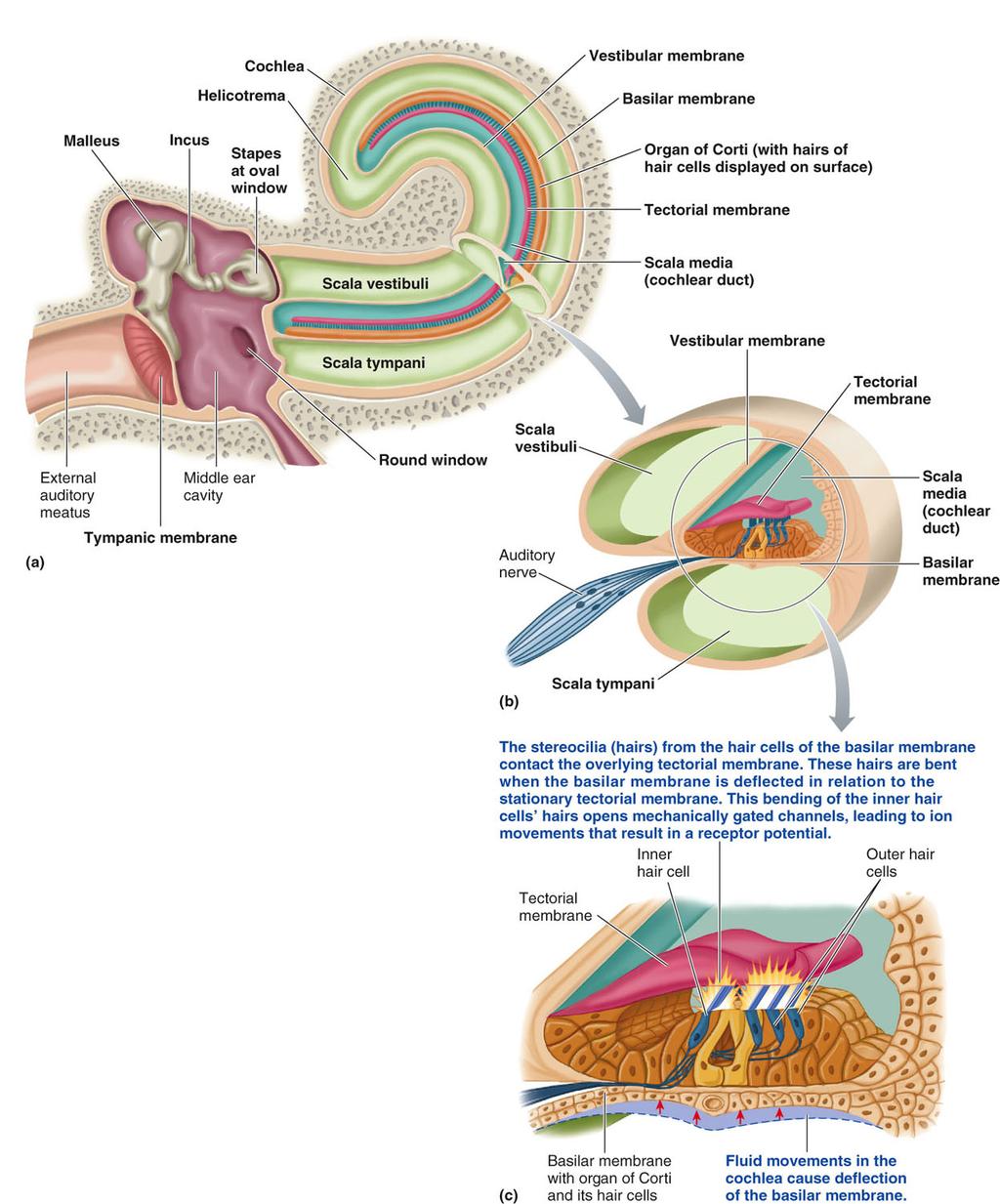
2 3. Inner Ear: Sense of Hearing (cochlea) a. three parts: cochlea, vestibule and semicircular canals 1. vestibule a. the stapes sits over a flexible connective membrane called the oval window b. the oval window is on the side of the vestibule; it vibrates when the stapes vibrates 2. semicircular canals involved with one s sense of balance or equilibrium 3. cochlea is involved with the sense of hearing b. cochlea: system of fluid-filled tubes within the osseous labyrinth of the temporal bone; it consists of the cochlear duct, vestibular duct (scala vestibuli), helicotrema, and tympanic duct (scala tympani); they are collectively referred to as the membranous labyrinth. A labyrinth is a network of winding passageways. 1. cochlear duct (also called the scala media) a. middle compartment b. filled with endolymph (viscous fluid) c. floor of cochlear duct is the basilar membrane. Hair cells and bipolar sensory neurons make up the organ of Corti that sits on top of the basilar membrane 2. vestibular duct (scala vestibuli) a. begins at the oval window which is on the side of the vestibule and covered by the stapes b. upper compartment (above cochlear duct) c. filled with perilymph (watery fluid) 2
3 d. it is continuous at the apex of the cochlea with the scala tympani; the region at the top of the cochlea where the fluids in the scala vestibuli and the scala tympani mix is called the helicotrema 3. helicotrema part of the cochlear labyrinth where the scala vestibuli and the scala tympani meet; it is at the apex of the cochlear duct 4. scala tympani (also called tympanic duct) a. ends at round window which is on the side of the cochlea b. the scala tympani contains perilymph (watery fluid) c. the round window on side of cochlea is a flexible connective tissue membrane 1. As the stapes footplate moves into the oval window, it pushes on the perilymph which pushes on the round window membrane causing it to move out. This allows movement of the fluid within the cochlea, leading to movement of the cochlear inner hair cells and thus hearing 2. the movement of the round window back and forth in response to fluid movements of the perilymph acts dissipate the energy of the fluid pressure waves to reduce deflection waves that might cause echoes. 3. pressure waves in perilymph of vestibular duct causes fluid waves in perilymph of tympanic duct and endolymph of cochlear duct that vibrates the basilar membrane up and down 4. If the round window were to be absent or rigidly fixed (as can happen in some congenital abnormalities), the stapes footplate would be pushing incompressible fluid against the unyielding walls of the cochlea. The fluid would therefore not move to any useful degree leading to a hearing loss of about 60dB. 5. Fluids can t be compressed. If the perilymph is pushed by the vibration of the oval window, then it can t move unless it can be moved to another area (vibrate the round window). If squeeze a water balloon, then it will bulge at other end. 4. the fluid-filled tubes make up the membranous labyrinth that is found inside an osseous labyrinth carved out of the inside of the temporal bone 5. the vestibular and tympanic ducts form a continuous duct that is filled with perilymph. The cochlear duct is filled with endolymph c. basilar membrane and Organ of Corti 1. forms the floor of the endolymph-filled cochlear duct separating it from the scala tympani 2. the Organ of Corti rests on top of basilar membrane throughout its entire length a. the Organ of Corti consists of hair cells, the dendritic ends of bipolar neurons, and support cells. 1. It does not include the basilar membrane which forms the foundation on which the organ of Corti sits. 2. The cell bodies and axons of the bipolar neurons are found in the cochlear nerve. 3. The hair cells are neurons that release the neurotransmitter glutamate 3

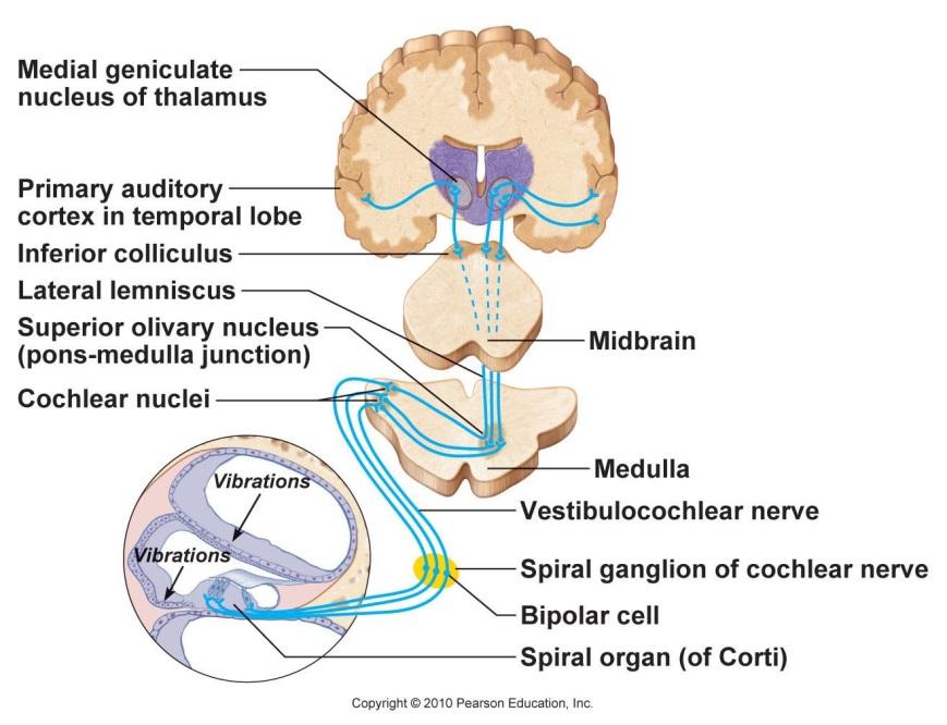
4 b. the Organ of Corti is made up of around 15,000 hair cells that stimulate bipolar sensory neurons 1. stereocilia are non-motile extensions of the plasma membrane and are more similar to microvilli than cilia. 2. Stereocilia contain actin filaments inside (cilia have a 9+2 arrangement of microtubules inside). 3. stereocilia are not cilia or microvilli, but they are closely related to microvilli. c. the hairs of hair cells are stereocilia that project up towards the bottom of the tectorial membrane d. the hair cells are the receptor cells that ear uses to hear. They are sensitive to fluid movements in perilymph caused by sound vibrations picked up by ear e. the hair cells are mechanoceptors f. pressure waves in the perilymph of the tympanic duct make the basilar membrane bounce up and down which causes the stereocilia of the hair cells to press against the tectorial membrane and distort (bend) in shape. g. the dendritic ends of bipolar neurons stimulated by the hair cells form the cochlear branch of the vestibulocochlear nerve (cranial nerve VIII, also called the auditory or cochlear nerve); the cell bodies of the bipolar neurons are in the spiral ganglion of the cochlear nerve and their axons go down cochlear nerve 1. the cochlear branch joins with the vestibular branch from the semicircular canals to form the vestibulocochlear nerve (VIII) 2. the vestibulocochlear nerve travels to pons of brainstem where the sensory neurons travel into the cochlear nucleus (at junction of pons and medulla) 3. interneurons in the cochlear nucleus project via the thalamus to a. primary auditory cortex in the temporal lobe of the cerebrum (hearing) b. cerebellum (balance or equilibrium) 3. the stationary tectorial membrane extends from the basilar membrane and hangs over the hair cells of the Organ of Corti cells like a porch awning. 4. The cells of the Organ of Corti are found beneath the tectorial membrane and run the length of the tectorial membrane. 4
5 5
6 6

7 Sound Pathway 1. sound waves move through air as jostling air molecules 2. sound waves are funneled by the pinna into the external auditory meatus 3. sound waves strike and vibrate the tympanum 4. the tympanum is physically attached to the malleus 5. the tympanum vibrates the malleus, which vibrates the incus and the stapes 6. the stapes is physically attached to the oval window on the side of the vestibule (pistonlike action of stapes on oval window) 7. the oval window is the beginning of the membrane-bound tube called the scala vestibuli which runs through the cochlea and becomes the scala tympani at the helicotrema 8. vibrations of the oval window vibrate perilymph fluids within the scala vestibuli at the same frequency 9. fluid waves (pressure waves) of the perilymph go down the scala vestibule, around the helicotrema and then down the scala tympani where the waves strike the round window which is on the side of the cochlea as it faces the middle ear cavity 10. waves within the perilymph going down the scala tympani vibrate the basilar membrane a. the basilar membrane then goes up and down b. the upward deflections press the stereocilia of the hair cells against the tectorial membrane and cause them to bend 7
 on the hair cells (special epithelial cells) causes the opening and closing of mechanically-gated K+ channels at the tips of the stereocilia")
8 11. movement of the stereocilia (mechanical deformation) stimulates the hair cells to stimulate the bipolar neurons to generate nerve impulses along their axons a. the back and forth displacement of the stereocilia (hairs) on the hair cells (special epithelial cells) causes the opening and closing of mechanically-gated K+ channels at the tips of the stereocilia of the hair cells b. when the mechanically-gated K+ channel gates open, K+ influx into the cell from the endolymph and depolarize the membrane by increasing the positivity of the ICF or cytoplasmic side of the membrane (this is a K+ influx which isn t what occurs during an action potential). K+ AT pumps create the df gradient for K+. c. the voltage change across the membrane as the K+ come in causes voltage-gated Ca2+ channels to open 1. when the voltage-gated Ca2+ channels open, Ca2+ go into the cell and cause the hair cells to release the neurotransmitter glutamate by exocytosis 2. glutamate binds to receptors on the dendritic end of bipolar neurons and stimulates them to generate nerve impulses 3. the dendrites of the bipolar neurons leave the basilar membrane and go into the cochlear nerve where their cell bodies are found in the spiral ganglion. The axons of the bipolar neurons travel in the cochlear nerve 12. the impulses generated by the hair cells that make up the Organ of Corti go out the cochlear nerve to the vestibulocochlear nerve (VIII) and then to the pons of the brainstem a. within the pons, the bipolar neurons stimulate interneurons that send impulses to auditory centers in the temporal lobe of the cerebrum where they are interpreted as meaningful sounds (hear) b. the vestibulocochlear nerve (cranial nerve VIII) transmits sound and equilibrium (balance) information from the inner ear to the pons of the brainstem where decussation occurs and from there to the thalamus and then to the primary auditory cortex of the cerebrum (hearing) or the cerebellum (balance) 13. the energy of the fluid waves is dissipated as the waves strike the round window and it bulges slowly out then back in 8
 stereocilia of hair cells in Organ of Corti move against tectorial membrane, (3) stereoocilia bend, (4) opens mechanicallygated K+ channels in stereocilia, (5) K+ efflux depolarizes")
9 Sound vibrates tympanum, vibrates ossicles, vibrate oval window, creates fluid pressure waves in perilymph of vestibular and tympanic ducts (1) pressure waves in fluids in perilymph move basilar membrane, (2) stereocilia of hair cells in Organ of Corti move against tectorial membrane, (3) stereoocilia bend, (4) opens mechanicallygated K+ channels in stereocilia, (5) K+ efflux depolarizes membrane, (6) opens voltage-gated Ca2+ channels, (7) Ca2+ influx triggers exocytosis of glutamate, (8) stimulate bipolar neurons 9
 (similar to gyroscope on airplanes) 1. each canal is filled with endolymph (watery fluid) 2.")
10 Inner Ear: Sense of Balance or Equilibrium (Semicircular Canals) 1. Vestibular Apparatus helps a person keep their balance during movements a. found in inner ear b. made up of 3 semicircular canals (SCC) (similar to gyroscope on airplanes) 1. each canal is filled with endolymph (watery fluid) 2. each canal oriented in a different 3-D plane (frontal, sagittal, and transverse) to create an x, y, z coordinate system for tracking fluid movements in the SCC s that occur when the head moves 3. hair cells with stereocilia are found at the base of each SCC 2. Function of Hair Cells trigger nerve impulses in sensory neurons a. special neurons that release the neurotransmitter glutamate by exocytosis b. hair cells have stereocilia (hairs) protruding from their apical ends that face the endolymph c. head movements cause the endolymph to move d. movement of the endolymph causes the stereocilia on the hair cells to bend back and forth e. movement of the hair cells back and forth opens mechanically-gated K+ channels in the tips of the stereocilia 10
11 f. K+ diffuse from the endolymph through the open channels into the cytoplasm of the hair cells (this is a K+ influx which is different than the K+ efflux that occurs during an action potential) g. the K+ influx depolarizes the inside of the plasma membrane and causes the opening of voltage-gated Ca2+ channels in the plasma membrane h. Ca2+ enter the cell and trigger the exocytosis of the neurotransmitter glutamate from secretory vesicles into the synaptic cleft. i. glutamate stimulates the dendrites of unipolar sensory neurons and the axons of the sensory neurons generate a nerve impulse j. the nerve impulse travels down the axons of the unipolar sensory neurons in the vestibular nerve that joins the cochlear nerve to become the vestibulocochlear nerve which goes to the pons of the brainstem k. the impulses on the axons of sensory neurons in stimulate the dendritic zones of interneurons in the pons l. interneurons in the pons then then send impulses to the cerebellum m. the cerebellum uses the information about the position of the head relative to the body to tell the motor centers of the cerebrum which skeletal muscles to contract to get the body under the head in order to be well-balanced. 11
 each of the 2 eyespots consists of less than 100 photoceptor cells lining a cup-like pit; allows Planaria to move away from light (under rocks in freshwater streams during")
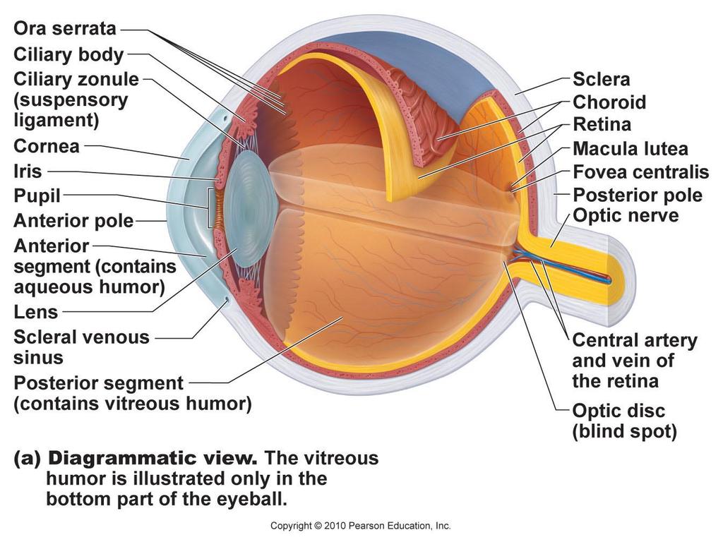
12 Photoreception: Eyes and Vision 1. over 90% of known animal species have some light sensors 2. light-sensing organs range from simple eyespots to complex eyes a. Planaria (flatworm) each of the 2 eyespots consists of less than 100 photoceptor cells lining a cup-like pit; allows Planaria to move away from light (under rocks in freshwater streams during daylight) b. complex eyes mammals and birds c. birds have the largest eyes relative to body size; they also have more photoceptor cells hence a sharp-eyed hawk can spot a mouse in a field from hundreds of meters away 3. the main function of the eye is to focus light on the photoceptor cells of the retina. The photoceptor cells then transduce light into nerve impulses that travel to centers in the cerebrum to form images Anatomy of the mammalian eye 3 Layers or tunics of the Mammalian Eye (fibrous, vascular, retina) 1. Fibrous tunic (layer) a. sclera 1. dense regular connective tissue that is rich in white-colored collagen fibers (structurally similar to tendons); very tough 2. white part of eye 3. about 1 in diameter (humans) b. cornea 1. transparent and avascular 12
13 2. consists of several layers of thin epithelial cells with some collagen fibers between the cells 3. curvature of cornea helps to focus light through lens to the retina; light bends as it goes through the cornea a. the cornea provides most of the eye s focusing power b. the curvature of the cornea is fixed, hence the fine-tuning of focus is done by the lens which can change shape to fine tune focus depending on the object s distance from the eye 4. densely innervated with pain receptors 2. Vascular tunic (middle layer) a. Choroid 1. highly pigmented with melanin; chocolate brown in color 2. vascular; blood vessels provide nutrients to the retina (photoceptor cells) b. ciliary body made up of ciliary muscles and ciliary processes 1. anterior extension of the choroid 2. aqueous humor a. blood vessels in the ciliary processes of the ciliary body continuously secrete a watery fluid called the aqueous humor that fills up the anterior cavity (between lens and cornea) b. supplies nutrients to the avascular lens and cornea 3. ciliary muscles control the tension on the suspensory ligaments; ciliary muscles are smooth muscle cells that make up most of the ciliary body c. iris 1. comes off the ciliary body 2. pigmented smooth muscle cells give color to the eye 3. contraction and relaxation of smooth muscle within the iris controls the diameter (aperture) of the pupil 4. the diameter of the pupil controls how much light reflecting off an object enters the eye and strikes the retina to form an image d. pupil 1. hole in the middle of the iris 2. it is not a physical structure 3. Pupil diameter 1/α amount of ambient light (dark > incr dia; bright > decr dia) e. lens 1. transparent and avascular; no nerves 2. made up mostly of thin epithelial cells (The lens itself lacks nerves, blood vessels, or connective tissue) 3. biconvex 4. lens can change shape to focus light on the retina to sharpen image 5. held in place by suspensory ligaments coming off the ciliary body that attach to the margin of the lens 3. Retina (inner tunic) pigmented epithelium and neural layer a. contains light-sensitive photoceptor cells (rods and cones) b. vitreous humor 13

14 1. clear, gel-like fluid (similar to jelly) that fills up the posterior cavity (space between the retina and lens) 2. formed during fetal life; we are born with all the vitreous humor that we will ever have c. Tapetum lucidum 1. reflective layer beneath the photoceptor cells that helps some animals see better in dim light 2. highly developed in crepuscular (twilight) and nocturnal (active at night) animals 3. tapetum reflects light that has passed through the retina back through it a second time to stimulate the photoceptor cells a second time 4. the cat eye is much more sensitive to light than the human eye 5. the tapetum accounts for the eye shine in animals that one sees when light is shown on their eyes in the dark Neural Structure of the retina (3 layers of neurons) 1. 3 layers of neurons in the retina: rods and cones, bipolar cells and ganglion cells a. photoceptor cells: rods and cones 1. about million rods (20x more rods than cones) 2. about 5-6 million cones b. bipolar cells c. ganglion cells (about 1 million ganglion cells in the retina) 14
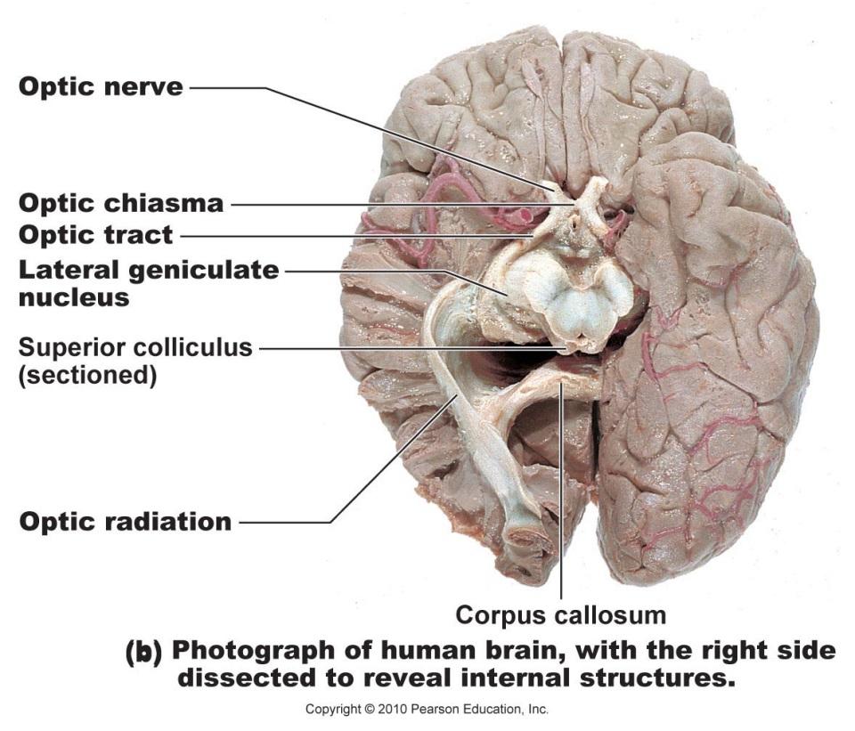
15 2. Photoceptor cells (special neurons) a. Rods (active in dim light, inactive in bright light) 1. contain a photopigment called rhodopsin (visual purple); rhodopsin requires Vitamin A for its synthesis 2. rods allow for indistinct grayscale image (black and white) formation in dim light; images lack sharp detail 3. rods are specialized for night vision; in dim light the rods produce an grayscale (black and white) image that is colorless and softly focused (lacks sharp details) c. cones (inactive in dim light, active in moderate to bright light) 1. cones allow for detailed color vision in moderate to bright light 2. cones are specialized for day vision and in well-lit rooms 3. fovea capitis (0.4 mm) in middle of the macula lutea of retina has the densest concentration of cones and forms the sharpest part of a color image d. photoceptor cells tonically secrete glutamate in the dark that inhibits any activity on the bipolar neurons e. light interacts with photopigments in rods and cones which causes them to stimulate bipolar cells 3. bipolar cells (bipolar neurons) glutamate is the neurotransmitter released by these cells 4. ganglion cells (ganglion neurons) a. axons of ganglion cells go across the surface of the retina and come together at the optic disk to form the optic nerve b. optic nerve goes through the optic canal in the sphenoid bone and exit on either side of the sella turcica c. optic nerves take impulses on axons of ganglion cells to the optic tract then to the thalamus of the diencephalon where axons synapse with interneurons that go to visual centers (primary visual cortex) in the occipital lobe of the cerebrum where the impulses are used to create images Photopigments 1. photopigments in rods and cones interact with light and undergo chemical changes a. dim light excites a photopigment called rhodopsin in rods b. moderate to bright light excites photopigments called scotopsins in cones; different scotopsins are sensitive to red, green and blue (RGB) wavelengths of the visibile spectrum that allow one to see in color; cones operate similar to an RGB color system (e.g., RGB monitor) 1. scotopsins of trichromatic RGB eyes (3-color vision) of primates are sensitive to red, blue, and green (RGB) wavelengths of light 2. 3 types of cone cells and each of them have their own form of scotopsin a. long (L) cones cones that are most sensitive to wavelengths of light around 560 nm give the perception of red b. middle (M) cones most sensitive to green wavelengths of light (around 530 nm) c. short (S) cones most sensitive to blue wavelengths of light (around 420 nm) 15

16 2. once stimulated by light, the rods and cones stimulate bipolar cells (neurons) to release a neurotransmitter that then stimulates ganglion cells 3. the axons of ganglion neurons run across the surface of the retina and converge at the optic disk to form the optic nerve. 4. the optic nerves (one from each eye) carry the axons of the ganglion neurons through the optic chiasma to the optic tracts 5. the optic tracts travel to the thalamus 6. the ganglion neurons synapse with interneurons within the thalamus (lenticulate nucleus) of the diencephalon 7. the interneurons then transmit the image information from the thalamus to the primary visual cortex of the occipital lobe of the cerebrum where the image forms 16
17 17


18 RGB color scheme. Sensitivity of the different types of cones to different wavelengths. Trichromatic eyes of primates. The ratios of stimulation of the 3 cone types are shown for 3 sample colors (red, green, yellow) Electromagnetic spectrum and visible light 1. visible light is a mix of wavelengths from violet-blue-cyan-green-yellow-orange-red Electromagnetic spectrum Visible spectrum 18
19 Transduction by Sensory Cells 1. Transduction conversion of one form of energy to another a. an external stimulus like touch, pressure, pain, temperature change is converted to nerve impulses by sensory cells b. transduction is associated with opening and closing of ion channels 2. 2 types of ion channels a. leak channels always open b. gated channels open/close in response to specific stimuli 3. 4 types of gated channels a. Mechanically Gated 1. stimulated by stretch; gates open when the sensory cell is stretched or deformed 2. opening of channels generates impulse on sensory cells b. chemically-gated 1. stimulated by chemicals (ligands) that bind to receptors 2. ligand-receptor binding opens ion channels and generates impulses on sensory cells c. voltage-gated channels d. thermally gated channels Perception 1. sensory information is perceived if it reaches the brain 2. perception animal s interpretation of the external world is created by the brain from a pattern of nerve impulses coming from sensory cells Sense of Hearing: Sound waves 1. hearing is the perception of sound waves through gas, solid, or liquid by the brain 2. sound waves travel through a medium as molecules are jostled against one another 3. many animals can detect sound a. fishes detect sound through lateral line system b. insects, amphibians and some reptiles hear with use of tympanic membrane (eardrum) on body surface that vibrates when struck by sound waves 4. sound travels fast a m/sec in water (6000 mph) b. 340 m/sec in air (760 mph) 5. Two main properties of a sound a. intensity (loudness) expressed in decibels (db) b. Pitch or tone frequency of the vibrations (cycles per second or Hertz) 1. humans can perceive sounds with frequencies from 20 to 20,000 Hz 2. dogs can hear sounds from 20 to 40,000 Hz (or even up to 60 khz). Higher frequency sound waves above 20 khz are called ultrasounds. Humans cannot hear them 3. dog whistles generate ultrasound frequencies with in a range of khz (humans only hear a quiet hissing sound) 19
Taste buds Gustatory cells extend taste hairs through a narrow taste pore
 The Special Senses Objectives Describe the sensory organs of smell, and olfaction. Identify the accessory and internal structures of the eye, and explain their function. Explain how light stimulates the
Copyright 2009 Pearson Education, Inc.
 Outline Nervous System Sensory Systems I. II. III. IV. V. VI. Biol 105 Lecture 11 Chapter 9 Senses Sensory receptors Touch Vision Hearing and balance Smell Senses Sensory receptor cells Sensory receptors
Chapter 17, Part 2! The Special Senses! Hearing and Equilibrium!
 Chapter 17, Part 2! The Special Senses! Hearing and Equilibrium! SECTION 17-5! Equilibrium sensations originate within the inner ear, while hearing involves the detection and interpretation of sound waves!
Chapter 17, Part 2! Chapter 17 Part 2 Special Senses! The Special Senses! Hearing and Equilibrium!
 Chapter 17, Part 2! The Special Senses! Hearing and Equilibrium! SECTION 17-5! Equilibrium sensations originate within the inner ear, while hearing involves the detection and interpretation of sound waves!
Presentation On SENSATION. Prof- Mrs.Kuldeep Kaur
 Presentation On SENSATION Prof- Mrs.Kuldeep Kaur INTRODUCTION:- Sensation is a specialty area within Psychology that works at understanding how are senses work and how we perceive stimuli in the environment.
-Detect heat or cold and help maintain body temperature
 Sensory Receptors -Transduce stimulus energy and transmit signals to the central nervous system -Reception occurs when a receptor detectd a stimulus -Perception occurs in the brain as this information
SPECIAL SENSES PART I: OLFACTION & GUSTATION
 SPECIAL SENSES PART I: OLFACTION & GUSTATION 5 Special Senses Olfaction Gustation Vision Equilibrium Hearing Olfactory Nerves Extend through cribriform plate into nasal cavity on both sides of nasal septum
is the clear, transparent part at the front of the eye. It allows light to enter the eye and it also refracts (focuses) the light onto the retina.
 the light onto the retina.") Senses- Vision Light is a small part (1/70th) of the total electromagnetic (EM) spectrum. The EM band extends from radio waves at one extreme to x-rays at the other. The eye detects light and converts
Senses- Vision Light is a small part (1/70th) of the total electromagnetic (EM) spectrum. The EM band extends from radio waves at one extreme to x-rays at the other. The eye detects light and converts
Introduction. Senses our perception of what is out there 2 groups. General senses Special senses
 Introduction Senses our perception of what is out there 2 groups General senses Special senses Central Processing and Adaptation Adaptation the loss of sensitivity after continuous stimulation Tonic receptors
Surgical Anatomy Ear and Eye. Presenters: Dr. Jim Hurrell and Dr. Dennis McCurnin
 Surgical Anatomy Ear and Eye Presenters: Dr. Jim Hurrell and Dr. Dennis McCurnin A Warm Welcome from My Faculty TEAM and Me!!! 2 The Pledge of Allegiance 3 The Senses 4 Hearing 3 Layers of Ear EXTERNAL
Special Senses. Mechanoreception Electroreception Chemoreception Others
 Special Senses Mechanoreception Electroreception Chemoreception Others Recall our receptor types Chemically regulated: Respond to particular chemicals Voltage regulated: respond to changing membrane potential
Biology. Slide 1 of 49. End Show. Copyright Pearson Prentice Hall
 Biology 1 of 49 2 of 49 Sensory Receptors Neurons that react directly to stimuli from the environment are called sensory receptors. Sensory receptors react to stimuli by sending impulses to other neurons
For this lab you will use parts of Exercise #18 in your Wise lab manual. Please be sure to read those sections before coming to lab
 Bio 322 Human Anatomy Objectives for the laboratory exercise The Eye and Ear Required reading before beginning this lab: Saladin, KS: Human Anatomy 5 th ed (2017) Chapter 17 For this lab you will use parts
Senses and Sense Organs
 Senses and Sense Organs SENSORY SYSTEMS Human experience is effected by both internal and external stimuli. Humans are able to distinguish among many different types of stimuli by means of a highly developed
Chapter 15 Lecture Outline
 Chapter 15 Lecture Outline See separate PowerPoint slides for all figures and tables preinserted into PowerPoint without notes. Copyright 2016 McGraw-Hill Education. Permission required for reproduction
Chapter 18 Senses SENSORY RECEPTION 10/21/2011. Sensory Receptors and Sensations. Sensory Receptors and Sensations. Sensory Receptors and Sensations
 SENSORY RECEPTION Chapter 18 Senses s convert stimulus energy to action potentials s 1. Are specialized cells, or 2. Specialized endings that detect stimuli All stimuli are forms of energy s in eyes detect
Special Senses. Accessory Structures of the Eye. The Eye and Vision. Accessory Structures of the Eye. Accessory Structures of the Eye
 8 PART A Special Senses PowerPoint Lecture Slide Presentation by Jerry L. Cook, Sam Houston University ESSENTIALS OF HUMAN ANATOMY & PHYSIOLOGY EIGHTH EDITION ELAINE N. MARIEB The Senses General senses
o A cushion of fat surrounds most of the eye
 Name Period SPECIAL SENSES The Senses of touch o Temperature o Pressure o Pain o Smell o Taste o Sight o Hearing o Equilibrium The Eye and Vision are in the eyes has over a o Most of the eye is enclosed
Sensory system. Dr. Carmen E. Rexach Anatomy 35 Mt San Antonio College
 Sensory system Dr. Carmen E. Rexach Anatomy 35 Mt San Antonio College Sensory receptors Detect stimuli Classified by structure Origin Distribution Modality Structural Classification naked nerve endings
The Senses. Chapter 10 7/8/11. Introduction
 Chapter 10 The Senses Introduction A. Sensory receptors detect changes in the environment and stimulate neurons to send nerve impulses to the brain. B. A sensation is formed based on the sensory input.
Chapter 10. The Senses
 Chapter 10 The Senses 1 Introduction A. Sensory receptors detect changes in the environment and stimulate neurons to send nerve impulses to the brain. B. A sensation is formed based on the sensory input.
 Question 1: Briefly describe the structure of the following: (a) Brain (b) Eye (c) Ear (A) Brain: Brain is the main coordinating centre of the body. It is a part of nervous system that controls and monitors
The cochlea: auditory sense. The cochlea: auditory sense
 Inner ear apparatus 1- Vestibule macula and sacculus sensing acceleration of the head and direction of gravity 2- Semicircular canals mainly for sensing direction of rotation of the head 1 3- cochlea in
ENT 318 Artificial Organs Physiology of Ear
 ENT 318 Artificial Organs Physiology of Ear Lecturer: Ahmad Nasrul Norali The Ear The Ear Components of hearing mechanism - Outer Ear - Middle Ear - Inner Ear - Central Auditory Nervous System Major Divisions
The Nervous System: General and Special Senses Pearson Education, Inc.
 18 The Nervous System: General and Special Senses Introduction Sensory information arrives at the CNS Information is picked up by sensory receptors Sensory receptors are the interface between the nervous
Ganglion Cells Blind Spot Cornea Pupil Visual Area of the Bipolar Cells Thalamus Rods and Cones Lens Visual cortex of the occipital lobe
 How We See How We See Cornea Ganglion Cells whose axons form the optic nerve Blind Spot the exit point at the back of the retina Pupil which is controlled by the iris Bipolar Cells Visual Area of the Thalamus
Physiology of human perception
 Physiology of human perception Vision Hearing Thermal and tactile sensations Basic introduction and the list and description of the tasks to be carried out Visible light: 400-700 nm. Vision or sight Anatomy
Ear. Utricle & saccule in the vestibule Connected to each other and to the endolymphatic sac by a utriculosaccular duct
 Rahaf Jreisat *You don t have to go back to the slides. Ear Inner Ear Membranous Labyrinth It is a reflection of bony labyrinth but inside. Membranous labyrinth = set of membranous tubes containing sensory
Auditory System. Barb Rohrer (SEI )
") Auditory System Barb Rohrer (SEI614 2-5086) Sounds arise from mechanical vibration (creating zones of compression and rarefaction; which ripple outwards) Transmitted through gaseous, aqueous or solid medium
Auditory System Barb Rohrer (SEI614 2-5086) Sounds arise from mechanical vibration (creating zones of compression and rarefaction; which ripple outwards) Transmitted through gaseous, aqueous or solid medium
4. Which letter in figure 9.1 points to the fovea centralis? Ans: b
 Chapter 9: The Sensory System 1. Proprioceptors are involved in the sense of A) pain. B) temperature. C) pressure. D) movement of limbs. 2. Which are chemoreceptors? A) taste B) olfactory C) proprioceptors
Unit VIII Problem 9 Physiology: Hearing
 Unit VIII Problem 9 Physiology: Hearing - We can hear a limited range of frequency between 20 Hz 20,000 Hz (human hearing acuity is between 1000 Hz 4000 Hz). - The ear is divided into 3 parts. Those are:
THE EAR AND HEARING Be sure you have read and understand Chapter 16 before beginning this lab. INTRODUCTION: hair cells outer ear tympanic membrane
 BIOLOGY 211: HUMAN ANATOMY & PHYSIOLOGY ****************************************************************************************************** THE EAR AND HEARING ******************************************************************************************************
The Sense Organs 10/13/2016. The Human Eye. 1. Sclera 2. Choroid 3. Retina. The eye is made up of three layers:
 The human body gathers information from the outside world by using the five senses of: The Sense Organs 12.3 Sight Hearing Taste Smell Touch This information is essential in helping the body maintain homeostasis.
Special Senses: The Eye
 Unit 4 Special Senses: The Eye ESSENTIALS OF HUMAN ANATOMY & PHYSIOLOGY The Senses General senses of touch Temperature Pressure Pain Special senses Smell Taste Sight Hearing Equilibrium The Eye and Vision
o A cushion of fat surrounds most of the eye
 Name Period SPECIAL SENSES The Senses General senses of touch o Temperature o Pressure o Pain Special senses o Smell o Taste o Sight o Hearing o Equilibrium The Eye and Vision 70 percent of all sensory
The olfactory epithelium is located at the roof of the nasal cavity. Nasal conchae cause turbulance of incoming air
 Special Senses I. Olfaction II. Gustation A. Anatomy and general info The olfactory epithelium is located at the roof of the nasal cavity Nasal conchae cause turbulance of incoming air Olfactory glands
The white of the eye and the part that maintains its shape is know n as the:
 Scrub In The white of the eye and the part that maintains its shape is know n as the: a. Cornea b. Pupil c. Retina d. Sclera The structure that is found in the ear and contains the organ of hearing is
AUDITORY APPARATUS. Mr. P Mazengenya. Tel 72204
 AUDITORY APPARATUS Mr. P Mazengenya Tel 72204 Describe the anatomical features of the external ear Describe the tympanic membrane (ear drum) Describe the walls of the middle ear Outline the structures
Hearing. By: Jimmy, Dana, and Karissa
 Hearing By: Jimmy, Dana, and Karissa Anatomy - The ear is divided up into three parts - Sound enters in through the outer ear and passes into the middle where the vibrations are received and sent to the
Overview of Sensory Receptors
 Sensory Systems Chapter 45 Overview of Sensory Receptors Sensory receptors provide information from our internal and external environments that is crucial for survival and success -Exteroceptors sense
Classifying receptors
 Sense organs Specialized nerves that detect changes in external environment Translate via nerve impulses to CNS Classifying receptors Chemoreceptors Electroreceptors Mechanoreceptors Photo (radiation)
SPECIAL SENSES: THE AUDITORY SYSTEM
 SPECIAL SENSES: THE AUDITORY SYSTEM REVISION OF PHYSICS: WAVES A wave is an oscillation of power, sound waves have two main characteristics: amplitude, which is the maximum displacement or the power of
Activity 1: Anatomy of the Eye and Ear Lab
 Activity 1: Anatomy of the Eye and Ear Lab 1. Launch the view! Launch Human Anatomy Atlas. Navigate to Quizzes/Lab Activities, find the Eye and Ear Lab section. Launch Augmented Reality mode and scan the
2. WINDOWS OF KNOWLEDGE
 CONTENT 2. WINDOWS OF KNOWLEDGE Vision - The protective measures of eyes. - Structure of human eye, Working of eye lens, - Photo receptors in the retina, Sense of vision. - Disorders & diseases of eyes,
Vision and Audition. This section concerns the anatomy of two important sensory systems, the visual and the auditory systems.
 Vision and Audition Vision and Audition This section concerns the anatomy of two important sensory systems, the visual and the auditory systems. The description of the organization of each begins with
Anatomy of the Ear Region. External ear Middle ear Internal ear
 Ear Lecture Objectives Make a list of structures making the external, middle, and internal ear. Discuss the features of the external auditory meatus and tympanic membrane. Describe the shape, position,
TASTE: Taste buds are the sense organs that respond to gustatory stimuli. Chemoreceptors that respond to chemicals broken down from food in the saliva
 UNIT 5: Nervous System- Senses Somatic Senses Somatic senses are associated with receptors in the skin, muscles, joints, and viscera (organs of the body) Include senses of touch, pressure, temperature,
Essential questions. What are the structures of the sensory system? 3.03 Remember the structures of the sensory system 2
 Essential questions What are the structures of the sensory system? 3.03 Remember the structures of the sensory system 2 The Senses Eyes Sight Ears Hearing Nose Smell Tongue Taste Skin Touch 3.03 Remember
Chapter 38 Active Reading Guide Nervous and Sensory Systems
 Name: AP Biology Mr. Croft Chapter 38 Active Reading Guide Nervous and Sensory Systems Section 1 1. This concept begins with a look at the evolution of nervous systems. You will want to study this to tie
Chapter 29 The Senses
 Chapter 29 The Senses PowerPoint Lectures for Biology: Concepts & Connections, Sixth Edition Campbell, Reece, Taylor, Simon, and Dickey Copyright 2009 Pearson Education, Inc. Lecture by Edward J. Zalisko
Sensory Physiology. Sensory Range Varies. Introduction to the Special Senses. How do we sense the world around us?
 Sensory Physiology How do we sense the world around us? We do not see things as they are; we see things as we are. --Anais Nin Anais Nin, French author 1903-1977 Sensory Range Varies Introduction to the
Senses- Ch. 12. Pain receptors- respond to tissue damage in all tissues except in the brain
 Senses- Ch. 12 5 general types of sensory neurons or receptors are known. These specialized neurons detect stimuli from the eyes, ears, nose, mouth, and skin. The stimuli are changed into electrical signals
20-20,000 Hertz range of human hearing
 20-20,000 Hertz range of human hearing accommodation automatic adjustment in focal length of the lens of the eye; changing the shape of the lens aqueous humor Watery fluid in the anterior chambers of the
Bi 121 Lab OLFACTION. olfactory bulb, olfactory nerve (=cranial nerve I), olfactory foramina, olfactory epithelium
, olfactory foramina, olfactory epithelium") Bi 121 Lab Week 9: THE SPECIAL SENSES The special senses include smell, taste, vision, hearing, and balance. In this laboratory exercise, we will look at many of the structures that provide for these senses.
Bi 121 Lab Week 9: THE SPECIAL SENSES The special senses include smell, taste, vision, hearing, and balance. In this laboratory exercise, we will look at many of the structures that provide for these senses.
Sensory Systems. BIOLOGY OF HUMANS Concepts, Applications, and Issues. Judith Goodenough Betty McGuire
 BIOLOGY OF HUMANS Concepts, Applications, and Issues Fifth Edition Judith Goodenough Betty McGuire 9 Sensory Systems Lecture Presentation Anne Gasc Hawaii Pacific University and University of Hawaii Honolulu
Biology 3201 The Nervous System Test
 Biology 3201 The Nervous System Test 1. The central nervous system consists of: a. Nerves and neurons c. spinal chord and nerves b. brain and neurons d. brain and spinal chord 2. This part of the brain
Biology. A Guide to the Natural World. Chapter 27 Lecture Outline Communication and Control 1: The Nervous System. Fifth Edition.
 Biology A Guide to the Natural World Chapter 27 Lecture Outline Communication and Control 1: The Nervous System Fifth Edition David Krogh The Nervous System Nervous tissue is composed of two kinds of cells:
Auditory System Feedback
 Feedback Auditory System Feedback Using all or a portion of the information from the output of a system to regulate or control the processes or inputs in order to modify the output. Central control of
MECHANISM OF HEARING
 MECHANISM OF HEARING Sound: Sound is a vibration that propagates as an audible wave of pressure, through a transmission medium such as gas, liquid or solid. Sound is produced from alternate compression
Sound. Audition. Physics of Sound. Properties of sound. Perception of sound works the same way as light.
 Sound Audition Perception of sound works the same way as light. Have receptors to convert a physical stimulus to action potentials Action potentials are organized in brain structures You apply some meaning
Audition. Sound. Physics of Sound. Perception of sound works the same way as light.
 Audition Sound Perception of sound works the same way as light. Have receptors to convert a physical stimulus to action potentials Action potentials are organized in brain structures You apply some meaning
Chap Senses. 1. Give an example of something a general sensory receptor would detect.
 Carl Christensen, PhD Chap. 17 - Senses Bio. 2304 Human Anatomy 1. Give an example of something a general sensory receptor would detect. 2. Classification of Sensory Receptors a. mechanoreceptors b. thermoreceptors
The Special Senses. Smell, taste, vision, hearing and equilibrium Housed in complex sensory organs
 The Special Senses Smell, taste, vision, hearing and equilibrium Housed in complex sensory organs Chemical Senses Interaction of molecules with receptor cells Olfaction (smell) and gustation (taste) Both
Gathering information the sensory systems; Vision
 Visual System Gathering information the sensory systems; Vision The retina is the light-sensitive receptor layer at the back of the eye. - Light passes through the cornea, the aqueous chamber, the lens,
Special Senses PART A
 8 Special Senses PART A PowerPoint Lecture Slide Presentation by Jerry L. Cook, Sam Houston University ESSENTIALS OF HUMAN ANATOMY & PHYSIOLOGY EIGHTH EDITION ELAINE N. MARIEB The Senses General senses
Sensation and Perception. A. Sensation: awareness of simple characteristics B. Perception: making complex interpretations
 I. Overview Sensation and Perception A. Sensation: awareness of simple characteristics B. Perception: making complex interpretations C. Top-Down vs Bottom-up Processing D. Psychophysics -- thresholds 1.
NERVOUS SYSTEM & SENSES TEACHER COPY
 NERVOUS SYSTEM & SENSES TEACHER COPY FUNCTIONS OF THE NERVOUS SYSTEM What are the three functions of the Nervous System? 1. Receives information about what is happening inside and outside of your body
Intro to Audition & Hearing
 Intro to Audition & Hearing Lecture 16 Chapter 9, part II Jonathan Pillow Sensation & Perception (PSY 345 / NEU 325) Fall 2017 1 Sine wave: one of the simplest kinds of sounds: sound for which pressure
Hearing. By Jack & Tori
 Hearing By Jack & Tori 3 Main Components of the Human Ear. Outer Ear. Middle Ear. Inner Ear Outer Ear Pinna: >Visible part of ear and ear canal -Acts as a funnel to direct sound Eardrum: >Airtight membrane
Chapter 50: Sensory and Motor Mechanisms
 Name Period As in Chapter 49, there are several topics in this chapter that we will emphasize only lightly. If your teacher stresses human anatomy and physiology, you may be expected to go into more depth.
Before we talk about the auditory system we will talk about the sound and waves
 The Auditory System PHYSIO: #3 DR.LOAI ZAGOUL 24/3/2014 Refer to the slides for some photos. Before we talk about the auditory system we will talk about the sound and waves All waves have basic characteristics:
a) Central sulcus- shallow groove that runs across brain sagitally
 Central sulcus- shallow groove that runs across brain sagitally") KEY BRAIN Brain Gross Anatomy Terms 1) Explain each of the following in terms of structure of the brain a) Central sulcus- shallow groove that runs across brain sagitally b) Lateral fissure- deep groove
KEY BRAIN Brain Gross Anatomy Terms 1) Explain each of the following in terms of structure of the brain a) Central sulcus- shallow groove that runs across brain sagitally b) Lateral fissure- deep groove
Structure, Energy Transmission and Function. Gross Anatomy. Structure, Function & Process. External Auditory Meatus or Canal (EAM, EAC) Outer Ear
 Outer Ear") Gross Anatomy Structure, Energy Transmission and Function IE N O ME 1 Structure, Function & Process 4 External Auditory Meatus or Canal (EAM, EAC) Outer third is cartilaginous Inner 2/3 is osseous Junction
Gross Anatomy Structure, Energy Transmission and Function IE N O ME 1 Structure, Function & Process 4 External Auditory Meatus or Canal (EAM, EAC) Outer third is cartilaginous Inner 2/3 is osseous Junction
Chapter 16B. The Special Senses. The Special Senses. Olfactory Epithelium. Chemical Senses
 The Special Senses Chapter 16B Smell, taste, vision, hearing and equilibrium Housed in complex sensory organs The Special Senses 1 2 Chemical Senses Interaction of molecules with chemoreceptor cells Olfaction
THE COCHLEA AND AUDITORY PATHWAY
 Dental Neuroanatomy Suzanne S. Stensaas, PhD February 23, 2012 Reading: Waxman, Chapter 16, Review pictures in a Histology book Computer Resources: http://www.cochlea.org/ - Promenade around the Cochlea
Chapter 18. The Senses SENSORY RECEPTION. Introduction: Superhuman Senses. Introduction: Superhuman Senses
 Introduction: Superhuman Senses Chapter 18 The Senses! Three senses found in some animals but not humans Echolocation locating objects by detecting echoes of emitted sound waves Electroreception ability
Introduction to Physiological Psychology
 Introduction to Physiological Psychology Vision ksweeney@cogsci.ucsd.edu cogsci.ucsd.edu/~ksweeney/psy260.html This class n Sensation vs. Perception n How light is translated into what we see n Structure
SENSORY SYSTEM VII THE EAR PART 1
 SENSORY SYSTEM VII THE EAR PART 1 Waves Sound is a compression wave The Ear Ear Outer Ear Pinna Outer ear: - Made up of the pinna and the auditory canal Auditory Canal Outer Ear Pinna (also called the
Sensory and Motor Mechanisms
 Chapter 50 Sensory and Motor Mechanisms PowerPoint Lecture Presentations for Biology Eighth Edition Neil Campbell and Jane Reece Lectures by Chris Romero, updated by Erin Barley with contributions from
13031_ch 10 8/15/08 10:01 AM Page 152. Overview
 13031_ch 10 8/15/08 10:01 AM Page 152 Overview The sensory system enables us to detect changes taking place both internally and externally. These changes are detected by specialized structures called receptors.
Essentials of Human Anatomy & Physiology. Chapter 8. Special Senses. Slides Lecture Slides in PowerPoint by Jerry L.
 Essentials of Human Anatomy & Physiology Elaine N. Marieb Seventh Edition Chapter 8 Special Senses Slides 8.1 8.19 Lecture Slides in PowerPoint by Jerry L. Cook Special Senses Title Somatosensation Essential
INTRODUCTION: ****************************************************************************************************
 BIOLOGY 211: HUMAN ANATOMY & PHYSIOLOGY **************************************************************************************************** EYES AND VISION ****************************************************************************************************
Principles of Anatomy and Physiology
 Principles of Anatomy and Physiology 14 th Edition CHAPTER 17 The Special Senses Olfaction: Sense of Smell Smell and taste are chemical senses. The human nose contains 10 million to 100 million receptors
Otoconia: Calcium carbonate crystals Gelatinous mass. Cilia. Hair cells. Vestibular nerve. Vestibular ganglion
 VESTIBULAR SYSTEM (Balance/Equilibrium) The vestibular stimulus is provided by Earth s, and. Located in the of the inner ear, in two components: 1. Vestibular sacs - gravity & head direction 2. Semicircular
[CHAPTER 12: THE NERVOUS SYSTEM] [ANSWER KEY]
![[CHAPTER 12: THE NERVOUS SYSTEM] [ANSWER KEY]](/thumbs/71/65616244.jpg "[CHAPTER 12: THE NERVOUS SYSTEM] [ANSWER KEY]") WORDBANK: Cholinesterase Dopamine Axon Choroid layer Cochlea Incus Action Potential Cataract Cornea Astigmatism Dendrite Malleus Alzheimer s Disease Central Excitatory Response Fovea Centralis Acetylcholine
WORDBANK: Cholinesterase Dopamine Axon Choroid layer Cochlea Incus Action Potential Cataract Cornea Astigmatism Dendrite Malleus Alzheimer s Disease Central Excitatory Response Fovea Centralis Acetylcholine
Receptors / physiology
 Hearing: physiology Receptors / physiology Energy transduction First goal of a sensory/perceptual system? Transduce environmental energy into neural energy (or energy that can be interpreted by perceptual
Systems Neuroscience Oct. 16, Auditory system. http:
 Systems Neuroscience Oct. 16, 2018 Auditory system http: www.ini.unizh.ch/~kiper/system_neurosci.html The physics of sound Measuring sound intensity We are sensitive to an enormous range of intensities,
Deafness and hearing impairment
 Auditory Physiology Deafness and hearing impairment About one in every 10 Americans has some degree of hearing loss. The great majority develop hearing loss as they age. Hearing impairment in very early
Visit For All NCERT solutions, CBSE sample papers, Question papers, Notes for Class 6 to 12. Chapter-21
 Chapter-21 NEURAL CONTROL AND COORDINATION POINTS TO REMEMBER Action potential : A sudden change in the electrical charges in the plasma membrane of a nerve fibre. Aqueous humour : The thin watery fluid
Hearing. istockphoto/thinkstock
 Hearing istockphoto/thinkstock Audition The sense or act of hearing The Stimulus Input: Sound Waves Sound waves are composed of changes in air pressure unfolding over time. Acoustical transduction: Conversion
Auditory Physiology Richard M. Costanzo, Ph.D.
 Auditory Physiology Richard M. Costanzo, Ph.D. OBJECTIVES After studying the material of this lecture, the student should be able to: 1. Describe the morphology and function of the following structures:
Printable version - Hearing - OpenLearn - The Open University
 Skip to content Accessibility Sign in Contact Search the OU The Open University Study at the OU Research at the OU OU Community About the OU Hearing Printable page generated Saturday, 12 November 2011,
Required Slide. Session Objectives
 Auditory Physiology Required Slide Session Objectives Auditory System: At the end of this session, students will be able to: 1. Characterize the range of normal human hearing. 2. Understand the components
SPECIAL SENSES. Anatomy & Physiology
 SPECIAL SENSES Anatomy & Physiology BELL WORK: DEFINE LACRIMAL ACHROMATIC OTOSCOPE TENNITIS VERTIGO STANDARD 25) Define key terms associated with vision disorders, ear disorders, nose disorders, and mouth
A&P 1. Ear, Hearing & Equilibrium Lab. Basic Concepts. These notes follow Carl s Talk at the beginning of lab
 A&P 1 Ear, Hearing & Equilibrium Lab Basic Concepts These notes follow Carl s Talk at the beginning of lab In this "Lab Exercise Guide", we will be looking at the basics of hearing and equilibrium. NOTE:
Chapter 15 Hearing & Equilibrium
 Chapter 15 Hearing & Equilibrium ANATOMY OF THE OUTER EAR EAR PINNA is the outer ear it is thin skin covering elastic cartilage. It directs incoming sound waves to the EXTERNAL AUDITORY CANAL, which is
FIGURES C. The interneurons form the olfactory tracts, which extend to the olfactory cortex in the temporal lobes. FIGURES 15.4 and 15.
 OLFACTION 1. Olfaction is the sense of smell. 2. Neural components. FIGURES 15.1-15.3 A. The olfactory neurons are bipolar neurons located in the olfactory epithelium in the superior part of the nasal
The Sensory Systems. Lesson 7.1: The Eye Lesson 7.2: The Ear Lesson 7.3: Smell and Taste
 7 The Sensory Systems Lesson 7.1: The Eye Lesson 7.2: The Ear Lesson 7.3: Smell and Taste Chapter 7: The Sensory Systems Lesson 7.1 The Eye The Eye anatomy of the eye external internal vision injuries,
Bio11 schedule. Chapter 13 and 14. The Nervous System. The Nervous System. Organization of Nervous Systems. Nerves. Nervous and Sensory Systems
 Bio11 schedule Lecture Nervous system and senses Lab Current events reports (10 pts) Urinalysis Lecture exam 2 Thursday Feb 24 Same format as before Study guide will be posted Your total points so far