Functional anatomy of the skull & muscles of the head. Dr. Oksana Ivanivna Petrichko Department of Human Anatomy and Histologi
|
|
|
- Jasmin Walton
- 5 years ago
- Views:
Transcription
1 Functional anatomy of the skull & muscles of the head Dr. Oksana Ivanivna Petrichko Department of Human Anatomy and Histologi
2 Plan of the lecture Development of the muscles of the head Morphofunctional characteristics of the muscles of the head Fasciae of the head muscles Topography of the head Biomechanics of the temporomandibular joint

3 Development of the muscles of the head & neck The main part of the muscles of the head and neck have brachiometric origin Sternohyoideus m., Sternothyreoideus m., Thyreohyoideus m., Omohyoideus m., Geniohyoideus m., Scalenic mm., Prevertebral mm.

4 General characteristics of the muscles of the head & neck Muscles of the head and some muscles of the neck are derivatives of the bronchial arches. Often they are included in category of the visceral muscles. Their function serves like criterion. These muscles are voluntary, at superior vertebrates they provide for: 1- mastication, 2- deglutition, 3- respiration, 4- phonation, 5- partially hearing, 6- articulate speech.

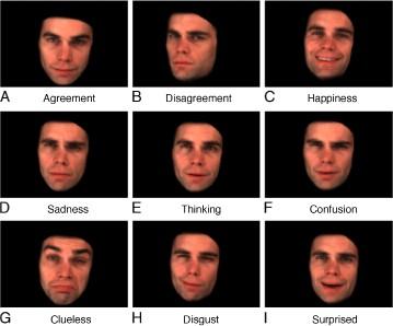
5 Groups of the facial expression muscles: I epicranial m. II- surrounding ear III- surrounding eye IV surrounding nose V- surrounding mouth
 orbit, (3) mouth, and (4) nose.")
6 Muscles of facial expression: Associated with (1) the forehead, (2) orbit, (3) mouth, and (4) nose.

7 Deep fascia exists in the regions of the parotid glands and the masseter muscles. It forms capsules around these structures. The other regions of the face have much subcutaneous tissue, but no deep fascia.

8 Characteristics of the muscles of facial expression They have bony origin. They are inserted into the skin /without the tendons/. They arrange themselves around the openings of the face as 2 groups: sphincter group & dilator group. They intermingle with each other. They are supplied by the facial nerve. Fascia covers the parotid gland and buccinator muscle only.


9 18 paired muscles participate in facial expression Major muscles involved in clinical assessment Frontalis Orbicularis oculi Zygomaticus major Orbicularis oris Lip depressors Functional elements Nasolabial fold (NLF) Smile dynamics


10 Galea aponeurotica It is interposed between the frontalis and occipitalis portions of the occipitofrontalis muscle. These muscles place the aponeurosis under tension so that deep transverse lacerations of the scalp gape widely.

11 Buccal fat pat & Buccinator muscle


12 Buccal fat pad Before After Buccal pad reduction
13 End

14 3 basic smile patterns Zygomaticus major smile Most common type (67%) Dominated by zygomaticus major and buccinator muscle Corners of the mouth elevate first Canine smile 2nd most common (31%) Mainly controlled by levator labii superioris contracting prior to zygomaticus major and buccinator Dominant upward elevation of lip followed by elevation of corners of the mouth Full-denture smile Least common (2%) Due to contraction of elevator and depressors of the lips and angles of the mouth Maxillary and mandibular teeth displayed

15 Full Denture Smile: 3% Canine Smile: 31% Zygomaticus major smile: 66%
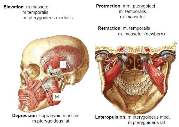
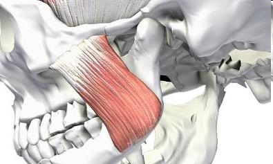
16 Muscles of mastication Arise from the temporal & infratemporal fossae Are inserted to the mandible Are supplied by mandibular nerve All of them elevate the mandible except the lateral pterygoid which lowers it by pulling forward on condylar process All of them protrude the mandible except Temporal m. which retracts it The pterygoid mm. move the mandible from side to side


17 Masticatory muscles Masseter Origin - inferior border and medial surface of zygomatic arch Insertion - lateral surface of ramus of mandible and angle of mandible Action: elevates mandible Temporalis Origin: temporal fossa Insertion: coronoid process of mandible Action: elevates and retracts mandible Lateral pterygoid Medial pterygoid
18 MASTICATION MUSCLES
19 True Muscles of mastication Accessory Muscles of mastication ADDUCTORS jaw closing/raising All V3 innervation 1. Temporalis m. 2. Masseter m. 3. Medial pterygoid 4. Lateral pterygoid upper head: to articular disc lower head: to neck of mandibular condyle Vital for normal chewing, but not mandibular adductors/protractors Buccinator VII Digastric V3 & VII Tongue XII
20 Fasciae of the head *Muscles of the facial expression don t have the fascia, except the buccinator muscle which is covered posteriorly with the thick buccopharyngeal fascia - it blends with the fatty tissue of the cheek (anteriorly) - it fuses with the pterigomandibular raphe (posteriorly) - is continuous with the connective tissue covering of the pharyngeal muscles *Muscles of the mastication possess of the fasciae: 1) Temporal fascia (fascia temporalis) -arises above from the temporal line -covers the temporal muscle 2) Masseteric fascia (fascia masseterica) = Parotideomasseteric Fascia - is a strong layer of fascia derived from the deep cervical fascia on the human head and neck. It covers the Masseter, and is firmly connected to it. Above, this fascia is attached to the lower border of the zygomatic arch, and behind, it invests the parotid gland. -arises above from the zygomatic arch -invests the lateral surface of the masseter muscle -attaches to the mandibular border (below) the mandibular branch (anteriorly+posteriorly) -is connected with the parotid fascia (fascia parotidea) which covers the salivary

21 Fascia of the Face Superficial fascia is copious and loose however, there is no discrete layer of deep fascia of the face except

22 Capsule of the parotid gland The parotid gland is enclosed within a welldefined capsule which extension of the investing fascia of the neck. The superficial capsule is thick and unyielding and tightly affixed to the underlying glandular tissue by numerous fibrous septa. The deep capsule id quite thin and easily disrupted.

23 Parallel to the carotid sheath and along its medial aspect the prevertebral fascia gives off a thin lamina, the buccopharyngeal fascia, which closely invests the constrictor muscles of the pha rynx and is continued forward from the constrictor pharyngis superior onto the buccinator. It is attached to the prevertebral layer by loose connective tissue only, and thus an easily distended space, the retropharyngeal space, is found between them.
24 1) Temporal fascia (fascia temporalis) *arises above from the temporal line *covers the temporal muscle *below the temporal line it attaches to the zygomatic arch and separates into 2 layers: 1. superficial / fuses with the external surface of the zygomatic arch / 2. deep / fuses with the internal surface of the zygomatic arch / Between them the interfascial space is formed /is filled with fatty tissue/

25 Temporal fascia

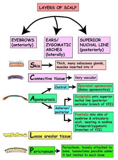
26 Topography of the head The soft tissue envelope of the cranial vault is called the scalp. The scalp extends from the external occipital protuberance and superior nuchal lines to the supraorbital margins. The scalp consists of 5 layers: - the skin, - connective tissue, - epicranial aponeurosis, - loose areolar tissue, - pericranium. Coronal section of skull, scalp & meninges The first 3 layers are bound together as a single unit. This single unit can move along the loose areolar tissue over the pericranium, which is adherent to the calvarium.
27
 haemorrhage is a serious.")
 subperiostal between the periosteum & external plate of the skull cap bones.")
28 Topography of the head I. Spaces of the skull cap a) subaponeurotic between the epicranian aponeurosis and periostium of the skull cap bones. Contents: loose connective tissue. The loose areolar tissue provides a potential subaponeurotic space that allows fluids and blood to pass from the scalp to the upper eyelids. Subgaleal (subaponeurotic) haemorrhage is a serious. Neonatal caregivers should be able to recognize the occurrence of this type of bleeding and to institute appropriate forms of management. b) subperiostal between the periosteum & external plate of the skull cap bones. It is placed at the limits of each cranial bone because the periosteum of the region of the skull sutures is fused with the bones. Along the suture lines, the pericranium becomes continuous with the endosteum. A subperiosteal hematoma, therefore, forms in the shape of the skull bones.


29 The skin has the greatest concentration of hair and sebaceous glands The superficial fascia is dense connective tissue that binds the skin strongly to the underlying galea aponeurotica The vasculature of the scalp runs primarily in it. lt is rich and widely anastomnotic Wounds of the scalp bleed profusely but heal well.


30 Subaponeurotic space (loose connective tissue) Extracranial hematoma, the result of bleeding in the subaponeurotic space, can extend over the cranium. lt can extend posteriorly, to the superior nuchal line; anteriorly, into the eyelids to produce the black eye ; and lateraliy, to the temporal line. The loose connective tissue layer provides the plane of separation of the scalp from the calvaria or for the surgeon elevating the scalp from the periosteum. Contains a rich network of deep arteries and veins. Therefore, this layer has been called the dangerous area. Infarction may spread to the substance of the bones, to venous channels within the cranial cavity, or to the brain.
 subaponeurotic between the fascia & şi temporal muscle. Contents: a part of the fat body of the cheek & vein of the m.")
31 Topography of the head II. Spaces of the temporal region a) interaponeurotic between the superficial & deep laminae of the temporal fascia which covers the temporal muscle. Contents: ţesutul adipos prin care trece artera şi vena temporală medie. b) subaponeurotic between the fascia & şi temporal muscle. Contents: a part of the fat body of the cheek & vein of the m.temporal muscle. c) deep temporal between the deep fascicles of the temporal muscle & periosteum. Downward this space is open, it is continuous with the temporo-pterygoid & suprapterygoid. Contents: deep temporal blood vessels and nerves.
32 The Temporal space is divided into two compartments by the temporalis muscle. The deep temporal space is the potential space that lie between the lateral surface of the skull and medial surface of the temporalis muscle. Whereas the superficial temporal space lies between the temporalis muscle and its overlying fascia. The deep and superficial temporal spaces are involved indirectly as a result of an infection spreading superiorly from the inferior pterygomandibular and submasseteric spaces, respectively.
 Space of the parotid gland Contents: parotid gland, facial & auriculotemporal")
33 Topography of the head III. Spaces of the lateral part of the face a) Space of the parotid gland Contents: parotid gland, facial & auriculotemporal nerves, external carotid artery with its branches, retromandibular vein & parotid lymph nodes.
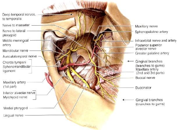
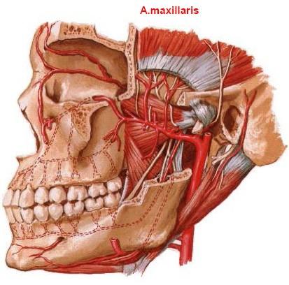
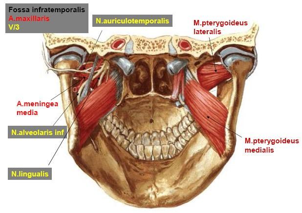
34 b) temporopterygoid between the lateral pterygoid & the temporal muscles. Contents: maxillary artery & the pterygoid venous plexus. Upward this space is continuous with the deep temporal space.

35 Masseter space c) masseteromandibular between the maseter m. & the branch of the mandible. Contents: connective tissue whith the neurovascular fascicle of the maseter muscle. Upward this space communicates with the temporopterygoid space under the zigomatic arch. mandible


36 Interpterygoid space d) interpterygoid between the lateral & medial pterygoid the temporal muscles Contents: inferior alveolar nerve with its branches, inferior alveolar artery & deep part of the pterigoid venous plexus.
37 e) Suprapterygoid space It lies between the superior head of the lateral pterygoid m. & infratemporală surface of the greater wing of the sphenoid bone. Contents: masseteric & deep temporal arteries & nerves. Communicates with the spaces : interpterygoid, temporo pterigoid & deep temporal.
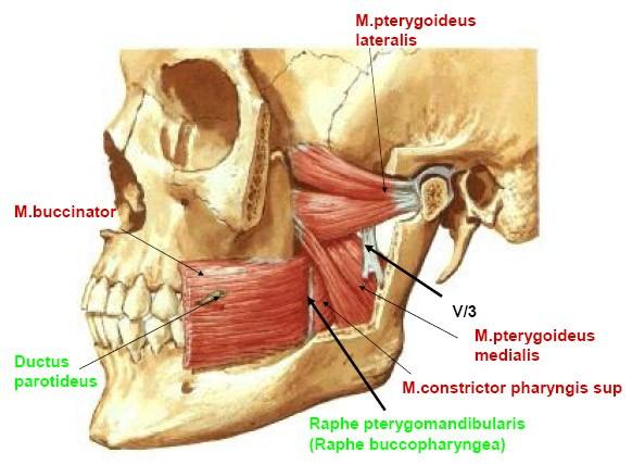
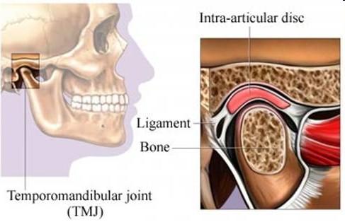
38 f) Pterygomandibular space It lies between the internal surface of the mandibular branch & medial pterigoid m. Contents: inferior alveolar nerve & homonymous artery & vein. Communications: interpterigoid space, temporo-pterigoid space & space of the bucal fat body.
39

40 Lies between ramus of mandible and medial pterygoid

41 g) Pterygopalatine fossa h) space of the bucal fat body in front of the anterior border of the buccinator m.. It gives the processes which enter: the subaponeurotic space of the temporal region, pterygopalatine fossa & pterygomandibular space. Contents: maxillary artery with its branches, pterygopalatine ganglion & venous plexus.
42

43 h) space of the bucal fat body It lies in front of the anterior border of the buccinator m.. It gives the processes which enter: the subaponeurotic space of the temporal region, pterygopalatine fossa & pterygomandibula r space.
44
45
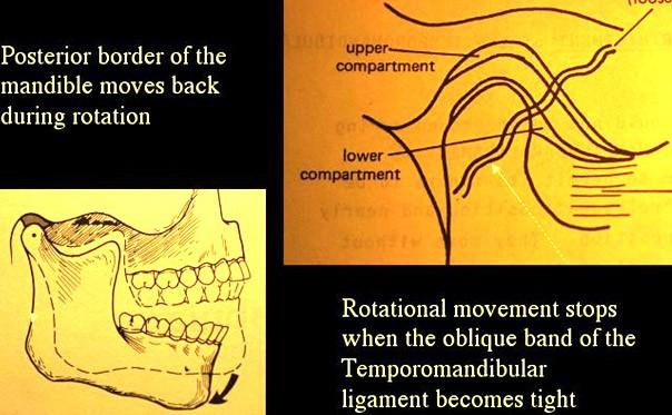
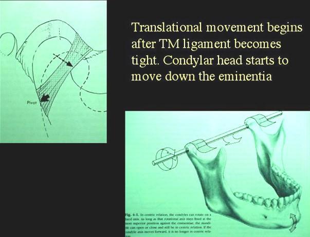
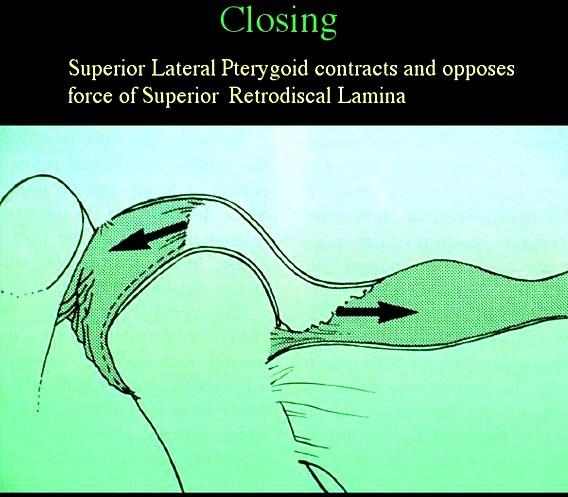
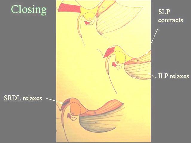
46
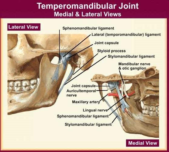
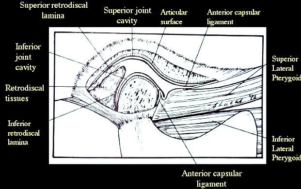
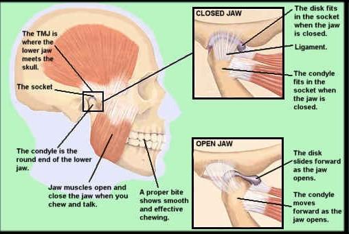
47 Normal TMJ Function When the mouth opens, two distinct motions occur at the joint. The first motion is rotation around a horizontal axis through the condylar heads. The second motion is translation. The condyle and meniscus move together anteriorly beneath the articular eminence. In the closed mouth position, the thick posterior band of the meniscus lies immediately above the condyle. As the condyle translates forward, the thinner intermediate zone of the meniscus becomes the articulating surface between the condyle and the articular eminence. When the mouth is fully open, the condyle may lie beneath the anterior band of the meniscus.

48 Functions of TMJ
49
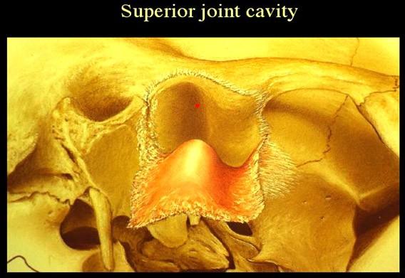
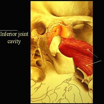
50 Internal structures of the TMJ 1. Articular disc -biconcave, thicker on outside edges - no vascular or nervous supply except at edges - made of fibrous tissue 2. Joint cavities - TMJ is a compound joint a. inferior - below the disc b. superior - above the disc 3. Retrodiscal tissue - superior retrodiscal lamina attaches to glenoid fossa and pulls disc back when mandible opens
51 Internal structure Of TMJ
52
53
54

55
56

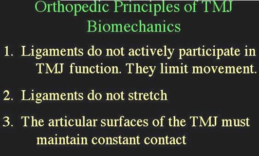
57 It acts as unique joint: - complex & combined
58 Biomechanics of the TMJ Two distinct joint systems: 1. Rotational movement - inferior joint cavity 2. Translational movement - superior joint cavity
59

60
61
62
63

64
65
66
67
68

69
70
71
72
73

74 Sublingual space Between mucous membrane of floor of mouth and mylohyoid and hyoglossus

75 The skin of face is very thin and connected to the facial bones by loose connective tissue. There is no deep fascia. The facial muscles lie in this connective tissue.
76 END
Tikrit University College of Dentistry Dr.Ban I.S. head & neck anatomy 2 nd y.
 Lec [3]/The scalp The scalp extends from the supraorbital margins anteriorly to the nuchal lines at the back of the skull and down to the temporal lines at the sides. The forehead, from eyebrows to hairline,
Head and Face Anatomy
 Head and Face Anatomy Epicranial region The Scalp The soft tissue that covers the vault of skull. Extends from supraorbital margin to superior nuchal line. Layers of the scalp S C A L P = skin = connective
The Skull and Temporomandibular joint II Prof. Abdulameer Al-Nuaimi. E. mail:
 The Skull and Temporomandibular joint II Prof. Abdulameer Al-Nuaimi E-mail: a.al-nuaimi@sheffield.ac.uk E. mail: abdulameerh@yahoo.com Temporal fossa The temporal fossa is a depression on the temporal
Tikrit University collage of dentistry Dr.Ban I.S. head & neck anatomy 2 nd y. Lec [5] / Temporal fossa :
![Tikrit University collage of dentistry Dr.Ban I.S. head & neck anatomy 2 nd y. Lec [5] / Temporal fossa :](/thumbs/88/115294566.jpg "Tikrit University collage of dentistry Dr.Ban I.S. head & neck anatomy 2 nd y. Lec [5] / Temporal fossa :") Lec [5] / Temporal fossa : Borders of the Temporal Fossa: Superior: Superior temporal line. Inferior: gap between zygomatic arch and infratemporal crest of sphenoid bone. Anterior: Frontal process of the
Lec [5] / Temporal fossa : Borders of the Temporal Fossa: Superior: Superior temporal line. Inferior: gap between zygomatic arch and infratemporal crest of sphenoid bone. Anterior: Frontal process of the
THIEME. Scalp and Superficial Temporal Region
 CHAPTER 2 Scalp and Superficial Temporal Region Scalp Learning Objectives At the end of the dissection of the scalp, you should be able to identify, understand and correlate the clinical aspects: Layers
Infratemporal fossa: Tikrit University college of Dentistry Dr.Ban I.S. head & neck Anatomy 2 nd y.
 Infratemporal fossa: This is a space lying beneath the base of the skull between the lateral wall of the pharynx and the ramus of the mandible. It is also referred to as the parapharyngeal or lateral pharyngeal
Parotid Gland. Parotid Gland. Largest of 3 paired salivary glands (submandibular; sublingual) Ramus of Mandible. Medial pterygoid.
 Ramus of Mandible. Medial pterygoid.") Parotid region Parotid Gland Largest of 3 paired salivary glands (submandibular; sublingual) Ramus of Mandible Medial pterygoid Cross section of mandible Masseter D S SCM Parotid Gland Mastoid Process
Parotid region Parotid Gland Largest of 3 paired salivary glands (submandibular; sublingual) Ramus of Mandible Medial pterygoid Cross section of mandible Masseter D S SCM Parotid Gland Mastoid Process
3. The Jaw and Related Structures
 Overview and objectives of this dissection 3. The Jaw and Related Structures The goal of this dissection is to observe the muscles of jaw raising. You will also have the opportunity to observe several
The SCALP. Prof. Dr. Muhammad Imran Qureshi
 The SCALP By Prof. Dr. Muhammad Imran Qureshi The SCALP includes FIVE layers external to the Calvaria. These are: S: Skin & Superficial Fascia C: Connective Tissue A: Aponeurosis (Epicranial) L: Loose
Temporal region. temporal & infratemporal fossae. Zhou Hong Ying Dept. of Anatomy
 Temporal region temporal & infratemporal fossae Zhou Hong Ying Dept. of Anatomy Temporal region is divided by zygomatic arch into temporal & infratemporal fossae. Temporal Fossa Infratemporal fossa Temporal
Face. Definition: The area between the two ears and from the chin to the eye brows. The muscles of the face
 Face Definition: The area between the two ears and from the chin to the eye brows. The muscles of the face The muscle of facial expression (include the muscle of the face and the scalp). All are derived
3-Deep fascia: is absent (except over the parotid gland & buccopharngeal fascia covering the buccinator muscle)
") The Face 1-Skin of the Face The skin of the face is: Elastic Vascular (bleed profusely however heal rapidly) Rich in sweat and sebaceous glands (can cause acne in adults) It is connected to the underlying
The Face 1-Skin of the Face The skin of the face is: Elastic Vascular (bleed profusely however heal rapidly) Rich in sweat and sebaceous glands (can cause acne in adults) It is connected to the underlying
Temporomandibular Joint. Dr Noman ullah wazir
 Temporomandibular Joint Dr Noman ullah wazir Type of Joint TMJ is a Synovial joint between : The condylar head of the mandible. The mandibular fossa of squamous part of temporal bone. The joint cavity
Oral cavity landmarks
 By: Dr. Ahmed Rabah Oral cavity landmarks The knowledge of oral anatomy and physiology will help the operator and provides enough landmarks to act as positive guide during denture construction. This subject
Temporal fossa Infratemporal fossa Pterygopalatine fossa Terminal branches of external carotid artery Pterygoid venous plexus
 Outline of content Temporal fossa Infratemporal fossa Pterygopalatine fossa Terminal branches of external carotid artery Pterygoid venous plexus Boundary Content Communication Mandibular division of trigeminal
The Neck the lower margin of the mandible above the suprasternal notch and the upper border of the clavicle
 The Neck is the region of the body that lies between the lower margin of the mandible above and the suprasternal notch and the upper border of the clavicle below Nerves of the neck Cervical Plexus Is formed
Parotid Gland, Temporomandibular Joint and Infratemporal Fossa
 M1 - Anatomy Parotid Gland, Temporomandibular Joint and Infratemporal Fossa Jeff Dupree Sanger 9-057 jldupree@vcu.edu Parotid gland: wraps around the mandible positioned between the mandible and the sphenoid
Dr.Ban I.S. head & neck anatomy 2 nd y. جامعة تكريت كلية طب االسنان مادة التشريح املرحلة الثانية أ.م.د. بان امساعيل صديق 6102/6102
 جامعة تكريت كلية طب االسنان مادة التشريح املرحلة الثانية أ.م.د. بان امساعيل صديق 6102/6102 The scalp The scalp extends from the supraorbital margins anteriorly to the nuchal lines at the back of the skull
Anatomy and Physiology. Bones, Sutures, Teeth, Processes and Foramina of the Human Skull
 Anatomy and Physiology Chapter 6 DRO Bones, Sutures, Teeth, Processes and Foramina of the Human Skull Name: Period: Bones of the Human Skull Bones of the Cranium: Frontal bone: forms the forehead and the
Veins of the Face and the Neck
 Veins of the Face and the Neck Facial Vein The facial vein is formed at the medial angle of the eye by the union of the supraorbital and supratrochlear veins. connected through the ophthalmic veins with
Superior View of the Skull (Norma Verticalis) Anteriorly the frontal bone articulates with the two parietal bones AT THE CORONAL SUTURE
 Anteriorly the frontal bone articulates with the two parietal bones AT THE CORONAL SUTURE") Superior View of the Skull (Norma Verticalis) Anteriorly the frontal bone articulates with the two parietal bones AT THE CORONAL SUTURE 1 The two parietal bones articulate in the midline AT THE SAGITTAL
Superior View of the Skull (Norma Verticalis) Anteriorly the frontal bone articulates with the two parietal bones AT THE CORONAL SUTURE 1 The two parietal bones articulate in the midline AT THE SAGITTAL
Bones Ethmoid bone Inferior nasal concha Lacrimal bone Maxilla Nasal bone Palatine bone Vomer Zygomatic bone Mandible
 splanchnocranium - Consists of part of skull that is derived from branchial arches - The facial bones are the bones of the anterior and lower human skull Bones Ethmoid bone Inferior nasal concha Lacrimal
Dr.Ban I.S. head & neck anatomy 2 nd y. جامعة تكريت كلية طب االسنان املرحلة الثانية أ.م.د. بان امساعيل صديق 6102/6102
 جامعة تكريت كلية طب االسنان التشريح مادة املرحلة الثانية أ.م.د. بان امساعيل صديق 6102/6102 Parotid region The part of the face in front of the ear and below the zygomatic arch is the parotid region. The
PTERYGOPALATINE FOSSA
 PTERYGOPALATINE FOSSA Outline Anatomical Structure and Boundaries Foramina and Communications with other spaces and cavities Contents Pterygopalatine Ganglion Especial emphasis on certain arteries and
Upper arch. 1Prosthodontics. Dr.Bassam Ali Al-Turaihi. Basic anatomy & & landmark of denture & mouth
 1Prosthodontics Lecture 2 Dr.Bassam Ali Al-Turaihi Basic anatomy & & landmark of denture & mouth Upper arch Palatine process of maxilla: it form the anterior three quarter of the hard palate. Horizontal
Anatomy & Physiology B. Chapter 6: Muscles
 Anatomy & Physiology B Chapter 6: Muscles Warm-up What are the three types of muscle tissue? Where are each located? Which are voluntary and which are involuntary? Which are striated which are unstriated?
Bony orbit Roof The orbital plate of the frontal bone Lateral wall: the zygomatic bone and the greater wing of the sphenoid
 Bony orbit Roof: Formed by: The orbital plate of the frontal bone, which separates the orbital cavity from the anterior cranial fossa and the frontal lobe of the cerebral hemisphere Lateral wall: Formed
Lec [8]: Mandibular nerve:
![Lec [8]: Mandibular nerve:](/thumbs/94/121295776.jpg "Lec [8]: Mandibular nerve:") Lec [8]: Mandibular nerve: The mandibular branch from the trigeminal ganglion lies in the middle cranial fossa lateral to the cavernous sinus. With the motor root of the trigeminal nerve [motor roots lies
Lec [8]: Mandibular nerve: The mandibular branch from the trigeminal ganglion lies in the middle cranial fossa lateral to the cavernous sinus. With the motor root of the trigeminal nerve [motor roots lies
ANTERIOR CERVICAL TRIANGLE (Fig. 2.1 )
") 2 Neck Anatomy ANTERIOR CERVICAL TRIANGLE (Fig. 2.1 ) The boundaries are: Lateral: sternocleidomastoid muscle Superior: inferior border of the mandible Medial: anterior midline of the neck This large triangle
2 Neck Anatomy ANTERIOR CERVICAL TRIANGLE (Fig. 2.1 ) The boundaries are: Lateral: sternocleidomastoid muscle Superior: inferior border of the mandible Medial: anterior midline of the neck This large triangle
Dr. Sami Zaqout Faculty of Medicine IUG
 Auricle External Ear External auditory meatus The Ear Middle Ear (Tympanic Cavity) Auditory ossicles Internal Ear (Labyrinth) Bony labyrinth Membranous labyrinth External Ear Auricle External auditory
The sebaceous glands (glands of Zeis) open directly into the eyelash follicles, ciliary glands (glands of Moll) are modified sweat glands that open
 open directly into the eyelash follicles, ciliary glands (glands of Moll) are modified sweat glands that open") The Orbital Region The orbits are a pair of bony cavities that contain the eyeballs; their associated muscles, nerves, vessels, and fat; and most of the lacrimal apparatus upper eyelid is larger and more
The Orbital Region The orbits are a pair of bony cavities that contain the eyeballs; their associated muscles, nerves, vessels, and fat; and most of the lacrimal apparatus upper eyelid is larger and more
OPEN ACCESS ATLAS OF OTOLARYNGOLOGY, HEAD & NECK OPERATIVE SURGERY
 OPEN ACCESS ATLAS OF OTOLARYNGOLOGY, HEAD & NECK OPERATIVE SURGERY BUCCINATOR MYOMUCOSAL FLAP The Buccinator Myomucosal Flap is an axial flap, based on the facial and/or buccal arteries. It is a flexible
Muscles of mastication [part 1]
![Muscles of mastication [part 1]](/thumbs/76/73586850.jpg "Muscles of mastication [part 1]") Muscles of mastication [part 1] In this lecture well have the muscles of mastication, neuromuscular function, and its relationship to the occlusion morphology. The fourth determinant of occlusion is the
Muscles of mastication [part 1] In this lecture well have the muscles of mastication, neuromuscular function, and its relationship to the occlusion morphology. The fourth determinant of occlusion is the
SCHOOL OF ANATOMICAL SCIENCES Mock Run Questions. 4 May 2012
 SCHOOL OF ANATOMICAL SCIENCES Mock Run Questions 4 May 2012 1. With regard to the muscles of the neck: a. the platysma muscle is supplied by the accessory nerve. b. the stylohyoid muscle is supplied by
Lab Activity 11: Group I
 Lab Activity 11: Group I Muscles Martini Chapter 11 Portland Community College BI 231 Origin and Insertion Origin: The place where the fixed end attaches to a bone, cartilage, or connective tissue. Insertion:
Face and Scalp 解剖學科鄭授德
 Face and Scalp 解剖學科鄭授德 本教材之圖片取自於 1 Gray s Anatomy for Students, 3rd ed, 2015, by Drake, Vogl, and Mitchell 2 Clinically Oriented Anatomy, 7th ed, 2014, by Moore, Dalley, and Agur 3 Clinically Oriented
Basic Anatomy and Physiology of the Lips and Oral Cavity. Dr. Faghih
 Basic Anatomy and Physiology of the Lips and Oral Cavity Dr. Faghih It is divided into seven specific subsites : 1. Lips 2. dentoalveolar ridges 3. oral tongue 4. retromolar trigone 5. floor of mouth 6.
Introduction to Occlusion and Mechanics of Mandibular Movement
 Introduction to Occlusion and Mechanics of Mandibular Movement Dr. Pauline Hayes Garrett Department of Endodontics, Prosthodontics, and Operative Dentistry University of Maryland, Baltimore Assigned reading
Temporomandibular Joint
 6 Temporomandibular Joint Most human bones are joined with one another. These connections of bones to each other are termed articulations or joints. Some joints are immovable as in the articulations of
Prevertebral Region, Pharynx and Soft Palate
 Unit 20: Prevertebral Region, Pharynx and Soft Palate Dissection Instructions: Step1 Step 2 Step 1: Insert your fingers posterior to the sternocleidomastoid muscle, vagus nerve, internal jugular vein,
Anatomy: head and Neck (6 questions) 1. Prevertebral Flexor Musculature (lying in front of the vertebrae) include all, EXCEPT: Longus Colli.
 1. Prevertebral Flexor Musculature (lying in front of the vertebrae) include all, EXCEPT: Longus Colli.") Anatomy: head and Neck (6 questions) 1. Prevertebral Flexor Musculature (lying in front of the vertebrae) include all, EXCEPT: Longus Colli. Rectus Capitis Anterior. Rectus Capitis Lateralis. Rectus Capitis
Anatomy: head and Neck (6 questions) 1. Prevertebral Flexor Musculature (lying in front of the vertebrae) include all, EXCEPT: Longus Colli. Rectus Capitis Anterior. Rectus Capitis Lateralis. Rectus Capitis
Anatomy and Physiology II. Review Spine and Neck
 Anatomy and Physiology II Review Spine and Neck Spine regions How many cervical vertibrae are there? 7 The curvature is the cervical region posterior? Concave posterior How many thoracic? And curvature?
University of Palestine. Midterm Exam 2013/2014 Total Grade:
 Course No: DNTS2208 Course Title: Head and Neck Anatomy Date: 09/11/2013 No. of Questions: (50) Time: 1hour Using Calculator (No) University of Palestine Midterm Exam 2013/2014 Total Grade: Instructor
Anatomy and physiology of Temporomandibular Joint
 Anatomy and physiology of Temporomandibular Joint Temporomandibular joint (TMJ): It is the articulation of the condyle of the mandible, and the inter-articular disc; with the mandibular fossa (glenoid
Mohammad Hisham Al-Mohtaseb. Lina Mansour. Reyad Jabiri. 0 P a g e
 2 Mohammad Hisham Al-Mohtaseb Lina Mansour Reyad Jabiri 0 P a g e This is only correction for the last year sheet according to our record. If you already studied this sheet just read the yellow notes which
Muscles of the Face, Head, and Neck
 Muscles of the Face, Head, and Neck 1 How Muscles Are Named Many muscles named using such features as Location Function Shape Direction of fibers Number of heads or divisions Points of attachment Size
Structure Location Function
 Frontal Bone Cranium forms the forehead and roof of the orbits Occipital Bone Cranium forms posterior and inferior portions of the cranium Temporal Bone Cranium inferior to the parietal bone forms the
Stracture The scalp consists of five layers,the 1st three of which are intimately bound together and move as a unit.to assist one in memorizing the
 Anatomy of Scalp Stracture The scalp consists of five layers,the 1st three of which are intimately bound together and move as a unit.to assist one in memorizing the names of the five layers of the scalp,use
The Pharynx. Dr. Nabil Khouri MD. MSc, Ph.D
 The Pharynx Dr. Nabil Khouri MD. MSc, Ph.D Introduction The pharynx is the Musculo-fascial halfcylinder that links the oral and nasal cavities in the head to the larynx and esophagus in the neck Common
Lips and labial mucosa
 Lips and labial mucosa External portion of the lips: the vermilion border and the skin Vermilion border : the exposed red portion of the lip, covered by mucous membrane, no mucous glands Boundary: the
Maxilla, ORBIT and infratemporal fossa. Neophytos C Demetriades MD, DDS, MSc Associate professor European University of Cyprus School of Medicine
 Maxilla, ORBIT and infratemporal fossa Neophytos C Demetriades MD, DDS, MSc Associate professor European University of Cyprus School of Medicine MAXILLA Superior, middle, and inferior meatus Frontal sinus
Human Anatomy and Physiology - Problem Drill 07: The Skeletal System Axial Skeleton
 Human Anatomy and Physiology - Problem Drill 07: The Skeletal System Axial Skeleton Question No. 1 of 10 Which of the following statements about the axial skeleton is correct? Question #01 A. The axial
The Ear The ear consists of : 1-THE EXTERNAL EAR 2-THE MIDDLE EAR, OR TYMPANIC CAVITY 3-THE INTERNAL EAR, OR LABYRINTH 1-THE EXTERNAL EAR.
 The Ear The ear consists of : 1-THE EXTERNAL EAR 2-THE MIDDLE EAR, OR TYMPANIC CAVITY 3-THE INTERNAL EAR, OR LABYRINTH 1-THE EXTERNAL EAR Made of A-AURICLE B-EXTERNAL AUDITORY MEATUS A-AURICLE It consists
be very thin and variable. Facial nerve branches that exit the parotid gland are deep to the SMAS.
 The Superficial musculoaponeurotic system (SMAS) fascia is a fanlike fascia that envelops the face and provides a suspensory sheet which distributes forces of facial expression.. The SMAS is continuous
The orbit-1. Dr. Heba Kalbouneh Assistant Professor of Anatomy and Histology
 The orbit-1 Dr. Heba Kalbouneh Assistant Professor of Anatomy and Histology Orbital plate of frontal bone Orbital plate of ethmoid bone Lesser wing of sphenoid Greater wing of sphenoid Lacrimal bone Orbital
Chapter 7 Part A The Skeleton
 Chapter 7 Part A The Skeleton Why This Matters Understanding the anatomy of the skeleton enables you to anticipate problems such as pelvic dimensions that may affect labor and delivery The Skeleton The
Oral cavity : consist of two parts: the oral vestibule and the oral cavity proper. Oral vestibule : is slit like space between.
 Oral cavity Oral cavity : consist of two parts: the oral vestibule and the oral cavity proper Oral vestibule : is slit like space between the teeth, buccal gingiva, lips, and cheeks 1 Oral cavity Oral
University of Palestine. Midterm Exam 2013/2014 Total Grade:
 [ Course No: DNTS2208 Course Title: Head and Neck Anatomy Date: 17/11/1024 No. of Questions: (52) Time: 2hours Using Calculator (No) University of Palestine Midterm Exam 2013/2014 Total Grade: Instructor
Subdivided into Vestibule & Oral cavity proper
 Extends from the lips to the oropharyngeal isthmus The oropharyngeal isthmus: Is the junction of mouth and pharynx. Is bounded: Above by the soft palate and the palatoglossal folds Below by the dorsum
Anatomic Relations Summary. Done by: Sohayyla Yasin Dababseh
 Anatomic Relations Summary Done by: Sohayyla Yasin Dababseh Anatomic Relations Lecture 1 Part-1 - The medial wall of the nose is the septum. - The vestibule lies directly inside the nostrils (Nares). -
FACE CN V & VII PAROTID GLAND. Jacek Baj, MD, PhD Department of Human Anatomy
 FACE CN V & VII PAROTID GLAND Jacek Baj, MD, PhD Department of Human Anatomy THE FACE THE SCALP The scalp is composed of five layers: Skin ConnecAve Assue Aponeurosis Loose connecave Assue Pericranium
Neck-2. Dr. Heba Kalbouneh Associate Professor of Anatomy and Histology
 Neck-2 ` Dr. Heba Kalbouneh Associate Professor of Anatomy and Histology Triangles of the neck Side of the neck Midline Lower border of mandible Line between angle of mandible and mastoid Superior nuchal
The Role of Muscles in Movement
 The Role of Muscles in Movement Muscles can t push, they can only pull as they contract, so most often body movements are the result of the activity of pairs or teams of muscles acting together or against
Anatomy of the Trigeminal Nerve
 19 Anatomy of the Trigeminal Nerve.1 Introduction 0. The Central Part of the Trigeminal Nerve 1..1 Origin 1.. Trigeminal Nuclei.3 The Peripheral Part of the Trigeminal Nerve 4.3.1 Ophthalmic Nerve 4.3.
Prosthodontics Dr.Yassen H.
 Prosthodontics Dr.Yassen H. Lecture -2- Anatomy & Physiology Related to Prosthodontics (Myology) Muscles are divided or classified into: 1. Muscles of facial expression. 2. Suprahyoid muscles. 3. Infrahyoid
Learning Outcomes. The Carotid 20/02/2013. Scalp, Face, Parotid. Layers of the Scalp. The Parotid Gland. The Scalp. The Carotid The Facial Artery
 Learning Outcomes The Scalp Layers of the Scalp Bleeding from the Scalp The Carotid The Facial Artery Major Muscles of the Face and Jaw(s) Muscles of Mastication Muscles of Facial Expression The Parotid
INTRODUCTION: ANATOMY UNDERLYING CLINICAL TESTS OF CRANIAL NERVES
 INTRODUCTION: ANATOMY UNDERLYING CLINICAL TESTS OF CRANIAL NERVES CRANIAL NERVE I - OLFACTORY I - OLFACTORY NERVE - SMELL TEST: SMELL ODORS (note: not ammonia; pain in nasal cavity CN5 DAMAGE: LOSS OF
Alexander C Vlantis. Selective Neck Dissection 33
 05 Modified Radical Neck Dissection Type II Alexander C Vlantis Selective Neck Dissection 33 Modified Radical Neck Dissection Type II INCISION Various incisions can be used for a neck dissection. The incision
Dr.Ban I.S. head & neck anatomy 2 nd y. جامعة تكريت كلية طب االسنان املرحلة الثانية
 جامعة تكريت كلية طب االسنان التشريح مادة املرحلة الثانية أ.م.د. بان امساعيل صديق 6102-6102 1 The Palate The palate forms the roof of the mouth and the floor of the nasal cavity. It is divided into two
Bones of the skull & face
 Bones of the skull & face Cranium= brain case or helmet Copyright The McGraw-Hill Companies, Inc. Permission required for reproduction or display. The cranium is composed of eight bones : frontal Occipital
The Neck. BY: Lina Abdullah & Rahaf Jreisat
 The Neck BY: Lina Abdullah & Rahaf Jreisat Boundaries of the Neck: generally from base of the skull to root of the neck Superior margin :From superior nuchal line of occipital bone up to mastoid process
Anatomy of Oral Cavity DR. MAAN AL-ABBASI
 Anatomy of Oral Cavity DR. MAAN AL-ABBASI By the end of this lecture you should be able to: 1. Differentiate different parts of the oral cavity 2. Describe the blood and nerve supply of mucosa and muscles
-Ibrahim Al-Naser. -Dr Al- Muhtaseb. 1 P a g e
 -1 -Ibrahim Al-Naser - -Dr Al- Muhtaseb 1 P a g e The Digestive System The doctor started the lecture by talking about the class rules. The GI system is an organ system, it is divided into: The Alimentary
The muscular system I Muscles of the head neck and trunk
 The muscular system I Muscles of the head neck and trunk Dr. Nabil Khouri Dr. Nabil Khouri MD MSc, PhD Interactions of Skeletal Muscles Skeletal muscles work together or in opposition Muscles only pull
Module 6 - The Muscular System Introduction to the Muscular System and Muscles of the Head, Neck and Shoulder
 Module 6 - The Muscular System Introduction to the Muscular System and Muscles of the Head, Neck and Shoulder There will be three modules to cover the muscle anatomy of the body. The first module will
This lab activity is aligned with Visible Body s Anatomy and Physiology app. Learn more at visiblebody.com/professors
 1 This lab activity is aligned with Visible Body s Anatomy and Physiology app. Learn more at visiblebody.com/professors 2 PRE-LAB EXERCISES A. Watch the video 13.1 Muscular System Overview and observe
SKULL AS A WHOLE + ANTERIOR CRANIAL FOSSA
 SKULL AS A WHOLE + ANTERIOR CRANIAL FOSSA LEARNING OBJECTIVES At the end of this lecture, the student should be able to know: Parts of skeleton (axial and appendicular) Parts of skull Sutures of skull
Trigeminal Nerve Anatomy. Dr. Mohamed Rahil Ali
 Trigeminal Nerve Anatomy Dr. Mohamed Rahil Ali Trigeminal nerve Largest cranial nerve Mixed nerve Small motor root and large sensory root Motor root Nucleus of motor root present in the pons and medulla
MAXILLA, ORBIT & PTERYGOPALATINE FOSSA. Neophytos C Demetriades MD, DDS, MSc Associate professor European University of Cyprus School of Medicine
 MAXILLA, ORBIT & PTERYGOPALATINE FOSSA Neophytos C Demetriades MD, DDS, MSc Associate professor European University of Cyprus School of Medicine Maxilla MAXILLA Superior, middle, and inferior meatus Frontal
Figure (2-6): Labial frenum and labial notch.
: Labial frenum and labial notch.") The anatomy of the edentulous ridge in the maxilla and mandible is very important for the design of a complete denture. The consistency of the mucosa and architecture of the underlying bone is different
The anatomy of the edentulous ridge in the maxilla and mandible is very important for the design of a complete denture. The consistency of the mucosa and architecture of the underlying bone is different
Axial Muscles of the Head, Neck, and Back
 OpenStax-CNX module: m46484 1 Axial Muscles of the Head, Neck, and Back OpenStax College This work is produced by OpenStax-CNX and licensed under the Creative Commons Attribution License 4.0 By the end
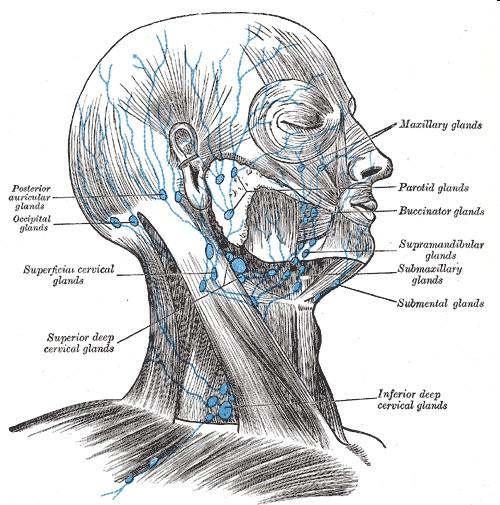
Lecture 07. Lymphatic's of Head & Neck. By: Dr Farooq Amanullah Khan PMC
 Lecture 07 Lymphatic's of Head & Neck By: Dr Farooq Amanullah Khan PMC Dated: 28.11.2017 Lymphatic Vessels Of the 800 lymph nodes in the human body, 300 are in the Head & neck region. The lymphatic vessels
For the following questions, indicate the letter that corresponds to the SINGLE MOST APPROPRIATE ANSWER
 GROSS ANATOMY EXAMINATION May 15, 2000 For the following questions, indicate the letter that corresponds to the SINGLE MOST APPROPRIATE ANSWER 1. Pain associated with an infection limited to the middle
2. In regards to the bones of the arm and forearm, which of the following is TRUE?
 Anatomy and Physiology Fall Exam II: Form A Name: 1. Use the following table to answer Question 1. I II III IV V The nasal bone articulates with the frontal process of the maxilla The mastoid process is
Dr.Noor Hashem Mohammad Lecture (5)
") Dr.Noor Hashem Mohammad Lecture (5) 2016-2017 If the mandible is discarded, the anterior part of this aspect of the skull is seen to be formed by the hard palate. The palatal processes of the maxillae
Dr.Noor Hashem Mohammad Lecture (5) 2016-2017 If the mandible is discarded, the anterior part of this aspect of the skull is seen to be formed by the hard palate. The palatal processes of the maxillae
Dr. Sami Zaqout, IUG Medical School
 The skull The skull is composed of several separate bones united at immobile joints called sutures. Exceptions? Frontal bone Occipital bone Vault Cranium Sphenoid bone Zygomatic bones Base Ethmoid bone
Anatomy images for MSS practical exam- 2019
 Anatomy images for MSS practical exam- 2019 Ilium Ischium Pubis Acetabulaum Iliac crest Iliac tubercle ASIS (muscle and ligament attached) AIIS (muscle attached) PSIS PIIS Ischial spine Ischial tuberosity
By : Prof Saeed Abuel Makarem & Dr.Sanaa Alshaarawi
 By : Prof Saeed Abuel Makarem & Dr.Sanaa Alshaarawi OBJECTIVES By the end of the lecture, students shouldbe able to: List the nuclei of the deep origin of the trigeminal and facial nerves in the brain
Cranial Nerve VII - Facial Nerve. The facial nerve has 3 main components with distinct functions
 Cranial Nerve VII - Facial Nerve The facial nerve has 3 main components with distinct functions Somatic motor efferent Supplies the muscles of facial expression; posterior belly of digastric muscle; stylohyoid,
ANATOMY & PHYSIOLOGY I Laboratory Version B Name Section. REVIEW SHEET Exercise 10 Axial Skeleton
 ANATOMY & PHYSIOLOGY I Laboratory Version B Name Section REVIEW SHEET Exercise 10 Axial Skeleton 1 POINT EACH. THE SKULL MULTIPLE CHOICE 1. The major components of the axial skeleton include the 7. The
Skull-2. Norma Basalis Interna Norma Basalis Externa. Dr. Heba Kalbouneh Associate Professor of Anatomy and Histology
 Skull-2 Norma Basalis Interna Norma Basalis Externa Dr. Heba Kalbouneh Associate Professor of Anatomy and Histology Norma basalis interna Base of the skull- superior view The interior of the base of the
The Scalp and Face Protocol. Julie Goodwin, BA, LMT
 The Scalp and Face Protocol Julie Goodwin, BA, LMT The Scalp and Face Protocol Julie Goodwin, BA, LMT Julie Goodwin 2014 2 Agenda Pertinent Anatomy and Physiology Treatment Planning Strokes, Techniques
Bone Practical. Labs Muscle Labs. Final Practical. Divisions of the Muscular System. Quiz format
 Bone Practical Labs 17 + 18 Muscles Wed 7/11 @ 8am 40 50 stations About half axial, half appendicular bones Disarticulated bones: Skulls, partial skulls, vertebrae, ribs, skeletons, arm bones, leg bones,
Biology 218 Human Anatomy. Adapted from Martini Human Anatomy 7th ed. Chapter 10 The Muscular System Axial Musculature
 Adapted from Martini Human Anatomy 7th ed. Chapter 10 The Muscular System Axial Musculature Introduction The skeletal muscle of the body can be subdivided into: Axial musculature Muscles that position
Neck of Condylar. Process. Anterior Border of Ramus. Mandibular. Foramen. Posterior Border of Ramus Incisive Fossa.
 Learning Outcomes The Mandible Surface Anatomy Muscle Attachments The (FOM) Muscles of the FOM The Tongue Muscles of the Tongue The Submandibular Region Submandibular Gland Sublingual Gland Lingual The
Trigeminal Nerve (V)
") Trigeminal Nerve (V) Lecture Objectives Discuss briefly how the face is developed. Follow up the course of trigeminal nerve from its point of central connections, exit and down to its target areas. Describe
Trigeminal Nerve (V) Lecture Objectives Discuss briefly how the face is developed. Follow up the course of trigeminal nerve from its point of central connections, exit and down to its target areas. Describe
REVIEW/PREVIEW OF HEAD AND NECK ANATOMY FOR ENT EXAM
 REVIEW/PREVIEW OF HEAD AND NECK ANATOMY FOR ENT EXAM - 2017 PALPATE CAROTID ARTERY: AT LEVEL OF CAROTID BIFURCATION VERTEBRAL LEVEL C4 Sternocleidomastoid Muscle INTERNAL CAROTID EXTERNAL CAROTID COMMON