Face and Scalp 解剖學科鄭授德
|
|
|
- Howard Gallagher
- 5 years ago
- Views:
Transcription
1 Face and Scalp 解剖學科鄭授德 本教材之圖片取自於 1 Gray s Anatomy for Students, 3rd ed, 2015, by Drake, Vogl, and Mitchell 2 Clinically Oriented Anatomy, 7th ed, 2014, by Moore, Dalley, and Agur 3 Clinically Oriented Anatomy, 4th ed, 1999, by Moore and Dalley 4 A Regional Atlas of the Human Body, 4th ed, 1997, by Clenente 5 Gunther von Hagens Body Worlds The Anatomical Exhibition of Real Human Bodies 4th ed, 2003
2


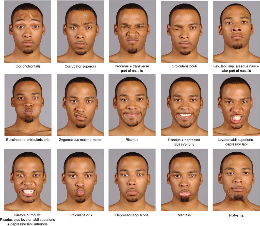
3 Face: Muscles - 1 Orbital group 1-1 Orbicularis oculi Orbital part Palpebral part 1-2 Corrugator supercilli


4 2-1 Nasalis Face: Muscles - 2 Nasal group Transverse part Alar part 2-2 Procerus 2-3 Depressor septi nasi


5 3-1 Orbicularis oris 3-2 Buccinator Face: Muscles - 3 Oral group

6 Face: Muscles - 4 Lower group oral muscles 4-1 Depressor anguli oris


7 Face: Muscles - 4 Lower group oral muscles 4-2 Depressor labii inferioris
8 Face: Muscles - 4 Lower group oral muscles 4-3 Mentalis


9 Face: Muscles - 5 Upper group oral muscles 5-1 Risorius 5-2 Zygomaticus major 5-3 Zygomaticus minor


10 Face: Muscles - 5 Upper group oral muscles 5-4 Levator labii superioris

11 Face: Muscles - 5 Upper group oral muscles 5-5 Levator labii superioris alaeque nasi 5-6 Levator anguli oris

12 Face: Muscles - 6 Other muscles 6-1 Platysma

13 Face: Muscles - 6 Other muscles 6-2 Auricular muscles
14 Face: Muscles - 6 Other muscles 6-3 Occipitofrontalis
15

16 Parotid gland Parotid duct opens into oral cavity near the 2nd upper molar tooth Structures passing through gland: 1 Facial nerve 2 External carotid artery and its branches 3 Retromandibular vein and its tributaries
 gives off postganglionic parasympathetic fibers carried by auriculotemporal nerve")
17 Parotid gland Innervation 1 Sensory innervation by auriculotemporal nerve (a br of CN V3) 2 Secretomotor fibers Branch of CN IX carries preganglionic parasympathetic fibers Postgnaglionic neurons at otic ganglion (associated with CN V3 inferior to foramen ovale) gives off postganglionic parasympathetic fibers carried by auriculotemporal nerve Parotid gland
18
19 Sensory innervation (of face) Trigeminal n (CN V) divides into Ophthalmic (CN V 1 ), maxillary (CN V 2 ) and mandibular nn (CN V 3 ) most of skin on face except: a small area covering angle and lower border of mandible and part of ear Skin on the ear by facial (CN VII), vagus (CN X), cervical nn
20
21 Ophthalmic nerve [V 1 ] Leaves skull through superior orbital fissure enters orbit brr innervating skin on face: Supraorbital and supratrochlear nerves Infratrochlear nerve Lacrimal nerve External nasal nerve
22 Maxillary nerve [V 2 ] Exits skull through foramen rotundum brr innervating skin on face: Zygomaticotemporal branch Zygomaticofacial branch Infra-orbital nerve
![Mandibular nerve [V 3 ] Exits skull through foramen ovale brr](/docs-images/96/129038538/images/23-0.jpg "innervating skin on face: Auriculotemporal nerve Buccal nerve")
23 Mandibular nerve [V 3 ] Exits skull through foramen ovale brr innervating skin on face: Auriculotemporal nerve Buccal nerve Mental nerve
24 Motor innervation (of face) mm of face, external ear, and scalp are innervated by CN VII (facial n) except: chewing muscles by brr of mandibular n
 posterior to middle ear (gives off chorda tympani n and n of stampedes) stylomastoid foramen (gives off")

25 Facial nerve Posterior cranial fossa internal acoustic meatus geniculate ganglion (gives off greater petrosal n) posterior to middle ear (gives off chorda tympani n and n of stampedes) stylomastoid foramen (gives off posterior auricular n) (gives off br for post belly of digastric and stylohyoid mm) enters parotid gland divides into temporofacial and cervicofacial brr forming parotid plexus five terminal brr emerge from parotid gland

26 5 terminal brr of facial nerve Temporal brr mm in temple, forehead, and supra-orbital area Zygomatic brr mm in infraorbital, nasal, and upper lip areas Buccal brr mm in cheek, upper lip, and corner of mouth Marginal mandibular brr mm of lower lip and chin Cervical brr platysma
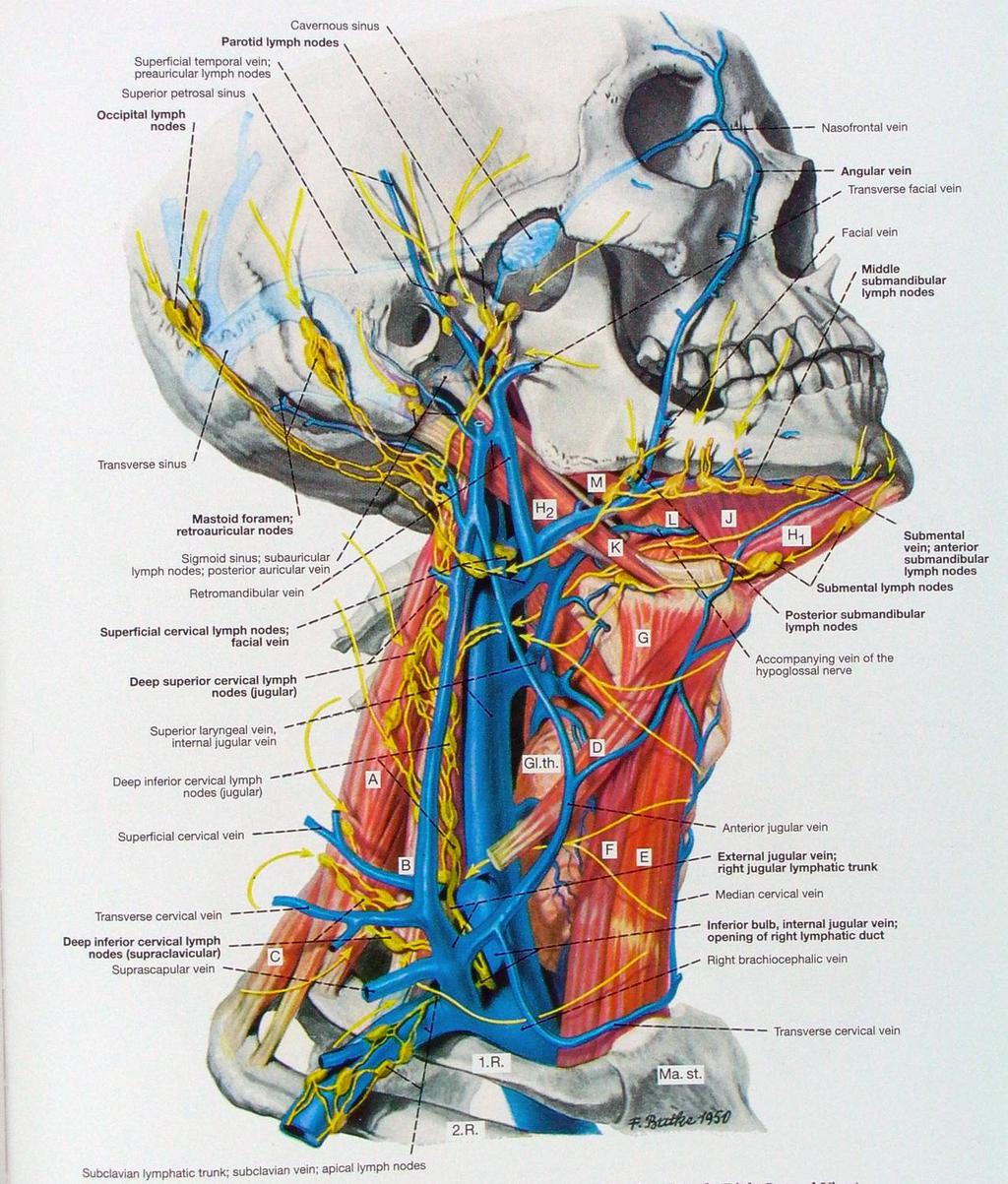
27 Arteries of face mostly from brr of ext carotid a with some limited brr from int carotid a 1 Facial a 2 Transverse facial a 3 Brr of maxillary a 4 Brr of ophthalmic a 2 3 1
28 Arteries of face - 1 Ext carotid a Facial a posterior to submandibular gland inferior border of mandible, ant to masseter, pulse can be felt here upward and medially in tortuous course corner of mouth gives off superior and inferior labial brr side of nose gives off lateral nasal br ends as angular a at med corner of eye *

29 Arteries of face - 2 Ext carotid a Superficial temporal a Transverse facial a across face, superficial to masseter m, b/w zygomatic arch and parotid duct
30 Arteries of face - 3 External carotid a Maxillary a Brr on face: Infra-orbital a through infra-orbital foremen lower eyelid, upper lip, and the area b/w Buccal a superficial to buccinator Mental a through mental foramen chin
31 Arteries of face - 4 Internal carotid a Ophthalmic a enters orbit Brr of ophthalmic a Lacrimal br through zygomaticofacial and zygomaticotemporal foramina face area over zygomatic bone Zygomaticofacial and zygomaticotemporal aa Terminal br of ophthalmic a Dorsal nasal a exits orbit in med corner dorsum of nose
32 Int carotid a Ext carotid a

33 from Gunther von Hagens Body Worlds The Anatomical Exhibition of Real Human Bodies
34 Veins of face Supratrochlear v + supra-orbital vv angular v at medial corner of eye Facial vein (the major v draining the face) receives tributaries from vv draining eyelids, external nose, lips, cheek, and chin posterior to facial a on inf border of mandible superficial to submandibular gland enters internal jugular v 2 Transverse facial vein Across face enters superficial temporal v in the parotid gland
35 Veins of face Intracranial venous connections Facial v connects with venous channels into deeper head regions 3-1 med corner of eye ophthalmic v 3-2 cheek vv enter infra-orbital foramen 3-3 deep facial vv enter pterygoid plexus All above connect with intracranial sinus: extracranial vv emissary vv intracranial vv
 Intracranial")
36 Veins of face - 3 No valves in facial v and these channels Infectious material in danger area on face region (above mouth) Intracranial direction
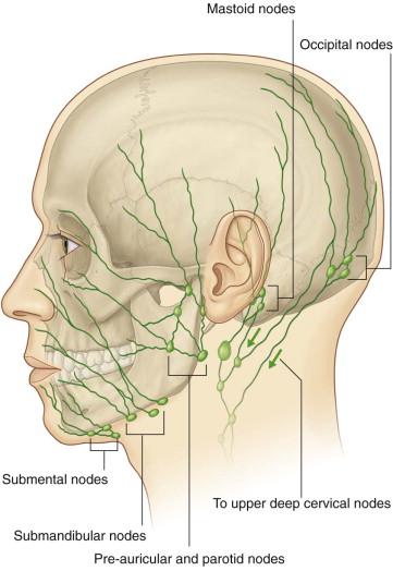
 Most of eyelids, a part of nose and lat cheek Pre-auricular and parotid nodes (anterior")
37 Lymphatic drainage of face Lymph on face drains toward: Medial part of lower lip and chin bilateral submental nodes (at inf and post to chin) Med corner of orbit, external nose, lat lower lip Submandibular nodes (superficial to submandibular gland, inferior to body of mandible) Most of eyelids, a part of nose and lat cheek Pre-auricular and parotid nodes (anterior to ear)
38
39 Facial nerve palsy (Bell s palsy) 1 Central lesions From both hemisphere Motor nucleus of facial n (in brainstem) ipsilateral upper face 2 Lesions at and around the geniculate ganglion Loss of motor function, taste sensation of anterior twothirds of tongue, and lacrimation of ipsilateral side 3 Lesions at and around the stylomastoid foramen Commonest abnormality, by viral inflammation of the nerve in bony canal Loss of motor function of ipsilateral side Taste sensation and lacrimation may not be affected if lesion is distal to greater petrosal and chorda tympani nn

40 2 1 X X 3 X
41 Trigeminal neuralgia Blood vessels adjacent to the sensory roots of mandibular and maxillary nerves may be involved
 b/w its two bellies Frontal belly attaches to skin of")

42 SCALP 1 Skin With large amount of hairs 2 Connective tissue Dense connective tissue with vv, aa, and nn 3 Aponeurotic layer Attaches firmly to skin, and consists of occipitofrontalis m Epicranial aponeurosis (gela aponeurotica) b/w its two bellies Frontal belly attaches to skin of eyebrows; innervated by temporal br of facial n Occipital belly arises from lateral part of superior nuchal line and mastoid processes; innervated by posterior auricular br 4 Loose connective tissue b/w aponeurotic layer and epicranium; infection may spread through 5 Pericranium Periosteum on calvaria
43 Innervation of scalp Anterior to the ears and the vertex by brr of trigeminal n: 1 Supratrochlear nerve exits from orbit, supplies front of forehead near midline 2 Supra-orbital nerve exits through supraorbital notch or foramen, passes through frontalis to vertex 3 Zygomaticotemporal nerve though foramen of zygomatic bone, to small ant area of temple 4 Auriculotemporal nerve through parotid gland, passes ant to ear, supplies scalp over temporal region
44 Innervation of scalp Posterior to the ears and the vertex by cervical nn from C2 and C3: 5 Great auricular nerve anterior rami of C2 and C3, to small area posterior to ear 6 Lesser occipital nerve anterior rami of C2, to small area posterior and superior to ear 7 Greater occipital nerve br posterior ramus of C2, supplies posterior scalp to vertex 8 Third occipital nerve br of posterior ramus of C3, supplies small area of lower scalp

45 3 1 2 * * 5
46 Arteries of scalp 1 Branches from the ophthalmic a Internal carotid a Ophthalmic a Supratrochlear and supra-orbital aa 2 Branches from the external carotid a 2-1 Posterior auricular a 2-2 Occipital a 2-3 Superficial temporal a
47 Veins of scalp Ant part of scalp (superciliary arches to vertex) Supratrochlear and supra-orbital vv ophthalmic v angular v facial v Lateral scalp Superficial temporal v Retromandibular v Scalp poterior to ear Posterior auricular v Retromandibular v Posterior scalp (ext occipital protuberance and sup nuchal line to vetex) Occipital v passes through mm of post neck venous plexus in suboccipital triangle
48 Lymphatic drainage of scalp Occipital region Occipital node (near attachment of trapezius m) Upper deep cervical nodes directly to Upper deep cervical nodes Upper scalp (posterior to vertex) directly to mastoid nodes (or retro-auricular node or posterior auricular node) Upper deep cervical nodes Upper scalp (anterior to vertex) Preauricular and parotid nodes Forehead Facial a Submandibular node
49
Head and Face Anatomy
 Head and Face Anatomy Epicranial region The Scalp The soft tissue that covers the vault of skull. Extends from supraorbital margin to superior nuchal line. Layers of the scalp S C A L P = skin = connective
3-Deep fascia: is absent (except over the parotid gland & buccopharngeal fascia covering the buccinator muscle)
") The Face 1-Skin of the Face The skin of the face is: Elastic Vascular (bleed profusely however heal rapidly) Rich in sweat and sebaceous glands (can cause acne in adults) It is connected to the underlying
The Face 1-Skin of the Face The skin of the face is: Elastic Vascular (bleed profusely however heal rapidly) Rich in sweat and sebaceous glands (can cause acne in adults) It is connected to the underlying
Face. Definition: The area between the two ears and from the chin to the eye brows. The muscles of the face
 Face Definition: The area between the two ears and from the chin to the eye brows. The muscles of the face The muscle of facial expression (include the muscle of the face and the scalp). All are derived
Tikrit University College of Dentistry Dr.Ban I.S. head & neck anatomy 2 nd y.
 Lec [3]/The scalp The scalp extends from the supraorbital margins anteriorly to the nuchal lines at the back of the skull and down to the temporal lines at the sides. The forehead, from eyebrows to hairline,
Parotid Gland, Temporomandibular Joint and Infratemporal Fossa
 M1 - Anatomy Parotid Gland, Temporomandibular Joint and Infratemporal Fossa Jeff Dupree Sanger 9-057 jldupree@vcu.edu Parotid gland: wraps around the mandible positioned between the mandible and the sphenoid
FACE CN V & VII PAROTID GLAND. Jacek Baj, MD, PhD Department of Human Anatomy
 FACE CN V & VII PAROTID GLAND Jacek Baj, MD, PhD Department of Human Anatomy THE FACE THE SCALP The scalp is composed of five layers: Skin ConnecAve Assue Aponeurosis Loose connecave Assue Pericranium
PTERYGOPALATINE FOSSA
 PTERYGOPALATINE FOSSA Outline Anatomical Structure and Boundaries Foramina and Communications with other spaces and cavities Contents Pterygopalatine Ganglion Especial emphasis on certain arteries and
THIEME. Scalp and Superficial Temporal Region
 CHAPTER 2 Scalp and Superficial Temporal Region Scalp Learning Objectives At the end of the dissection of the scalp, you should be able to identify, understand and correlate the clinical aspects: Layers
Dr.Ban I.S. head & neck anatomy 2 nd y. جامعة تكريت كلية طب االسنان املرحلة الثانية أ.م.د. بان امساعيل صديق 6102/6102
 جامعة تكريت كلية طب االسنان التشريح مادة املرحلة الثانية أ.م.د. بان امساعيل صديق 6102/6102 Parotid region The part of the face in front of the ear and below the zygomatic arch is the parotid region. The
By : Prof Saeed Abuel Makarem & Dr.Sanaa Alshaarawi
 By : Prof Saeed Abuel Makarem & Dr.Sanaa Alshaarawi OBJECTIVES By the end of the lecture, students shouldbe able to: List the nuclei of the deep origin of the trigeminal and facial nerves in the brain
Lec [8]: Mandibular nerve:
![Lec [8]: Mandibular nerve:](/thumbs/94/121295776.jpg "Lec [8]: Mandibular nerve:") Lec [8]: Mandibular nerve: The mandibular branch from the trigeminal ganglion lies in the middle cranial fossa lateral to the cavernous sinus. With the motor root of the trigeminal nerve [motor roots lies
Lec [8]: Mandibular nerve: The mandibular branch from the trigeminal ganglion lies in the middle cranial fossa lateral to the cavernous sinus. With the motor root of the trigeminal nerve [motor roots lies
function - sensory & postganglionic sympathetic [communication from the internal carotid plexus in the cavernous sinus] innervation of the mucosa of
![function - sensory & postganglionic sympathetic [communication from the internal carotid plexus in the cavernous sinus] innervation of the mucosa of](/thumbs/74/71276096.jpg "function - sensory & postganglionic sympathetic [communication from the internal carotid plexus in the cavernous sinus] innervation of the mucosa of") Nerves I. Cranial nerves A. Olfactory (CN I) 1. Olfactory bulb 2. Olfactory tract B. Optic n. (CNII) function - carries visual sensory information from the neural retina to the diencephalon & midbrain
Nerves I. Cranial nerves A. Olfactory (CN I) 1. Olfactory bulb 2. Olfactory tract B. Optic n. (CNII) function - carries visual sensory information from the neural retina to the diencephalon & midbrain
Temporal region. temporal & infratemporal fossae. Zhou Hong Ying Dept. of Anatomy
 Temporal region temporal & infratemporal fossae Zhou Hong Ying Dept. of Anatomy Temporal region is divided by zygomatic arch into temporal & infratemporal fossae. Temporal Fossa Infratemporal fossa Temporal
Temporal fossa Infratemporal fossa Pterygopalatine fossa Terminal branches of external carotid artery Pterygoid venous plexus
 Outline of content Temporal fossa Infratemporal fossa Pterygopalatine fossa Terminal branches of external carotid artery Pterygoid venous plexus Boundary Content Communication Mandibular division of trigeminal
Functional components
 Facial Nerve VII cranial nerve Emerges from Pons Two roots Functional components: 1. GSA (general somatic afferent) 2. SA (Somatic afferent) 3. GVE (general visceral efferent) 4. BE (Special visceral/branchial
Tikrit University collage of dentistry Dr.Ban I.S. head & neck anatomy 2 nd y. Lec [5] / Temporal fossa :
![Tikrit University collage of dentistry Dr.Ban I.S. head & neck anatomy 2 nd y. Lec [5] / Temporal fossa :](/thumbs/88/115294566.jpg "Tikrit University collage of dentistry Dr.Ban I.S. head & neck anatomy 2 nd y. Lec [5] / Temporal fossa :") Lec [5] / Temporal fossa : Borders of the Temporal Fossa: Superior: Superior temporal line. Inferior: gap between zygomatic arch and infratemporal crest of sphenoid bone. Anterior: Frontal process of the
Lec [5] / Temporal fossa : Borders of the Temporal Fossa: Superior: Superior temporal line. Inferior: gap between zygomatic arch and infratemporal crest of sphenoid bone. Anterior: Frontal process of the
Veins of the Face and the Neck
 Veins of the Face and the Neck Facial Vein The facial vein is formed at the medial angle of the eye by the union of the supraorbital and supratrochlear veins. connected through the ophthalmic veins with
Mohammad Hisham Al-Mohtaseb. Lina Mansour. Reyad Jabiri. 0 P a g e
 2 Mohammad Hisham Al-Mohtaseb Lina Mansour Reyad Jabiri 0 P a g e This is only correction for the last year sheet according to our record. If you already studied this sheet just read the yellow notes which
Parotid Gland. Parotid Gland. Largest of 3 paired salivary glands (submandibular; sublingual) Ramus of Mandible. Medial pterygoid.
 Ramus of Mandible. Medial pterygoid.") Parotid region Parotid Gland Largest of 3 paired salivary glands (submandibular; sublingual) Ramus of Mandible Medial pterygoid Cross section of mandible Masseter D S SCM Parotid Gland Mastoid Process
Parotid region Parotid Gland Largest of 3 paired salivary glands (submandibular; sublingual) Ramus of Mandible Medial pterygoid Cross section of mandible Masseter D S SCM Parotid Gland Mastoid Process
The SCALP. Prof. Dr. Muhammad Imran Qureshi
 The SCALP By Prof. Dr. Muhammad Imran Qureshi The SCALP includes FIVE layers external to the Calvaria. These are: S: Skin & Superficial Fascia C: Connective Tissue A: Aponeurosis (Epicranial) L: Loose
Cranial Nerve VII - Facial Nerve. The facial nerve has 3 main components with distinct functions
 Cranial Nerve VII - Facial Nerve The facial nerve has 3 main components with distinct functions Somatic motor efferent Supplies the muscles of facial expression; posterior belly of digastric muscle; stylohyoid,
Trigeminal Nerve Worksheets, Distributions Page 1
 Trigeminal Nerve Worksheet #1 Distribution by Nerve Dr. Darren Hoffmann Dental Gross Anatomy, Spring 2013 We have drawn out each of the branches of CN V in lecture and you have an idea now for their basic
Tracing the Cranial Nerves Osteologically
 CN I II III IV V 1 Supra-orbital ethmoidal nn. Ext. nasal V 2 Tracing the Cranial Nerves Osteologically Nucleus of Origin Olfactory tracts of frontal lobe of cerebrum Optic tracts from optic chiasma and
Introduction to Local Anesthesia and Review of Anatomy
 5-Sep Introduction and Anatomy Review 12-Sep Neurophysiology and Pain 19-Sep Physiology and Pharmacology part 1 26-Sep Physiology and Pharmacology part 2 Introduction to Local Anesthesia and Review of
Anatomy and Physiology. Bones, Sutures, Teeth, Processes and Foramina of the Human Skull
 Anatomy and Physiology Chapter 6 DRO Bones, Sutures, Teeth, Processes and Foramina of the Human Skull Name: Period: Bones of the Human Skull Bones of the Cranium: Frontal bone: forms the forehead and the
Omran Saeed. Luma Taweel. Mohammad Almohtaseb. 1 P a g e
 2 Omran Saeed Luma Taweel Mohammad Almohtaseb 1 P a g e I didn t include all the photos in this sheet in order to keep it as small as possible so if you need more clarification please refer to slides In
Bony orbit Roof The orbital plate of the frontal bone Lateral wall: the zygomatic bone and the greater wing of the sphenoid
 Bony orbit Roof: Formed by: The orbital plate of the frontal bone, which separates the orbital cavity from the anterior cranial fossa and the frontal lobe of the cerebral hemisphere Lateral wall: Formed
INTRODUCTION: ANATOMY UNDERLYING CLINICAL TESTS OF CRANIAL NERVES
 INTRODUCTION: ANATOMY UNDERLYING CLINICAL TESTS OF CRANIAL NERVES CRANIAL NERVE I - OLFACTORY I - OLFACTORY NERVE - SMELL TEST: SMELL ODORS (note: not ammonia; pain in nasal cavity CN5 DAMAGE: LOSS OF
Dr.Ban I.S. head & neck anatomy 2 nd y. جامعة تكريت كلية طب االسنان مادة التشريح املرحلة الثانية أ.م.د. بان امساعيل صديق 6102/6102
 جامعة تكريت كلية طب االسنان مادة التشريح املرحلة الثانية أ.م.د. بان امساعيل صديق 6102/6102 The scalp The scalp extends from the supraorbital margins anteriorly to the nuchal lines at the back of the skull
For the following questions, indicate the letter that corresponds to the SINGLE MOST APPROPRIATE ANSWER
 GROSS ANATOMY EXAMINATION May 15, 2000 For the following questions, indicate the letter that corresponds to the SINGLE MOST APPROPRIATE ANSWER 1. Pain associated with an infection limited to the middle
Maxilla, ORBIT and infratemporal fossa. Neophytos C Demetriades MD, DDS, MSc Associate professor European University of Cyprus School of Medicine
 Maxilla, ORBIT and infratemporal fossa Neophytos C Demetriades MD, DDS, MSc Associate professor European University of Cyprus School of Medicine MAXILLA Superior, middle, and inferior meatus Frontal sinus
Infratemporal fossa: Tikrit University college of Dentistry Dr.Ban I.S. head & neck Anatomy 2 nd y.
 Infratemporal fossa: This is a space lying beneath the base of the skull between the lateral wall of the pharynx and the ramus of the mandible. It is also referred to as the parapharyngeal or lateral pharyngeal
The sebaceous glands (glands of Zeis) open directly into the eyelash follicles, ciliary glands (glands of Moll) are modified sweat glands that open
 open directly into the eyelash follicles, ciliary glands (glands of Moll) are modified sweat glands that open") The Orbital Region The orbits are a pair of bony cavities that contain the eyeballs; their associated muscles, nerves, vessels, and fat; and most of the lacrimal apparatus upper eyelid is larger and more
The Orbital Region The orbits are a pair of bony cavities that contain the eyeballs; their associated muscles, nerves, vessels, and fat; and most of the lacrimal apparatus upper eyelid is larger and more
MAXILLA, ORBIT & PTERYGOPALATINE FOSSA. Neophytos C Demetriades MD, DDS, MSc Associate professor European University of Cyprus School of Medicine
 MAXILLA, ORBIT & PTERYGOPALATINE FOSSA Neophytos C Demetriades MD, DDS, MSc Associate professor European University of Cyprus School of Medicine Maxilla MAXILLA Superior, middle, and inferior meatus Frontal
Bones Ethmoid bone Inferior nasal concha Lacrimal bone Maxilla Nasal bone Palatine bone Vomer Zygomatic bone Mandible
 splanchnocranium - Consists of part of skull that is derived from branchial arches - The facial bones are the bones of the anterior and lower human skull Bones Ethmoid bone Inferior nasal concha Lacrimal
Learning Outcomes. The Carotid 20/02/2013. Scalp, Face, Parotid. Layers of the Scalp. The Parotid Gland. The Scalp. The Carotid The Facial Artery
 Learning Outcomes The Scalp Layers of the Scalp Bleeding from the Scalp The Carotid The Facial Artery Major Muscles of the Face and Jaw(s) Muscles of Mastication Muscles of Facial Expression The Parotid
Anatomy of the Trigeminal Nerve
 19 Anatomy of the Trigeminal Nerve.1 Introduction 0. The Central Part of the Trigeminal Nerve 1..1 Origin 1.. Trigeminal Nuclei.3 The Peripheral Part of the Trigeminal Nerve 4.3.1 Ophthalmic Nerve 4.3.
University of Palestine. Final Exam 1 st Semester 2014/2015 Total Grade: 60
 Question One: MCQ: 1- The coronal suture joins the a) frontal and parietal bones. b) left and right parietal bones. c) parietal and occipital bones. d) parietal, squamous temporal and greater wing of the
SCHOOL OF ANATOMICAL SCIENCES Mock Run Questions. 4 May 2012
 SCHOOL OF ANATOMICAL SCIENCES Mock Run Questions 4 May 2012 1. With regard to the muscles of the neck: a. the platysma muscle is supplied by the accessory nerve. b. the stylohyoid muscle is supplied by
Cranial nerves.
 Cranial nerves eaglezhyxzy@163.com Key Points of Learning Name Components Passing through Peripheral distribution Central connection Function Cranial nerves Ⅰ olfactory Ⅱ optic Ⅲ occulomotor Ⅳ trochlear
The Neck the lower margin of the mandible above the suprasternal notch and the upper border of the clavicle
 The Neck is the region of the body that lies between the lower margin of the mandible above and the suprasternal notch and the upper border of the clavicle below Nerves of the neck Cervical Plexus Is formed
The Seventh Cranial Nerve The Facial By Prof. Dr. Muhammad Imran Qureshi
 The Seventh Cranial Nerve The Facial By Prof. Dr. Muhammad Imran Qureshi Functional Components: SVE: Fibers originate from nucleus of facial nerve, and supply facial muscles GVE: Fibers derived from superior
External Occipital Protuberance
 Osteology Exterior Skull Frontal Bone Glabella Superciliary Arch Supraorbital Notch/Foramen Nasion (junction w/ Nasal bone) Frontal/Metopic Suture (usually absent in adult, b/w ossification centers of
human anatomy 2016 lecture fifteen Dr meethak ali ahmed neurosurgeon
 Cranial Nerves Organization of the Cranial Nerves The cranial nerves are named as follows: I. Olfactory II. Optic III. Oculomotor IV. Trochlear V. Trigeminal VI. Abducent VII. Facial VIII. Vestibulocochlear
HEAD & NECK BY NUMBERS THIRD EDITION Copyright 2013, Anatomy Numbers (Phoenix, AZ) All rights reserved
 All rights reserved") HEAD & NECK BY NUMBERS THIRD EDITION Copyright 2013, Anatomy Numbers (Phoenix, AZ) All rights reserved No part of this publication may be reproduced or transmitted in any form or by any means, electronic
HEAD & NECK BY NUMBERS THIRD EDITION Copyright 2013, Anatomy Numbers (Phoenix, AZ) All rights reserved No part of this publication may be reproduced or transmitted in any form or by any means, electronic
Lecture 07. Lymphatic's of Head & Neck. By: Dr Farooq Amanullah Khan PMC
 Lecture 07 Lymphatic's of Head & Neck By: Dr Farooq Amanullah Khan PMC Dated: 28.11.2017 Lymphatic Vessels Of the 800 lymph nodes in the human body, 300 are in the Head & neck region. The lymphatic vessels
PART 1. Dermatologic Surgery COPYRIGHTED MATERIAL
 1 PART 1 Dermatologic Surgery COPYRIGHTED MATERIAL 1 CHAPTER 1 Cutaneous anatomy in dermatologic surgery Diana Bolotin1, Lucille White2 3, and Murad Alam 1 Section of Dermatology, University of Chicago,
Trigeminal Nerve Anatomy. Dr. Mohamed Rahil Ali
 Trigeminal Nerve Anatomy Dr. Mohamed Rahil Ali Trigeminal nerve Largest cranial nerve Mixed nerve Small motor root and large sensory root Motor root Nucleus of motor root present in the pons and medulla
Introduction to Head and Neck Anatomy
 Introduction to Head and Neck Anatomy Nervous Tissue Controls and integrates all body activities within limits that maintain life Three basic functions 1. sensing changes with sensory receptors 2. interpreting
University of Palestine. Midterm Exam 2013/2014 Total Grade:
 Course No: DNTS2208 Course Title: Head and Neck Anatomy Date: 09/11/2013 No. of Questions: (50) Time: 1hour Using Calculator (No) University of Palestine Midterm Exam 2013/2014 Total Grade: Instructor
Muscles of the Face, Head, and Neck
 Muscles of the Face, Head, and Neck 1 How Muscles Are Named Many muscles named using such features as Location Function Shape Direction of fibers Number of heads or divisions Points of attachment Size
University of Palestine. Midterm Exam 2013/2014 Total Grade:
 [ Course No: DNTS2208 Course Title: Head and Neck Anatomy Date: 17/11/1024 No. of Questions: (52) Time: 2hours Using Calculator (No) University of Palestine Midterm Exam 2013/2014 Total Grade: Instructor
Bisection of Head & Nasal Cavity 頭部對切以及鼻腔. 解剖學科馮琮涵副教授 分機
 Bisection of Head & Nasal Cavity 頭部對切以及鼻腔 解剖學科馮琮涵副教授 分機 3250 E-mail: thfong@tmu.edu.tw Outline: The structure of nose The concha and meatus in nasal cavity The openings of paranasal sinuses Canals, foramens
Stracture The scalp consists of five layers,the 1st three of which are intimately bound together and move as a unit.to assist one in memorizing the
 Anatomy of Scalp Stracture The scalp consists of five layers,the 1st three of which are intimately bound together and move as a unit.to assist one in memorizing the names of the five layers of the scalp,use
*in general the blood supply of the nose comes from branches of the internal and external carotid arteries.
 In the previous lecture we talked about the anatomy of the nasal cavity, today we will talk about its blood supply, venous drainage, innervations, and finally about the paranasal sinuses. When we describe
Dr.Ban I.S. head & neck anatomy 2 nd y جامعة تكريت كلية طب االسنان مادة التشريح املرحلة الثانية أ.م.د. بان امساعيل صديق 6102/6102
 جامعة تكريت كلية طب االسنان مادة التشريح املرحلة الثانية أ.م.د. بان امساعيل صديق 6102/6102 Pterygopalatine fossa: The pterygopalatine fossa is a cone-shaped depression, It is located between the maxilla,
Trigeminal Nerve (V)
") Trigeminal Nerve (V) Lecture Objectives Discuss briefly how the face is developed. Follow up the course of trigeminal nerve from its point of central connections, exit and down to its target areas. Describe
Trigeminal Nerve (V) Lecture Objectives Discuss briefly how the face is developed. Follow up the course of trigeminal nerve from its point of central connections, exit and down to its target areas. Describe
CN I Olfactory. CN II Optic. CN III Oculomotor. Special Sensory Efferent fibers to Olfactory Bulb. Cribiform Plate of Ethmoid
 CN I Olfactory Efferent fibers to Olfactory Bulb Olfactory Tract Olfactory Bulb Cribiform Plate of Ethmoid Anosmia Loss of sense of smell Uncinate Fits olfactory hallucinations To Olfactory Epithelium
Anatomy images for MSS practical exam- 2019
 Anatomy images for MSS practical exam- 2019 Ilium Ischium Pubis Acetabulaum Iliac crest Iliac tubercle ASIS (muscle and ligament attached) AIIS (muscle attached) PSIS PIIS Ischial spine Ischial tuberosity
Laith Sorour. Facial nerve (vii):
:") Laith Sorour Cranial nerves 7 & 8 Hello, there are edited slides please go back to them to see pictures, they are not that much important in this lecture but still, and yes slides are included :p Let s
Laith Sorour Cranial nerves 7 & 8 Hello, there are edited slides please go back to them to see pictures, they are not that much important in this lecture but still, and yes slides are included :p Let s
Bony orbit. Sup. Med. Inf. Lat. frontal bone. frontal process of maxilla. zygomatic process of maxilla zygomatic bone
 Orbit 解剖學科鄭授德 本教材之圖片取自於 1. Gray s Anatomy for Students, 3rd ed., 2015, by Drake, Vogl, and Mitchell 2. Clinically Oriented Anatomy, 7th ed., 2014, by Moore, Dalley, and Agur 3. Anatomy, an Essential Textbook,
Dr. Sami Zaqout Faculty of Medicine IUG
 Auricle External Ear External auditory meatus The Ear Middle Ear (Tympanic Cavity) Auditory ossicles Internal Ear (Labyrinth) Bony labyrinth Membranous labyrinth External Ear Auricle External auditory
The Scalp and Face Protocol. Julie Goodwin, BA, LMT
 The Scalp and Face Protocol Julie Goodwin, BA, LMT The Scalp and Face Protocol Julie Goodwin, BA, LMT Julie Goodwin 2014 2 Agenda Pertinent Anatomy and Physiology Treatment Planning Strokes, Techniques
Anatomic Relations Summary. Done by: Sohayyla Yasin Dababseh
 Anatomic Relations Summary Done by: Sohayyla Yasin Dababseh Anatomic Relations Lecture 1 Part-1 - The medial wall of the nose is the septum. - The vestibule lies directly inside the nostrils (Nares). -
Basic Anatomy and Physiology of the Lips and Oral Cavity. Dr. Faghih
 Basic Anatomy and Physiology of the Lips and Oral Cavity Dr. Faghih It is divided into seven specific subsites : 1. Lips 2. dentoalveolar ridges 3. oral tongue 4. retromolar trigone 5. floor of mouth 6.
Skull-2. Norma Basalis Interna Norma Basalis Externa. Dr. Heba Kalbouneh Associate Professor of Anatomy and Histology
 Skull-2 Norma Basalis Interna Norma Basalis Externa Dr. Heba Kalbouneh Associate Professor of Anatomy and Histology Norma basalis interna Base of the skull- superior view The interior of the base of the
Anatomy: head and Neck (6 questions) 1. Prevertebral Flexor Musculature (lying in front of the vertebrae) include all, EXCEPT: Longus Colli.
 1. Prevertebral Flexor Musculature (lying in front of the vertebrae) include all, EXCEPT: Longus Colli.") Anatomy: head and Neck (6 questions) 1. Prevertebral Flexor Musculature (lying in front of the vertebrae) include all, EXCEPT: Longus Colli. Rectus Capitis Anterior. Rectus Capitis Lateralis. Rectus Capitis
Anatomy: head and Neck (6 questions) 1. Prevertebral Flexor Musculature (lying in front of the vertebrae) include all, EXCEPT: Longus Colli. Rectus Capitis Anterior. Rectus Capitis Lateralis. Rectus Capitis
Unit VIII Problem 9 Anatomy of The Ear
 Unit VIII Problem 9 Anatomy of The Ear - The ear is an organ with 2 functions: Hearing. Maintenance of equilibrium/balance. - The ear is divided into 3 parts: External ear. Middle ear (which is also known
Anatomy of Oral Cavity DR. MAAN AL-ABBASI
 Anatomy of Oral Cavity DR. MAAN AL-ABBASI By the end of this lecture you should be able to: 1. Differentiate different parts of the oral cavity 2. Describe the blood and nerve supply of mucosa and muscles
Trigeminal nerve. Slide in bold and please go back to see the pictures, if I skipped any part of record that because it wasn t clear to me
 Trigeminal nerve Slide in bold and please go back to see the pictures, if I skipped any part of record that because it wasn t clear to me Hala nsour 2/26/2018 P a g e 1 this lecture contain two topics
Major Anatomic Components of the Orbit
 Major Anatomic Components of the Orbit 1. Osseous Framework 2. Globe 3. Optic nerve and sheath 4. Extraocular muscles Bony Orbit Seven Bones Frontal bone Zygomatic bone Maxillary bone Ethmoid bone Sphenoid
Perineural Tumor Spread (PNS) Perineural Tumor Spread (PNS) PNS Anatomic Considerations. Perineural Tumor Spread-Imaging
 Perineural Tumor Spread (PNS) PNS Anatomic Considerations. Perineural Tumor Spread-Imaging") Imaging of Perineural Tumor Spread in Head and Neck Cancer Lawrence E. Ginsberg, MD Departments of Diagnostic Radiology and Head and Neck Surgery University of Texas M.D. Anderson Cancer Center Houston,
Imaging of Perineural Tumor Spread in Head and Neck Cancer Lawrence E. Ginsberg, MD Departments of Diagnostic Radiology and Head and Neck Surgery University of Texas M.D. Anderson Cancer Center Houston,
Nose & Mouth OUTLINE. Nose. - Nasal Cavity & Its Walls. - Paranasal Sinuses. - Neurovascular Structures. Mouth. - Oral Cavity & Its Contents
 Dept. of Human Anatomy, Si Chuan University Zhou hongying eaglezhyxzy@163.com Nose & Mouth OUTLINE Nose - Nasal Cavity & Its Walls - Paranasal Sinuses - Neurovascular Structures Mouth - Oral Cavity & Its
Anatomy of. External NOSE. By Dr Farooq Aman Ullah Khan PMC
 Anatomy of External NOSE By Dr Farooq Aman Ullah Khan PMC 24 th Nov. 2017 The External Nose Descriptions of the nose always begin with that part of it which is covered by the skin, i.e., the EXPOSED PART
Structure Location Function
 Frontal Bone Cranium forms the forehead and roof of the orbits Occipital Bone Cranium forms posterior and inferior portions of the cranium Temporal Bone Cranium inferior to the parietal bone forms the
Oral cavity : consist of two parts: the oral vestibule and the oral cavity proper. Oral vestibule : is slit like space between.
 Oral cavity Oral cavity : consist of two parts: the oral vestibule and the oral cavity proper Oral vestibule : is slit like space between the teeth, buccal gingiva, lips, and cheeks 1 Oral cavity Oral
HEAD AND NECK ANATOMY PRACTICE QUESTIONS
 HEAD AND NECK ANATOMY PRACTICE QUESTIONS 1. A patient complains that he has lost sensation on his face and that the skin of his face feels numb. The physician tests tactile acuity by touching the forehead
The Ear The ear consists of : 1-THE EXTERNAL EAR 2-THE MIDDLE EAR, OR TYMPANIC CAVITY 3-THE INTERNAL EAR, OR LABYRINTH 1-THE EXTERNAL EAR.
 The Ear The ear consists of : 1-THE EXTERNAL EAR 2-THE MIDDLE EAR, OR TYMPANIC CAVITY 3-THE INTERNAL EAR, OR LABYRINTH 1-THE EXTERNAL EAR Made of A-AURICLE B-EXTERNAL AUDITORY MEATUS A-AURICLE It consists
The orbit-2. Dr. Heba Kalbouneh Assistant Professor of Anatomy and Histology
 The orbit-2 Dr. Heba Kalbouneh Assistant Professor of Anatomy and Histology Eyelids The eyelids (act like the curtains) protect the eye from injury and excessive light by their closure The upper eyelid
Superior View of the Skull (Norma Verticalis) Anteriorly the frontal bone articulates with the two parietal bones AT THE CORONAL SUTURE
 Anteriorly the frontal bone articulates with the two parietal bones AT THE CORONAL SUTURE") Superior View of the Skull (Norma Verticalis) Anteriorly the frontal bone articulates with the two parietal bones AT THE CORONAL SUTURE 1 The two parietal bones articulate in the midline AT THE SAGITTAL
Superior View of the Skull (Norma Verticalis) Anteriorly the frontal bone articulates with the two parietal bones AT THE CORONAL SUTURE 1 The two parietal bones articulate in the midline AT THE SAGITTAL
Posterior Triangle of the Neck By Prof. Dr. Muhammad Imran Qureshi
 Posterior Triangle of the Neck By Prof. Dr. Muhammad Imran Qureshi For the purpose of anatomical description the neck is sub divided into two major triangles, the Anterior and the Posterior by muscle bellies
REVIEW/PREVIEW OF HEAD AND NECK ANATOMY FOR ENT EXAM
 REVIEW/PREVIEW OF HEAD AND NECK ANATOMY FOR ENT EXAM - 2017 PALPATE CAROTID ARTERY: AT LEVEL OF CAROTID BIFURCATION VERTEBRAL LEVEL C4 Sternocleidomastoid Muscle INTERNAL CAROTID EXTERNAL CAROTID COMMON
Biology 218 Human Anatomy. Adapted from Martini Human Anatomy 7th ed. Chapter 6 The Skeletal System: Axial Division
 Adapted from Martini Human Anatomy 7th ed. Chapter 6 The Skeletal System: Axial Division Introduction The axial skeleton: Composed of bones along the central axis of the body Divided into three regions:
PERIPHERAL NERVOUS SYSTEM
 CHAPTER 13 PERIPHERAL NERVOUS SYSTEM Functional division of nervous system = afferent info to the CNS ascending spinal cord = efferent info from CNS descending spinal cord somatic skin, muscles visceral
Biology 323 Human Anatomy for Biology Majors Week 10; Lecture 1; Tuesday Dr. Stuart S. Sumida. Cranial Nerves and Soft Tissues of the Skull
 Biology 323 Human Anatomy for Biology Majors Week 10; Lecture 1; Tuesday Dr. Stuart S. Sumida Cranial Nerves and Soft Tissues of the Skull FOREBRAIN MIDBRAIN HINDBRAIN Forebrain: Cerebrum Perception,
C h a p t e r PowerPoint Lecture Slides prepared by Jason LaPres North Harris College Houston, Texas
 C h a p t e r 15 The Nervous System: The Brain and Cranial Nerves PowerPoint Lecture Slides prepared by Jason LaPres North Harris College Houston, Texas Copyright 2009 Pearson Education, Inc., publishing
HEAD & NECK ANATOMY - MCQ HEAD & NECK ANATOMY
 . ' HEAD & NECK ANATOMY I. Deep investing layer of cervical fascia splits to enclose: A. Sternocleidomastoid B. Trapezius C. Parotid gland D. Omohyoid 2. Regarding the prevertebral fascia, the following
1. EPINEPHRINE 2. PREDNISONE 3. BENADRYL 4. HYALURONIDASE 5. BABY ASPIRIN 6. NITROPASTE 7. VIAGRA 8. CANNULAS. Must Haves for Injection Safety
 1. EPINEPHRINE 2. PREDNISONE 3. BENADRYL 4. HYALURONIDASE 5. BABY ASPIRIN 6. NITROPASTE 7. VIAGRA 8. CANNULAS Must Haves for Injection Safety Facial artery: This artery stems from the external carotid
Bones of the skull & face
 Bones of the skull & face Cranium= brain case or helmet Copyright The McGraw-Hill Companies, Inc. Permission required for reproduction or display. The cranium is composed of eight bones : frontal Occipital
The Neck. BY: Lina Abdullah & Rahaf Jreisat
 The Neck BY: Lina Abdullah & Rahaf Jreisat Boundaries of the Neck: generally from base of the skull to root of the neck Superior margin :From superior nuchal line of occipital bone up to mastoid process
HBA THE BODY Head & Neck Written Examination October 23, 2014
 HBA 531 - THE BODY Head & Neck Written Examination October 23, 2014 Name: NOTE 2: When asked to trace nerve, artery, or vein pathways, do so by using arrows, e.g., structure a structure b structure c...
ANTERIOR CERVICAL TRIANGLE (Fig. 2.1 )
") 2 Neck Anatomy ANTERIOR CERVICAL TRIANGLE (Fig. 2.1 ) The boundaries are: Lateral: sternocleidomastoid muscle Superior: inferior border of the mandible Medial: anterior midline of the neck This large triangle
2 Neck Anatomy ANTERIOR CERVICAL TRIANGLE (Fig. 2.1 ) The boundaries are: Lateral: sternocleidomastoid muscle Superior: inferior border of the mandible Medial: anterior midline of the neck This large triangle
Cranial Cavity REFERENCES: OBJECTIVES OSTEOLOGY. Stephen A. Gudas, PT, PhD
 Stephen A. Gudas, PT, PhD Cranial Cavity REFERENCES: Moore and Agur, Essential Clinical Anatomy (ECA), 3rd ed., pp. 496 498; 500 507; 512 514 Grant s Atlas 12 th ed., Figs 7.6; 7.19 7.30. Grant s Dissector
Chapter 7 Part A The Skeleton
 Chapter 7 Part A The Skeleton Why This Matters Understanding the anatomy of the skeleton enables you to anticipate problems such as pelvic dimensions that may affect labor and delivery The Skeleton The
Functional anatomy of the skull & muscles of the head. Dr. Oksana Ivanivna Petrichko Department of Human Anatomy and Histologi
 Functional anatomy of the skull & muscles of the head Dr. Oksana Ivanivna Petrichko Department of Human Anatomy and Histologi Plan of the lecture 1. 2. 3. 4. 5. Development of the muscles of the head Morphofunctional
SKULL / CRANIUM BONES OF THE NEUROCRANIUM (7) Occipital bone (1) Sphenoid bone (1) Temporal bone (2) Frontal bone (1) Parietal bone (2)
 Occipital bone (1) Sphenoid bone (1) Temporal bone (2) Frontal bone (1) Parietal bone (2)") Important! 1. Memorizing these pages only does not guarantee the succesfull passing of the midterm test or the semifinal exam. 2. The handout has not been supervised, and I can not guarantee, that these
Important! 1. Memorizing these pages only does not guarantee the succesfull passing of the midterm test or the semifinal exam. 2. The handout has not been supervised, and I can not guarantee, that these
This lab activity is aligned with Visible Body s Anatomy and Physiology app. Learn more at visiblebody.com/professors
 1 This lab activity is aligned with Visible Body s Anatomy and Physiology app. Learn more at visiblebody.com/professors 2 PRE-LAB EXERCISES A. Watch the video 13.1 Muscular System Overview and observe