A transgenic animal model resembling amelogenesis imperfecta related to. ameloblastin over-expression
|
|
|
- Jocelin Leonard
- 6 years ago
- Views:
Transcription
1 JBC Papers in Press. Published on March 25, 2003 as Manuscript M A transgenic animal model resembling amelogenesis imperfecta related to ameloblastin over-expression Michael L. Painea, Hong-Jun Wanga, Wen Luoa, Paul H. Krebsbachb and Malcolm L. Sneada a University of Southern California, School of Dentistry, Center for Craniofacial Molecular Biology, 2250 Alcazar Street, CSA Room 142, Los Angeles, California b University of Michigan, School of Dentistry, Room 4207, Ann Arbor, Michigan To whom correspondences should be addressed: phone (323) ; paine@usc.edu Short title: Enamel matrix protein interactions Keywords: amelin; ameloblastin; amelogenin; amelogenesis imperfecta; biomineralization; odontogenesis; scanning electron microscopy; sheathlin; transgenics. This work was supported by Grants DE06988, DE12502, DE13045 and DE13404 from the National Institute of Dental and Craniofacial Research (NIDCR). Copyright 2003 by The American Society for Biochemistry and Molecular Biology, Inc.
2 Genetic diseases that affect tooth enamel are grouped under the classification of amelogenesis imperfecta. Human pedigrees, and experiments on transgenic and null mice, have all demonstrated that mutations to the secreted proteins amelogenin, enamelin and enamelysin result in visibly, structurally or mechanically defective enamel. We have produced transgenic mice that mis-express the ameloblastin gene in an attempt to better define a physiologic function for ameloblastin during enamel formation. These transgenic animals exhibit imperfections in their enamel that is evident at the nanoscale level. Specifically, ameloblastin over-expression influences enamel crystallite habit and enamel rod morphology. These findings suggest enamel crystallite habit and rod morphology are influenced by the temporal- and spatial-expression of ameloblastin, and may implicate the role of the ameloblastin gene locus in the etiology of a number of undiagnosed autosomally dominant cases of amelegenesis imperfecta. 2
3 INTRODUCTION In 1996 a new member of the non-amelogenin, non-enamelin class of enamel proteins was simultaneously characterized by three different groups of investigators, two groups using rat incisors and one group using porcine teeth. In the United States of America, Krebsbach and colleagues named it ameloblastin (1); in Sweden, Cerny and colleagues named it amelin (2); and in a joint study between investigators in Japan and the United States of America, Hu and colleagues named it sheathlin (3). A definitive physiologic role for the ameloblastin protein in tooth development remains unknown. Immunologic identification of ameloblastin during secretory amelogenesis, the developmental stage at which the extracellular matrix is deposited and organized, reveals an ameloblastin distribution (within the enamel extracellular matrix) that follows the ameloblast outline, resulting in a fish-net partitioning (3). Ameloblastin can also be immuno-localized to Tome s processes, the highly specialized plasma membrane component of secretory ameloblast cells (4). Localization to Tomes processes has also lead to the speculation that ameloblastin has a role to play in crystal nucleation (4,5). The ameloblastin molecule has a DGEA domain that has been identified in collagen type I as a recognition site for alpha 2 beta 1 integrin (2,6). Ameloblastin also contains a trombospondin-like cell adhesion domain, VTKG (7). These findings have lead to the speculation that ameloblastin might serve as part of the linkage between ameloblasts and the enamel extracellular matrix (2). Ameloblastin may be critical for retaining a registration between the secretory ameloblasts and the enamel organic matrix since a single ameloblast is responsible for the creation of its corresponding matrix. Amelogenesis imperfecta (AI) is an inherited dental disease that affects enamel. Linkage 3
4 analysis was performed on three Swedish families that where affected with an autosomal dominant variant of amelogenesis imperfecta (ADAI) which was clinically noted as a localize hypoplastic enamel phenotype (8). DNA microsatellite markers linked all three families to a defect at chromosome 4q (8). In the human genome, both ameloblastin and enamelin are localized to chromosome 4q11-q21 (1,9-11), thus making both genes potential candidates responsible for an amelogenesis imperfecta. A gene mutation in enamelin has been recently been identified in a localized hypoplastic autosomal dominant amelogenesis imperfecta (AIH2) family pedigree (12). To date, no definitive evidence links the ameloblastin gene to defective enamel. The human enamelysin gene maps to chromosome 11q22.3, and while no linkage data yet has implicated enamelysin to an amelogenesis imperfecta phenotype, enamelysin null animals do have defective enamel and a weakened dentin enamel junction (13). Over-expressing a gene in a tissue-specific manner is an effective method for determining the role of a protein in the context of a developing animal. We targeted ameloblastin over-expression to ameloblasts by generating transgenic animals in which ameloblastin expression was under the control of the ameloblast-specific amelogenin promoter. With this transgenic animal model we provide evidence that altering the expression profile of ameloblastin has an adverse effect on enamel formation that results in imperfect enamel. These data suggest that ameloblastin has a significant physiologic role to play in enamel formation, and ameloblastin should be considered as a candidate gene when discussing the genetics of amelogenesis imperfecta. 4
5 MATERIALS AND METHODS DNA cassette Briefly, the 2.3kb amelogenin promoter (14) was used to express the rat ameloblastin transgene (GenBank accession U35097) (1). Also included, as part of the promoter, is intron 1 of amelogenin which is included to ensure that in vivo RNA processing events are appropriate and functional (15). Located at the amino-terminus of the transgene product are; the mouse dentin sialophosphoprotein (DSPP) signal peptide to ensure transport to the extracellular space (16); and the vesicular stomatitis virus glycoprotein (VSV-G) (17) and human c-myc epitopes (Roche, Indianapolis, IN) (18) which are used to follow transgene expression (19). Finally, the mouse DSPP 3 untranslated region follows the transgene coding region. For this particular construct, the selection of the mouse DSPP signal peptide, the DSPP 3 untranslated region and the inclusion of multiple VSV-G and c-myc epitopes was primarily based on ease of construction from available plasmids and relates to other ongoing transgenic studies (unpublished). Transgenic animals All vertebrate animal manipulation complied with institutional and federal guidelines. Transgenic mice lines were prepared as described elsewhere (15). Animals were analyzed for transgene status by Southern blot hybridization of genomic DNA (14). Hybridization was to random primed 32 P-labeled PCR generated DNA to the VSV-G and c-myc region of the transgenic construct. In addition, animal transgene status was confirmed by PCR using a forward primer within the ameloblastin coding region (PA56; 5-5
6 GCCTGTGCATCCCCCACCTCTCCC) and a reverse primer within the DSPP 3 untranslated region (PA57; 5 - AGCTCGGCTCCACTCCCTATGAGC) to give a DNA product of 1082 base pairs (data not included). Five independent transgenic animal lines were bred beyond three generations and showed a similar transgene expression pattern observed by immuno-detection as reported in this paper and previously (14,15). SEM studies were performed on animals bearing the transgene following five additional matings among transgenic siblings or parent founder lines; this to reduce any genetic variability. Aged-matched, non-transgenic animal controls were taken from the same breeding stock and used for SEM studies. Immunolocalization Tissue sections of four-day post-natal heterozygous transgenic mouse pups, or their normal (non-transgenic) littermates, were prepared as described previously (14). An anti-cmyc monoclonal antibody (Roche, Indianapolis, IN; catalogue # ) was used to demonstrate tissue specific expression of the introduced ameloblastin transgene. Concentration and reaction conditions used for this antibody was as recommended by the manufacturer. Polyclonal rabbit antibodies against recombinant rat ameloblastin were prepared, purified and used as previously described at a dilution of 1: 2,000 (1,4). Immunohistochemistry methodology has been described elsewhere (20). Scanning electron microscopy Methodology for sample preparation and imaging by SEM were previously reported (21-23). Six-week old animals were sacrificed for SEM imaging. A single animal from each of five 6
7 unique transgene lines was subjected to SEM analysis, and all lines gave similar results. The figures prepared are representative of the defects noted in all of these transgenic animals. 7
8 RESULTS Establishment of transgenic lines The details of the transgene DNA construct are illustrated (Figure 1). The mouse X- chromosomal derived amelogenin promoter is used to drive the expression of the ameloblastin gene that is marked as a transgene by the presence of the c-myc epitope. No gross abnormalities were detected in the dentition of any of the transgenic mouse lines at the time of the eruption of the incisor or molar teeth. No gross abnormalities were observed for the molar or the incisor teeth at 6-weeks of age (Figure 2). The animals diet was constant for both non-transgenic and transgenic animals and transgenic animals appeared to develop normally with no evidence of malnutrition. Transgene expression is restricted to secretory ameloblast cells and the developing enamel matrix Using a monoclonal antibody against the c-myc epitope (18), 4-day old mouse incisors were chosen for immunohistochemical detection of the transgene protein since at this developmental stage of mouse tooth formation ameloblastin expression is robust (4). The transgenic protein product was identified in the transition zone (Figure 3) of developing enamel of a lower incisor tooth using the c-myc epitope tag (Figures 4). This strategy allowed us to identify and distinguish the protein corresponding to the transgene, as opposed to protein derived from the endogenous wild-type ameloblastin gene. Four-day old mouse incisors were chosen for immunohistochemical detection of the transgene protein. At this developmental stage of mouse tooth formation high levels of ameloblastin expression are expected (4). The transgenic 8
9 protein was identified in the cytoplasm of ameloblasts and also within the newly secreted enamel organic extracellular matrix (Figure 4). Using an antibody to amelogenin (4), no gross disturbances to the expression pattern for the endogenous amelogenin gene were observed (data not shown). Ameloblastin gene expression in transgenic and non-transgenic animals using polyclonal antibodies against rat ameloblastin Using polyclonal antibodies to rat ameloblastin (1,4), 4-day old mouse incisors were chosen for immunohistochemical detection of both the endogenous and transgenic ameloblastin proteins. The transgenic protein is identified in the cytoplasm of ameloblasts and also within the newly secreted enamel organic extracellular matrix (Figure 5). Of particular note are the greater levels of ameloblastin expression observed in transgenic animals (Figure 5, panel C) when compared to the non-transgenic animals (Figure 5, panel B). This increased level of ameloblastin expression is consistent with expression levels observed for the amelogenin gene whose promoter is regulating expression of the transgene. Localization of the ameloblastin protein is identical in transgenic animals when compared to non-transgenic animals; that is it is present in the cytoplasm of secretory ameloblasts, it appears evenly distributed in the enamel matrix in the transition zone of the incisor (Figure 5, panels Bc and Cc) and in the maturing enamel (Figure 5, panels Bb and Cb) and mature enamel (Figure 5, panels Ba and Ca). In addition, immuno-reactivity is observed at the dentin-enamel junction, a unique junction linking the enamel to the underlying dentin (Figure 5, panels Ca and Cb). Within the maturing enamel, ameloblastin immuno-reactivity is significantly less than is 9
10 observed at either the dentin-enamel junction or the ameloblast cells (Figure 5, panels Ba and Ca). This decrease in observed ameloblastin is consistent with the history of ameloblastin processing, namely the hydrolysis of ameloblastin protein subsequent to its delivery to the enamel extracellular matrix environment with a consequential loss of epitopes. Enamel rod and interrod structure by SEM SEM analysis of six-week post-natal mouse incisor teeth showed the consequences of the ameloblastin protein up-regulation on enamel morphology. A 6-week old mouse incisor indicating the transition zone of the enamel from which all SEM images were collected for this study is provided (Figure 3). For this study incisor teeth were fractured coronally through the enamel transition zone (Figure 3). There was no acid-etching of the samples done prior to SEM analysis. The results shown are representative of findings observed consistently in each of five independent transgenic founder lines. Because in each of the multiple founder lines the observed phenotype is relatively constant, it appears unlikely that transgene integration disrupted a gene required for enamel formation. The enamel formed in the mice homozygous for the transgene appeared to be more porous (Figure 6, panels E and F). There was severe enamel-rod dysmorphology (Figure 6, panels C through F) when compared to age-matched non-transgenic control animals (Figure 6, panels A and B). While some regions of transgenic enamel appeared relatively normal (Figure 6, panel C compared to panel A), perhaps more apparent in the incisor of transgenic animals were vast areas of enamel showing no rod architecture, and with interrod enamel dominating (Figure 6, panel D). Another feature noted in the enamel of these incisor teeth of transgenic animals was 10
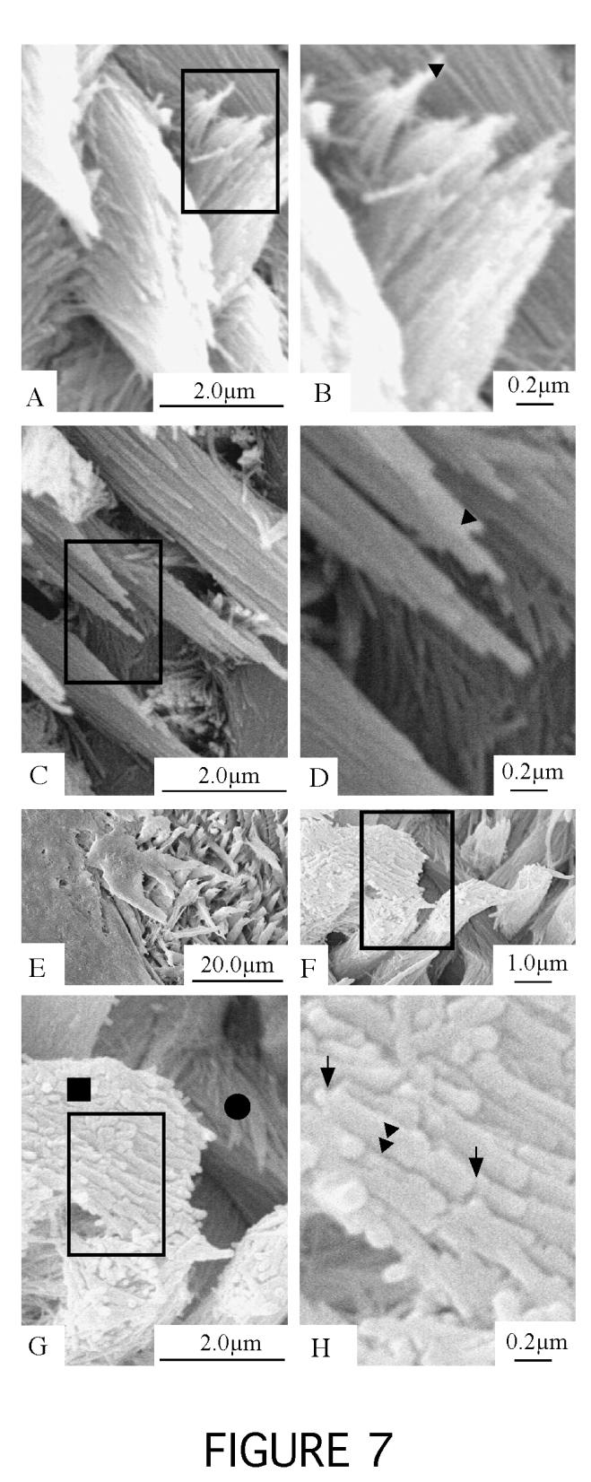
11 the absence of an aprismatic surface layer (as noted in the majority of sections studied) (Figure 6, panels D, E and F). This feature would equate to an increase in surface porosity at this stage of development. Enamel crystallites as observed by SEM Individual crystallites were visualized by SEM and compared to crystallites from nontransgenic mice. All samples were from the transition zone of incisor teeth (Figure 3). Within the enamel of transgenic animas it was clear that regions of relatively normal rod and interrod structure could be found directly neighboring homogeneous regions of interrod-only enamel (Figure 7, panel E, right side verses the left side). For transgenic animals, in some regions where rod enamel architecture was apparent, crystallite dimensions were comparable to non-transgenic animals (Figure 7, panels C and D when compared to panels A and B). Individual enamel rods of transgenic animals had either relatively normal enamel crystallites (Figure 7, panel D and panel G for the enamel rod identified with a solid circle), or relatively abnormal crystallites (Figure 7, panel G for the enamel rod identified with a solid square) predominating. These abnormal enamel crystallites, seen only in the enamel of transgenic animals, had a diameter of approximately twice that of the enamel crystallites for the non-transgenic controls. In addition, the c-axis of these abnormal crystallites measured approximately 1µm, as opposed to being a continuous inorganic unit-structure spanning the entire enamel thickness. 11
12 DISCUSSION Genetic linkage analysis is a valuable tool when tracking genes responsible for particular genetic diseases. With any genetic disease, having identified a genetic locus and potential candidate genes, classically the emphasis has been to then identify the responsible mutations that can gave rise to a particular phenotype. This requires the sequencing of a gene until a mutation can be identified and characterized. Another approach to gene function discovery of would be to create a mutant animal null for a particular gene. A third approach used to define gene function, and to show a genotype to phenotype relationship, is to produce transgenic animals that overexpress a particular gene product within a particular tissue or organ. We present data (Figure 4) which shows that the ameloblastin transgene expression in this study is limited to ameloblast cells and the enamel matrix; that is there is no ectopic expression noted in surrounding tissues, including odontoblasts and dentin. This data highlights the specificity of the amelogenin promoter to ameloblasts, and this has been noted and discussed previously (14,15). In addition, the inclusion of c-myc and VSV-G epitopes to the transgene at the amino-terminus appears to have negligible affects on protein localization (Figure 5). This combined data (immunolocalization and tissue specificity) is also suggestive that ameloblastin protein function is not significantly affected by inclusion of the marker epitopes. This transgenic animal model that over-expresses ameloblastin should help define a physiologic function for ameloblastin. With an investigative focus on the enamel organ, significant strides have been made over the past decade in defining protein components of the matrix (1-4,24-29), matrix assembly (29-31), the matrix disassembly (13,32,33) and environmental influences on enamel crystallite habit (34-36). In general, data generated from both in vitro and in vivo model systems have been 12
13 complementary. Animals null for amelogenin produce mineralized enamel that is both hypoplastic, and absent of a well-defined rod and interrod architecture (37). Human pedigrees with mutations to the amelogenin gene have a phenotype that is either categorized as hypoplastic or hypomineralized (38), and there is emerging data that would suggest that disruptions to the amino-terminal of amelogenin (that leave the carboxyl-terminal intact) result in a primarily hypomineralized enamel, while mutations to the carboxyl-terminus primarily result in a hypoplastic enamel (38). Heterogeneous mutations of the enamelin gene are also responsible for a number of amelogenesis imperfecta family pedigrees, where affected individuals display a hypoplastic enamel phenotype (12,39). Animals null for enamelysin have a weakened dentin enamel junction such that enamel can be sheared from the underlying dentin with relative ease (13). To date, no linkage data has implicated ameloblastin to an amelogenesis imperfecta phenotype (11), nor have any animal models been offered which show a correlation between wild-type ameloblastin expression and the development of an unaffected enamel organ. Clearly, an ameloblastin null animal would be of great value in defining a physiological role for ameloblastin. Here we present in vivo data that would suggest ameloblastin has a clear and definable relationship with the enamel crystals, and also that ameloblastin has an influence on enamel rod organization. While our data has not attempted to answer the question about hypoplasticity verses hypomineralization of enamel, our data does suggest that ameloblastin over-expression results in crystallite defects at the nanoscale level, and that the animals engineered for this study have clear rod and interrod morphological differences; that is the ameloblastin over-expressing animals have an increase in interrod enamel at the expense of rod 13
14 enamel. ACKNOWLEDGMENTS The authors would like to thank Dr. Mary MacDougall for kindly providing the mouse DSPP cdna, and all of our colleagues at the University of Southern California and elsewhere for valued discussions over the years. The authors would also like to thank the anonymous reviewers of this manuscript for their helpful critique that has improved the manuscript presentation in this final version. 14
15 REFERENCES 1. Krebsbach, P. H., Lee, S. K., Matsuki, Y., Kozac, C., Yamada, K. M., and Yamada, Y. (1996) J. Biol. Chem. 271, Cerny, R., Slaby, I., Hammarstrom, L., and Wurtz, T. (1996) J Bone Miner Res 11, Hu, C.-C., Fukae, M., Uchida, T., Qian, Q., Zhang, C. H., Ryu, O. H., Tanabe, T., Yamakoshi, Y., Murakami, C., Dohi, N., Shimizu, M., and Simmer, P. J. (1997) J. Dent. Res. 76, Nanci, A., Zalzal, S., Lavoie, P., Kunikata, M., Chen, W., Krebsbach, P. H., Yamada, Y., Hammarstrom, L., Simmer, J. P., Fincham, A. G., Snead, M. L., and Smith, C. E. (1998) J. Histochem. Cytochem. 46, Dhamija, S., and Krebsbach, P. H. (2001) J. Biol. Chem. 276, Staatz, W. D., Fok, K. F., Zutter, M. M., Adams, S. P., Rodriguez, B. A., and Santoro, S. A. (1991) J. Biol. Chem. 266, Yamada, Y., and Kleinman, H. K. (1992) Curr. Opin. Cell Biol. 4, Forsman, K., Lind, L., Backman, B., Westermark, E., and Holmgren, G. (1994) Hum. Mol. Genet. 3, Hu, C. C., Hart, T. C., Dupont, B. R., Chen, J. J., Sun, X., Qian, Q., Zhang, C. H., Jiang, H., Mattern, V. L., Wright, J. T., and Simmer, J. P. (2000) J. Dent. Res. 79, Dong, J., Gu, T. T., Simmons, D., and MacDougall, M. (2000) Eur. J. Oral Sci. 108, Mardh, C. K., Backman, B., Simmons, D., Golovleva, I., Gu, T. T., Holmgren, G., 15
16 MacDougall, M., and Forsman-Semb, K. (2001) Eur. J. Oral Sci. 109, Mardh, C. K., Backman, B., Holmgren, G., Hu, J. C., Simmer, J. P., and Forsman-Semb, K. (2002) Hum. Mol. Genet. 11, Caterina, J. J., Skobe, Z., Shi, J., Ding, Y., Simmer, J. P., Birkedal-Hansen, H., and Bartlett, J. D. (2002) J. Biol. Chem. 277, Snead, M. L., Paine, M. L., Chen, L. S., Yoshida, B., Luo, W., Zhu, D.-H., Lei, Y.-P., Liu, Y.-H., and Maxson, R. E. J. (1996) Connect. Tissue Res. 35, Paine, M. L., Zhu, D. H., Luo, W., Bringas, P. J., Goldberg, M., White, S. N., Lei, Y. P., Sarikaya, M., Fong, H. K., and Snead, M. L. (2000) J. Struct. Biol. 132, MacDougall, M., Simmons, D., Luan, X., Nydegger, J., Feng, J., and Gu, T. T. (1997) J. Biol. Chem. 272, Kreis, T. E. (1986) EMBO J. 5, Evan, G. I., Lewis, G. K., Ramsay, G., and Bishop, J. M. (1985) Mol. Cell Biol. 5, Kolodziej, P. A., and Young, R. A. (1991) Methods Enzymol. 194, Paine, C. T., Paine, M. L., Luo, W., Okamoto, C. T., Lyngstadaas, S. P., and Snead, M. L. (2000) J. Biol. Chem. 275, Risnes, S. (1985) Scand. J. Dent. Res. 93, Lyngstadaas, S. P., Risnes, S., Sproat, B. S., Thrane, P. S., and Prydz, H. P. (1995) EMBO J. 14, Snead, M. L., Paine, M. L., Luo, W., Zhu, D.-H., Yoshida, B., Lei, Y.-P., Chen, L. S., Paine, C. T., Burstein, J. M., Jitpukdeebudintra, S., White, S. N., and Bringas, P. J. 16
17 (1998) Connect. Tissue Res. 38, Snead, M. L., Lau, E. C., Zeichner-David, M., Fincham, A. G., Woo, S. L., and Slavkin, H. C. (1985) Biochem. Biophys. Res. Commun. 129, Robinson, C., Mann, C. J., and Kirkham, J. (1986) J. Dent. Res. 65, Lee, S. K., Krebsbach, P., Matsuki, Y., Nanci, A., Yamada, K. M., and Yamada, Y. (1996) Int. J. Dev. Biol. 40, Hu, C.-C., Fukae, M., Uchida, T., Qian, Q., Zhang, C. H., Ryu, O. H., Tanabe, T., Yamakoshi, Y., Murakami, C., Dohi, N., Shimizu, M., and Simmer, J. P. (1997) J. Dent. Res. 76, Smith, C. (1998) Crit. Rev. Oral Biol. Med. 9, Paine, M. L., White, S. N., Luo, W., Fong, H., Sarikaya, M., and Snead, M. L. (2001) Matrix Biol. 20, Fincham, A. G., Moradian-Oldak, J., Diekwisch, T. G. H., Lyaruu, D. M., Wright, J. T., Bringas Jr., P., and Slavkin, H. C. (1995) J. Struct. Biol. 115, Paine, M. L., Krebsbach, P. H., Chen, L. S., Paine, C. T., Yamada, Y., Deutsch, D., and Snead, M. L. (1998) J. Dent. Res. 77, Bartlett, J. D., and Simmer, J. P. (1999) Crit. Rev. Oral Biol. Med. 10, Li, W., Gibson, C. W., Abrams, D. W., Andrews, D. W., and DenBesten, P. K. (2001) Matrix Biology 19, Hayashi, Y., Bianco, P., Shimokawa, H., Termine, J. D., and Bonucci, E. (1986) Basic Appl. Histochem. 30, Warshawsky, H. (1987) Scanning Microsc. 1,
18 36. Iijima, M., Moriwaki, Y., Wen, H. B., Fincham, A. G., and Moradian-Oldak, J. (2002) J. Dent. Res. 81, Gibson, C. W., Yuan, Z. A., Hall, B., Longenecker, G., Chen, E., Thyagarajan, T., Sreenath, T., Wright, J. T., Decker, S., Piddington, R., Harrison, G., and Kulkarni, A. B. (2001) J. Biol. Chem. 276, Hart, P. S., Aldred, M. J., Crawford, P. J., Wright, N. J., Hart, T. C., and Wright, J. T. (2002) Arch Oral Biol. 47, Kida, M., Ariga, T., Shirakawa, T., Oguchi, H., and Sakiyama, Y. (2002) J. Dent. Res. 81,
19 FIGURE LEGENDS Figure 1. PANEL A: SCHEMATIC OF THE AMELOBLASTIN TRANSGENE. Significant features are identified. Restriction enzymes Pvu I and Bam HI were used to remove the pgem 7zf(+) vector backbone. PANEL B: THE PREDICTED TRANSLATED AND UNMODIFIED TRANSGENE PRODUCT. The introduced signal peptide and eptiopes are underlined. The design origins of the regions identified by labeling a-g are as follows: a = first amino-acid of dentin sialophosphoprotein after the signal peptide; b and d = an interrupted VSV-G epitope; c = part of the pgem 7zf(+) multi-cloning site employed in the construction of the transgene construct and; e = an introduced region containing multiple restriction sites for cloning manipulations. PANEL C: THE NUCLEOTIDE SEQUENCE FROM REGION E OF PANEL B. Four blunt-end restriction sites are identified. Figure 2. SIX-WEEK OLD MICE INCISOR AND FIRST MOLAR. Panels A through D are from a nontransgenic animal, and E through H are from a transgenic animal homozygous to the overexpressing ameloblastin transgene. Images A and E of incisors are taken from the distal direction and B and F are taken from the mesial direction. Images C and G are taken of the buccal side of the molar and D and H are taken of the lingual side of the molar. 19
20 Figure 3. SAGITTAL VIEW OF A MANDIBLE FROM A 6-WEEK OLD MICE. The mature (M) end and growing end (GE) of the incisor tooth are identified, as are the secretory zone (sz), transitional zone (tz) and mature (m) enamel. Figure 4. EXPRESSION OF AMELOBLASTIN TRANSGENE. Panel B is a third generation transgenic animal and panel A is a non-transgenic littermate control animal. The monoclonal antibody against c-myc was used to detect transgene expression in secretory ameloblast cells of the transition zone on a 4-day old mouse. Transgene expression is limited to the cytoplasm of the ameloblast cells and also the enamel matrix. The red line is to the dentin-enamel junction with dentine being superior (between the arrow-head and the red arrow) and enamel being inferior (between the red and black arrows). Odontoblasts (Od), ameloblasts (Am) and cells of the stratum intermedium (Si) are labeled. The scale bar for panel B is also true for panel A. Figure 5. EXPRESSION OF AMELOBLASTIN IN INCISOR TEETH OF 4-DAY OLD MICE TRANSGENIC AND NON-TRANSGENIC ANIMALS USING POLYCLONAL ANTIBODIES TO RAT AMELOBLASTIN. Panel A; transgenic control animal exposed to secondary, but no primary antibody. Panel B; non-transgenic animal, and panel C; transgenic animal. All three sections were prepared under identical conditions. Subset panels (Aa-d, Ba-d and Ca-d) are enlarged regions from the parent figure (A, B and C) taken at the approximate regions identified as a, b, c and d in panel A. Regions a and b are of mature enamel; region c of 20
21 transition zone ameloblasts and enamel; and region d is of ameloblasts of the secretory zone (prior to any evidence of the enamel matrix). The red line in panels Aa, Bb, Ca and Cb is to the dentin-enamel junction with dentine being superior (between the arrow-head and the red arrow as identified in panel Aa and Cb) and enamel being inferior (between the red and black arrows as identified in panel Aa and Cb). The mature end (M) and growing end (GE) of the incisor is identified, as are the odontoblasts (Od), ameloblasts (Am) and cells of the stratum intermedium (Si). The bar scale in panel A is also true for panels B and C. The bar scale in panel Aa is also true from panels Ab-d, Ba-d and Ca-d. Figure 6. ENAMEL ROD AND INTERROD MORPHOLOGY. Scanning electron microscopy images of 6-week old mouse incisor teeth that were fractured coronally through the transitional zone. Fractured samples were not acid etched during their preparation. Panels A and B are nontransgenic control pups, and panels C through F are from transgenic animals that are overexpressing ameloblastin. Arrow is to the dentin-enamel junction in each panel. Arrowhead is to the junction between presumed interrod (IR) enamel (superior) and enamel with a predominantly rod-like (R) architecture. Enamel rod (R), interrod (IR) and dentin (De) are identified. A scale bar is included in each panel. Figure 7. HETEROGENEOUS NATURE OF THE ENAMEL CRYSTALLITE HABIT IN THE MATURING ENAMEL OF TRANSGENIC ANIMALS. Scanning electron microscopy images 21
22 of 6-week old mouse incisor teeth that were fractured coronally through the transitional zone. Fractured samples were not acid etched during their preparation. Panel B is an enlarged region identified in panel A; panel D is an enlarged region identified in panel C; and panels G and H are enlarged regions identified in either panel F or panel G. Panel A (and B) is a non-transgenic control pups, and panels C (and D), E and F (and G and H) are from transgenic animals. A black triangle spans the width of a single crystallite in panels B and D, and the same dimension triangle covers approximately half of the diameter of individual crystallites shown in panel H. An image of an enamel rod composed of normal crystallites (panel G, solid circle) that is neighboring an enamel rod of abnormal crystallites (panel G, solid square) is presented. Arrows point to the extremes of the c-axis for a single crystallite with an approximate c-axis length of 1µm (panel H). A scale bar is included in each panel. 22
23

24

25

26

27

28
29
Protein-to-Protein Interactions:
 J Dent Res 77(3): 496-502, March, 1998 Protein-to-Protein Interactions: Criteria Defining the Assembly of the Enamel Organic Matrix M.L. Painel*, P.H. Krebsbach2, L.S. Chen', C.T. Paine', Y. Yamada3, D.
Fibers and extracellular matrix of hard tissues - Collagen and non-collagen proteins in hard tissues
 Fibers and extracellular matrix of hard tissues - Collagen and non-collagen proteins in hard tissues Dr. Gábor Varga Department of Oral Biology February, 2016 Radiograph of teeth remarkable harmony of
Mutational Spectrum of FAM83H: The C-Terminal Portion is Required for Tooth Enamel Calcification
 HUMAN MUTATION Mutation in Brief #1014, 29:E95-E99, (2008) Online MUTATION IN BRIEF Mutational Spectrum of FAM83H: The C-Terminal Portion is Required for Tooth Enamel Calcification Sook-Kyung Lee 1, Jan
THE GENETICS OF AMELOGENESIS IMPERFECTA. A REVIEW OF THE LITERATURE
 www.fob.usp.br/revista or www.scielo.br/jaos J Appl Oral Sci 2005; 13(3): 212-7 THE GENETICS OF AMELOGENESIS IMPERFECTA. A REVIEW OF THE LITERATURE GENÉTICA DA AMELOGÊNESE IMPERFEITA - UMA REVISÃO DA LITERATURA
Benefits of conducting research while completing the DDS program Critical thinking skills and opportunity to publish scientific papers NIH/NIDCR
 Benefits of conducting research while completing the DDS program Critical thinking skills and opportunity to publish scientific papers NIH/NIDCR training opportunities (basic and clinical research) Presentation
S erine protease functionality is based on nucleophilic
 545 LETTER TO JMG Mutation in kallikrein 4 causes autosomal recessive hypomaturation amelogenesis imperfecta P S Hart, T C Hart, M D Michalec, O H Ryu, D Simmons, S Hong, J T Wright... S erine protease
NIH Public Access Author Manuscript Biomaterials. Author manuscript; available in PMC 2012 April 1.
 NIH Public Access Author Manuscript Published in final edited form as: Biomaterials. 2011 April ; 32(12): 3151 3157. doi:10.1016/j.biomaterials.2011.01.024. A simplified genetic design for mammalian enamel
AMELOGENESIS. Prof. Shaleen Chandra
 AMELOGENESIS Epithelial Enamel Organ Outer Enamel Epithelium Stellate Reticulum Stratum Intermedium Inner Enamel Epithelium Cervical Loop Life Cycle of Ameloblasts Morphogenic stage Organizing Stage Formative
The effects of acid-etching on enamel from different clinical variants of amelogenesis imperfecta: an SEM study
 The effects of acid-etching on enamel from different clinical variants of amelogenesis imperfecta: an SEM study W. Kim Seow MDSc, DDSc, PhD, FRACDS A. Amaratunge RDS, PhD Abstract fissure sealants, and
Evidence for a genetic disorder affecting tooth
 Studies on the Early Paleolithic site of Melka Kunture, Ethiopia - 2004: 703-713. Paleoanthropology Evidence for a genetic disorder affecting tooth formation in the Garba IV child Uri Zilberman 1,Patricia
Dentin Formation(Dentinogenesis)
") Lecture four Dr. Wajnaa Oral Histology Dentin Formation(Dentinogenesis) Dentinogenesis begins at the cusp tips after the odontoblasts have differentiated and begin collagen production. Dentinogenesis growth
Lecture four Dr. Wajnaa Oral Histology Dentin Formation(Dentinogenesis) Dentinogenesis begins at the cusp tips after the odontoblasts have differentiated and begin collagen production. Dentinogenesis growth
Enamelin Is Critical for Ameloblast Integrity and Enamel Ultrastructure Formation
 Enamelin Is Critical for Ameloblast Integrity and Enamel Ultrastructure Formation Jan C.-C. Hu 1 *, Yuanyuan Hu 1, Yuhe Lu 1, Charles E. Smith 1,2, Rangsiyakorn Lertlam 1, John Timothy Wright 3, Cynthia
TGF-ß Regulates Enamel Mineralization and Maturation through KLK4 Expression
 TGF-ß Regulates Enamel Mineralization and Maturation through KLK4 Expression The Harvard community has made this article openly available. Please share how this access benefits you. Your story matters.
Amelogenesis Imperfecta: Review of Literature with a case report
 Case Report Journal of Applied Dental and Medical Sciences NLM ID: 101671413 ISSN:2454-2288 Volume 2 Issue 1 January - March 2016 Amelogenesis Imperfecta: Review of Literature with a case report Vijender
06 Tooth Development and Eruption
 + 06 Tooth Development and Eruption Tooth development Root development PDL and alveolar bone development Primary tooth eruption and shedding Permanent tooth eruption Q. Where and how tooth starts to form?
SUPPLEMENTARY INFORMATION
 doi:10.1038/nature10643 Supplementary Table 1. Identification of hecw-1 coding polymorphisms at amino acid positions 322 and 325 in 162 strains of C. elegans. WWW.NATURE.COM/NATURE 1 Supplementary Figure
Amelogenesis imperfecta caused by N-terminal enamelin point mutations in mice and men is driven by endoplasmic reticulum stress
 Human Molecular Genetics, 2017, Vol. 26, No. 10 1863 1876 doi: 10.1093/hmg/ddx090 Advance Access Publication Date: 11 March 2017 Original Article ORIGINAL ARTICLE Amelogenesis imperfecta caused by N-terminal
Supplementary Appendix
 Supplementary Appendix This appendix has been provided by the authors to give readers additional information about their work. Supplement to: Choi YL, Soda M, Yamashita Y, et al. EML4-ALK mutations in
Lack of cadherins Celsr2 and Celsr3 impairs ependymal ciliogenesis, leading to fatal
 Lack of cadherins Celsr2 and Celsr3 impairs ependymal ciliogenesis, leading to fatal hydrocephalus Fadel TISSIR, Yibo QU, Mireille MONTCOUQUIOL, Libing ZHOU, Kouji KOMATSU, Dongbo SHI, Toshihiko FUJIMORI,
Amelogenesis Imperfecta- A Review. Pankhuri Nigam, Vijay Pal Singh, Krishnadeo Prasad, Jalaj Tak, Anju Sinha, Parveen Grewal
 Review Article Amelogenesis Imperfecta- A Review Pankhuri Nigam, Vijay Pal Singh, Krishnadeo Prasad, Jalaj Tak, Anju Sinha, Parveen Grewal Department of Oral Pathology, S.B.B.D.C, Ghaizabad, U.P, India
BIOL2005 WORKSHEET 2008
 BIOL2005 WORKSHEET 2008 Answer all 6 questions in the space provided using additional sheets where necessary. Hand your completed answers in to the Biology office by 3 p.m. Friday 8th February. 1. Your
Expression of acid base transporters in the kidney collecting duct in Slc2a7 -/-
 Supplemental Material Results. Expression of acid base transporters in the kidney collecting duct in Slc2a7 -/- and Slc2a7 -/- mice. The expression of AE1 in the kidney was examined in Slc26a7 KO mice.
Muscular Dystrophy. Biol 405 Molecular Medicine
 Muscular Dystrophy Biol 405 Molecular Medicine Duchenne muscular dystrophy Duchenne muscular dystrophy is a neuromuscular disease that occurs in ~ 1/3,500 male births. The disease causes developmental
Dentinogenesis and dentin permeability
 Dentinogenesis and dentin permeability Dr. Gábor Varga February, 2016 Department of Oral Biology Faculty of Dentistry, Semmelweis University Radiograph of teeth dentin is the major component Molar longitudinal
Supplementary Figure 1. AdipoR1 silencing and overexpression controls. (a) Representative blots (upper and lower panels) showing the AdipoR1 protein
 Representative blots (upper and lower panels) showing the AdipoR1 protein") Supplementary Figure 1. AdipoR1 silencing and overexpression controls. (a) Representative blots (upper and lower panels) showing the AdipoR1 protein content relative to GAPDH in two independent experiments.
Supplementary Figure 1. AdipoR1 silencing and overexpression controls. (a) Representative blots (upper and lower panels) showing the AdipoR1 protein content relative to GAPDH in two independent experiments.
Enamel: Composition, Formation & Structure PROF. DR. KARTHIKEYAN RAMALINGAM
 Enamel: Composition, Formation & Structure PROF. DR. KARTHIKEYAN RAMALINGAM ENAMEL It is the hardest calcified matrix in the body. Ameloblasts are the cells responsible for enamel formation. These cells
Supporting Online Material for
 www.sciencemag.org/cgi/content/full/1171320/dc1 Supporting Online Material for A Frazzled/DCC-Dependent Transcriptional Switch Regulates Midline Axon Guidance Long Yang, David S. Garbe, Greg J. Bashaw*
distinctively expressed during murine dental differentiation
 Eur J Oral Sci 1998; 106: 963 970 Copyright Eur J Oral Sci 1998 Printed in UK. All rights reserved EUROPEAN JOURNAL OF ORAL SCIENCES ISSN 0909-8836 Dentin sialoprotein, dentin phosphoprotein, enamelysin
An atomic force microscopic study of the ultrastructure of dental enamel afflicted with amelogenesis imperfecta
 J. Biomater. Sci. Polymer Edn, Vol. 13, No. 3, pp. 337 348 (2002) VSP 2002. An atomic force microscopic study of the ultrastructure of dental enamel afflicted with amelogenesis imperfecta N. BATINA 1,
The autoimmune disease-associated PTPN22 variant promotes calpain-mediated Lyp/Pep
 SUPPLEMENTARY INFORMATION The autoimmune disease-associated PTPN22 variant promotes calpain-mediated Lyp/Pep degradation associated with lymphocyte and dendritic cell hyperresponsiveness Jinyi Zhang, Naima
ODONTOGENESIS- A HIGHLY COMPLEX CELL-CELL INTERACTION PROCESS
 ODONTOGENESIS- A HIGHLY COMPLEX CELL-CELL INTERACTION PROCESS AMBRISH KAUSHAL, MALA KAMBOJ Department of Oral and Maxillofacial Pathology Career Post Graduate Institute of Dental Sciences and Hospital
Anisotropy of Tensile Strengths of Bovine Dentin Regarding Dentinal Tubule Orientation and Location
 Original paper Dental Materials Journal 21 (1): 32-43, 2002 Anisotropy of Tensile Strengths of Bovine Dentin Regarding Dentinal Tubule Orientation and Location Toshiko INOUE, Hidekazu TAKAHASHI and Fumio
Psych 3102 Lecture 3. Mendelian Genetics
 Psych 3102 Lecture 3 Mendelian Genetics Gregor Mendel 1822 1884, paper read 1865-66 Augustinian monk genotype alleles present at a locus can we identify this? phenotype expressed trait/characteristic can
The subcortical maternal complex controls symmetric division of mouse zygotes by
 The subcortical maternal complex controls symmetric division of mouse zygotes by regulating F-actin dynamics Xing-Jiang Yu 1,2, Zhaohong Yi 1, Zheng Gao 1,2, Dan-dan Qin 1,2, Yanhua Zhai 1, Xue Chen 1,
Differential expression patterns of the tight junction-associated proteins occludin and claudins in secretory and mature ameloblasts in mouse incisor
 Med Mol Morphol (2010) 43:102 106 The Japanese Society for Clinical Molecular Morphology 2010 DOI 10.1007/s00795-009-0482-7 ORIGINAL PAPER Masaki Hata Tadafumi Kawamoto Mariko Kawai Toshio Yamamoto Differential
MODULE NO.14: Y-Chromosome Testing
 SUBJECT Paper No. and Title Module No. and Title Module Tag FORENSIC SIENCE PAPER No.13: DNA Forensics MODULE No.21: Y-Chromosome Testing FSC_P13_M21 TABLE OF CONTENTS 1. Learning Outcome 2. Introduction:
Significance of the MHC
 CHAPTER 7 Major Histocompatibility Complex (MHC) What is is MHC? HLA H-2 Minor histocompatibility antigens Peter Gorer & George Sneell (1940) Significance of the MHC role in immune response role in organ
Supplemental Information. Otic Mesenchyme Cells Regulate. Spiral Ganglion Axon Fasciculation. through a Pou3f4/EphA4 Signaling Pathway
 Neuron, Volume 73 Supplemental Information Otic Mesenchyme Cells Regulate Spiral Ganglion Axon Fasciculation through a Pou3f4/EphA4 Signaling Pathway Thomas M. Coate, Steven Raft, Xiumei Zhao, Aimee K.
Ch 8 Practice Questions
 Ch 8 Practice Questions Multiple Choice Identify the choice that best completes the statement or answers the question. 1. What fraction of offspring of the cross Aa Aa is homozygous for the dominant allele?
Temporal Analysis of Ectopic Enamel Production in Incisors From Sprouty Mutant Mice
 JOURNAL OF EXPERIMENTAL ZOOLOGY (MOL DEV EVOL) 312B (2009) Temporal Analysis of Ectopic Enamel Production in Incisors From Sprouty Mutant Mice TOMAS BORAN 1,2, RENATA PETERKOVA 1, HERVE LESOT 3,4,5, DAVID
BCL11B Regulates Epithelial Proliferation and Asymmetric Development of the Mouse Mandibular Incisor
 BCL11B Regulates Epithelial Proliferation and Asymmetric Development of the Mouse Mandibular Incisor Kateryna Kyrylkova 1, Sergiy Kyryachenko 1, Brian Biehs 2 *, Ophir Klein 2, Chrissa Kioussi 1 *, Mark
DENTIN It a hard vital tissue, surrounds the pulp & underlies the enamel on the crown & the cementum on the roots of the teeth.
 Lec. 7 Dr. Ali H.Murad DENTIN It a hard vital tissue, surrounds the pulp & underlies the enamel on the crown & the cementum on the roots of the teeth. Physical properties: 1-Dentin is pale yellow in color,
SalvinOss Xenograft Bone Graft Material In Vivo Testing Summary
 SalvinOss Xenograft Bone Graft Material In Vivo Testing Summary Summary of In Vivo Use Of Bioresorbable Xenograft Bone Graft Materials In The Treatment Of One-Walled Intrabony Defects In A Canine Model
Amelogenesis Imperfecta: A Series of
 Case Report Amelogenesis Imperfecta: A Series of Case Report Nuzula Begum 1, Gowri P Bhandarkar 2, Raghavendra Kini 3, Vathsala Naik 4, K Rashmi 1, Lizzy Carol D Souza 1 1 Post Graduate Student, Department
Amelogenin: lessons from evolution
 Archives of Oral Biology (2005) 50, 205 212 www.intl.elsevierhealth.com/journals/arob Amelogenin: lessons from evolution Jean-Yves Sire a, *, Sidney Delgado a, Delphine Fromentin a, Marc Girondot b a Equipe
CANCER. Inherited Cancer Syndromes. Affects 25% of US population. Kills 19% of US population (2nd largest killer after heart disease)
") CANCER Affects 25% of US population Kills 19% of US population (2nd largest killer after heart disease) NOT one disease but 200-300 different defects Etiologic Factors In Cancer: Relative contributions
CANCER Affects 25% of US population Kills 19% of US population (2nd largest killer after heart disease) NOT one disease but 200-300 different defects Etiologic Factors In Cancer: Relative contributions
HLA and antigen presentation. Department of Immunology Charles University, 2nd Medical School University Hospital Motol
 HLA and antigen presentation Department of Immunology Charles University, 2nd Medical School University Hospital Motol MHC in adaptive immunity Characteristics Specificity Innate For structures shared
Tooth eruption and movement
 Tooth eruption and movement Dr. Krisztián Nagy Diphydont dentition Deciduous dentition primary dentition Diphydont dentition Permanent dentition secondary dentition Mixed Dentition: Presence of both dentitions
Enamel protein in smooth hypoplastic amelogenesis imperfecta
 Enamel protein in smooth hypoplastic amelogenesis imperfecta John Timothy Wright,DDS, MS Colin Robinson, PhD Jennifer Kirkham, PhD Abstract Amelogenesis imperfecta (AI) remains a poorly understood group
Name: PS#: Biol 3301 Midterm 1 Spring 2012
 Name: PS#: Biol 3301 Midterm 1 Spring 2012 Multiple Choice. Circle the single best answer. (4 pts each) 1. Which of the following changes in the DNA sequence of a gene will produce a new allele? a) base
Original Article KLK4 is synchronically expressed to CTSC in ameloblasts during amelogenesis molars
 Int J Clin Exp Pathol 2017;10(5):5751-5757 www.ijcep.com /ISSN:1936-2625/IJCEP0047909 Original Article KLK4 is synchronically expressed to CTSC in ameloblasts during amelogenesis molars Lijie Wang 1,2*,
Supplementary Figures
 Supplementary Figures Supplementary Figure 1. nrg1 bns101/bns101 embryos develop a functional heart and survive to adulthood (a-b) Cartoon of Talen-induced nrg1 mutation with a 14-base-pair deletion in
The passing of traits from parents to offspring. The scientific study of the inheritance
 Inheritance The passing of traits from parents to offspring Genetics The scientific study of the inheritance Gregor Mendel -Father of modern genetics -Used peas to successfully identify the laws of heredity
READ ORPHA.NET WEBSITE ABOUT BETA-SARCOGLYOCANOPATHY LIMB-GIRDLE MUSCULAR DYSTROPHIES
 READ ORPHA.NET WEBSITE ABOUT BETA-SARCOGLYOCANOPATHY LIMB-GIRDLE MUSCULAR DYSTROPHIES (LGMD) Limb-girdle muscular dystrophies (LGMD) are a heterogeneous group of genetically determined disorders with a
Developmental Biology of Cementum
 Int. J. Dev. Biol. 45: 695-706 (2001) Review Developmental Biology of Cementum THOMAS G.H. DIEKWISCH* Allan G. Brodie Laboratory for Craniofacial Genetics, University of Illinois at Chicago, USA CONTENTS
Genetics All somatic cells contain 23 pairs of chromosomes 22 pairs of autosomes 1 pair of sex chromosomes Genes contained in each pair of chromosomes
 Chapter 6 Genetics and Inheritance Lecture 1: Genetics and Patterns of Inheritance Asexual reproduction = daughter cells genetically identical to parent (clones) Sexual reproduction = offspring are genetic
2.79J/3.96J/BE.441/HST522J DENTAL TISSUE REPLACEMENT AND REGENERATION
 Massachusetts Institute of Technology Harvard Medical School Brigham and Women s/massachusetts General Hosp. VA Boston Healthcare System 2.79J/3.96J/BE.441/HST522J DENTAL TISSUE REPLACEMENT AND REGENERATION
SUPPLEMENTARY INFORMATION
 DOI: 10.1038/ncb2988 Supplementary Figure 1 Kif7 L130P encodes a stable protein that does not localize to cilia tips. (a) Immunoblot with KIF7 antibody in cell lysates of wild-type, Kif7 L130P and Kif7
THE JOURNAL OF BIOLOGICAL CHEMISTRY Vol. 279, No. 18, Issue of April 30, pp , 2004 Printed in U.S.A.
 THE JOURNAL OF BIOLOGICAL CHEMISTRY Vol. 279, No. 18, Issue of April 30, pp. 19141 19148, 2004 Printed in U.S.A. Deletion of Dentin Matrix Protein-1 Leads to a Partial Failure of Maturation of Predentin
SUPPLEMENTARY INFORMATION
 DOI: 10.1038/ncb2566 Figure S1 CDKL5 protein expression pattern and localization in mouse brain. (a) Multiple-tissue western blot from a postnatal day (P) 21 mouse probed with an antibody against CDKL5.
SUPPLEMENTARY INFORMATION
 doi:10.1038/nature10353 Supplementary Figure 1. Mutations of UBQLN2 in patients with ALS and ALS/dementia. (a) A mutation, c.1489c>t (p.p497s), was identified in F#9975. The pedigree is shown on the left
Supplementary Figure 1. Generation of knockin mice expressing L-selectinN138G. (a) Schematics of the Sellg allele (top), the targeting vector, the
 Schematics of the Sellg allele (top), the targeting vector, the") Supplementary Figure 1. Generation of knockin mice expressing L-selectinN138G. (a) Schematics of the Sellg allele (top), the targeting vector, the targeted allele in ES cells, and the mutant allele in
Supplementary Figure 1. Generation of knockin mice expressing L-selectinN138G. (a) Schematics of the Sellg allele (top), the targeting vector, the targeted allele in ES cells, and the mutant allele in
Clinical and molecular analysis of the enamelin gene ENAM in Colombian families with autosomal dominant amelogenesis imperfecta
 Research Article Genetics and Molecular Biology, 35, 3, 557-566 (2012) Copyright 2012, Sociedade Brasileira de Genética. Printed in Brazil www.sbg.org.br Clinical and molecular analysis of the enamelin
Expression of kallikrein-related peptidase 4 in dental and non-dental tissues
 Eur J Oral Sci 2011; 119 (Suppl. 1): 226 233 DOI: 10.1111/j.1600-0722.2011.00834.x Printed in Singapore. All rights reserved Ó 2011 Eur J Oral Sci European Journal of Oral Sciences Expression of kallikrein-related
Diseases of the skeleton
 Diseases of the skeleton Flexibility Protection of vital organs Strength Skeletal defects impact human health Developmental diseases Degenerative diseases Fins regenerate and grow rapidly following amputation
Frictional Behavior and Surface Failure of Human Enamel
 Frictional Behavior and Surface Failure of Human Enamel J. M. POWERS, R. G. CRAIG, and K. C. LUDEMA School of Dentistry, University of Michigan, Ann Arbor, Michigan 48104, USA, and College of Engineering,
The Determination of the Genetic Order and Genetic Map for the Eye Color, Wing Size, and Bristle Morphology in Drosophila melanogaster
 Kudlac 1 Kaitie Kudlac March 24, 2015 Professor Ma Genetics 356 The Determination of the Genetic Order and Genetic Map for the Eye Color, Wing Size, and Bristle Morphology in Drosophila melanogaster Abstract:
Genetics and Genomics in Medicine Chapter 8 Questions
 Genetics and Genomics in Medicine Chapter 8 Questions Linkage Analysis Question Question 8.1 Affected members of the pedigree above have an autosomal dominant disorder, and cytogenetic analyses using conventional
Genome - Wide Linkage Mapping
 Biological Sciences Initiative HHMI Genome - Wide Linkage Mapping Introduction This activity is based on the work of Dr. Christine Seidman et al that was published in Circulation, 1998, vol 97, pgs 2043-2048.
Supplementary Information. Preferential associations between co-regulated genes reveal a. transcriptional interactome in erythroid cells
 Supplementary Information Preferential associations between co-regulated genes reveal a transcriptional interactome in erythroid cells Stefan Schoenfelder, * Tom Sexton, * Lyubomira Chakalova, * Nathan
Supporting Information
 Supporting Information Franco et al. 10.1073/pnas.1015557108 SI Materials and Methods Drug Administration. PD352901 was dissolved in 0.5% (wt/vol) hydroxyl-propyl-methylcellulose, 0.2% (vol/vol) Tween
By Mir Mohammed Abbas II PCMB 'A' CHAPTER CONCEPT NOTES
 Chapter Notes- Genetics By Mir Mohammed Abbas II PCMB 'A' 1 CHAPTER CONCEPT NOTES Relationship between genes and chromosome of diploid organism and the terms used to describe them Know the terms Terms
D uring mammalian tooth formation, two proteinases are
 271 LEER O JM MMP-20 mutation in autosomal recessive pigmented hypomaturation amelogenesis imperfecta J-W Kim, J P Simmer, Hart, P S Hart, M D Ramaswami, J D Bartlett, J - Hu... D uring mammalian tooth
Immunolocalization of gene products responsible for Amelogenesis Imperfecta and Dentinogenesis Imperfecta in mice
 Boston University OpenBU Theses & Dissertations http://open.bu.edu Boston University Theses & Dissertations 2016 Immunolocalization of gene products responsible for Amelogenesis Imperfecta and Dentinogenesis
Breeding scheme, transgenes, histological analysis and site distribution of SB-mutagenized osteosarcoma.
 Supplementary Figure 1 Breeding scheme, transgenes, histological analysis and site distribution of SB-mutagenized osteosarcoma. (a) Breeding scheme. R26-LSL-SB11 homozygous mice were bred to Trp53 LSL-R270H/+
SUPPLEMENTARY FIGURES
 SUPPLEMENTARY FIGURES Figure S1. Clinical significance of ZNF322A overexpression in Caucasian lung cancer patients. (A) Representative immunohistochemistry images of ZNF322A protein expression in tissue
Nature Neuroscience: doi: /nn Supplementary Figure 1
 Supplementary Figure 1 Subcellular segregation of VGluT2-IR and TH-IR within the same VGluT2-TH axon (wild type rats). (a-e) Serial sections of a dual VGluT2-TH labeled axon. This axon (blue outline) has
A gene is a sequence of DNA that resides at a particular site on a chromosome the locus (plural loci). Genetic linkage of genes on a single
. Genetic linkage of genes on a single") 8.3 A gene is a sequence of DNA that resides at a particular site on a chromosome the locus (plural loci). Genetic linkage of genes on a single chromosome can alter their pattern of inheritance from those
8.3 A gene is a sequence of DNA that resides at a particular site on a chromosome the locus (plural loci). Genetic linkage of genes on a single chromosome can alter their pattern of inheritance from those
Significance of the MHC
 CHAPTER 8 Major Histocompatibility Complex (MHC) What is is MHC? HLA H-2 Minor histocompatibility antigens Peter Gorer & George Sneell (1940) Significance of the MHC role in immune response role in organ
Alveolar Growth in Japanese Infants: A Comparison between Now and 40 Years ago
 Bull Tokyo Dent Coll (2017) 58(1): 9 18 Original Article doi:10.2209/tdcpublication.2016-0500 Alveolar Growth in Japanese Infants: A Comparison between Now and 40 Years ago Hiroki Imai 1), Tetsuhide Makiguchi
Index. Note: Page numbers of article titles are in boldface type.
 Index Note: Page numbers of article titles are in boldface type. A Alginate, tooth-shaped, for constructs, encapsulated pulp cells in, 589 590 Antibiotic paste, triple, change in root length and width
Medical NBDE-II. Dental Board Exams Part I.
 Medical NBDE-II Dental Board Exams Part I http://killexams.com/exam-detail/nbde-ii Question: 149 Anatomically, the term "clinical root" can be defined as which of the following: A. The space in the tooth
We are IntechOpen, the world s leading publisher of Open Access books Built by scientists, for scientists. International authors and editors
 We are IntechOpen, the world s leading publisher of Open Access books Built by scientists, for scientists 4,000 116,000 120M Open access books available International authors and editors Downloads Our
The laws of Heredity. Allele: is the copy (or a version) of the gene that control the same characteristics.
 of the gene that control the same characteristics.") The laws of Heredity 1. Definition: Heredity: The passing of traits from parents to their offspring by means of the genes from the parents. Gene: Part or portion of a chromosome that carries genetic information
The laws of Heredity 1. Definition: Heredity: The passing of traits from parents to their offspring by means of the genes from the parents. Gene: Part or portion of a chromosome that carries genetic information
WDR62 is associated with the spindle pole and mutated in human microcephaly
 WDR62 is associated with the spindle pole and mutated in human microcephaly Adeline K. Nicholas, Maryam Khurshid, Julie Désir, Ofélia P. Carvalho, James J. Cox, Gemma Thornton, Rizwana Kausar, Muhammad
Nature Medicine doi: /nm.2860
 Supplemental Figure Legends Supplemental Figure 1: Hypomorphic expression of IFT88 results in olfactory signaling proteins no longer localizing to the ciliary layer. (a) ACIII localizes to the cilia and
Doctor of Philosophy
 Regulation of Gene Expression of the 25-Hydroxyvitamin D la-hydroxylase (CYP27BI) Promoter: Study of A Transgenic Mouse Model Ivanka Hendrix School of Molecular and Biomedical Science The University of
(a) Significant biological processes (upper panel) and disease biomarkers (lower panel)
 Significant biological processes (upper panel) and disease biomarkers (lower panel)") Supplementary Figure 1. Functional enrichment analyses of secretomic proteins. (a) Significant biological processes (upper panel) and disease biomarkers (lower panel) 2 involved by hrab37-mediated secretory
Supplementary Figure 1. Functional enrichment analyses of secretomic proteins. (a) Significant biological processes (upper panel) and disease biomarkers (lower panel) 2 involved by hrab37-mediated secretory
Lec. 11 & 12 Dr. Ali H. Murad Dental pulp 1- Coronal pulp
 Lec. 11 & 12 Dr. Ali H. Murad Dental pulp Is the soft connective tissue located in the central portion of each tooth. All pulps have similar morphologic characteristic, such as a soft, gelatinous consistency
HLA and antigen presentation. Department of Immunology Charles University, 2nd Medical School University Hospital Motol
 HLA and antigen presentation Department of Immunology Charles University, 2nd Medical School University Hospital Motol MHC in adaptive immunity Characteristics Specificity Innate For structures shared
Role of Paired Box9 (PAX9) (rs ) and Muscle Segment Homeobox1 (MSX1) (581C>T) Gene Polymorphisms in Tooth Agenesis
 (rs ) and Muscle Segment Homeobox1 (MSX1) (581C>T) Gene Polymorphisms in Tooth Agenesis") EC Dental Science Special Issue - 2017 Role of Paired Box9 (PAX9) (rs2073245) and Muscle Segment Homeobox1 (MSX1) (581C>T) Gene Polymorphisms in Tooth Agenesis Research Article Dr. Sonam Sethi 1, Dr. Anmol
EC Dental Science Special Issue - 2017 Role of Paired Box9 (PAX9) (rs2073245) and Muscle Segment Homeobox1 (MSX1) (581C>T) Gene Polymorphisms in Tooth Agenesis Research Article Dr. Sonam Sethi 1, Dr. Anmol
AGENTS WITHIN A DEVELOPMENTAL COMPLEX ADAPTIVE SYSTEM: INTRAUTERINE MALE HORMONES INFLUENCE HUMAN TOOTH SIZE AND SHAPE
 F. Lam, et al., Int. J. of Design & Nature and Ecodynamics. Vol. 11, No. 4 (2016) 696 702 AGENTS WITHIN A DEVELOPMENTAL COMPLEX ADAPTIVE SYSTEM: INTRAUTERINE MALE HORMONES INFLUENCE HUMAN TOOTH SIZE AND
QB365 Important Questions - Principles of Inheritance and Variation
 QB36 Important Questions - Principles of Inheritance and Variation th Standard CBSE Biology Reg.No. : Time : 0:00:00 Hrs Section - A ) Heterozygous purple flower is crossed with recessive white flower.
Minou Nirvani. Faculty of Dentistry, University of Oslo, Norway. Oslo, Supervisor: Amer Sehic
 Minou Nirvani Faculty of Dentistry, University of Oslo, Norway Oslo, 2012 Supervisor: Amer Sehic 0 CONTENTS INTRODUCTION 2 PART I: TOOTH DEVELOPMENT 5 ENAMEL FORMATION AND STRUCTURE 12 GROWTH TRACKS IN
Supplementary Figure 1
 Supplementary Figure 1 Asymmetrical function of 5p and 3p arms of mir-181 and mir-30 families and mir-142 and mir-154. (a) Control experiments using mirna sensor vector and empty pri-mirna overexpression
A Method for Rapid Demineralization of Teeth and Bones
 The Open Dentistry Journal, 2010, 4, 223-229 223 A Method for Rapid Demineralization of Teeth and Bones Open Access Andrew Cho 1, Shigeki Suzuki 1, Junko Hatakeyama 1, Naoto Haruyama 1, and Ashok B. Kulkarni
THE EFFECT OF PACAP (PITUITARY ADENYLATE
 DOCTORAL (PH.D.) THESIS THE EFFECT OF PACAP (PITUITARY ADENYLATE CYCLASE ACTIVATING POLYPEPTIDE) ON TOOTH DEVELOPMENT IN ANIMAL MODEL Balazs Sandor D.M.D Tutors: Andrea Tamas M.D., Ph.D., Habil., associate
Genome-editing via Oviductal Nucleic Acids Delivery (GONAD) system: a novel microinjection-independent genome engineering method in mice
 system: a novel microinjection-independent genome engineering method in mice") Supplementary Information Genome-editing via Oviductal Nucleic Acids Delivery (GONAD) system: a novel microinjection-independent genome engineering method in mice Gou Takahashi, Channabasavaiah B Gurumurthy,
Supplementary Information Genome-editing via Oviductal Nucleic Acids Delivery (GONAD) system: a novel microinjection-independent genome engineering method in mice Gou Takahashi, Channabasavaiah B Gurumurthy,
Development of teeth. 5.DM - Pedo
 Development of teeth 5.DM - Pedo Tooth development process of continuous changes in predetermined order starts from dental lamina A band of ectodermal cells growing from the epithelium of the embryonic
Dan Koller, Ph.D. Medical and Molecular Genetics
 Design of Genetic Studies Dan Koller, Ph.D. Research Assistant Professor Medical and Molecular Genetics Genetics and Medicine Over the past decade, advances from genetics have permeated medicine Identification