THE SPECTRUM OF HISTOPATHOLOGICAL FEATURES IN ACUTE GENERALISED EXANTHEMATOUS PUSTULOSIS: A STUDY OF 102 CASES
|
|
|
- Leo King
- 5 years ago
- Views:
Transcription
1 These articles have been accepted for publication in the British Journal of Dermatology and are currently being edited and typeset. Readers should note that articles published below have been fully refereed, but have not been through the copy-editing and proof correction process. Wiley-Blackwell and the British Association of Dermatologists cannot be held responsible for errors or consequences arising from the use of information contained in these articles; nor do the views and opinions expressed necessarily reflect those of Wiley-Blackwell or the British Association of Dermatologists Accepted Date : 26-Jul-2010 Article type : Original Article THE SPECTRUM OF HISTOPATHOLOGICAL FEATURES IN ACUTE GENERALISED EXANTHEMATOUS PUSTULOSIS: A STUDY OF 102 CASES S. Halevy 1 MD, S. H. Kardaun 2 MD, B. Davidovici 1 MD, J. Wechsler 3 MD, for the EuroSCAR and RegiSCAR study group 1 Department of Dermatology and Venereology, Soroka Medical Center, Faculty of Health Sciences, Ben-Gurion University of the Negev, Beer-Sheva, Israel. 2 Department of Dermatology, University Medical Center Groningen, University of Groningen, Groningen, the Netherlands. 3 Department of Pathology, Henri-Mondor Hospital, Faculty of Medicine, Paris XII University, Creteil, France Running head: Histopathological features in AGEP Keywords: Acute Generalised Exanthematous Pustulosis; AGEP; Cutaneous Adverse Drug Reaction; Histopathology addresses: halevy@bgu.ac.il; s.h.kardaun@derm.umcg.nl; bdavidovici@yahoo.com; jwechsler@noos.fr Corresponding author: Sylvia H. Kardaun, MD University Medical Center Groningen, dept. Dermatology Hanzeplein 1, 9713 GZ Groningen, The Netherlands. Tel Fax (s.h.kardaun@derm.umcg.nl) Previously presented as: 1. Halevy S, Kardaun S, Davidovici B, Wechsler J. Histopathology of the skin in Acute generalized Exanthematous Pustulosis (AGEP) A study of 102 cases. 6th Annual European Society for Dermatological Research Meeting, September 7-9, 2006, Paris, France. The Journal of Investigative Dermatology 2006;126, (Suppl. 3):114s (Abstract # 671) 2. Halevy S, Kardaun S, Davidovici B, Wechsler J. Histopathology of the skin in Acute generalized Exanthematous Pustulosis (AGEP) A study of 102 Cases. 32th Annual Meeting of the Israel Society of Dermatology and Venereology, June 18-19, 2008, Tel-Aviv, Israel. (Abstract Book)
2 Funding/Support: The EuroSCAR study was funded by the following institutions/companies: ADIR & Cie, Bayer Pharma / AG / Vital, Boehringer Ingelheim, Cassenne, Ciba Geigy / Novartis, Cilag GmbH, Dr. Willmar Schwabe, Goedecke Parke Davis, Glaxo Wellcome / GlaxoSmithKline, Hoechst AG / Hoechst Marion Roussel / Aventis, Hoffmann-La-Roche, IRIS Servier, Jouveinal Lab., LEO, LILLY, MSD Sharp & Dohme, Pfizer, Rhone Poulenc Rorer, Sanofi Winthrop / Sanofi Synthelabo GmbH, Schering AG. Funding from Pharmaceutical Companies in France was managed through contract with INSERM (Institut National de la Santé et de la Recherche Médicale), French Ministry of Health (PHRC AOM 98027). The German Registry (the basis for EuroSCAR in Germany) was funded in part by the Federal Institute for Drugs and Medical Devices (BfArM). The RegiSCAR study was funded by grants from the European Commission (QLRT ), GIS-Institut des Maladies Rares and INSERM (4CH09G) in France, DFG (FOR 534) in Germany and by a consortium of Pharmaceutical companies (Bayer Vital, Boehringer-Ingelheim, GlaxoSmithKline, MSD Sharp and Dohme, Merck Novartis, Pfizer, Roche, Sanofi-Aventis, Servier). Role of the Sponsors: The sponsors had no role in the design or conduct of the study, in the collection, analysis, and interpretation of data, or in the preparation, review, or approval of the manuscript. Financial Disclosure: None reported. Acknowledgment: We are indebted to all SCAR-patients and all the collaborating hospitals and colleagues for their important contribution to this work. The authors also thank the following EuroSCAR and RegiSCAR principal study investigators for their important contributory comments and assistance: Prof. Jean-Claude Roujeau, Department of Dermatology, Henri Mondor Hospital, Paris XII University, Créteil, France; Mrs. Peggy Sekula, Institute of Medical Biometry and Informatics (IMBI), University Medical Center Freiburg, Freiburg, Germany; Mrs. Ariane Dunant, Biostatistics and Epidemiology Unit, Institut Gustave-Roussy, Villejuif and Reference Center for Toxic and Autoimmune Blistering Diseases, Department of Dermatology, Henri-Mondor Hospital, Paris What's already known about this topic? Knowledge of the histopathology of AGEP is based primarily on case reports and a few clinical studies. What does this study add? The present study includes a large collection of validated AGEP cases that were collected in a systematized manner within two international studies. The histopathological evaluation of AGEP was based on a standardised grading system. The paper shows the prevalence of the various histopathological criteria and provides unique diagnostic clues that support the diagnosis of AGEP.
3 SUMMARY Background: Acute Generalised Exanthematous Pustulosis (AGEP) is a rare severe pustular reaction pattern with a typical clinical picture. Objectives: To characterise the histopathological features of AGEP in a large series of cases with a validated diagnosis. Patients and methods: A multi-national retrospective histopathological study was conducted. It included 102 hospitalised patients (recruited within the EuroSCAR and RegiSCAR studies) with a validated diagnosis of probable or definite AGEP. A systematic description of the histopathologic features in AGEP was done based on a standardised grading system. Results: Sub/intracorneal pustules (41%), intraepidermal pustules (20%) or combinations of them (38%) were observed in 102 cases. The pustules were usually large (>15 keratinocytes) (82% and 89%, respectively) and regularly contained eosinophils (36% and 32%, respectively). Spongiform features were less prominent in the sub/intracorneal pustules compared to the intraepidermal pustules (44% and 95%, respectively). The main epidermal features were necrotic keratinocytes (67%), including incidental segmental necrosis (7%), and spongiosis (80%) with neutrophil exocytosis (77%). The main dermal features were papillary oedema (88%) and mixed superficial (100%), interstitial (93%), and mid/deepdermal infiltrates (95%) containing neutrophils (100%) and eosinophils (81%). Follicular pustules were also seen (23%), but vasculitis generally was absent. Classical features of plaque-type psoriasis were infrequent and usually mild. No significant differences were observed between a sub-group of 16 cases with and 86 cases without psoriasis. Conclusions: The present histopathologic study concerns a large series of cases with a validated diagnosis of AGEP. It provides diagnostic clues in favour of AGEP in patients with a pustular eruption.
4 INTRODUCTION Acute generalised exanthematous pustulosis (AGEP) is a rare, severe, acute-onset pustular reaction pattern characterised by a typical clinical picture and course. AGEP is attributed mostly to drugs, although other etiologies such as viral infections due to human parvovirus B19, cytomegalovirus, and Coxsackie B4, hypersensitivity to mercury and spider bite have been implicated Clinically, AGEP is characterised by the sudden appearance of dozens of sterile, nonfollicular pinhead sized pustules arising on oedematous erythema with a predilection to the big folds, or widespread distribution. Mild, non-erosive mucous membrane involvement (mostly oral) may occur in about 20% of the cases. Other skin symptoms, such as marked oedema of the face, purpura, atypical target-like lesions and blisters have been described but are not typical for AGEP. The course of AGEP is characterised in most of the cases by fever ( 38 C) and elevated blood neutrophil count ( 7000/µ/L). Mild eosinophilia may be present in about one third of the patients. 13 Pustules resolve spontaneously within a few days, followed typically by post-pustular, pin-point desquamation. The reaction resolves fully in 15 days. Internal organs are generally not involved and the disease has a favourable prognosis, although secondary infection might pose a danger to patients in poor general medical condition. The reported mortality is 5%. 14 Eruptions similar to AGEP have been described in the literature as toxic pustuloderma or pustular drug eruption, or have been interpreted as special variants of other pustular diseases, such as exanthematic pustular psoriasis (PP), suspected to be triggered by drugs or infections. 22,23 Knowledge of the histopathology of AGEP is based primarily on case reports and a few clinical studies. 1,3,4,13,24-29
5 The aim of the present study was to characterise the histopathological features in a large series of cases with a validated diagnosis of AGEP. PATIENTS AND METHODS Source of patients The AGEP patients came from two multi-national studies devoted to Severe Cutaneous Adverse Reactions (SCAR): The EuroSCAR study, conducted in France, Germany, Italy, the Netherlands, Austria, Spain and Israel during the years ,30 and the RegiSCAR study, conducted in France, Germany, Italy, the Netherlands, Austria and Israel since ,32 In both studies AGEP cases were actively detected in a network of hospitals in Europe and Israel. Potential AGEP cases were patients admitted to hospital due to acute pustular skin reactions (i.e. community cases) or who developed such reactions during a hospital stay (i.e. hospital cases). They had dozens of pustules that could not be attributed to another definitive diagnosis. All patients signed informed consent to participate in those studies. The study was approved by the Helsinki Committee of each participating center that recruited patients. Case validation An international committee of experts validated the diagnosis of AGEP based on a special standardised scoring system that was developed in the EuroSCAR study, the AGEP validation score. 2,33 Based on the score, patients were either excluded from the study or classified as definite, probable or possible cases. Inclusion of cases The present study population was comprised of patients with a definite or probable diagnosis of AGEP and a skin biopsy with slides available for histopathological investigation. Histopathologic evaluation The histopathological study was performed on haematoxylin-eosin (HE) stained sections.
6 All four readers viewed the same slide with a multi-headed microscope and discussed it together at that time. When several sections were available for a particular patient only the most informative specimen was chosen, based on the proper representation of the epidermis and dermis and the presence of an acute inflammatory process, preferentially including a pustule. When several pustules were present in a section, the largest was evaluated. Evaluation was based on a standardised list of histopathological parameters used for the diagnosis of AGEP. A severity scale of the various histopathological parameters, ranging from 0 to 3 was developed (Table 1), and the degree of severity was determined by consensus. Analysis and statistics Data were analysed using SPSS (version 12, SPSS, Chicago, IL). The frequencies of different variables in two subgroups (with and without a background of psoriasis) were compared using the t-test for continuous variables, or the chi-square or Fisher's exact test for differences in proportions, as appropriate. RESULTS The present study included 102 cases with a definite or probable diagnosis of AGEP: 86 of 134 cases from the EuroSCAR study and 16 of 70 cases from phase I of the RegiSCAR study (cases enrolled in the study until the end of 2004). Seventy cases (68.6%) originated from France, 22 (21.6%) from Israel (see reference no. 5 for the clinical profile of nine cases), and 10 (9.8%) from the Netherlands. A personal history of psoriasis was recorded in 16 cases (15.6%) (11 from the EuroSCAR study and five from the RegiSCAR study). The skin biopsies were taken from a known clinical lesion in only 45 of the 102 cases (44%): 40 biopsies (39%) were obtained from pustules (sometimes associated with erythema, oedema, or purpura) and five (5%) from non-pustular clinical lesions, described as erythema or oedema.
7 The prevalence rates of the broad range of histopathological parameters seen in the 102 cases are presented in Table 2. Pustules were found in 94 cases (92%) and the location of the pustules was sub/intracorneal in 41%, intraepidermal in 20%, and combined in 38%. In addition to subcorneal pustules, subcorneal pustules contiguous with intracorneal pustules, and intracorneal pustules were also seen. The intraepidermal pustules were located in the upper part of the epidermis, most often contiguous with subcorneal or sub/intracorneal pustules. The sub/intracorneal and intraepidermal pustules were usually large (>15 keratinocytes) in 82% and 89%, respectively, and regularly contained eosinophils (36% and 32%, respectively). Spongiform features were less prominent in the sub/intracorneal pustules compared to the intraepidermal pustules (44% and 95%, respectively). Follicular pustules were seen in 22 cases (23%). They were accessory, predominant, or alone. The main epidermal features (Figures 1, 2, 3) were necrotic keratinocytes (67%) including segmental necrosis (7%), and spongiosis (80%) with neutrophil exocytosis (77%). The main dermal features were papillary oedema (88%), mixed superficial (100%), interstitial (93%), and mid/deep-dermal infiltrates (95%) containing neutrophils (100%) and eosinophils (81%). Erythrocyte extravasation (54%) was also observed, but vasculitis occurred only once (1%). Classical features of plaque-type psoriasis were infrequent and usually mild. These included the presence of Munro abscesses (17%), parakeratosis (62%), suprapapillary plate thinning (7%), tortuous and dilated blood vessels (16%), and absence of the granular layer (3%). The calculated mean mitosis was 0.95/high-power field at magnification x40. Three levels of prevalence were observed. All AGEP cases showed superficial infiltrates (mostly moderate) and dermal neutrophils (mostly scattered). Additional features observed in 80-99% of AGEP cases were large sub/intracorneal or intraepidermal pustules, spongiform intraepidermal pustules, spongiosis (mostly mild), papillary oedema, mid/deep-dermal infiltrates (mostly discrete), interstitial infiltrates (mostly discrete to moderate), dermal
8 eosinophils (usually just a few), absence of vasculitis, presence of granular cell layer, rete ridges fusion (mild to moderate), and absence of classical features of plaque-type psoriasis (Munro abscesses, suprapapillary plate thinning, tortuous and dilated blood vessels, and a high mitotic rate). Additional features observed in 50-79% of AGEP cases were necrotic keratinocytes, parakeratosis, extravasation of erythrocytes, exocytosis of neutrophils (usually just a few), rete ridge elongation (mild to moderate), and clubbing (mostly mild). There were no statistically significant differences in the prevalence of histopathological parameters between a sub-group of 16 AGEP cases (15.6%) with a personal history of psoriasis and the 86 AGEP cases with no personal history of psoriasis (data not shown). DISCUSSION The present study, which focused on the histopathological evaluation of AGEP, included unique features in design and methodology: a) the study population consisted of patients recruited in two multi-national studies with a validated diagnosis of probable or definite AGEP; b) validation of the diagnosis was based on a special standardised scoring system, the AGEP validation score; 2,33 c) it included the largest series of AGEP cases; d) the histopathological evaluation of AGEP was based on direct investigation of the slides by four investigators using a multi-headed microscope; and e) evaluation was based on a standardised grading system related to pustules, epidermis, dermis, and psoriasis-like changes developed by the authors. The main histopathological findings, in a previous clinical study of 63 cases of AGEP 13 with 64 biopsies from 48 patients reviewed by two investigators, were superficial spongiform pustules (66%), papillary oedema (61%), polymorphous perivascular infiltrate with eosinophils (34%), and leukocytoclastic vasculitis with fibrinoid deposits (20%). Focal necrosis of keratinocytes was observed in 25% and the epidermis was normal or spongiotic without psoriasiform hyperplasia in 61%.
9 In comparison, the present study of 102 AGEP cases disclosed several unique histopathological features: 1) sub/intracorneal pustules and intraepidermal pustules, often contiguous with sub/intracorneal pustules; 2) the pustules showed a higher prevalence of spongiform features (95% of intraepidermal pustules); 3) a higher prevalence of necrotic keratinocytes (67%), papillary oedema (88%), and dermal eosinophils (81%); (4) a marked prevalence of interstitial and mid/deep-dermal infiltrates (93% and 95%, respectively) and of dermal neutrophils (100%), not emphasised previously; 5) psoriasiform hyperplasia (rete ridge elongation, clubbing, and fusion at rates of 76%, 51%, 81%, respectively) was usually mild, although more common than previously reported. Munro abscesses, which are generally associated with psoriasis, were observed in 17% of the cases; 6) on the other hand, vasculitis, which was strictly defined by the presence of vascular fibrinoid alteration and leucocytoclasia, was observed in the present study only once, indicating that vasculitis is not a diagnostic feature of AGEP. Since erythrocyte extravasation occurred in 54% of the cases, the previously reported high rate of vasculitis in AGEP might be attributed to misinterpretation of leukocytoclasia and/or erythrocyte extravasation as vasculitis, or to a diagnostic confusion of AGEP with pustular vasculitis. 34 ; 7) histologically, follicular pustules were found in 23% of the cases. Although the distribution of the pustular eruption in AGEP is mostly non-follicular, 13 the occurrence of follicular pustules in association with non-follicular pustules has been reported. 3,5 Thus, the presence of follicular pustules would appear not to exclude the diagnosis of AGEP. Differences between the histopathological features of AGEP reported in various case reports and clinical studies might be attributed to case definition or different stages in the evolution of the skin lesions analysed. It was shown in a study of 21 AGEP cases 26 that the histopathological features vary in relation to the age of the skin lesion. Thus, biopsies of early lesions showed marked to moderate papillary dermal oedema and a mixed dermal
10 inflammatory infiltrate, often with erythrocyte extravasation, and some leukocytoclasia. Biopsies of well-developed lesions showed spongiform pustules within the epidermis and occasional dyskeratotic cells with residual perivascular dermal oedema. Although no definitive vasculitis was seen, leukocytoclasia was observed within the dermal infiltrate in the majority of biopsy specimens obtained more than 48 hours after the onset of the eruption. A wide spectrum of pustular reactions can easily be differentiated from AGEP both clinically and histologically (e.g. bacterial folliculitis, acne, dermatophyte infections, impetigo, infantile chronic acropustulosis, Sweet s syndrome, IgA pemphigus, necrolytic migratory erythema, bowel bypass syndrome, Behçet s disease, and staphylococcal scalded skin syndrome). However, the differential diagnosis between AGEP and generalised PP, especially the acute von Zumbusch type, may be difficult clinically and histologically.. Various histological features in PP bear similarity to AGEP, including superficial spongiform pustules, neutrophils beneath the stratum corneum, acanthosis, and papillary oedema. On the other hand, characteristic for PP is the spongiform macropustule, arising from neutrophils that migrate from the dermal papillary capillaries into the epidermis, while dermal infiltrates are superficial and lymphocytic, usually lacking eosinophils. In addition, classical epidermal changes of psoriasis vulgaris vary and may be rather prominent in PP. 35,36 Several histopathological features that were observed in the present study, may point to the diagnosis of AGEP. These include superficial spongiform pustules, spongiosis, exocytosis of neutrophils, necrotic keratinocytes, papillary oedema, mixed dermal infiltrates, including mid/deep-dermal and interstitial infiltrates, containing neutrophils and eosinophils, and the paucity of classical plaque-type psoriatic changes (i.e., Munro abscesses, absence of granular layer, suprapapillary plate thinning, tortuous and dilated blood vessels). The diagnosis of AGEP may be based on these key histopathological features combined with clinical signs in favour of AGEP including an abrupt onset, a short duration ( 15 days), association with
11 recently introduced drugs, spontaneous resolution after withdrawal of the culprit drugs, and a non-recurrent tendency. 2,6,13 It has been reported that AGEP may occur in patients with psoriasis. 2,13 Accordingly, AGEP has been alleged to be a variant of PP, that could be triggered by a variety of exogenous triggers such as drugs or infections. 13,22-25,37 In the present study a personal history of psoriasis was recorded in 16 (15.6%) of the 102 AGEP cases. No significant differences were observed between the sub-group of 16 AGEP cases with a personal history of psoriasis and the other 86 AGEP cases. Nevertheless, our study does not support the assumption that any acute pustular eruption, occurring in patients with a psoriatic background, is necessarily PP. Several of the prevalent key features in favour of AGEP may imply its aetiopathogenesis: a) The prominent presence of eosinophils in the skin of AGEP patients, both within the pustules and in the dermis, is in agreement with the presence of blood eosinophilia observed in about a third of AGEP patients. 13 The presence of tissue and blood eosinophilia, which is a hallmark of many drug-induced allergic reactions, suggests that AGEP is a hypersensitivity reaction, probably drug-induced. 38,39 Eosinophilia observed in AGEP may be attributed to the rare presence of IL8/CXCL8 producing T-cell clones, which display a Th2-type cytokine profile with high IL-4 and IL-5 secretion b) The presence of necrotic keratinocytes in AGEP, has been reported in other drug eruptions including exanthematic drug eruptions and drug eruptions characterised primarily by interface dermatitis such as lichenoid drug eruptions, Stevens Johnson syndrome (SJS), toxic epidermal necrolysis (TEN), and fixed drug eruptions. 43 Although SJS or TEN are drug-induced reactions manifested by full thickness epidermal necrosis and only a very sparse inflammatory infiltrate, some similarity
12 may exist between AGEP and SJS or TEN. 27,44 The necrotic keratinocytes observed in AGEP can be induced by cytotoxic drug-specific T-cells (CD8+ or CD4+). 45 c) The neutrophilic inflammation observed in AGEP is unusual in allergic drug reactions. The prominent presence of dermal neutrophils in AGEP may reflect their recruitment by the potent neutrophil-attracting chemokine IL-8/CXCL8, secreted by drug-specific T-cells (CD4+ and CD8+) and keratinocytes. Factors produced by the IL8/CXCL8-producing T-cells reduce neutrophil apoptosis, thus enhancing neutrophil survival and leading to the sterile pustular eruption found in AGEP d) The mid/deep-dermal perivascular infiltrates and extravasation of erythrocytes, which were observed in AGEP have been reported in other drug-induced eruptions, even in the absence of vasculitis, and may point to a drug etiology. 38,46 In conclusion, the present study, conducted in a large series of AGEP patients with a validated diagnosis, disclosed a spectrum of histopathological features that provides additional support for the concept that AGEP is a separate entity that can occur as an acute episode, even in patients with psoriasis.
13 REFERENCES 1 Beylot C, Bioulac P, Doutre MS. [Acute generalized exanthematic pustuloses (four cases) (author's transl)]. Ann Dermatol Venereol 1980; 107: Sidoroff A, Dunant A, Viboud C et al. Risk factors for acute generalized exanthematous pustulosis (AGEP)-results of a multinational case-control study (EuroSCAR). Br J Dermatol 2007; 157: Davidovici BB, Pavel D, Cagnano E et al. Acute generalized exanthematous pustulosis following a spider bite: report of 3 cases. J Am Acad Dermatol 2006; 55: Davidovici BB, Naveh HP, Cagnano E et al. [Acute generalized exanthematous pustulosis (AGEP) following intake of furosemide]. Harefuah 2006; 145: Davidovici B, Dodiuk-Gad R, Rozenman D et al. Profile of acute generalized exanthematous pustulosis in Israel during : results of the RegiSCAR Study. Isr Med Assoc J 2008; 10: Halevy S. Acute generalized exanthematous pustulosis. Curr Opin Allergy Clin Immunol 2009; 9: Rouchouse B, Bonnefoy M, Pallot B et al. Acute generalized exanthematous pustular dermatitis and viral infection. Dermatologica 1986; 173: Naides SJ, Piette W, Veach LA et al. Human parvovirus B19-induced vesiculopustular skin eruption. Am J Med 1988; 84: Haro-Gabaldon V, Sanchez-Sanchez-Vizcaino J, Ruiz-Avila P et al. Acute generalized exanthematous pustulosis with cytomegalovirus infection. Int J Dermatol 1996; 35: Feio AB, Apetato M, Costa MM et al. [Acute generalized exanthematous pustulosis due to Coxsackie B4 virus]. Acta Med Port 1997; 10: Lerch M, Bircher AJ. Systemically induced allergic exanthem from mercury. Contact Dermatitis 2004; 50:
14 12 Belhadjali H, Mandhouj S, Moussa A et al. Mercury-induced acute generalized exanthematous pustulosis misdiagnosed as a drug-related case. Contact Dermatitis 2008; 59: Roujeau JC, Bioulac-Sage P, Bourseau C et al. Acute generalized exanthematous pustulosis. Analysis of 63 cases. Arch Dermatol 1991; 127: Roujeau JC. Clinical heterogeneity of drug hypersensitivity. Toxicology 2005; 209: Staughton RC, Payne CM, Harper JI et al. Toxic pustuloderma--a new entity? J Royal Soc Med 1984; 77 Suppl 4: Rustin MH, Robinson TW, Dowd PM. Toxic pustuloderma: a self-limiting eruption. Br J Dermatol 1990; 123: Bissonnette R, Tousignant J, Allaire G. Drug-induced toxic pustuloderma. Int J Dermatol 1992; 31: Macmillan AL. Generalised pustular drug rash. Dermatologica 1973; 146: Shelley ED, Shelley WB. The subcorneal pustular drug eruption: an example induced by norfloxacin. Cutis 1988; 42: Lotem M, Ingber A, Segal R et al. Generalized pustular drug rash induced by hydroxychloroquine. Acta Derm Venereol 1990; 70: Lazarov A, Livni E, Halevy S. Generalized pustular drug eruptions: confirmation by in vitro tests. J Eur Acad Dermatol Venereol 1998; 10: Baker H, Ryan TJ. Generalized pustular psoriasis. A clinical and epidemiological study of 104 cases. Br J Dermatol 1968; 80: Whittam LR, Wakelin SH, Barker JN. Generalized pustular psoriasis or drug-induced toxic pustuloderma? The use of patch testing. Clin Exp Dermatol 2000; 25: Burrows NP, Russell Jones RR. Pustular drug eruptions: a histopathological spectrum. Histopathology 1993; 22: Spencer JM, Silvers DN, Grossman ME. Pustular eruption after drug exposure: is it pustular psoriasis or a pustular drug eruption? Br J Dermatol 1994; 130:
15 26 Smith K, Norwood C, Skelton H. Do the physical and histologic features and time course in acute generalized exanthematous pustulosis reflect a pattern of cytokine dysregulation? J Cutan Med Surg 2003; 7: Cohen AD, Cagnano E, Halevy S. Acute generalized exanthematous pustulosis mimicking toxic epidermal necrolysis. Int J Dermatol 2001; 40: Kardaun SH, de Monchy JG. Acute generalized exanthematous pustulosis caused by morphine, confirmed by positive patch test and lymphocyte transformation test. J Am Acad Dermatol 2006; 55: S Paradisi A, Bugatti L, Sisto T et al. Acute generalized exanthematous pustulosis induced by hydroxychloroquine: three cases and a review of the literature. Clin Ther 2008; 30: Mockenhaupt M, Viboud C, Dunant A et al. Stevens-Johnson syndrome and toxic epidermal necrolysis: Assessment of medication risks with emphasis on recently marketed drugs. The EuroSCAR-study. J Invest Dermatol 2007: (in press). 31 Kardaun SH, Sidoroff A, Valeyrie-Allanore L et al. Variability in the clinical pattern of cutaneous side-effects of drugs with systemic symptoms: does a DRESS syndrome really exist? Br J Dermatol 2007; 156: Valeyrie-Allanore L, Poulalhon N, Fagot JP et al. Stevens-Johnson syndrome and toxic epidermal necrolysis induced by amifostine during head and neck radiotherapy. Radiother Oncol 2008; 87: Sidoroff A, Halevy S, Bavinck JN et al. Acute generalized exanthematous pustulosis (AGEP)--a clinical reaction pattern. J Cutan Pathol 2001; 28: Rockl H. [Leukocytoclastic vasculitis due to drug allergy presenting as generalized pustular exanthema]. Hautarzt 1981; 32: Mobini N, Toussaint S, Kamino H. Noninfectious erythemathous, papular, and squamous diseases. In: Lever's Histology of the Skin (Elder DE, ed), 9th edn. Philadelphia, PA: Lippincott Williams & Wilkins Ackerman AB, Böer A, Bennin B, Gottlieb GJ. Histologic diagnosis of inflammatory skin diseases. 3rd ed. New York:Ardor Scribendi;2005.
16 37 Beylot C, Doutre MS, Beylot-Barry M. Acute generalized exanthematous pustulosis. Semin Cutan Med Surg 1996; 15: Ramdial PK, Naidoo DK. Drug-induced cutaneous pathology. J Clin Pathol 2009; 62: Pichler WJ, Yawalkar N, Britschgi M et al. Cellular and molecular pathophysiology of cutaneous drug reactions. Am J Clin Dermatol 2002; 3: Britschgi M, Pichler WJ. Acute generalized exanthematous pustulosis, a clue to neutrophil-mediated inflammatory processes orchestrated by T cells. Curr Opin Allergy Clin Immunol 2002; 2: Britschgi M, Steiner UC, Schmid S et al. T-cell involvement in drug-induced acute generalized exanthematous pustulosis. J Clin Invest 2001; 107: Schaerli P, Britschgi M, Keller M et al. Characterization of human T cells that regulate neutrophilic skin inflammation. J Immunol 2004; 173: Horn TD, Hiatt KM. Cutaneous toxicities of drugs. In: Lever's Histology of the Skin (Elder DE, ed), 9th edn. Philadelphia, PA: Lippincott Williams & Wilkins Meiss F, Helmbold P, Meykadeh N et al. Overlap of acute generalized exanthematous pustulosis and toxic epidermal necrolysis: response to antitumour necrosis factor-alpha antibody infliximab: report of three cases. J Eur Acad Dermatol Venereol 2007; 21: Schmid S, Kuechler PC, Britschgi M et al. Acute generalized exanthematous pustulosis: role of cytotoxic T cells in pustule formation. Am J Pathol 2002; 161: Pang BK, Su D, Ratnam KV. Drug-induced purpura simplex: clinical and histological characteristics. Ann Acad Med Singapore 1993; 22:
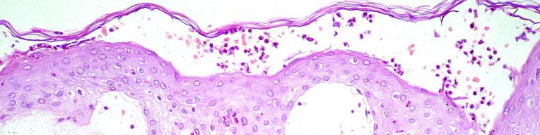
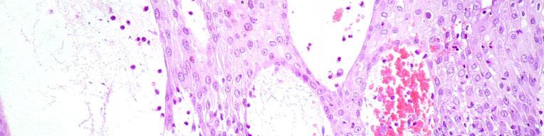
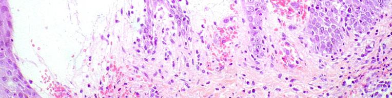
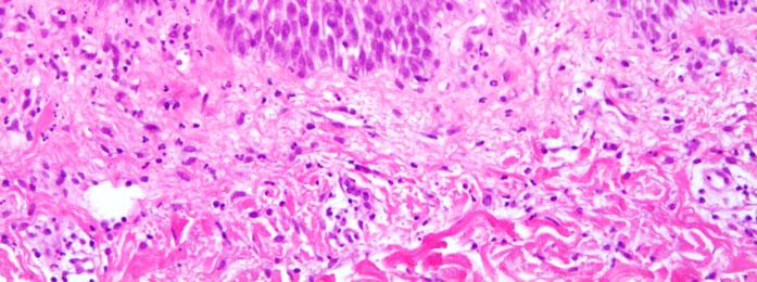
17 LEGENDS TO FIGURES Figure 1. Large non-spongiform subcorneal pustule, papillary oedema, and erythrocyte extravasation (haematoxylin-eosin stain, original magnification x 200). Figure 2. Small subcorneal pustule, presence of neutrophils and eosinophils in the epidermis and in the superficial dermis (haematoxylin-eosin stain, original magnification x 400). Figure 3. Large spongiform intraepidermal pustule with necrotic keratinocytes and spongiosis in the lower part of the epidermis. In the dermis there is discrete leucocytoclasia, but no vasculitis (haematoxylin-eosin stain, original magnification x 200).
18


19
20
21 Table 1: Histopathological parameters used in the evaluation of AGEP Degree Histological parameter of severity Pustule size* Histological parameter Degree of severity Eosinophils (pustule or dermis)/erythrocyte extravasation small (<10 keratinocytes) 1 1 or 2 cells 1 medium (10 15 keratinocytes) 2 >2 2 large (> 15 keratinocytes) 3 many 3 Spongiform pustule Munro abscess mild moderate prominent 3 >2 3 Follicular pustule Hyperkeratosis accessory 1 mild 1 predominant 2 moderate 2 solitary 3 prominent 3 Granular cell layer/parakeratosis absent 0 mild 1 up to 1/3 the length of the biopsy 1 prominent 2 up to 2/3 the length of the biopsy 2 vesicles 3 almost total 3 Spongiosis Exocytosis of neutrophils Suprapapillary plate thinning a few 1 1 papilla 1 scattered 2 2 papillae 2 many 3 >2 papillae 3 Necrotic keratinocytes Tortuous and dilated blood vessels/vasculitis 1 or vessel 1 >2 2 2 vessels 2 segmental necrosis or more 3 >2 vessels 3 Papillary oedema Rete ridges elongation/clubbing/fusion discrete moderate severe 3 >2 3 Infiltrates: superficial, interstitial, mid/deep-dermal Mitosis discrete 1 number per high power field moderate 2 (x40 magnification) dense 3 Dermal neutrophils a few 1 scattered 2 full fields 3 * In case of several pustules, the largest is used. Accumulation of micro-aggregates of neutrophils separated by degenerated and thinned keratinocytes. In conjunction with other types of pustules only. Without other types of pustules. Parakeratosis/granular layer above a pustule is not included. Suprapapillary plate thinning of the epidermis is defined as less than 3 layers of keratinocytes above the papillae. Leucocytoclastic vasculitis.
22 Table 2: The prevalence of histopathological parameters in 102 AGEP patients Histopathological parameter Degree of severity Prevalence (%) Pustules Sub/intracorneal and intraepidermal pustules 94 (92) Sub/intracorneal pustules 39 (41) large 3 32 (82) small 1 4 (10) spongiform 1,2,3 17 (44) presence of eosinophils 1,2,3 14 (36) Intraepidermal pustules 19 (20) large 3 17 (89) small 1 2 (10) spongiform 1,2,3 18 (95) presence of eosinophils 1,2,3 6 (32) Combined (sub/intracorneal and intraepidermal) 36 (38) Follicular pustules 22 (23) accessory (in conjunction with other pustules) 1 8 (36) predominant 2 9 (41) solitary 3 5 (23) Epidermis Spongiosis 82 (80) mild 1 62 (76) Exocytosis of neutrophils 79 (77) A few 1 53 (67) Necrotic keratinocytes 68 (67) 1-2 keratinocytes 1 32 (47) >2 keratinocytes 2 31 (46) segmental necrosis 3 5 (7) Dermis Papillary oedema 90 (88) discrete 1 41 (45) moderate 2 24 (27) severe 3 25 (28) Superficial infiltrates 102 (100) moderate 2 76 (74) Mid/deep-dermal infiltrates 97 (95) discrete 1 64 (66) Interstitial infiltrates 95 (93) discrete 1 38 (40) moderate 2 36 (38) Dermal neutrophils 102 (100) a few 1 24 (23) scattered 2 60 (59) Dermal eosinophils 83 (81) a few 1 61 (73) Vasculitis 1 (1) Erythrocyte extravasation 55 (54) 1-2 cells 1 27 (49) >2 cells 2 26 (47) many 3 2 (4)
23 Histopathological parameter Degree of severity Prevalence (%) Classical plaque-type psoriatic changes Munro abscess 17 (17) (59) 2 2,3 7 (41) Granular cell layer 99 (97) none 0 3 (3) Parakeratosis 63 (62) mild 1 35 (55) moderate 2 24 (38) Hyperkeratosis 26 (25) mild 1 25 (96) Suprapapillary plate thinning 7 (7) mild 1 7 (100) Tortuous and dilated blood vessels 16 (16) mild 1 8 (50) moderate 2 8 (50) Rete ridge elongation 78 (76) mild 1 38 (49) moderate 2 32 (41) Rete ridge clubbing 52 (51) mild 1 39 (75) Rete ridge fusion 83 (81) mild 1 35 (42) moderate 2 39 (47) Mitosis number per high-power field (x40 magnification) mean 0.95
The histopathological spectrum of acute generalized exanthematous pustulosis (AGEP) and its differentiation from generalized pustular psoriasis
 and its differentiation from generalized pustular psoriasis") J Cutan Pathol 2010: 37: 1220 1229 doi: 10.1111/j.1600-0560.2010.01612.x John Wiley & Sons. Printed in Singapore Copyright 2010 John Wiley & Sons A/S Journal of Cutaneous Pathology The histopathological
J Cutan Pathol 2010: 37: 1220 1229 doi: 10.1111/j.1600-0560.2010.01612.x John Wiley & Sons. Printed in Singapore Copyright 2010 John Wiley & Sons A/S Journal of Cutaneous Pathology The histopathological
Retrospective 10 years review of 100 patients with psoriasis in the Kingdom of Saudi Arabia (KSA)
") Retrospective 10 years review of 100 patients with psoriasis in the Kingdom of Saudi Arabia (KSA) Ahmed Abdullah Alhumidi King saud university, Riyadh, kingdom of Saudi Arabia Abstract Background: This
Retrospective 10 years review of 100 patients with psoriasis in the Kingdom of Saudi Arabia (KSA) Ahmed Abdullah Alhumidi King saud university, Riyadh, kingdom of Saudi Arabia Abstract Background: This
Guttate psoriasis =ﻒدﺼﻠا ﻲﻄﻘﻨﻠا
 1 / 69 Psoriasis Psoriasis may be divided into psoriasis vulgaris, generalized pustular psoriasis, and localized pustular ps Psoriasis Vulgaris Clinical Features 2 / 69 Psoriasis vulgaris is a common chronic
Psoraisis = ﻒدﺼﻠا 1 / 84
 1 / 84 2 / 84 3 / 84 4 / 84 5 / 84 6 / 84 Psoriasis Psoriasis may be divided into psoriasis vulgaris, generalized pustular psoriasis, and localized pustular ps Psoriasis Vulgaris Clinical Features 7 /
Agenda* The 9th International Congress on Cutaneous Adverse Drug Reactions (iscar 2015)
") 1 of 5 Agenda* The 9th International Congress on Cutaneous Adverse Drug Reactions (iscar 2015) Co-chairs: Dr. Neil H. Shear at the 23rd World Congress of Dermatology in Vancouver, Canada. Division of Dermatology,
1 of 5 Agenda* The 9th International Congress on Cutaneous Adverse Drug Reactions (iscar 2015) Co-chairs: Dr. Neil H. Shear at the 23rd World Congress of Dermatology in Vancouver, Canada. Division of Dermatology,
Acute generalized exanthematous pustulosis in the ambulatory care setting
 www.edoriumjournals.com CASE REPORT PEER REVIEWED OPEN ACCESS Acute generalized exanthematous pustulosis in the ambulatory care setting LT Mark C. Hubbard, LT John C. Walsh, LCDR Megan A. Brelsford ABSTRACT
Original Article. Palmoplantar Psoriasis versus Eczema: Major Histopathologic Clues for Diagnosis
 Iranian Journal of Pathology (2014) 9 (4), 251-256 251 Original Article Palmoplantar Psoriasis versus Eczema: Major Histopathologic Clues for Diagnosis Kambiz Kamyab Hesari, Zahra Safaei Naraghi, Azita
Actinic keratosis (AK): Dr Sarma s simple guide
: Dr Sarma s simple guide") Actinic keratosis (AK): Dr Sarma s simple guide Actinic keratosis is a very common lesion that you will see in your day-to-day practice. First, let me explain the name Actinic keratosis. It means keratosis
Actinic keratosis (AK): Dr Sarma s simple guide Actinic keratosis is a very common lesion that you will see in your day-to-day practice. First, let me explain the name Actinic keratosis. It means keratosis
University of Groningen
 University of Groningen Toxic epidermal necrolysis, DRESS, AGEP Bouvresse, Sophie; Valeyrie-Allanore, Laurence; Ortonne, Nicolas; Konstantinou, Marie Pauline; Kardaun, Sylvia H.; Bagot, Martine; Wolkenstein,
A Histopathologic Study of Papulosquamous Lesions of Skin
 Original 404 Article Indian Journal of Pathology: Research and Practice Volume 6 Number 2, April - June 2017 (Part 2) DOI: http://dx.doi.org/10.21088/ijprp.2278.148x.6217.11 A Histopathologic Study of
ORIGINAL ARTICLE. Siew Eng Choon, FRCP 1,2, Yi Shan Der 1, Nai Liang Joel Lai 1, Evelyn Sing Ee Yu 1, Xiao Ling Yap 1, Nalini Nanu Madhavan, MBBS 2
 ORIGINAL ARTICLE Clinical characteristics, culprit drugs and outcome of patients with Acute Generalised Exanthematous Pustulosis seen in Hospital Sultanah Aminah, Johor Bahru Siew Eng Choon, FRCP 1,2,
Citation The Journal of Dermatology, 37(8), available at
, available at") NAOSITE: Nagasaki University's Ac Title Two cases of blaschkitis with promi Author(s) Utani, Atsushi Citation The Journal of Dermatology, 37(8), Issue Date 2010-08 URL Right http://hdl.handle.net/10069/25634
NAOSITE: Nagasaki University's Ac Title Two cases of blaschkitis with promi Author(s) Utani, Atsushi Citation The Journal of Dermatology, 37(8), Issue Date 2010-08 URL Right http://hdl.handle.net/10069/25634
REGISTRY OF SEVERE CUTANEOUS ADVERSE REACTIONS TO DRUGS AND COLLECTION OF BIOLOGICAL SAMPLES. R e g i S C A R PATIENT'S DATA. Age country of birth
 REGISTRY OF SEVERE CUTANEOUS ADVERSE REACTIONS TO DRUGS AND COLLECTION OF BIOLOGICAL SAMPLES R e g i S C A R PATIENT'S DATA Initials of the patient date of birth Age country of birth Gender male female
Egyptian Dermatology Online Journal Vol. 6 No 1: 14, June 2010
 Wells Syndrome H. Gammaz, H. Amer, A. Adly and S. Mahmoud Egyptian Dermatology Online Journal 6 (1): 14 Al-Haud Al-Marsoud Hospital, Cairo, Egypt e-mail: hananderma@hotmail.com Submitted: April 15, 2010
Department of Dermatology, Christian Medical College and Hospital, Ludhiana, Punjab, India.
 Bullous pemphigoid mimicking granulomatous inflammation Abhilasha Williams, Emy Abi Thomas. Department of Dermatology, Christian Medical College and Hospital, Ludhiana, Punjab, India. Egyptian Dermatology
=ﻰﻤاﻤﺤﻠا ﺔﻴﻘﻠﺤﻠا ﺔذﺒاﻨﻠا
 1 / 15 Erythema Annulare Centrifugum and Other Figurate Erythemas The figurate erythemas include a variety of eruptions characterized by annular and polycyclic lesions. Classification of this group has
MECHANISMS OF HUMAN DISEASE: LABORATORY SESSION PATHOLOGY OF THE SKIN LAB. Friday, February 12, :30 am 11:00 am
 MECHANISMS OF HUMAN DISEASE: LABORATORY SESSION PATHOLOGY OF THE SKIN LAB Friday, February 12, 2012 9:30 am 11:00 am FACULTY COPY GOALS: Describe the basic clinical and morphologic features of various
Paradoxial Safety Signals from Biologics
 Paradoxial Safety Signals from Biologics Christopher Ritchlin, MD, MPH Professor of Medicine Director, Translational Immunology Research Center University of Rochester Medical Center Rochester, NY Disclosures
Case Rep Dermatol 2009;1:66 70 DOI: / Key Words Coma Blister Barbiturate Overdose Meningoencephalitis
 66 Coma Blisters Joana Rocha a Teresa Pereira a Filipa Ventura a Fernando Pardal b Celeste Brito a Departments of a Dermatology and b Pathology, Hospital de São Marcos, Braga, Portugal Key Words Coma Blister
Title: Erythema annulare centrifugum associated with chronic lymphocytic leukaemia. Authors: Helbling I, Walewska R, Dyer MJS, Bamford M, Harman KE
 Title: Erythema annulare centrifugum associated with chronic lymphocytic leukaemia Authors: Helbling I, Walewska R, Dyer MJS, Bamford M, Harman KE Sir, A wide range of conditions have been described as
Supplementary Online Content
 Supplementary Online Content Ross NA, Chung H-J, Li Q, Andrews JP, Keller MS, Uitto J. Pityriasis rubra pilaris: a case series of patients. Published online March 9, 26. JAMA Dermatol. doi:./jamadermatol.26.9.
CD30 + cells in benign inflammatory infiltrate of some dermatological diseases. Abstract. Latef M. El Balshy. Benha University-Benha, Egypt.
 CD30 + cells in benign inflammatory infiltrate of some dermatological diseases 1 Asmaa M. El Refaeie, 1 Osama H. Abdel Salam, 1 Sherine H.Abd EL-Rahman and 2 Abdel Latef M. El Balshy. 1 Dermatology & Andrology
Vancomycin-induced acute generalized exanthematous pustulosis (AGEP) masquerading septic shock an unusual presentation of a rare disease
 masquerading septic shock an unusual presentation of a rare disease") Mawri et al. Journal of Intensive Care (2015) 3:47 DOI 10.1186/s40560-015-0114-3 CASE REPORT Vancomycin-induced acute generalized exanthematous pustulosis (AGEP) masquerading septic shock an unusual presentation
Mawri et al. Journal of Intensive Care (2015) 3:47 DOI 10.1186/s40560-015-0114-3 CASE REPORT Vancomycin-induced acute generalized exanthematous pustulosis (AGEP) masquerading septic shock an unusual presentation
THERE IS A GROUP OF PAtients. Defining Urticarial Dermatitis. A Subset of Dermal Hypersensitivity Reaction Pattern
 STUDY Defining Urticarial Dermatitis A Subset of Dermal Hypersensitivity Reaction Pattern Steven Kossard, FACD; Ian Hamann, FACD; Barbara Wilkinson, BSc Background: Urticarial dermatitis may represent
A Descriptive Study on Patients of Papulosquamous Lesion at Tertiary Care Institute
 MVP Journal of Medical Sciences, Vol 1(1), 30 35, January 2014 A Descriptive Study on Patients of Papulosquamous Lesion at Tertiary Care Institute S. D. Chavhan 1*, S. V. Mahajan 2 and A. J. Vankudre 3
Histopathology: skin pathology
 Histopathology: skin pathology These presentations are to help you identify, and to test yourself on identifying, basic histopathological features. They do not contain the additional factual information
Drug Allergy A Guide to Diagnosis and Management
 Drug Allergy A Guide to Diagnosis and Management (Version 1 April 2015 updated April 2018) Author: Jed Hewitt Chief Pharmacist, Governance & Professional Practice Date of Preparation: April 2015 Updated:
Mucinoses Diverse group of disorders which have in common deposition of basophilic, finely granular and stringy material in the connective tissues of
 Cutaneous Mucinoses Nathan C. Walk, M.D. Mucinoses Diverse group of disorders which have in common deposition of basophilic, finely granular and stringy material in the connective tissues of the dermis.
Pathology of the skin. Dr Fónyad László, 1sz. Patológiai és Kísérleti Rákkutató Intézet, SE
 Pathology of the skin Dr Fónyad László, 1sz. Patológiai és Kísérleti Rákkutató Intézet, SE The skin Biggest organ Kb. 1.8 nm Kb. 10 kg Most frequent site for tumor development (BCC) Pathology of the skin
CUTANEOUS DRUG REACTIONS OR I WOULDN T HAVE SEEN IT, IF I HADN T BELIEVED IT Edmund J. Rosser Jr., DVM, DACVD
 CUTANEOUS DRUG REACTIONS OR I WOULDN T HAVE SEEN IT, IF I HADN T BELIEVED IT Edmund J. Rosser Jr., DVM, DACVD DERMATOLOGY Pathogenesis Immunologic: can involve Type I, II, III, IV hypersensitivity reactions.
MECHANISMS OF HUMAN DISEASE: LABORATORY SESSION PATHOLOGY OF THE SKIN LAB. Friday, February 13, :30 am 11:00 am
 MECHANISMS OF HUMAN DISEASE: LABORATORY SESSION PATHOLOGY OF THE SKIN LAB Friday, February 13, 2009 9:30 am 11:00 am FACULTY COPY GOALS: Describe the basic clinical and morphologic features of various
Pimples and Boils!! Dr Nathan Harvey Anatomical Pathology, PathWest
 Pimples and Boils!! Dr Nathan Harvey Anatomical Pathology, PathWest Overview & Learning Objectives Review the cardinal signs/symptoms of acute inflammation Review the histological features of acute inflammation
Histopathological spectrum of non-infectious erythematous, papulo-squamous lesions
 Histopathological spectrum of non-infectious erythematous, papulo-squamous lesions B. Rajasekhar Reddy 1*, Nalini Krishna.M 2 1 Associate Professor, Department of Pathology, Mediciti Institute of Medical
HEMORRHAGIC BULLOUS HENOCH- SCHONLEIN PURPURA: A CASE REPORT
 HEMORRHAGIC BULLOUS HENOCH- SCHONLEIN PURPURA: A CASE REPORT Nirmala Ponnuthurai, Sabeera Begum, Lee Bang Rom Paediatric Dermatology Unit, Institute of Paediatric, Hospital Kuala Lumpur, Malaysia Abstract
ISPUB.COM. Seborrheic Keratosis: A Pictorial Review of the Histopathologic Variations. D Sarma, S Repertinger
 ISPUB.COM The Internet Journal of Dermatology Volume 7 Number 2 Seborrheic Keratosis: A Pictorial Review of the Histopathologic Variations D Sarma, S Repertinger Citation D Sarma, S Repertinger.. The Internet
Chronology of lichen planus-like keratosis features by dermoscopy: a summary of 17 cases
 DERMATOLOGY PRACTICAL & CONCEPTUAL www.derm101.com Chronology of lichen planus-like keratosis features by dermoscopy: a summary of 17 cases Soko Watanabe 1, Mizuki Sawada 1, Itaru Dekio 1, Sumiko Ishizaki
BSD Self Assessment Workshop 7 th July 2013 CASE 27 RAC6123
 BSD Self Assessment Workshop 7 th July 2013 CASE 27 RAC6123 M55. 4/7 tender lesions on knee, legs and arms. Also iritis/ weight loss/headache, synovitis.?vasculitis. Sarcoidosis. Biopsy from left elbow
Rashes Not To Be Missed In Children
 May 2016 Rashes Not To Be Missed In Children Dr Chan Yuin Chew Dermatologist Dermatology Associates Gleneagles Medical Centre Scope of presentation Focus on rashes May lead to significant morbidity if
Inflammatory skin disease I Jade Wititsuwannakul, MD Chulalongkorn University, Thailand
 Inflammatory skin disease I Jade Wititsuwannakul, MD Chulalongkorn University, Thailand Superficial Perivascular Dermatitis Interface Dermatitis Vacuolar Dermatitis Lichenoid Dermatitis Barnhill Textbook
Important Decisions in Dermatopathology: The Clinico- Pathologic Correlation. Dermatopathology Specialists Needed. Changing Trends
 Important Decisions in Dermatopathology: The Clinico- Pathologic Correlation Uma Sundram, MD, PhD Departments of Pathology and Dermatology Stanford University May 29, 2008 Dermatopathology Specialists
A case of rosacea fulminans in a pregnant woman
 Hong Kong J. Dermatol. Venereol. (2018) 26, 122-126 Views and Practice A case of rosacea fulminans in a pregnant woman JE Seol, SH Park, JU Kim, GJ Cho, SH Moon, H Kim Introduction Rosacea fulminans (RF)
Cutaneous drug reactions
 Maintenance of Certification clinical management series Series editor: James T. Li, MD, PhD Cutaneous drug reactions David A. Khan, MD Dallas, Tex INSTRUCTIONS Credit can now be obtained, free for a limited
OXCARBAZEPINE-INDUCED STEVENS-JOHNSON SYNDROME: A CASE REPORT
 OXCARBAZEPINE-INDUCED STEVENS-JOHNSON SYNDROME: A CASE REPORT Lung-Chang Lin, 1,2 Ping-Chin Lai, 3 Sheau-Fang Yang, 4 and Rei-Cheng Yang 1,5 Departments of 1 Pediatrics and 4 Pathology, Kaohsiung Medical
They are updated regularly as new NICE guidance is published. To view the latest version of this NICE Pathway see:
 bring together everything NICE says on a topic in an interactive flowchart. are interactive and designed to be used online. They are updated regularly as new NICE guidance is published. To view the latest
Emergency Dermatology Dr Melissa Barkham
 Emergency Dermatology Dr Melissa Barkham Spotlight Seminar 30 th September 2010 Why is this important? Urgent recognition and treatment of dermatologic emergencies can be life saving and prevent long term
CPC. Chutika Srisuttiyakorn, M.D. Kobkul Aunhachoke, M.D. Phramongkutklao Hospital Bangkok, Thailand
 CPC Chutika Srisuttiyakorn, M.D. Kobkul Aunhachoke, M.D. Phramongkutklao Hospital Bangkok, Thailand A 53 year-old woman with fever, facial swelling and rashes on face, trunk and upper extremities for 3
Benign Lichenoid Keratosis
 Benign Lichenoid Keratosis ALAN F. FRIGY, M.D. AND PHILIP H. COOPER, M.D. The microscopic spectrum of benign lichenoid keratosis (BLK) was studied by examination of 30 examples. BLK consists of a segment
Annex I. Scientific conclusions and grounds for the variation to the terms of the Marketing Authorisation(s)
") Annex I Scientific conclusions and grounds for the variation to the terms of the Marketing Authorisation(s) 1 Scientific conclusions Taking into account the PRAC Assessment Report on the PSUR(s) for ibuprofen
Annex I Scientific conclusions and grounds for the variation to the terms of the Marketing Authorisation(s) 1 Scientific conclusions Taking into account the PRAC Assessment Report on the PSUR(s) for ibuprofen
Grover s disease: A case report.
 320 Case report Thai J Dermatol, October-December 2011 ABSTRACT: Grover s disease: A case report. Supicha Chavanich MD, Praneet Sajjachareonpong MD. CHAVANICH C, SAJJACHAREONPONG P. GROVER S DISEASE: A
Hospital-based Dermatopathology. Janis M. Taube, MD Director of Dermatopathology Johns Hopkins University SOM
 Hospital-based Dermatopathology Janis M. Taube, MD Director of Dermatopathology Johns Hopkins University SOM Overview Drug-eruptions Erythroderma Manifestations of renal disease Blistering disorders Vasculitis/Vasculopathy
Microscopic study of the normal skin in cases of mycosis fungoides
 Microscopic study of the normal skin in cases of mycosis fungoides M. El Darouti, S. Marzook, M. Bosseila, O. Abu Zeid, O. El- Safouri, A. Zayed, A. El-Ramly Egyptian Dermatology Online Journal 2 (1):
Proceedings of the Southern European Veterinary Conference - SEVC -
 Close this window to return to IVIS www.ivis.org Proceedings of the Southern European Veterinary Conference - SEVC - Sep. 30-Oct. 3, 2010, Barcelona, Spain Next SEVC Conference: Sep. 30-Oct. 2, 2011 -
New product information wording Extracts from PRAC recommendations on signals
 12 October 2017 EMA/PRAC/610988/2017 Pharmacovigilance Risk Assessment Committee (PRAC) New product information wording Extracts from PRAC recommendations on signals Adopted at the 25-29 September 2017
Prevalence and pattern of adverse cutaneous drug reactions presenting to a tertiary care hospital
 International Journal of Research in Dermatology Janardhan B et al. Int J Res Dermatol. 2017 Mar;3(1):74-78 http://www.ijord.com Original Research Article DOI: http://dx.doi.org/10.18203/issn.2455-4529.intjresdermatol20164248
EFAVIRENZ ASSOCIATED STEVENS-JOHNSON SYNDROME
 EFAVIRENZ ASSOCIATED STEVENS-JOHNSON SYNDROME To the Editor: Persons with human immunodeficiency virus (HIV) infection are highly susceptible to adverse dermatological reactions to specific medications
Psoriasiform pemphigus foliaceus: a report of two cases
 J Cutan Pathol 2012: 39: 549 553 doi: 10.1111/j.1600-0560.2012.01866.x John Wiley & Sons. Printed in Singapore Copyright 2012 John Wiley & Sons A/S Journal of Cutaneous Pathology Psoriasiform pemphigus
Original Contribution
 Direct Immunofluorescence Test of Skin Biopsy Samples Results of 204 Cases Kabir AN, 1 Das RK, 2 Kamal M 3 Direct immunofluorescence (DIF) test of skin and renal biopsy specimens is being done on regular
Comparative microanatomy of the normal skin with that of immunobullous condition
 Original article: Comparative microanatomy of the normal skin with that of immunobullous condition 1Dr BananiKundu, 2 DrAnirban Sadhu, 3 DrRudradev Meyur, 4 Dr SauravKundu, 5 Dr Alpana De, 6Dr SatabdiSarkar
ISPUB.COM. A Case of Actinic Lichen Planus. K Choi, H Kim, H Kim, Y Park INTRODUCTION CASE REPORT
 ISPUB.COM The Internet Journal of Dermatology Volume 8 Number K Choi, H Kim, H Kim, Y Park Citation K Choi, H Kim, H Kim, Y Park.. The Internet Journal of Dermatology. 2009 Volume 8 Number. Abstract The
Skin Manifestations of Drug Reactions
 Skin Manifestations of Drug Reactions Dr Carol Hlela, Division of Dermatology Department of Medicine, University of Cape Town and Red Cross Children s Hospital What are the Skin Manifestations of Drug
CLINCOPATHOLOGICAL CASE
 CLINCOPATHOLOGICAL CASE Generalized vesiculo-bullous and pustular eruption in an adult man Hassab El-Naby H, MD, El-Khalawany M, MD Department of Dermatology, Al-Azhar University, Cairo, Egypt CLINICAL
A. Erythema multiforme and related diseases
 Go Back to the Top To Order, Visit the Purchasing Page for Details Chapter Erythema, Erythroderma (Exfoliative Dermatitis) Erythema is caused by telangiectasia or hyperemia in the papillary and reticular
insight Exceptional SKINTELL in-depth, non-invasive dermatological solution HealthCare
 Optical Coherence Tomography insight Exceptional SKINTELL in-depth, non-invasive dermatological solution HealthCare From outside, exceptional insight inside SKINTELL from Agfa HealthCare enables you to
BJD. Summary. British Journal of Dermatology THERAPEUTICS
 THERAPEUTICS BJD British Journal of Dermatology Drug reaction with eosinophilia and systemic symptoms: is cutaneous phenotype a prognostic marker for outcome? A review of clinicopathological features of
Dilated Lymphatics in Gottron s Papules
 2010;18(2):99-103 CASE REPORT Dilated Lymphatics in Gottron s Papules Angel Fernandez-Flores Service of Cellular Pathology, Clinica Ponferrada, Ponferrada, Spain Corresponding author: Angel Fernandez-Flores,
Lymphomatoid Papulosis 3 Case Reports
 IOSR Journal of Dental and Medical Sciences (IOSR-JDMS) e-issn: 2279-0853, p-issn: 2279-0861.Volume 14, Issue 7 Ver. III (July. 2015), PP 31-35 www.iosrjournals.org Lymphomatoid Papulosis 3 Case Reports
DISTRIBUTION OF HUMAN Β-DEFENSIN 2, TNF-ALPHA, IL-1 ALPHA, IL-6 AND IL-8 IN PSORIATIC SKIN
 Papers on Anthropology XX, 2011, pp. 289 302 DISTRIBUTION OF HUMAN Β-DEFENSIN 2, TNF-ALPHA, IL-1 ALPHA, IL-6 AND IL-8 IN PSORIATIC SKIN E. Mozeika 1,2, M. Pilmane 1, J. Kisis 2 1 Institute of Anatomy and
Observations on the Pathology of Lesions Associated with Stephanofilaria dinniki Round, 1964 from the Black Rhinoceros (Diceros bicornis)
") Journal of Helminthology, ~ol. XXXVIII, Nos. 1/2, 1964, pp. 171-174. Observations on the Pathology of Lesions Associated with Stephanofilaria dinniki Round, 1964 from the Black Rhinoceros (Diceros bicornis)
Journal of Helminthology, ~ol. XXXVIII, Nos. 1/2, 1964, pp. 171-174. Observations on the Pathology of Lesions Associated with Stephanofilaria dinniki Round, 1964 from the Black Rhinoceros (Diceros bicornis)
A Chronic or Recurring Pattern of Esophagitis Resembling Allergic Contact Dermatitis
 Anatomic Pathology / Lymphocytic Esophagitis Lymphocytic Esophagitis A Chronic or Recurring Pattern of Esophagitis Resembling Allergic Contact Dermatitis Julianne K. Purdy, MD, Henry D. Appelman, MD, Christopher
Basal cell carcinoma 5/28/2011
 Goal of this Presentation A practical approach to the diagnosis of cutaneous carcinomas and their mimics Thaddeus Mully, MD University of California San Francisco To review common non-melanoma skin cancers
A 40-year old male with follicular papule and pustule at central face area for 3 months
 A 40-year old male with follicular papule and pustule at central face area for 3 months GMS- Neg AFB-Neg Fite stain - neg HISTOPATHOLOGICAL DIFFERENTIAL DIAGNOSIS CASEOUS GRANULOMA INFECTION -MYCOBACTERIUM
An Evidenced-Based Approach to the Adult with a Morbilliform Eruption
 Relatively An Evidenced-Based Approach to the Adult with a Morbilliform Eruption Ben Kaffenberger, MD Assistant Professor, Dermatology Director, Inpatient Dermatology Consult Service Ohio State University
Original Article ABSTRACT
 Original Article Drug reaction with eosinophilia and systemic symptoms (DRESS): A histopathology based analysis Sarita Sasidharanpillai, Aparna Govindan 1, Najeeba Riyaz, Manikoth P. Binitha, Kunnummal
Clinicopathologic Evaluation of Nodular Cutaneous Lesions of Behçet Syndrome
 Anatomic Pathology / NODULAR CUTANEOUS LESIONS OF BEHÇET SYNDROME Clinicopathologic Evaluation of Nodular Cutaneous Lesions of Behçet Syndrome Cuyan Demirkesen, MD, 1 Nükhet Tüzüner, MD, 1 Cem Mat, MD,
A case of Sweet's syndrome with marked facial swelling and fluid collection
 Hong Kong J. Dermatol. Venereol. (2017) 25, 128-132 Case Report A case of Sweet's syndrome with marked facial swelling and fluid collection JE Seol, SH Park, DH Kim, JN Kang, H Kim A 43-year-old woman
My Method for Approaching Skin Biopsies
 My Method for Approaching Skin Biopsies P A U L H A U N, MD, MS, F A A D A S S I S T A N T P R O F E S S O R D E R M A T O L O G Y A N D D E R M A T O P A T H O L O G Y D E P A R T M E N T O F D E R M
COPYRIGHTED MATERIAL. Introduction CHAPTER 1. Introduction
 CHAPTER 1 Introduction OVERVIEW The clinical features of skin lesions are related to the underlying pathological processes. Broadly skin conditions fall into three clinical groups: (a) those with a well-defined
Gastrooesophageal reflux disease. Jera Jeruc Institute of pathology, Faculty of Medicine, Ljubljana, Slovenia
 Gastrooesophageal reflux disease Jera Jeruc Institute of pathology, Faculty of Medicine, Ljubljana, Slovenia Reflux esophagitis (RE) GERD: a spectrum of clinical conditions and histologic alterations resulting
Oral Manifestations of Dermatologic Disease: A Focus on Lichenoid Lesions. Proceedings of the NASHNP Companion Meeting, March, 2011, San Antonio, TX
 1 Oral Manifestations of Dermatologic Disease: A Focus on Lichenoid Lesions Proceedings of the NASHNP Companion Meeting, March, 2011, San Antonio, TX Susan Müller, DMD, MS Professor Department of Pathology
المركب النموذج--- سبيتز وحمة = Type Spitz's Nevus, Compound SPITZ NEVUS 1 / 7
 SPITZ NEVUS 1 / 7 Epidemiology An annual incidence rate of 1.4 cases of Spitz nevus per 100,000 individuals has been estimated in Australia, compared with 25.4 per 100,000 individuals for cutaneous melanoma
CASE REPORT ATYPICAL BULLOUS PYODERMA GANGRENOSUM WITH EARLY LESIONS MIMICKING CHICKEN POX
 ATYPICAL BULLOUS PYODERMA GANGRENOSUM WITH EARLY LESIONS MIMICKING CHICKEN POX Ramesh M 1, Kavya Raju Nayak 2, M.G. Gopal 3, Sharath Kumar B.C 4, Nandini A.S 5 HOW TO CITE THIS ARTICLE: Ramesh M, Kavya
Case No. 5; Slide No. B13/8956/2
 Interface diseases Case No. 5; Slide No. B13/8956/2 Histological findings Severe hydropic vacuolation of epidermal and follicular basal cells/ interface dermatitis Multifocally apoptotic keratinocytes
The Role of Plasmacytoid Dendritic Cells in Psoriasis
 6 ème Journée du Groupe de Recherche sur le Psoriasis Paris, 9 novembre 2012 The Role of Plasmacytoid Dendritic Cells in Psoriasis Dr Curdin Conrad, PD & MER Department of Dermatology Psoriasis: Clinical
A dinical and histopathologic entity associated with an increased risk of nonmelanoma skin cancer
 PUVA keratosis A dinical and histopathologic entity associated with an increased risk of nonmelanoma skin cancer M. C. G. van Praag, MD, a J. N. Bouwes Bavinck, MD, a W. Bergman, MD, PhD, a F. R. Rosendaal,
Patterns and mechanisms of inflammatory skin conditions: the pathologist s survival kit SALVADOR J. DIAZ-CANO BAHRAIN, APRIL 2017
 Patterns and mechanisms of inflammatory skin conditions: the pathologist s survival kit SALVADOR J. DIAZ-CANO 0000-0003-1245-2859 BAHRAIN, APRIL 2017 Basic Elements of Lesions Repair Injury Time & Intensity
Warfarin-induced toxic epidermal necrolysis in combination therapy of Henoch- Schönlein purpura nephritis: a case report
 Kasahara et al. BMC Nephrology (2017) 18:237 DOI 10.1186/s12882-017-0648-9 CASE REPORT Open Access Warfarin-induced toxic epidermal necrolysis in combination therapy of Henoch- Schönlein purpura nephritis:
Original Research Article
 CLINICOPATHOLOGICAL STUDY OF PAPULOSQUAMOUS SKIN LESIONS Chowdari Balaji 1, Metta Parvathi 2, Seeram Satish Kumar 3, Gunta Divya Lekha 4, Latchupatula Lavanya 5, Mantripragada Vidya Soundarya Lahari 6,
Cutaneous Drug Reactions
 Cutaneous Drug Reactions Andrei Metelitsa, MD, FRCPC, FAAD Co-Director, Institute for Skin Advancement Clinical Associate Professor, Dermatology University of Calgary, Canada Copyright 2017 by Sea Courses
Purpuric drug eruption without leukocytoclastic vasculitis associated with vancomycin
 Asian Pacific Journal of Allergy and Immunology CASE REPORT Purpuric drug eruption without leukocytoclastic vasculitis associated with vancomycin Tamaki Nakamura, 1 Hiroyuki Wakiguchi, 1 Fumiko Okazaki,
Psoriasis Penis - A Two Case Report
 Article ID: WMC003176 ISSN 2046-1690 Psoriasis Penis - A Two Case Report Corresponding Author: Dr. Ilko Bakardzhiev, MD, PhD, Medical College, Medical University of Varna, 9000 - Bulgaria Submitting Author:
Pathology of the skin. 2nd Department of Pathology, Semmelweis University
 Pathology of the skin 2nd Department of Pathology, Semmelweis University Histology of the skin Epidermis: Stratum corneum Stratum granulosum Stratum spinosum Stratum basale Dermis: papillary and reticular
Future of Pediatrics: Blisters, Hives and Other Tales from the Emergency Room June 14 th, 2016
 A. Yasmine Kirkorian MD Assistant Professor of Dermatology & Pediatrics Children s National Health System George Washington University School of Medicine & Health Sciences Future of Pediatrics: Blisters,
Classification: 1. Infective: 2. Traumatic: 3. Idiopathic: Recurrent Aphthous Stomatitis (RAS) 4. Associated with systemic disease:
 4. Associated with systemic disease:") Classification: 1. Infective: 2. Traumatic: 3. Idiopathic: Recurrent Aphthous Stomatitis (RAS) 4. Associated with systemic disease: Hematological GIT Behcet s HIV 5. Associated with dermatological diseases:
Classification: 1. Infective: 2. Traumatic: 3. Idiopathic: Recurrent Aphthous Stomatitis (RAS) 4. Associated with systemic disease: Hematological GIT Behcet s HIV 5. Associated with dermatological diseases:
The Utility Of Congo Red Stain And Cytokeratin Immunostain In The Detection Of Primary Cutaneous Amyloidosis
 ISPUB.COM The Internet Journal of Pathology Volume 17 Number 1 The Utility Of Congo Red Stain And Cytokeratin Immunostain In The Detection Of Primary Cutaneous A,A Citation A, A.. The Internet Journal
Some skin conditions
 Some skin conditions Some skin conditions Acute Inflammatory Dermatoses Chronic Inflammatory Dermatoses Blistering (Bullous) Diseases Panniculitis Disorders of Epidermal Appendages -Urticaria -Acute eczematous
33 rd Annual Meeting of the Israel Society of Dermatology and Venereology Meridian Hotel, Dead Sea, Israel, June 16-17, 2010
 First Announcement and Call for Papers Dear Colleagues, The Department of of Soroka University Medical Center, takes great pleasure to invite you to attend the 33rd Annual Meeting of the Israel Society
Inpatient dermatopathology. Inpatient dermatopathology. Inpatient dermatopathology: Histologic patterns that matter most
 Inpatient dermatopathology: Histologic patterns that matter most Beth S. Ruben, M.D. Associate Clinical Professor University of California, San Francisco Departments of Dermatology and Pathology Dermatopathology
Comment on Association of bullous pemphigoid with malignancy: A systematic review and meta-analysis
 Accepted Manuscript Comment on Association of bullous pemphigoid with malignancy: A systematic review and meta-analysis Maglie Roberto, MD, Antiga Emiliano, MD, PhD, Caproni Marzia, MD, PhD PII: S0190-9622(17)32812-8
CHAPTER 22 Brocq s alopecia (pseudopelade of Brocq) and burnt out scarring alopecia
 and burnt out scarring alopecia") CHAPTER 22 Brocq s alopecia (pseudopelade of Brocq) and burnt out scarring alopecia BROCQ S ALOPECIA The term pseudopelade of Brocq is a source of much confusion and fruitless debate, and should be abandoned.
CHAPTER 22 Brocq s alopecia (pseudopelade of Brocq) and burnt out scarring alopecia BROCQ S ALOPECIA The term pseudopelade of Brocq is a source of much confusion and fruitless debate, and should be abandoned.
Early View Article: Online published version of an accepted article before publication in the final form.
 : Online published version of an accepted article before publication in the final form. Journal Name: International Journal of Case Reports and Images (IJCRI) Type of Article: Case Report Title: A Case