Novel LMNA mutations in patients with Emery-Dreifuss muscular dystrophy and functional characterization of four LMNA mutations
|
|
|
- Gabriel McDowell
- 5 years ago
- Views:
Transcription

, pp.. <0.00/humu.>.")

1 Novel LMNA mutations in patients with Emery-Dreifuss muscular dystrophy and functional characterization of four LMNA mutations Juergen Scharner, Charlotte A Brown, Matthew Bower, Susan T Iannaccone, Ismail A Khatri, Diana Escolar, Erynn Gordon, Kevin Felice, Carol A Crowe, Carla Grosmann, et al. To cite this version: Juergen Scharner, Charlotte A Brown, Matthew Bower, Susan T Iannaccone, Ismail A Khatri, et al.. Novel LMNA mutations in patients with Emery-Dreifuss muscular dystrophy and functional characterization of four LMNA mutations., Wiley, 0, (), pp.. <0.00/humu.>. <hal-000> HAL Id: hal Submitted on Jul 0 HAL is a multi-disciplinary open access archive for the deposit and dissemination of scientific research documents, whether they are published or not. The documents may come from teaching and research institutions in France or abroad, or from public or private research centers. L archive ouverte pluridisciplinaire HAL, est destinée au dépôt et à la diffusion de documents scientifiques de niveau recherche, publiés ou non, émanant des établissements d enseignement et de recherche français ou étrangers, des laboratoires publics ou privés.
2 Novel LMNA mutations in patients with Emery-Dreifuss muscular dystrophy and functional characterization of four LMNA mutations Journal: Manuscript ID: humu-00-0.r Wiley - Manuscript type: Research Article Date Submitted by the Author: -Aug-00 Complete List of Authors: Scharner, Juergen; King's College London, Randall Division of Cell and Molecular Biophysics Brown, Charlotte; BD Diagnostics Bower, Matthew; Fairview-University Medical Center, Department of Pediatrics Iannaccone, Susan; UT Southwestern Medical Center, Texas Scottish Rite Hospital for Children Khatri, Ismail; Shifa International Hospital, Division of Neurology Escolar, Diana; Children s National Medical Center, Research Center for Genetic Medicine Gordon, Erynn; Coriell Institute for Medical Research Felice, Kevin; Hospital for Special Care, Department of Neuromuscular Medicine Crowe, Carol; MetroHealth Medical Center, Department of Pediatrics Grosmann, Carla; Rady Children s Hospital San Diego, Department of Neurology Meriggioli, Matthew; University of Illinois, Department of Neurology and Rehabilitation Asamoah, Alexander; Weisskopf Child Evaluation Center, University of Louisville, Department of Pediatrics Gordon, Ora; Cedar-Sinai Medical Center, Medical Genetics Institute Gnocchi, Viola; King's College London, Randall Division of Cell and Molecular Biophysics Ellis, Juliet; King's College London, Randall Division of Cell and Molecular Biophysics Mendell, Jerry; Ohio State University, and the Research Institute at Nationwide Children s Hospital, Departments of Pediatrics and Neurology Zammit, Peter; king's College London, Randall Division of Cell and Molecular Biophysics Key Words: lamin A, lamin B, emerin, EDMD, LGMD, L-CMD, nuclear envelope, nuclear lamina, laminopathy, skeletal muscle
3 Page of
4 Page of Novel LMNA mutations in patients with Emery-Dreifuss muscular dystrophy and functional characterization of four LMNA mutations Juergen Scharner,, Charlotte A. Brown,, Matthew Bower, Susan T. Iannaccone, Ismail A. Khatri, Diana Escolar, Erynn Gordon, Kevin Felice, Carol A. Crowe, Carla Grosmann, 0 Matthew N. Meriggioli, Alexander Asamoah, Ora Gordon, Viola F. Gnocchi, Juliet A. Ellis, Jerry R. Mendell, * Peter S. Zammit * Randall Division of Cell and Molecular Biophysics, King s College London, UK; BD Diagnostics, Durham, NC, USA; Department of Pediatrics, Fairview-University Medical Center, Minneapolis, MN; Texas Scottish Rite Hospital for Children, UT Southwestern Medical Center, Dallas, TX; Division of Neurology, Shifa International Hospital, Islamabad, Pakistan; Research Center for Genetic Medicine, Children s National Medical Center, Washington, D.C.; Coriell Institute for Medical Research, Camden, NJ Department of Neuromuscular Medicine, Hospital for Special Care, New Britain, CT; Department of Pediatrics, MetroHealth Medical Center, Cleveland, OH; 0 Department of Neurology, Rady Children s Hospital San Diego, San Diego, CA; Department of Neurology and Rehabilitation, University of Illinois, Chicago, IL; Department of Pediatrics, Weisskopf Child Evaluation Center, University of Louisville, KY; Medical Genetics Institute, Cedar-Sinai Medical Center, Los Angeles, CA; Department of Pediatrics and Neurology, Ohio State University, and the Research Institute at Nationwide Children s Hospital, Columbus, OH; These authors contributed equally to this work * These authors are joint last authors. Correspondence to: peter.zammit@kcl.ac.uk; jerry.mendell@nationwidechildrens.org
5 Page of Abstract Mutations in LMNA cause a variety of diseases affecting striated muscle including autosomal- Emery-Dreifuss muscular dystrophy (EDMD), LMNA-associated congenital muscular dystrophy (L-CMD) and limb-girdle muscular dystrophy type B (LGMDB). Here, we describe novel and recurrent LMNA mutations identified in 0 patients from the USA and Canada, which is the first report of the distribution of LMNA mutations from a large cohort outside Europe. This augments the number of LMNA mutations known to cause EDMD by.%, equating to an increase of.% in the total known LMNA mutations. Eight patients presented with p.rw/q or p.ek mutations and an early onset EDMD phenotype: two mutations recently associated with L-CMD. Importantly, mutations are novel and include eight missense mutations (p.rp, p.f0l, p.sp, p.sp, p.ek, p.gd, p.lp and p.wr), three splice site mutations (c.ivs+g>a, c.ivs-a>g, c.ivs+g>a), one duplication/in frame insertion (p.r0dup), one deletion (p.qdel) and two silent mutations (p.rr and p.k0k). Analysis of of our lamin A mutations showed that some caused nuclear deformations and lamin B re-distribution in a mutation specific manner. Together, this study significantly augments the number of EDMD patients on the database and describes novel mutations that underlie EDMD, which will contribute to establishing genotype-phenotype correlations. Keywords: lamin A, lamin B, emerin, EDMD, LGMD, L-CMD, nuclear envelope, nuclear lamina, laminopathy, skeletal muscle
6 Page of Introduction Emery-Dreifuss muscular dystrophy (EDMD) manifests in childhood with slowly progressive muscle weakness with a scapulo-humeroperoneal distribution, associated with contractures of the Achilles tendon, neck and elbow. Cardiac involvement is a consistent feature associated with nearly all patients by the third decade, initially involving arrhythmias, progressing towards complete heart block with a substantial risk of sudden death in middle age (Emery, ). Both X-linked and autosomal forms of EDMD have been described. Mutations in the EMD gene (Xq, MIM# 00) have been identified in patients with X-linked EDMD (X- EDMD, MIM# 000) (Bione, et al., ). EMD encodes emerin, a small integral membrane protein located at the inner nuclear membrane and a member of the LEM domain proteins (Wagner and Krohne, 00). Mutations in the LMNA gene (q., MIM# 00), encoding the A-type lamins - lamin A and C by alternative splicing (Figure A), have been found in patients with autosomal dominant (EDMD, MIM# 0) (Bonne, et al., ) and autosomal recessive (EDMD, MIM# 0) (Raffaele Di Barletta, et al., 000) forms of EDMD. LMNA mutations are also associated with a congenital muscular dystrophy termed LMNA-related congenital muscular dystrophy (L-CMD, MIM# 0) (Quijano-Roy, et al., 00). Lamins A and C are type V intermediate filament proteins, and together with B-type lamins, are major components of the nuclear lamina: a proteinaceous meshwork underlying the inner nuclear membrane thought to provide a structural framework for the nuclear envelope and an anchoring site at the nuclear periphery for interphase chromosomes. Recently, it has been shown that mutations in the FHL gene (Xq., MIM# 00) are also associated with EDMD (Gueneau, et al., 00). FHL encodes a LIM domain protein
7 Page of containing zinc fingers in tandem, which has been implicated in sarcomere assembly (Gueneau, et al., 00; McGrath, et al., 00). Emerin and lamin A/C are thought to have a close functional relationship, even though the phenotypes caused by EMD and LMNA mutations are clinically different (Bonne, et al., 00; Meune, et al., 00). In fact, direct interaction between emerin and lamin A has been demonstrated by multiple experimental techniques (Clements, et al., 000; Lee, et al., 00) and emerin localization has been shown to be affected by mutant lamin A (Holt, et al., 00). LMNA mutations have also been detected in patients with autosomal dominant limb-girdle muscular dystrophy with atrioventricular conduction disturbances (LGMDB, MIM# 00) (Muchir, et al., 000). While the clinical phenotype of LGMDB overlaps with EDMD, there are differences, for example, LGMDB is characterized by slowly progressive pelvic girdle weakness, with a sparing of the lower leg muscles. In addition, contractures and cardiac disturbances tend to occur later in life in LGMDB, compared to EDMD patients (Muchir, et al., 000). In addition to EDMD, LGMDB, L-CMD and isolated dilated cardiomyopathy and conduction-system disease (CMDA, MIM# 00) (Fatkin, et al., ) that primarily affect striated muscle, laminopathies also affect other tissues. Such laminopathies include Dunningan-type familial partial lipodystrophy (FPLD, MIM# 0) (Hegele, et al., 000; Shackleton, et al., 000; Speckman, et al., 000), autosomal recessive Charcot-Marie-Tooth Disorder Type B (CMTB, MIM# 0) (De Sandre-Giovannoli, et al., 00), mandibuloacral dysplasia (MAD, MIM# 0) (Novelli, et al., 00; Shen, et al., 00), Hutchinson-Gilford progeria syndrome (HGPS, MIM# 0) (De Sandre-Giovannoli, et al., 00; Eriksson, et al., 00), and autosomal dominant atypical Werner s syndrome
8 Page of (WRN, MIM# 00) (Chen, et al., 00). In addition, both intra-familial and inter-familial variability of phenotypes associated with LMNA mutations is a common feature (Shen, et al., 00; Simha, et al., 00; Speckman, et al., 000; Vytopil, et al., 00). A clear genotype-phenotype correlation has not been identified for most laminopathies, although HGPS tends to correlate with C-terminal splicing defects, while other conditions (i.e. MAD, WRN and CMTB) result from hot spot missense mutations. For other laminopathies including EDMD, the pathogenic LMNA mutations are distributed throughout the gene (Scharner, et al., 00). The LMNA gene contains exons and encodes both lamin A and C by alternative splicing, both sharing the first amino acids (Lin and Worman, ). In addition to their role in the nuclear lamina, lamins also interact with numerous integral proteins of the inner nuclear membrane and proteins of nuclear pore complexes (Schirmer and Foisner, 00), orchestrating a number of cellular and molecular processes including nuclear envelope assembly, maintenance of nuclear architecture, DNA synthesis, chromatin organization, gene transcription, cell cycle progression, cell differentiation and migration and response to DNA damage (Broers, et al., 00; Prokocimer, et al., 00; Verstraeten, et al., 00), processes that could be differentially affected by specific mutations. This report describes novel and recurrent LMNA mutations identified in 0 patients referred for clinical testing for a suspected diagnosis of EDMD or LGMD in the USA and Canada. This is also the first report of the distribution of LMNA mutations from a large patient cohort from outside Europe, and augments the EDMD mutation database by ~%. Importantly, amongst these were novel pathogenic LMNA mutations that include eight missense mutations (p.rp, p.f0l, p.sp, p.sp, p.ek, p.gd, p.lp and
9 Page of p.wr), three splice site mutations (c.ivs+g>a, c.ivs-a>g, c.ivs+g>a), one duplication/in frame insertion (p.r0dup), one deletion (p.qdel) and two silent mutations (p.rr and p.k0k). Examining LMNA of our mutations further, we found that mutations located in various protein domains differentially affect basic nuclear characteristics including nuclear size, shape and protein distribution. Taken together, we provide clinical and in vitro data to confirm the complex pathogenicity associated with LMNA mutations, suggesting that genetic background is also a major player in the severity of the expressed phenotype of autosomal EDMD.
10 Page of Material and Methods Patients The patients reported in this manuscript were referred to either the DNA Diagnostic Laboratory at Carolinas Medical Center (-00) or the Molecular Diagnostics Laboratory at the University of Minnesota (00-00) from the United States of America and Canada for mutation analysis of the LMNA gene (GenBank Accession Number RefSeq NM_00.). The clinical diagnoses of patients referred for testing included muscular dystrophies of the EDMD, LGMD, congenital, Becker (BMD) and fascioscapulohumeral (FSHD) types, as well as spinal muscular atrophy (SMA). This study was approved by the Institutional Review Board at both Carolinas Medical Center and the University of Minnesota. DNA Isolation and Polymerase Chain Reaction DNA extracted from peripheral blood by either standard proteinase K/phenol-chloroform extraction procedures on a Nucleic Acid Extractor (Perkin-Elmer, Norwalk, CT) or using a QIamp DNA Blood Mini Kit (Qiagen, Valencia, CA). Exons - of LMNA were amplified by PCR as previously described (Brown et al. 00). Primers used for the PCR reaction are listed in table. Sequencing Amplimers were extracted from agarose gel using a Gel Extraction Kit (Qiagen, Valencia, CA) and each exon sequenced directly from both strands using the Prism Ready Reaction DyeDeoxy Terminator Cycle Sequencing Kit (Applied Biosystems, Foster City, CA) with analysis of sequenced products on either an ABI or ABI00 Genetic Analyzer.
11 Page of Generated sequences were compared to the published EMD or LMNA gene sequence using the Sequence Navigator (Applied Biosystems, Foster City, CA) or Sequencher (Gene Codes Corporation, Ann Arbor, MI) software packages. Nucleotide numbering reflects cdna numbering with + corresponding to the A of the ATG translation initiation codon in the reference sequence, according to journal guidelines ( The initiation codon is codon. Retroviral expression vectors The retroviral (RV) backbone pmscv-puro (Clontech, Mountain View, CA) was modified to replace the puromycin selection gene with egfp, to create pmscv-ires-egfp, which served as the RV control vector (Zammit, et al., 00). Wild type (Motsch, et al., 00) and four mutant human lamin A pegfp-c cdnas (RP, RW, NI and RP) were sub-cloned into pmscv-ires-egfp to generate control pmscv-lamina-ires-egfp and pmscv-lamina-rp-ires-egfp, pmscv-lamina-rw-ires-egfp, pmscv-lamina- NI-IRES-eGFP and pmscv-lamina-rp-ires-egfp, producing either wildtype lamin A as a control, or mutant lamin A species, as a bicistronic message with egfp (Figure C). Retroviral constructs, together with an ecotropic packaging plasmid, were transiently cotransfected into T cells to produce non-replicating retrovirus and the supernatant harvested, 0 and hours later. Cell Culture and Retroviral infection CC mouse myoblast cells were grown and maintained in DMEM-Glutamax (Invitrogen, Paisley, UK) supplemented with 0% FBS (PAA, Somerset, UK) and % Penicillin/Streptomycin (Sigma, Dorset, UK). A total of 0,000 CC cells were plated into one well of a -well plate and exposed to retroviral supernatant (diluted : in growth
12 Page 0 of medium supplemented with µg/ml polybrene) for at least hours, before they were analyzed at least hours later. Morphometric Analysis To quantify the morphological abnormalities of nuclei such as elongation or lobulation, we measured the perimeter and the area of at least 0 nuclei from independent experiments and calculated the nuclear roundness or contour ratio (π area/circumference ) (Goldman, et al., 00). The contour ratio of a circle is and the value gets smaller with an increased degree of lobulation. Micrographs of infected CC cells stained for lamin B were analyzed using the NIH ImageJ software ( To calculate the perimeter and area of the nuclei, they were outlined using the polygon selection tool and measured with ImageJ. A two-tailed Student s t-test was used to calculate the statistical significance of the result. Immunostaining Cells were fixed with % paraformaldehyde (PFA) for 0 minutes, permeabilized with 0.% Triton X-00 in PBS for 0 minutes and blocked with % normal goat serum and % normal swine serum (or % horse serum and % normal swine serum when primary antibodies raised in goat were used) in PBS/0.0% Tween0 for hour at room temperature. After incubation with primary antibody in % serum (goat anti-lamin A C0 :00, goat anti-lamin A/C N :00, goat anti-lamin B (recognises all forms) M0 :00 (Santa Cruz Biotechnology, Santa Cruz, CA); rabbit anti-emerin AP :00 (Ellis, et al., ); rabbit anti-gfp :00 (Molecular Probes, Eugene, OR) and mouse anti-gfp.&. :00 (Roche, Mannheim, Germany)), the cells were washed and exposed to fluorescently coupled secondary antibodies (Alexa Fluor - Molecular Probes, Eugene, OR) at a concentration of
13 Page of :00 and mounted in DAPI containing mounting medium. F-actin was stained by exposing the PFA-fixed cells to Alexa Fluor conjugated phalloidin (Molecular Probes, Eugene, OR) (diluted :0 in PBS+0.0% Tween0) for 0 minutes prior washing and mounting the slides with DAPI containing mounting medium. Fluorescence intensity measurement Representative nuclei stained for DAPI, lamin A and lamin B respectively were analyzed using the Plot Profile tool in ImageJ. The x, y coordinates were imported in MS Excel to compile the histogram. Western Blot Harvested cells were re-suspended in suspension buffer (00mM NaCl, 0mM TrisHCl ph., mm EDTA) supplemented with proteinase inhibitors and lysed in loading buffer (Laemmli, 0). The protein concentration was determined with the Bio-Rad DC Protein Assay based on the Lowry method (Lowry, et al., ). Samples were immunoblotted with the following antibodies: goat anti-lamin A/C N :000 (Santa Cruz Biotechnology, Santa Cruz, CA), rabbit anti-gfp :000 (Molecular Probes, Eugene, OR), mouse anti β-tubulin :000 (DSHB, Iowa City, IA), and then probed with HRP-conjugated secondary antibodies (ECL sheep anti mouse or rabbit :000, GE Healthcare, Uppsala, Sweden; sheep anti goat SO :000, Abcam, Cambridge, UK) and developed using ECL from GE Healthcare. Band intensity was quantified using ImageJ. 0
14 Page of Results Clinical and genetic information on 0 patients This is the first report on the distribution of LMNA mutations from a large patient cohort from the USA and Canada. Among the referrals, patients (.0%) were found to possess EMD gene mutations (manuscript in preparation) and individuals (.%) were found to have LMNA mutations. Clinical and genetic information on of the patients with LMNA mutations has been published previously (Brown, et al., 00). The mutations identified in the remaining 0 patients and their clinical information are presented in Tables and. Amongst the 0 individuals found to carry a LMNA mutation, different mutations (in patients) were detected, scattered throughout the gene (Figure A and Table and ). The majority of LMNA mutations identified here are missense mutations (/;.0%), with an additional mutations being small in-frame insertions or deletions (/;.%). Seven mutations were found to be deleterious mutations at conserved donor or acceptor splice site sequences (/;.%) or silent mutations (/;.%). The final mutation in patient was the only frame shift mutation, presumably encoding both truncated lamin A and lamin C protein species (/;.%). This patient had a predominantly cardiac phenotype with only mild skeletal muscle weakness developing at age 0. These results are consistent with the phenotypes of other patients found to possess truncating mutations (Benedetti, et al., 00). The most common previously reported LMNA mutation, c.c>t (p.rw), was also the most frequent in our study group, being found in (.0%) of the patients. Two mutations were found to be confined to the lamin A protein, p.g0s and p.rc, suggesting that alterations in the far C-terminus of the lamin A protein alone is sufficient to cause EDMD.
15 Page of A few of the patients with p.rw and p.ek and p.ns presented at <- years old (Table ), which is below the average onset age for a severe EDMD case ( months). Interestingly, these mutations have recently been reported to be associated with the congenital muscular dystrophy L-CMD, where the age of onset is from birth to year. Five patients (,,, and 0) had mutations located in non-coding regions at the exon-intron or intron-exon junctions affecting the universally conserved GT and AG di-nucleotides. Therefore, these mutations likely affect pre-mrna splicing although RNA analysis was not performed. These patients with intronic splice site mutations presented with an EDMD phenotype (Table ). A further three patients (, and 0) had two silent mutations located on either the first or last nucleotides of an exon, which could also modify splicing. Of the patients for whom a family history was available, there was an approximately even split between cases that appeared to be familial in origin (/ [.%]) compared to cases that appeared to be sporadic (/ [.%]). Indeed, for our commonest mutation for which we have information (p.rw), cases were familial and cases were sporadic. DNA analysis was only performed on the family members of cases classed as sporadic (patients,, and ), which confirmed the de novo origin of mutations in these individuals. It is possible of course, that apparently sporadic cases may have relatives who are currently asymptomatic gene mutation carriers. Novel pathogenic LMNA mutations Of these LMNA mutations, are novel pathogenic LMNA mutations (highlighted in red in Figure A and B and Table and ), not previously published or listed in any of the
16 Page of following databases: The Universal Mutation Database (UMD) ( Human Intermediate Filament Database ( (Szeverenyi, et al., 00) and the Leiden Open Variation Database (LOVD) ( This augments the number of LMNA mutations known to cause EDMD by.%, equating to an increase of.% in the total known LMNA mutations. These mutations include eight missense mutations (p.rp, p.f0l, p.sp, p.sp, p.ek, p.gd, p.lp and p.wr), three splice site mutations (c.ivs+g>a, c.ivs-a>g, c.ivs+g>a), one duplication/in frame insertion (p.r0dup), one deletion (p.qdel) and two silent mutations (p.rr and p.k0k). The majority of patients carrying novel mutations (/) show symptoms associated with EDMD, while patient 0 carrying the silent mutation c.0g>a (p.k0k), was initially diagnosed with BMD, prior to screening for LMNA mutations. Patient carrying the silent mutation c.c>t (p.rr) presented with an LGMDB phenotype with cardiac involvement. Of the mutations located in the coding region of LMNA, four are in coil B, five are in coil of the central rod domain, and the remaining three are located on conserved residues in the Ig-like fold of the tail of the lamin A/C protein (Figure A). The LMNA gene is well conserved with % homology of the coding region between D. melanogaster and Homo sapiens. The majority of our novel missense mutations and one insertion (/0) affect residues conserved between species from C. elegans to Homo sapiens, whilst the remaining two missense mutations and one deletion (p.rp, p.sp and p.qdel) are all located in the central rod domain, and affect residues that are less conserved between species (Figure B). Effects of lamin A mutants on myogenic cells
17 Page of Having described pathogenic LMNA mutations in patients, we next sought to gain insight into the effects of several mutant human lamin A species on myogenic cells. We chose four mutations from this (p.rw and p.rp) and our previous study (p.rp and p.ni) (Brown, et al., 00) that span the LMNA gene and affect different domains of the protein. The majority of pathogenic lamin A mutations are dominant, with usually only one allele being affected in patients. This suggests that in cases that are not caused by haploinsufficiency, the amount of mutated protein that is necessary to create a phenotype is relatively small. Importantly, patient myoblasts are not readily available and a commonly used DNA delivery method is cell transfection, which results in dramatic over-expression (up to fold) of mutant lamin A (Bechert, et al., 00). Retroviral infection is efficient in mouse CC myogenic cells with an infection rate of >% routinely obtained (data not shown). Retroviral-mediated constitutive expression of wild-type lamin A clearly shows the co-expression of lamin A and egfp, with cells with higher levels of egfp generally also expressing higher levels of lamin A (Figure A). In addition to efficiently targeting >% cells, retroviral-mediated constitutive expression also has the advantage that the levels of exogenous proteins are generally only moderately elevated compared to endogenous proteins. Western blot analysis confirmed increased levels of exogenous wild type and mutant lamin A was only on average.-. fold higher than controls (Figure B) resembling a more physiological condition. Endogenous expression level of lamin C was not affected (Figure B). The presence of lamin A-RP and lamin A-RW results in abnormal nuclear morphology
18 Page of We observed large differences in nuclear size depending on the lamin variant expressed. While a small increase in the nuclear cross-sectional area was observed in cells treated with the RV control, the average nuclear size was further significantly increased in the presence of lamin A-RP (% increase) and lamin A-RW (% increase) compared to lamin A- wild type infected cells (Figure D). Constitutive expression of lamin A-wild type did not have an effect on nuclear size. The dramatic increase in nuclear size coincides with changes in nuclear morphology. Normal ovoid nuclear morphology was observed when cells were infected with RV control (contour ratio 0.±0.00) or lamin A-wild-type (0.±0.00). The presence of either lamin A-RP or lamin A-RW however, resulted in abnormal nuclear morphology with simple or multiple lobules of the nuclear envelope. The contour ratio was significantly smaller in cells expressing lamin A-RP (0.±0.00) and lamin A-RW (0.±0.00), reflecting these nuclear distortions (Figure E). Changes in both nuclear size and contour ratio occurred concomitantly. This effect was mutation specific however, since the presence of lamin A-NI or lamin A-RP did not alter nuclear size or shape. Mutant lamin A protein species are mislocalised in the nucleus Abnormalities in mutant lamin A protein distribution were also observed (Figure and A- C). Although all the mutant lamin A proteins were present to some degree at the nuclear periphery, there was frequent disorganisation of the lattice, with evidence of lamin A capping, particularly in lamin A-RP and lamin A-RW-expressing nuclei (lamin A mislocalization is shown by arrows in Figure ). There were also more lamin A-positive nuclear speckles present, with a greater size distribution compared with cells expressing lamin A-wild type. Prominent lamin A speckles distributed throughout the nucleus were particularly evident in many cells expressing the lamin A-NI and lamin A-RP mutant proteins, (lamin A-NI,.%±.0%; lamin A-RP,.0%±.%) than with the other
19 Page of mutants (Figure A and C). Lamin A speckles observed in lamin A-NI infected cells were generally larger in size compared to those observed in lamin A-RP infected cells (Figure A). When the fluorescence intensity of lamin A together with that of DAPI was plotted on a histogram, lamin A-positive foci did not co-stain with the chromatin (Figure B). Nuclear lamin A foci were not analyzed in cells infected with RV control because the fixation protocol used to preserve cellular morphology resulted in endogenous mouse lamin A being weakly detected (Figure ), compared to the robust immunostaining to the human lamin A species encoded by RVs. Endogenous lamin B mis-localization in the presence of lamin A-RP and RW To investigate whether other endogenous nuclear envelope components were mislocalised in the presence of the mutant lamin A proteins, we analyzed the expression of several components of the nuclear envelope by immunocytochemistry (Figures and D-F). Lamin A and C directly bind emerin in vitro (Vaughan, et al., 00) and have been shown to colocalize with mutant lamin A (Ostlund, et al., 00). We therefore determined if the expression of our constructs had an effect on emerin localization, and if lamin A-NI in particular, could cause nuclear foci that strongly immunostain for emerin. However, none of the lamin A mutants induced nuclear foci that contained emerin, confirming previous descriptions (Capanni, et al., 00; Muchir, et al., 00) (Figure ). F-actin staining by standard immunofluorescence microscopy appeared normal, except in cells expressing the p.rp mutation, where fewer stress fibres were observed (Figure ). The localization of lamin B however was changed by the expression of two mutant lamins. In nuclei of cells expressing lamin A-RP and lamin A-RW, B-type lamins were lost from one or more poles in some cells (Figure., arrowed and Figure D), which was particularly
20 Page of severe with the p.rw mutant. The DAPI staining pattern implies the nuclei remain intact, but the redistribution of lamin B is suggestive of nuclear membrane herniation. Figure D shows a detailed micrograph of lamin B stained cells infected with either lamin A-wild type, lamin A-RP or lamin A-RW. Fluorescent intensity profiles of DAPI and B-type lamin staining across a central axis of the nuclei were calculated (Figure E). Whereas the signal for lamin B in cells expressing lamin A-wild type (shown by the histogram) remained constant across the nucleus, in the presence of lamin A-RW, where the central axis bisects one of the poles, the fluorescence intensity gradually reduced to background levels as the pole was approached. Nuclei of cells infected with lamin A-RP showed an intermediate phenotype between lamin A-wild type and lamin A-RW. While less than 0% of nuclei infected with lamin A-wild type, lamin A-NI and lamin A-RP were affected, the number of nuclei with a polar loss of lamin B was significantly higher in presence of lamin A-RP (.%±.%) and especially lamin A-RW (.%±.%), with cells containing the RW mutation being the most severely affected (Figure F).
21 Page of Discussion In this study we report patients with novel, and patients with recurring LMNA mutations, increasing the known LMNA mutation database by ~.% (The Universal Mutation Database (UMD)). Since laminopathies are rare diseases, this significant increase in patient data will hopefully aid genotype/phenotype correlations. Variable expressivity of the EDMD phenotype is noted in many families illustrating the importance of the genetic background for clinical presentation. One example of highly variable expressivity in EDMD is demonstrated by the family of patient 0, found to be heterozygous for the common p.rw lamin A/C mutation. The proband was initially diagnosed with a mild form of LGMDB at age, and at age, he had mild proximal muscle weakness, contractures and cardiac involvement. The probands father died of heart failure at age, at which time he had neck and elbow contractures, but minimal, if any, muscle weakness. The proband s paternal grandmother was not considered to be affected with EDMD until an autopsy performed at age revealed a skeletal muscle myopathy consistent with this disorder, but a cardiac muscle biopsy exhibited no significant pathology. Another example of the phenomenon of variable expressivity is demonstrated by patient, who was found to be heterozygous for an intron splice junction mutation. Whilst both patient and his sister were known to be affected with EDMD upon referral for genetic testing, neither of their parents showed any clinical signs of this disorder. Molecular testing indicated that their father carried the splice junction mutation and shortly after receiving this information, presented with cardiac arrhythmia.
22 Page 0 of Two patients were found to have a missense mutation within codon. Patient, with a predicted p.rp mutation within lamin A/C was diagnosed with LGMDB. She has two affected children, a daughter diagnosed with mild LGMDB and a son with a severe form of EDMD. A p.rs mutation was detected in patient who required a heart transplant at age due to severe DCM. Other members of this family possessing the p.rs mutation were diagnosed with either dilated cardiomyopathy or LGMDB, while female mutation carriers had an unusual habitus due to loss of adipose tissue. We previously reported patient who is heterozygous for both p.ek and p.rh mutations (Brown, et al., 00). We hypothesized that the compound heterozygous mutations found in this patient may account for her particularly severe phenotype, with the lamin A- specific mutation being either recessive or dominant with incomplete penetrance, as it was detected in her clinically unaffected father. Of note, three other patients in this study were found to be heterozygous for just the p.ek mutation, and all appear to be severely affected with EDMD, making the role of the second, lamin A-specific mutation, in patient unclear. In this study, compound heterozygous patients were found, both presenting with an early onset phenotype (marked by # in table ). Patient, with a severe form of EDMD, was compound heterozygous for c.ivs-g>t and p.dy, two mutations that have been described before in a patient with early onset EDMD (Bakay, et al., 00). A second severely affected patient (patient ) was also found to be a compound heterozygote carrying p.rw and p.rc mutations. Interestingly, our study population included patients carrying each of these sequence alterations individually, with patient being heterozygous for the p.rw alteration and patient being heterozygous for the lamin A- specific p.rc mutation. As the two latter patients with single mutations are affected with early age-of-onset disease, both of these mutations appear to act in a dominant fashion. This
23 Page of paradox suggests further studies are required to determine the true reason behind modifying effects, if any, of compound heterozygosity on clinical phenotype. In 00, Quijano-Roy et al. reported a new form of congenital muscular dystrophy associated with eleven different mutations in the LMNA gene (Quijano-Roy, et al., 00). Notably, affected patients presented with muscle weakness between birth and the first year of life (rather than at an average age of months as seen for EDMD). L-CMD patients additionally display a dropped head, also not observed in EDMD. The muscle weakness in L-CMD expresses as selective axial weakness and wasting of the cervicoaxial muscles with a predominantly proximal upper and distal lower limb involvement. Of 0 patients reported in our study, (marked by in table ) associated with different singular mutations originating from familial and sporadic origin, expressed a severe EDMD-like phenotype characterised by an onset of < years of age or early cardiac involvement. Three mutations (p.ns, p.rw and p.ek) with an early onset were also reported to cause L-CMD (Quijano-Roy, et al., 00). Three further mutations described here (c.ivs+g>a, p.gd and p.wr) causing early onset EDMD are novel. The remaining five mutations (p.rq, p.s0p, p.rp, p.tr and p.rc) have been previously described as causing EDMD (Benedetti, et al., 00; Bonne, et al., ; Bonne, et al., 000; Boriani, et al., 00; Brown, et al., 00; Fokkema, et al., 00; Muchir, et al., 00; Onishi, et al., 00; Raffaele Di Barletta, et al., 000; van der Kooi, et al., 00; Vytopil, et al., 00) with RC causing a large variety of phenotypes including EDMD (Brown, et al., 00; Csoka, et al., 00; Mercuri, et al., 00; Rankin, et al., 00). We conclude that additional mechanisms must contribute to the disease severity at presentation such as modifier genes or digenism. Patients with mutations in both EMD and LMNA genes present with a more severe 0
24 Page of phenotype (Ben Yaou, et al., 00), but all our patients were also screened for EMD mutations and found to be negative. Taking the entire lamin A protein, approximately 0% of all pathogenic mutations affect residues that are conserved from man to C. elegans. Although mutations in the conserved residues are more likely to cause a phenotype, to date no clear genotype-phenotype correlation has been established. Six of eight novel amino acid substitutions described in this report affect highly conserved residues of the lamin A protein. Two affected residues reported to be pathogenic (p.r and p.s) are located in the central rod domain of lamin A and are not conserved in evolution. Interestingly, both non-conserved residues are mutated to a proline and located adjacent to pathogenic residues. These are p.r0, associated with DCM when mutated to Trp or Gln (Perrot, et al., 00; Sylvius, et al., 00), and p.y, associated with EDMD and DCM when mutated to His or Cys (Carboni, et al., 00; Vytopil, et al., 00). Proline is not found in the centre of a straight alpha-helix, due to its helix breaking attributes (Richardson and Richardson, ). Interestingly, /0 (0%) of all pathogenic, non-conserved residues in the central rod domain, are substituted to a proline. Eight of these cause either EDMD or DCM while one (p.ap) has been reported to cause WRN (Chen, et al., 00). Our data therefore confirms the assumption that the introduction of a proline is more likely to be pathogenic irrespective of amino acid conservation. Eight patients were found to have mutations located on the exon-intron or intron-exon junctions, suggesting they may affect pre-mrna splicing. In five (,,, and 0), the universally conserved GT and AG di-nucleotides are affected. The majority of intronic splice site mutations published so far are associated with DCM or LGMDB (Chrestian, et al., 00; Muchir, et al., 000; Otomo, et al., 00) while others have been
25 Page of shown to result in EDMD (Bakay, et al., 00). In our study however, all patients with intronic splice site mutations presented with an EDMD phenotype (Table ). We also had three patients with mutations located on either the first nucleotide of exon or the last nucleotide of exon. Patient 0, who presented with DCM, and patient, who presented with an EDMD phenotype with cardiac involvement, both had the last nucleotide of exon mutated to an A (c.0g>a; p.k0k). In patient, who presented with an LGMD phenotype with cardiac involvement, the first nucleotide in exon is mutated to a T (c.c>t; p.rr) (Figure A). Such mutation-types are very rare mutational events and only one other such mutation has been described so far in the LMNA gene. It is located on the last nucleotide of exon (c.g>a; p.kk) and was found in four patients diagnosed with LGMDB (Todorova, et al., 00). mrna analysis on these patients showed that this results in a partial skipping of the canonical splice site in intron and the alternative use of a cryptic GT donor site within intron leading to an insertion of additional amino acids between exon and (Todorova, et al., 00). Cells isolated from patients with LMNA mutations, or cells over-expressing mutant lamin A, often display aberrant localization of nuclear envelope proteins and changes in nuclear morphology. In our study, we observe both phenomena: constitutive lamin A-RP and lamin A-RW expression caused severe changes in nuclear shape and size, while lamin A- NI and lamin A-RP resulted in nuclear lamin A-positive foci. Nuclear foci staining for lamin A are frequently described when over-expressing mutant and lamin A-wild type in vitro (Bechert, et al., 00; Holt, et al., 00; Ostlund, et al., 00). In contrast, such foci are not very common in primary cells isolated from patients with LMNA mutations (Emerson, et al., 00; Markiewicz, et al., 00; Muchir, et al., 00; Taimen, et al., 00; Vigouroux, et al., 00; Zhang, et al., 00), suggesting that lamin A-positive foci formation can be
26 Page of attributed to lamin A over-expression. Patient fibroblasts that do show lamin A-positive foci usually carry FPLD but also EDMD causing mutations in the Ig-fold domain (Capanni, et al., 00; Muchir, et al., 00). We see similar effects using mouse CC myogenic cells. Cells expressing lamin A-RP and lamin A-RW do not form nuclear foci staining for lamin A. However, constitutive expression of the lamin A variants with a mutation in the Ig-like fold (lamin A-NI) and adjacent to the Ig-like fold (lamin A-RP) induces lamin A foci formation in % and 0% of all infected nuclei respectively. Although nuclear foci appear to be smaller in cells infected with lamin A-RP, these results may suggest that the molecular mechanism inducing these foci and perhaps the underlying disease mechanism, is similar for the two mutations in the tail domain of lamin A analyzed in this study. In contrast, constitutive lamin A-RP and lamin A-RW expression in mouse myogenic cells caused severe changes in nuclear shape and size. Nuclear blebbing is a type of nuclear distortion commonly found in progeria and other laminopathies (Eriksson, et al., 00; Goldman, et al., 00; Jacob and Garg, 00; Taimen, et al., 00). Khatau et al. report that the nuclear morphology can be regulated by a perinuclear actin cap which is disrupted in cells from lmna -/- and lmna L0P/L0P mice (Khatau, et al., 00). To test if the abnormal nuclear morphology we observed was associated with a misarranged actin-cytoskeleton, we stained infected CC cells for F-actin. However, while there were perturbed F-actin stress fibres with lamin A-RP, as has been described previously for other mutations in patient fibroblasts (Emerson, et al., 00), confocal imaging did not reveal differences in the perinuclear actin cap associated with this, or any of our other mutant lamin A-species (data not shown). This suggests that there might be other factors involved in nuclear deformation in this case. One possibility of nuclear deformation is a misregulation of nesprin or other members of the LINC complex which physically connects the nuclear lamin with the
27 Page of cytoskeleton (Razafsky and Hodzic, 00). The expression pattern of these interaction partners will be the subject of future investigations. Interestingly, aberrant nuclear morphology in both mutants (lamin A-RP and lamin A- RW) is accompanied by a polar loss of lamin B expression in approximately 0-0% of nuclei. The same phenomenon has been observed by others in cells from HGPS patients with the p.ek mutation (Taimen, et al., 00) and in fibroblasts isolated from FPLD patients (Vigouroux, et al., 00). Mouse embryonic fibroblasts from mouse embryos homozygous for a truncated, non-functional form of lamin B show severely misshaped nuclei too, suggesting that the loss of lamin B is a causative event for the observed nuclear lobulation (Vergnes, et al., 00). How mutated lamin A affects lamin B localization is unclear and if a correction of lamin B expression can restore a normal nuclear morphology remains to be determined. It would also be very interesting to see if there are any LMNA single nucleotide polymorphisms (SNPs) that can affect lamin B localization and nuclear morphology. Generally, patient fibroblasts with mutations associated with severe forms of laminopathies such as MAD (p.lr, p.rc, p.rh) (Agarwal, et al., 00; Nguyen, et al., 00; Novelli, et al., 00), WRN (p.rl, p.l0r) (Caux, et al., 00; Chen, et al., 00) and FPLD (p.rc) (Verstraeten, et al., 00) all show the most profound changes in nuclear morphology. Taimen et al. have shown that nuclei of dermal fibroblast isolated from HGPS patients with the lamin A-EK mutation show drastic changes in morphology and are severely lobulated (Taimen, et al., 00). It has been suggested that the underlying mechanism of the pathogenicity of HGPS causing lamin A-EK is a disrupted filament formation (Taimen, et al., 00). However, mutant lamin A variants that caused an effect on nuclear morphology in our study result in either typical EDMD (lamin A-RP) or an early
28 Page of onset/l-cmd phenotype (lamin A-RW), but whether they affect filament formation to differing degrees is unknown. A-type lamins are expressed in mouse satellite cells: the resident stem cells of skeletal muscle (Gnocchi, et al., 00). Therefore perturbed lamin function does not only affect the myonuclei of muscle fibres, but could also compromise satellite cell-mediated muscle maintenance and repair. A disrupted nuclear morphology is likely to affect basic cellular processes, such as proliferation and differentiation of myoblasts (Gnocchi, et al., 00). It is generally accepted that mutations in lamin A can affect cell cycle kinetics (Emerson, et al., 00; Johnson, et al., 00). Fibroblasts from HGPS patients demonstrate a decreased cell growth rate (Goldman, et al., 00) and fibroblasts isolated from EDMD but not DCM patients have been shown to have an increase in cell proliferation (Emerson, et al., 00). However, it has also been reported that cell cycle progression is dependent on nuclear size (Roca-Cusachs, et al., 00; Yen and Pardee, ). It is therefore likely that cells expressing lamin A-RP and lamin A-RW show altered cell cycle progression and proliferation. In conclusion, we describe novel LMNA mutations that underlie a striated muscle phenotype, increasing the mutation spectrum by ~%. These will contribute towards formulating more accurate genotype-phenotype correlations in the laminopathies. Our analysis of LMNA mutations shows that those located in the central rod domain perturb lamin B polarization and nuclear morphology, whilst mutations in the Ig-like fold domain increase lamin A-positive nuclear foci. We conclude that although the molecular mechanisms underlying the LMNA-generated pathogenic cascade in a particular tissue, with respect to intrinsic cellular processes such as cell cycle and cell signalling etc., may be similar for a given laminopathy, the disease penetrance and clinical variability within a phenotype may
29 Page of arise from the combined contribution of genetic background, digenism and environmental factors. Acknowledgements This work was supported by grant No. A0 from the Muscular Dystrophy Association, awarded to JAE and CAB. JS was partially funded by the MYORES Network of Excellence (contract ) from the European Commission th Framework Programme, VFG was funded by The Medical Research Council (grant G0000). The laboratory of PSZ is also supported by The Muscular Dystrophy Campaign, the Association of International Cancer Research, Association Française contre les Myopathies, The Wellcome Trust and OPTISTEM (contract 0), through the European Union th Framework Programme.
30 Page of References Agarwal AK, Kazachkova I, Ten S, Garg A. 00. Severe mandibuloacral dysplasiaassociated lipodystrophy and progeria in a young girl with a novel homozygous ArgCys LMNA mutation. J Clin Endocrinol Metab ():-. Arbustini EA, Pasotti M, Pilotto A, Repetto A, Grasso M, Diegoli M. 00. Gene symbol: CMDA. Disease: Dilated cardiomyopathy associated with conduction system disease. Hum Genet (-):. Bakay M, Wang Z, Melcon G, Schiltz L, Xuan J, Zhao P, Sartorelli V, Seo J, Pegoraro E, Angelini C and others. 00. Nuclear envelope dystrophies show a transcriptional fingerprint suggesting disruption of Rb-MyoD pathways in muscle regeneration. Brain (Pt ):-0. Bechert K, Lagos-Quintana M, Harborth J, Weber K, Osborn M. 00. Effects of expressing lamin A mutant protein causing Emery-Dreifuss muscular dystrophy and familial partial lipodystrophy in HeLa cells. Exp Cell Res ():-. Ben Yaou R, Toutain A, Arimura T, Demay L, Massart C, Peccate C, Muchir A, Llense S, Deburgrave N, Leturcq F and others. 00. Multitissular involvement in a family with LMNA and EMD mutations: Role of digenic mechanism? Neurology ():-. Benedetti S, Menditto I, Degano M, Rodolico C, Merlini L, D'Amico A, Palmucci L, Berardinelli A, Pegoraro E, Trevisan CP and others. 00. Phenotypic clustering of lamin A/C mutations in neuromuscular patients. Neurology ():-. Bione S, Maestrini E, Rivella S, Mancini M, Regis S, Romeo G, Toniolo D.. Identification of a novel X-linked gene responsible for Emery-Dreifuss muscular dystrophy. Nat Genet ():-. Bonne G, Di Barletta MR, Varnous S, Becane HM, Hammouda EH, Merlini L, Muntoni F, Greenberg CR, Gary F, Urtizberea JA and others.. Mutations in the gene encoding lamin A/C cause autosomal dominant Emery-Dreifuss muscular dystrophy. Nat Genet ():-. Bonne G, Mercuri E, Muchir A, Urtizberea A, Becane HM, Recan D, Merlini L, Wehnert M, Boor R, Reuner U and others Clinical and molecular genetic spectrum of autosomal dominant Emery-Dreifuss muscular dystrophy due to mutations of the lamin A/C gene. Ann Neurol ():0-0. Bonne G, Yaou RB, Beroud C, Boriani G, Brown S, de Visser M, Duboc D, Ellis J, Hausmanowa-Petrusewicz I, Lattanzi G and others th ENMC International Workshop, rd Workshop of the MYO-CLUSTER project: EUROMEN, th International Emery-Dreifuss Muscular Dystrophy (EDMD) Workshop, - September 00, Naarden, The Netherlands. Neuromuscul Disord ():0-.
31 Page of Boriani G, Gallina M, Merlini L, Bonne G, Toniolo D, Amati S, Biffi M, Martignani C, Frabetti L, Bonvicini M and others. 00. Clinical relevance of atrial fibrillation/flutter, stroke, pacemaker implant, and heart failure in Emery-Dreifuss muscular dystrophy: a long-term longitudinal study. Stroke ():0-. Brette S, Penisson-Besnier I, Dupuis JM, Bonne G, Victor J. 00. [Cardiac manifestations of laminopathies]. Arch Mal Coeur Vaiss (0):-. Broers JL, Ramaekers FC, Bonne G, Yaou RB, Hutchison CJ. 00. Nuclear lamins: laminopathies and their role in premature ageing. Physiol Rev ():-00. Brown CA, Lanning RW, McKinney KQ, Salvino AR, Cherniske E, Crowe CA, Darras BT, Gominak S, Greenberg CR, Grosmann C and others. 00. Novel and recurrent mutations in lamin A/C in patients with Emery-Dreifuss muscular dystrophy. Am J Med Genet 0():-. Capanni C, Cenni V, Mattioli E, Sabatelli P, Ognibene A, Columbaro M, Parnaik VK, Wehnert M, Maraldi NM, Squarzoni S and others. 00. Failure of lamin A/C to functionally assemble in RL mutated familial partial lipodystrophy fibroblasts: altered intermolecular interaction with emerin and implications for gene transcription. Exp Cell Res ():-. Carboni N, Mura M, Marrosu G, Cocco E, Ahmad M, Solla E, Mateddu A, Maioli MA, Marini S, Nissardi V and others. 00. Muscle MRI findings in patients with an apparently exclusive cardiac phenotype due to a novel LMNA gene mutation. Neuromuscul Disord ():-. Caux F, Dubosclard E, Lascols O, Buendia B, Chazouilleres O, Cohen A, Courvalin JC, Laroche L, Capeau J, Vigouroux C and others. 00. A new clinical condition linked to a novel mutation in lamins A and C with generalized lipoatrophy, insulin-resistant diabetes, disseminated leukomelanodermic papules, liver steatosis, and cardiomyopathy. J Clin Endocrinol Metab ():00-. Chen L, Lee L, Kudlow BA, Dos Santos HG, Sletvold O, Shafeghati Y, Botha EG, Garg A, Hanson NB, Martin GM and others. 00. LMNA mutations in atypical Werner's syndrome. Lancet ():0-. Chrestian N, Valdmanis PN, Echahidi N, Brunet D, Bouchard JP, Gould P, Rouleau GA, Champagne J, Dupre N. 00. A novel mutation in a large French-Canadian family with LGMDB. Can J Neurol Sci ():-. Clements L, Manilal S, Love DR, Morris GE Direct interaction between emerin and lamin A. Biochem Biophys Res Commun ():0-. Colomer J, Iturriaga C, Bonne G, Schwartz K, Manilal S, Morris GE, Puche M, Fernandez- Alvarez E. 00. Autosomal dominant Emery-Dreifuss muscular dystrophy: a new family with late diagnosis. Neuromuscul Disord ():-.
32 Page 0 of Csoka AB, Cao H, Sammak PJ, Constantinescu D, Schatten GP, Hegele RA. 00. Novel lamin A/C gene (LMNA) mutations in atypical progeroid syndromes. J Med Genet ():0-. De Sandre-Giovannoli A, Bernard R, Cau P, Navarro C, Amiel J, Boccaccio I, Lyonnet S, Stewart CL, Munnich A, Le Merrer M and others. 00. Lamin a truncation in Hutchinson-Gilford progeria. Science 00():0. De Sandre-Giovannoli A, Chaouch M, Kozlov S, Vallat JM, Tazir M, Kassouri N, Szepetowski P, Hammadouche T, Vandenberghe A, Stewart CL and others. 00. Homozygous defects in LMNA, encoding lamin A/C nuclear-envelope proteins, cause autosomal recessive axonal neuropathy in human (Charcot-Marie-Tooth disorder type ) and mouse. Am J Hum Genet 0():-. Ellis JA, Craxton M, Yates JR, Kendrick-Jones J.. Aberrant intracellular targeting and cell cycle-dependent phosphorylation of emerin contribute to the Emery-Dreifuss muscular dystrophy phenotype. J Cell Sci ( Pt ):-. Emerson LJ, Holt MR, Wheeler MA, Wehnert M, Parsons M, Ellis JA. 00. Defects in cell spreading and ERK/ activation in fibroblasts with lamin A/C mutations. Biochim Biophys Acta ():0-. Emery AE.. Emery-Dreifuss syndrome. J Med Genet (0):-. Eriksson M, Brown WT, Gordon LB, Glynn MW, Singer J, Scott L, Erdos MR, Robbins CM, Moses TY, Berglund P and others. 00. Recurrent de novo point mutations in lamin A cause Hutchinson-Gilford progeria syndrome. Nature ():-. Fatkin D, MacRae C, Sasaki T, Wolff MR, Porcu M, Frenneaux M, Atherton J, Vidaillet HJ, Jr., Spudich S, De Girolami U and others.. Missense mutations in the rod domain of the lamin A/C gene as causes of dilated cardiomyopathy and conductionsystem disease. N Engl J Med ():-. Felice KJ, Schwartz RC, Brown CA, Leicher CR, Grunnet ML Autosomal dominant Emery-Dreifuss dystrophy due to mutations in rod domain of the lamin A/C gene. Neurology ():-0. Fidzianska A, Glinka Z. 00. Rimmed vacuoles with beta-amyloid and tau protein deposits in the muscle of children with hereditary myopathy. Acta Neuropathol ():-. Fokkema IF, den Dunnen JT, Taschner PE. 00. LOVD: easy creation of a locus-specific sequence variation database using an "LSDB-in-a-box" approach. Hum Mutat ():-. Genschel J, Bochow B, Kuepferling S, Ewert R, Hetzer R, Lochs H, Schmidt H. 00. A RC mutation within lamin A extends the mutations causing dilated cardiomyopathy. Hum Mutat ():.
33 Page of Gnocchi VF, Ellis JA, Zammit PS. 00. Does satellite cell dysfunction contribute to disease progression in Emery-Dreifuss muscular dystrophy? Biochem Soc Trans (Pt ):-. Gnocchi VF, White RB, Ono Y, Ellis JA, Zammit PS. 00. Further characterisation of the molecular signature of quiescent and activated mouse muscle satellite cells. PLoS One ():e0. Goldman RD, Shumaker DK, Erdos MR, Eriksson M, Goldman AE, Gordon LB, Gruenbaum Y, Khuon S, Mendez M, Varga R and others. 00. Accumulation of mutant lamin A causes progressive changes in nuclear architecture in Hutchinson-Gilford progeria syndrome. Proc Natl Acad Sci U S A 0():-. Golzio PG, Chiribiri A, Gaita F. 00. 'Unexpected' sudden death avoided by implantable cardioverter defibrillator in Emery Dreifuss patient. Europace ():-0. Gueneau L, Bertrand AT, Jais JP, Salih MA, Stojkovic T, Wehnert M, Hoeltzenbein M, Spuler S, Saitoh S, Verschueren A and others. 00. Mutations of the FHL gene cause Emery-Dreifuss muscular dystrophy. Am J Hum Genet ():-. Hegele RA, Anderson CM, Cao H Lamin A/C mutation in a woman and her two daughters with Dunnigan-type partial lipodystrophy and insulin resistance. Diabetes Care ():-. Holt I, Clements L, Manilal S, Brown SC, Morris GE. 00. The RQ lamin A/C mutation that causes lipodystrophy does not prevent nuclear targeting of lamin A in adipocytes or its interaction with emerin. Eur J Hum Genet ():0-. Holt I, Ostlund C, Stewart CL, Man N, Worman HJ, Morris GE. 00. Effect of pathogenic mis-sense mutations in lamin A on its interaction with emerin in vivo. J Cell Sci (Pt ):0-. Jacob KN, Garg A. 00. Laminopathies: multisystem dystrophy syndromes. Mol Genet Metab ():-0. Johnson BR, Nitta RT, Frock RL, Mounkes L, Barbie DA, Stewart CL, Harlow E, Kennedy BK. 00. A-type lamins regulate retinoblastoma protein function by promoting subnuclear localization and preventing proteasomal degradation. Proc Natl Acad Sci U S A 0():-. Khatau SB, Hale CM, Stewart-Hutchinson PJ, Patel MS, Stewart CL, Searson PC, Hodzic D, Wirtz D. 00. A perinuclear actin cap regulates nuclear shape. Proc Natl Acad Sci U S A 0():0-. Ki CS, Hong JS, Jeong GY, Ahn KJ, Choi KM, Kim DK, Kim JW. 00. Identification of lamin A/C ( LMNA) gene mutations in Korean patients with autosomal dominant 0
34 Page of Emery-Dreifuss muscular dystrophy and limb-girdle muscular dystrophy B. J Hum Genet ():-. Kichuk Chrisant MR, Drummond-Webb J, Hallowell S, Friedman NR. 00. Cardiac transplantation in twins with autosomal dominant Emery-Dreifuss muscular dystrophy. J Heart Lung Transplant ():-. Laemmli UK. 0. Cleavage of structural proteins during the assembly of the head of bacteriophage T. Nature ():0-. Lee KK, Haraguchi T, Lee RS, Koujin T, Hiraoka Y, Wilson KL. 00. Distinct functional domains in emerin bind lamin A and DNA-bridging protein BAF. J Cell Sci (Pt ):-. Lin F, Worman HJ.. Structural organization of the human gene encoding nuclear lamin A and nuclear lamin C. J Biol Chem ():-. Lowry OH, Rosebrough NJ, Farr AL, Randall RJ.. Protein measurement with the Folin phenol reagent. J Biol Chem ():-. Markiewicz E, Venables R, Mauricio Alvarez R, Quinlan R, Dorobek M, Hausmanowa- Petrucewicz I, Hutchison C. 00. Increased solubility of lamins and redistribution of lamin C in X-linked Emery-Dreifuss muscular dystrophy fibroblasts. J Struct Biol 0(-):-. McGrath MJ, Cottle DL, Nguyen MA, Dyson JM, Coghill ID, Robinson PA, Holdsworth M, Cowling BS, Hardeman EC, Mitchell CA and others. 00. Four and a half LIM protein binds myosin-binding protein C and regulates myosin filament formation and sarcomere assembly. J Biol Chem ():-. Mercuri E, Brown SC, Nihoyannopoulos P, Poulton J, Kinali M, Richard P, Piercy RJ, Messina S, Sewry C, Burke MM and others. 00. Extreme variability of skeletal and cardiac muscle involvement in patients with mutations in exon of the lamin A/C gene. Muscle Nerve ():0-. Mercuri E, Poppe M, Quinlivan R, Messina S, Kinali M, Demay L, Bourke J, Richard P, Sewry C, Pike M and others. 00. Extreme variability of phenotype in patients with an identical missense mutation in the lamin A/C gene: from congenital onset with severe phenotype to milder classic Emery-Dreifuss variant. Arch Neurol ():0-. Meune C, Van Berlo JH, Anselme F, Bonne G, Pinto YM, Duboc D. 00. Primary prevention of sudden death in patients with lamin A/C gene mutations. N Engl J Med ():0-0. Motsch I, Kaluarachchi M, Emerson LJ, Brown CA, Brown SC, Dabauvalle MC, Ellis JA. 00. Lamins A and C are differentially dysfunctional in autosomal dominant Emery- Dreifuss muscular dystrophy. Eur J Cell Biol ():-.
35 Page of Muchir A, Bonne G, van der Kooi AJ, van Meegen M, Baas F, Bolhuis PA, de Visser M, Schwartz K Identification of mutations in the gene encoding lamins A/C in autosomal dominant limb girdle muscular dystrophy with atrioventricular conduction disturbances (LGMDB). Hum Mol Genet ():-. Muchir A, Medioni J, Laluc M, Massart C, Arimura T, van der Kooi AJ, Desguerre I, Mayer M, Ferrer X, Briault S and others. 00. Nuclear envelope alterations in fibroblasts from patients with muscular dystrophy, cardiomyopathy, and partial lipodystrophy carrying lamin A/C gene mutations. Muscle Nerve 0():-0. Muntoni F, Bonne G, Goldfarb LG, Mercuri E, Piercy RJ, Burke M, Yaou RB, Richard P, Recan D, Shatunov A and others. 00. Disease severity in dominant Emery Dreifuss is increased by mutations in both emerin and desmin proteins. Brain (Pt ):0-. Nguyen D, Leistritz DF, Turner L, MacGregor D, Ohson K, Dancey P, Martin GM, Oshima J. 00. Collagen expression in fibroblasts with a novel LMNA mutation. Biochem Biophys Res Commun ():0-. Novelli G, Muchir A, Sangiuolo F, Helbling-Leclerc A, D'Apice MR, Massart C, Capon F, Sbraccia P, Federici M, Lauro R and others. 00. Mandibuloacral dysplasia is caused by a mutation in LMNA-encoding lamin A/C. Am J Hum Genet ():-. Onishi Y, Higuchi J, Ogawa T, Namekawa A, Hayashi H, Odakura H, Goto K, Hayashi YK. 00. [The first Japanese case of autosomal dominant Emery-Dreifuss muscular dystrophy with a novel mutation in the lamin A/C gene]. Rinsho Shinkeigaku ():0-. Ostlund C, Bonne G, Schwartz K, Worman HJ. 00. Properties of lamin A mutants found in Emery-Dreifuss muscular dystrophy, cardiomyopathy and Dunnigan-type partial lipodystrophy. J Cell Sci (Pt ):-. Otomo J, Kure S, Shiba T, Karibe A, Shinozaki T, Yagi T, Naganuma H, Tezuka F, Miura M, Ito M and others. 00. Electrophysiological and histopathological characteristics of progressive atrioventricular block accompanied by familial dilated cardiomyopathy caused by a novel mutation of lamin A/C gene. J Cardiovasc Electrophysiol ():-. Pasotti M, Klersy C, Pilotto A, Marziliano N, Rapezzi C, Serio A, Mannarino S, Gambarin F, Favalli V, Grasso M and others. 00. Long-term outcome and risk stratification in dilated cardiolaminopathies. J Am Coll Cardiol ():0-0. Perrot A, Hussein S, Ruppert V, Schmidt HH, Wehnert MS, Duong NT, Posch MG, Panek A, Dietz R, Kindermann I and others. 00. Identification of mutational hot spots in LMNA encoding lamin A/C in patients with familial dilated cardiomyopathy. Basic Res Cardiol 0():0-.
36 Page of Prokocimer M, Davidovich M, Nissim-Rafinia M, Wiesel-Motiuk N, Bar D, Barkan R, Meshorer E, Gruenbaum Y. 00. Nuclear lamins: key regulators of nuclear structure and activities. J Cell Mol Med. Quijano-Roy S, Mbieleu B, Bonnemann CG, Jeannet PY, Colomer J, Clarke NF, Cuisset JM, Roper H, De Meirleir L, D'Amico A and others. 00. De novo LMNA mutations cause a new form of congenital muscular dystrophy. Ann Neurol ():-. Raffaele Di Barletta M, Ricci E, Galluzzi G, Tonali P, Mora M, Morandi L, Romorini A, Voit T, Orstavik KH, Merlini L and others Different mutations in the LMNA gene cause autosomal dominant and autosomal recessive Emery-Dreifuss muscular dystrophy. Am J Hum Genet ():0-. Rankin J, Auer-Grumbach M, Bagg W, Colclough K, Nguyen TD, Fenton-May J, Hattersley A, Hudson J, Jardine P, Josifova D and others. 00. Extreme phenotypic diversity and nonpenetrance in families with the LMNA gene mutation RC. Am J Med Genet A A():0-. Razafsky D, Hodzic D. 00. Bringing KASH under the SUN: the many faces of nucleocytoskeletal connections. J Cell Biol ():-. Richardson JS, Richardson DC.. Amino acid preferences for specific locations at the ends of alpha helices. Science 0():-. Roca-Cusachs P, Alcaraz J, Sunyer R, Samitier J, Farre R, Navajas D. 00. Micropatterning of single endothelial cell shape reveals a tight coupling between nuclear volume in G and proliferation. Biophys J ():-. Rudenskaya GE, Polyakov AV, Tverskaya SM, Zaklyazminskaya EV, Chukhrova AL, Groznova OE, Ginter EK. 00. Laminopathies in Russian families. Clin Genet ():-. Scharner J, Gnocchi VF, Ellis JA, Zammit PS. 00. Genotype-phenotype correlations in laminopathies: how does fate translate? Biochem Soc Trans (Pt ):-. Schirmer EC, Foisner R. 00. Proteins that associate with lamins: many faces, many functions. Exp Cell Res (0):-. Shackleton S, Lloyd DJ, Jackson SN, Evans R, Niermeijer MF, Singh BM, Schmidt H, Brabant G, Kumar S, Durrington PN and others LMNA, encoding lamin A/C, is mutated in partial lipodystrophy. Nat Genet ():-. Shen JJ, Brown CA, Lupski JR, Potocki L. 00. Mandibuloacral dysplasia caused by homozygosity for the RH mutation in lamin A/C. J Med Genet 0():-.
37 Page of Simha V, Agarwal AK, Oral EA, Fryns JP, Garg A. 00. Genetic and phenotypic heterogeneity in patients with mandibuloacral dysplasia-associated lipodystrophy. J Clin Endocrinol Metab ():-. Speckman RA, Garg A, Du F, Bennett L, Veile R, Arioglu E, Taylor SI, Lovett M, Bowcock AM Mutational and haplotype analyses of families with familial partial lipodystrophy (Dunnigan variety) reveal recurrent missense mutations in the globular C-terminal domain of lamin A/C. Am J Hum Genet ():-. Sylvius N, Bilinska ZT, Veinot JP, Fidzianska A, Bolongo PM, Poon S, McKeown P, Davies RA, Chan KL, Tang AS and others. 00. In vivo and in vitro examination of the functional significances of novel lamin gene mutations in heart failure patients. J Med Genet ():-. Szeverenyi I, Ramamoorthy R, Teo ZW, Luan HF, Ma ZG, Ramachandran S. 00. Largescale systematic study on stability of the Ds element and timing of transposition in rice. Plant Cell Physiol ():-. Taimen P, Pfleghaar K, Shimi T, Moller D, Ben-Harush K, Erdos MR, Adam SA, Herrmann H, Medalia O, Collins FS and others. 00. A progeria mutation reveals functions for lamin A in nuclear assembly, architecture, and chromosome organization. Proc Natl Acad Sci U S A. Todorova A, Halliger-Keller B, Walter MC, Dabauvalle MC, Lochmuller H, Muller CR. 00. A synonymous codon change in the LMNA gene alters mrna splicing and causes limb girdle muscular dystrophy type B. J Med Genet 0(0):e. van der Kooi AJ, Bonne G, Eymard B, Duboc D, Talim B, Van der Valk M, Reiss P, Richard P, Demay L, Merlini L and others. 00. Lamin A/C mutations with lipodystrophy, cardiac abnormalities, and muscular dystrophy. Neurology ():0-. van Tintelen JP, Hofstra RM, Katerberg H, Rossenbacker T, Wiesfeld AC, du Marchie Sarvaas GJ, Wilde AA, van Langen IM, Nannenberg EA, van der Kooi AJ and others. 00. High yield of LMNA mutations in patients with dilated cardiomyopathy and/or conduction disease referred to cardiogenetics outpatient clinics. Am Heart J ():0-. Vaughan A, Alvarez-Reyes M, Bridger JM, Broers JL, Ramaekers FC, Wehnert M, Morris GE, Whitfield WGF, Hutchison CJ. 00. Both emerin and lamin C depend on lamin A for localization at the nuclear envelope. J Cell Sci (Pt ):-0. Vergnes L, Peterfy M, Bergo MO, Young SG, Reue K. 00. Lamin B is required for mouse development and nuclear integrity. Proc Natl Acad Sci U S A 0():0-. Verstraeten VL, Broers JL, Ramaekers FC, van Steensel MA. 00. The nuclear envelope, a key structure in cellular integrity and gene expression. Curr Med Chem ():-.
38 Page of Verstraeten VL, Caputo S, van Steensel MA, Duband-Goulet I, Zinn-Justin S, Kamps M, Kuijpers HJ, Ostlund C, Worman HJ, Briede JJ and others. 00. The RC mutation in LMNA causes lamin oligomerization and susceptibility to oxidative stress. J Cell Mol Med ():-. Vigouroux C, Auclair M, Dubosclard E, Pouchelet M, Capeau J, Courvalin JC, Buendia B. 00. Nuclear envelope disorganization in fibroblasts from lipodystrophic patients with heterozygous RQ/W mutations in the lamin A/C gene. J Cell Sci (Pt ):-. Vytopil M, Benedetti S, Ricci E, Galluzzi G, Dello Russo A, Merlini L, Boriani G, Gallina M, Morandi L, Politano L and others. 00. Mutation analysis of the lamin A/C gene (LMNA) among patients with different cardiomuscular phenotypes. J Med Genet 0():e. Vytopil M, Ricci E, Dello Russo A, Hanisch F, Neudecker S, Zierz S, Ricotti R, Demay L, Richard P, Wehnert M and others. 00. Frequent low penetrance mutations in the Lamin A/C gene, causing Emery Dreifuss muscular dystrophy. Neuromuscul Disord (0):-. Wagner N, Krohne G. 00. LEM-Domain proteins: new insights into lamin-interacting proteins. Int Rev Cytol :-. Yen A, Pardee AB.. Role of nuclear size in cell growth initiation. Science 0():-. Young J, Morbois-Trabut L, Couzinet B, Lascols O, Dion E, Bereziat V, Feve B, Richard I, Capeau J, Chanson P and others. 00. Type A insulin resistance syndrome revealing a novel lamin A mutation. Diabetes ():-. Zammit PS, Relaix F, Nagata Y, Ruiz AP, Collins CA, Partridge TA, Beauchamp JR. 00. Pax and myogenic progression in skeletal muscle satellite cells. J Cell Sci (Pt ):-. Zhang Q, Bethmann C, Worth NF, Davies JD, Wasner C, Feuer A, Ragnauth CD, Yi Q, Mellad JA, Warren DT and others. 00. Nesprin- and - are involved in the pathogenesis of Emery Dreifuss muscular dystrophy and are critical for nuclear envelope integrity. Hum Mol Genet ():-.
39 Page of Figure Legends Figure. Schematic of the LMNA gene and lamin A protein indicating mutations (A) Schematic of the LMNA gene and lamin A protein. Lamin A is encoded by exons - while lamin C terminates at exon 0 with unique amino acids at its C-terminal. The alternative splice site for lamin A is indicated. Sequence variations affecting splice donor or acceptor sites which lead to disease are shown for IVS,, and. Missense mutations, insertions and deletions are indicated on the lamin A protein, where the head, central rod and tail domain (incorporating the nuclear localisation signal (NLS), and Ig-like fold) are indicated. Novel sequence variants are shown above the gene/protein (red) and mutations used for in vitro investigation are underlined (blue), while recurring mutations are shown beneath the gene/protein. Table provides a detailed genetic description of the sequence variants. (B) Illustration of evolutionary conservation of residues associated with novel missense mutations, insertions and deletions located in the coding region of LMNA and those used in our in vitro analysis (blue/underlined). Sequences were obtained from the online databank swissprot ( and aligned with the online tool ClustalW ( Partial sequence only, with // denoting non-consecutive sequence, * indicating identical residues in all aligned sequences; : showing conserved substitutions; and. marking semi-conserved substitutions. If viewed in colour: residues with nonpolar side chains are indicated in red, acidic residues are shown in blue, basic residues shown in magenta and amino acids with uncharged polar side chains are indicated in green. Figure. Mutant lamin A species affect nuclear size and shape in myogenic cells (A) Proliferating mouse CC cells infected with control pmscv-lamina-ires-egfp encoding wild type lamin A-IRES-eGFP and immunostained for egfp and lamin A. Cells
40 Page of expressing high levels of GFP show a increased expression of lamin A-wild type (arrowheads) compared to cells expressing low levels of GFP (arrows); Scale bar 00µm. (B) Western blot of uninfected CC cells and cells infected with pmscv-ires-egfp (RV control), pmscv-lamina-ires-egfp (Lamin A), pmscv-lamina-rp-ires-egfp (RP), pmscv-lamina-rw-ires-egfp (RW), pmscv-lamina-ni-ires-egfp (NI) and pmscv-lamina-rp-ires-egfp (RP). A representative blot probed for lamin A/C (N) is shown. The relative quantities of lamin A normalized to β-tubulin are shown below, and range from.. fold endogenous lamin A levels in the RV control sample. The endogenous level of lamin C does not change with constitutive expression of wild-type lamin A or the mutant forms. (C) Schematic of the pmscv-lamina-ires-egfp vector used in this study, indicating the and long terminal repeats (LTR), Ψ packaging signal, the lamin A (wild type or mutant versions) and egfp cdnas, separated by an internal ribosome entry site (IRES). (D) Nuclear size (cross-sectional area) of cells with lamin A-RP and lamin A-RW increased by %±.% and %±.% respectively, compared to lamin A- wild type infected cells. (E) The contour ratio was significantly decreased in nuclei of cells expressing lamin A-RP (0.±0.00) and lamin A-RW (0.±0.00), but not with the other two mutant lamin A species. Values represent the mean ± SEM; * p<0.0; ** p<0.0 compared to lamin A-wild type infected cells. Figure. Distribution of nuclear envelope-associated components in myogenic cells infected with mutant lamin A Cells infected with pmscv-ires-egfp (RV control), pmscv-lamina-ires-egfp (Lamin A), pmscv-lamina-rp-ires-egfp (RP), pmscv-lamina-rw-ires-egfp (RW), pmscv-lamina-ni-ires-egfp (NI) and pmscv-lamina-rp-iresegfp (RP) were immunostained for nuclear envelope components (lamin A, lamin A/C,
41 Page of lamin B, emerin; Scale bar = 0µm) and F-actin (red) (egfp positive infected cells are indicated by arrowheads; Scale bar = 0µm). A representative selection of nuclei and cells are shown. Lamin A-positive nuclear speckles and lamin A capping (arrows) as well as a loss of lamin B from nuclear poles was evident to varying degrees in a mutant-dependent fashion. The localization of emerin is normal in all instances. Figure. Nuclear abnormalities in myogenic cells infected with mutant lamin A (A) Prominent lamin A foci distributed throughout the nucleus were frequently observed in cells infected with lamin A-NI and lamin A-RP. Nuclei with representative lamin A foci are shown. Scale bar = 0µm. (B) The fluorescence intensity of DAPI and lamin A along the central axis indicated by a dashed line is shown in the adjacent histogram. (C) Quantification of nuclei with prominent lamin A foci distributed throughout the nucleus. Values represent the mean ± SEM; * p<0.0, ** p<0.0 compared to wild type lamin A infected cells (wild type). (D) Loss of lamin B expression from one or multiple poles is frequently observed in cells expressing lamin A-RP and lamin A-RW. Scale bar = 0µm. (E) The fluorescence intensity of DAPI and lamin B along the central axis indicated by a dashed line is shown in the histogram. (F) Quantification of nuclei where a polar loss of lamin B expression was observed. Values represent the mean ± SEM; * p<0.0, ** p<0.0 compared to wild type lamin A infected cells.
42 Page 0 of Table : List of primers used for PCR and sequencing Exon Primer Name Sequence ( - ) F* -TCTCTGTCCTTCGACCCGAG- R* -CCTCTCCACTCCCCGCCA- F* - ACAGACTCCTTCTCTTAAATCTAC- R* -GTAGAAGAGTGAGTGTACATGTG- F* -ACCTCTCAGCTTCCTTCAAGTT- R -CTAGCCCAGCCCAAGTCTGT- F -GCCTCCCAGGAACTAATTCTG- R* -CGTGGGTAAGGGTAGGGCTG- F* -ATGCCCAACTCAGGCCTGTG- R -CGTTCCAGCCTGCATCCGG- F -TCCCTCCTTCCCCATACTTAG- R -CCAGTTGCCGGGCCAGAG- F -CCCCACTTGGTCTCCCTCTCC- R* -CCCTGATGCAGCTGTATCCCC- / F* -CAAGATACACCCAAGAGCCTG- R -CTCGTCCAGCAAGCAGCCAG- 0 0F -TCTTGTACAACCCTTCCCTGG- 0R* -GGGTTCCCTGTTCAAGGTATA- F* -GTTGGGCCTGAGTGGTCAG- R* -CACCTCGTCCTACCCCTCG- F* -GGCTGGAGTGTGGAGGGATG- R* -CCTCCCATGACGTGCAGGG- * Primer used for PCR; Primer used for sequencing
43 Page of Table. Summary of clinical description of patients with LMNA mutations ID# LMNA mutation Novel mutations Gender Age of onset p.rr M Childhood Age of diagnosis Died at age Origin Skeletal muscle involvement Contractures Cardiac involvement Other information Familial Progressive proximal leg weakness; LGMD phenotype Heel cord Mild dilated cardiomyopathy; LVEF, % and cardiac conduction disease; pacemaker at age ; died of heart failure at age Weakness and pacemaker in mother; sudden cardiac death in brother (age ); cardiac arrest (in 0 s) in maternal grandfather, uncle and aunt p.rp M 0 Sporadic* Limb-girdle, proximal upper Neck and elbow (age ) Primary AV block and palpitations at age n.a. p.r0dup F n.a. 0 Familial Generalized None Atrial standstill, ventricular arrythmia n.a. p.f0l M n.a. Familial Generalized n.a. Cardiac conduction defect n.a. p.sp M n.a. Familial Proximal lower weakness Elbow and achilles tendon n.a. n.a. 0 p.k0k M n.a. n.a. n.a. n.a. Cardiomyopathy and conduction disorder Initially diagnosed with BMD p.k0k M Sporadic* Cardiac involvement age, prolonged PR-interval which slowly EDMD phenotype; Limb-girdle, proximal Elbow and ankle lengthened until at age developed atrial fibrillation and upper, distal lower cardiomyopathy; pacemaker at age Muscle biopsy age myocyte apoptosis. c.ivs+g>a (c.0+g>a) M Familial Proximal and distal weakness Elbow, neck and rigid spine n.a. n.a. p.sp F n.a. Familial Proximal weakness Generalized Cardiomyopathy with conduction system defects n.a. p.qdel F n.a. Familial p.ek F < Familial EDMD phenotype c.ivs-a>g (c.-a>g) M n.a. Sporadic p.gd M Familial 0 p.lp F Sporadic p.wr F < Sporadic 0 c.ivs+g>a (c.+g>a) Recurrent mutations Atrophy of deltoid, triceps, trapezius and proximal legs muscles and calf hypertrophy EDMD phenotype; moderate proximal upper, distal lower and limb-girdle weakness Limb-girdle, proximal upper and distal lower weakness Limb-girdle, proximal upper and distal lower weakness Limb-girdle, proximal upper and distal lower weakness n.a. Ankle, achilles tendon and elbow Elbow (severe), neck and ankle (minimal) Neck, elbow (age ) and ankle (age ) Ankle (age ), elbow and neck (age ) Ankle (<age ) and elbow (age ) First degree heart block at age 0; mild AV dysfunction on bundle studies n.a. Cardiac conduction defect, pacemaker fitted n.a. n.a. Cardiomyopathy in mother who received a heart transplant Cardiac involvement at age Non-ambulatory at age M n.a. Sporadic n.a. n.a. Pacemaker at age which was replaced at age 0 n.a. n.a. n.a. Clinical diagnosis of LGMD vs. CMD Patient needed a cane and a scoot for ambulation; died in his early 0s (death possibly unrelated to EDMD) p.ns M <0 Died at Limb-girdle, proximal upper and distal Ankle (<age ), knee (age Right bundle branch block, AV block, atrial flutter, atrial fibrillation In wheelchair at age ; patient died at age Sporadic* months age lower weakness ), neck and elbow (age ) age, pacemaker at age of cardiac failure p.yc M n.a. Sporadic n.a. n.a. Severe dilated cardiomyopathy associated with muscular dystrophy, atrial flutter/fibrillation n.a. p.t0p F n.a. Familial Progressive limb weakness Neck and elbow Cardiac arrhythmia at age n.a. p.rw M n.a. Familial? Wasting of humeral muscles Neck n.a. n.a. p.rw M 0 n.a. Initially diagnosed with FSHD; Wasting/weakness of biceps, triceps, Elbow and ankle n.a. asymptomatic mother, grandfather with brachii cardiac abnormalities p.rw F < Sporadic # p.rw; p.rc Limb-girdle, proximal upper and distal lower weakness Ankle (<age ), elbow (age ) and neck (age ) F < Sporadic n.a. Elbow and knee (age ) p.rq F n.a. Sporadic Proximal and distal upper and lower extremity weakness Primary AV block, prolonged PR-interval (age ), prolapse of mitral valve with mitral insufficiency Mild ventricular enlargement with normal function at age 0, no cardiac disease or atrial arrythmias at age Neck, elbows and knees n.a. n.a. Initially diagnosed with Spinal muscular atrophy Non-ambulatory at age
44 Page of p.rq F < Familial CMD phenotype; distal and upper lower extremity weakness, facial weakness Elbow n.a. Congenital cataracts, nystagmus, glaucoma p.lp M n.a. Familial Humeroperoneal weakness Elbow and tibialis anterior Cardiomypathy n.a. p.qp F n.a. Familial EDMD phenotype n.a. n.a. n.a. p.s0p M < Sporadic Mild proximal upper and lower weakness n.a. Normal sinus rhythm borderline prolonged PR-interval with occasional premature supraventricular complexes, poor R-wave n.a. progression 0 p.ek M n.a. Sporadic Proximal upper, distal lower and limbgirdle weakness; Neck, elbow, ankle and knee Dysrhythmia with prolonged PR-interval (age ) Wheelchair at age p.ek F n.a. Familial Severe EDMD phenotype n.a. n.a. n.a. p.ek M Sporadic Proximal and distal lower extremity weakness Elbow, ankle, knee and hip n.a. n.a. p.rk M n.a. 0 Sporadic n.a. n.a. n.a. n.a. p.rk F n.a. n.a. EDMD phenotype n.a. n.a. n.a. 0 p.rw M n.a. Familial Mild proximal weakness; Neck and heel cord Echocardiogram shows mildly enlarged left atrium and ventricle, mild global hypokinesis and reduced systolic function, estimated CK level 0U/L LVEF % p.rw M n.a. n.a. EDMD phenotype n.a. First degree heart block n.a. p.rw M n.a. Sporadic Limb-girdle, proximal upper, distal lower Neck, elbow, ankle and knee Beta blockers at age, pacemaker at age Wheelchair at age p.rw M < Sporadic Limb-girdle, proximal upper, distal lower Neck, elbow and ankle No cardiac involvement n.a. p.rw F Familial Leg weakness (foot-drop like gait) Elbow EMG-diffuse myopathic process; cardiac involvement Spinal fusion for sever thoracic-lumbar scoliosis (age 0); normal muscle biopsy (age ) and normal CK level Increased CK level (0U/L); muscle p.rw M Sporadic LGMD phenotype; proximal lower biopsy showed scattered hyper-contracted Hip Normal electrocardiography extremity weakness fibers often associated with a membrane protein defect Neck extensor, elbow flexor c.ivs-a>g M n.a. Sporadic EDMD phenotype; muscular atrophy and ankle (severe), knee (c.-a>g) (minimal) n.a. Scoliosis requiring surgery c.ivs-g>t # Cardiac catheterization (age ); suggestive of probably early (c.-g>t); F < Familial Proximal upper and limb-girdle weakness Neck, elbow, ankle and knee cardiomyopathy p.dy n.a. Arrythmia onset in 0s, pacemaker placement recommended at p.t0yfsx M n.a. Sporadic Mild proximal upper weakness at age 0 Mild elbow contractures age, placed at age when patient experienced incomplete n.a. heart block, heart transplant at age p.rp M < Familial Proximal upper and limb-girdle weakness None Right bundle branch block diagnosed <age n.a. p.tk M n.a. Sporadic n.a. n.a. n.a. n.a. 00 p.tr M n.a. n.a. n.a. n.a. n.a. n.a. p.tr F Sporadic Congenital myopathy phenotype; distal Bilateral elbow contractures Progressive arthrogryposis, increased CK Normal EMG, myopathic biopsy, no cardiac issues at age lower weakness (age ), neck, ankle level p.rs M n.a. Familial n.a. n.a. p.rp F Childhood Familial LGMDB phenotype; proximal upper, limb-girdle, distal lower weakness, loss of muscle mass (age ) p.g0s M n.a. Sporadic Limb-girdle weakness (age ) p.rc M Birth Sporadic Weakness and hypotonia of the proximal musculature of the upper and lower extremities and broad based gait (age ) Dilated cardiomyopathy; congestive heart failure requiring transplant Family history of various degrees of DCM and LGMDB, loss of adipose tissue in females Elbow, knee Atrial fibrillation One child with EDMD, other with LGMDB Hamstrings, elbows, neck and rigid spine Elbow and ankle Cardiac defect (atrial septal defect) Patients with a severe phenotype and single LMNA mutation # Compound heterozygous patients with a severe phenotype * Patients confirmed to be sporadic cases of EDMD by molecular analysis of family members including parents and siblings Abbreviations: n.a., data not available; AV, Atrioventricular; CK, Creatine kinase; EMG, Electromyography; LVEF, Left ventricular ejection fraction n.a. n.a. Progression until age 0, then stabilization; CK level 00U/L
45 Page of Table. Summary of LMNA mutations and encoded protein ID# LMNA mutation Novel mutations LMNA exon/ intron Predicted amino acid change Mutation type Protein domain affected c.c>t p.rr Silent Coil b Novel c._delinscc p.rp Missense Coil b Novel c._0dup p.r0dup Duplication (In frame Coil b Novel insertion) c.c>g p.f0l Missense Coil b Novel c.0t>c p.sp Missense Coil b Novel 0 c.0g>a p.k0k Silent Coil b Novel c.0g>a p.k0k Silent Coil b Novel c.ivs+g>a (c.0+g>a) IVS Splice site Novel c.t>c p.sp Missense Coil b Novel c.0_0del p.qdel In frame deletion Coil b Novel c.0g>a p.ek Missense Coil b Novel c.ivs-a>g (c.-a>g) IVS Splice site Novel c.g>a p.gd Missense Tail (Ig-fold) Novel 0 c.t>c p.lp Missense Tail (Ig-fold) Novel c.t>c p.wr Missense Tail (Ig-fold) Novel 0 c.ivs+g>a (c.+g>a) IVS Splice site Novel Recurrent mutations Ref. for published mutations c.a>g p.ns Missense Coil a (Benedetti, et al., 00; Quijano-Roy, et al., 00) c.a>g p.yc Missense Coil a (Bonne, et al., 000) c.a>c p.t0p Missense Coil b (Felice, et al., 000) c.c>t p.rw Missense Coil a (Quijano-Roy, et al., 00) c.c>t p.rw Missense Coil a same as above c.c>t p.rw Missense Coil a same as above c.c>t; c.0c>t ; p.rw; p.rc Missense Coil a; Tail (Lamin A specific) c.g>a p.rq Missense Coil a (Quijano-Roy, et al., 00); (Csoka, et al., 00; Genschel, et al., 00; Mercuri, et al., 00; Muntoni, et al., 00; Pasotti, et al., 00; Perrot, et al., 00; Rankin, et al., 00) (Benedetti, et al., 00; Bonne, et al., 000; Boriani, et al., 00; Brown, et al., 00; Ki, et al., 00; Muchir, et al., 00; Raffaele Di Barletta, et al., 000; Rudenskaya, et al., 00; Vytopil, et al., 00) c.g>a p.rq Missense Coil a same as above c.t>c p.lp Missense Coil b (Kichuk Chrisant, et al., 00) c.a>c p.qp Missense Coil b (Bonne, et al., 000) c.0t>c p.s0p Missense Coil b (Onishi, et al., 00) 0 c.0g>a p.ek Missense Coil b (Benedetti, et al., 00; Bonne, et al., 000; Brown, et al., 00; Mercuri, et al., 00; Quijano-Roy, et al., 00) c.0g>a p.ek Missense Coil b same as above c.0g>a p.ek Missense Coil b same as above c.g>a p.rk Missense Coil b (Bonne, et al., 000; Boriani, et al., 00) c.g>a p.rk Missense Coil b same as above 0 c.c>t p.rw Missense Tail (Ig-fold) (Benedetti, et al., 00; Bonne, et al., ; Bonne, et al., 000; Brette, et al., 00; Colomer, et al., 00; Fidzianska and Glinka, 00; Golzio, et al., 00; Muchir, et al., 00; Raffaele Di Barletta, et al., 000; Vytopil, et al., 00) c.c>t p.rw Missense Tail (Ig-fold) same as above c.c>t p.rw Missense Tail (Ig-fold) same as above c.c>t p.rw Missense Tail (Ig-fold) same as above c.c>t p.rw Missense Tail (Ig-fold) same as above c.c>t p.rw Missense Tail (Ig-fold) same as above c.ivs-a>g (c.-a>g) IVS Splice site (Quijano-Roy, et al., 00) c.ivs-g>t (c.-g>t); c.g>t IVS / exon p.dy Splice site; Missense Tail (Ig-fold) (Bakay, et al., 00) c.dup p.t0yfsx Frame shift Tail (Ig-fold) (Arbustini, et al., 00) c.0g>c p.rp Missense Tail (Ig-fold) (Benedetti, et al., 00; Bonne, et al., ; Bonne, et al., 000; Brown, et al., 00; Raffaele Di Barletta, et al., 000; van der Kooi, et al., 00) c.c>a p.tk Missense Tail (Ig-fold) (Benedetti, et al., 00; Bonne, et al., 000; Fokkema, et al., 00; Raffaele Di Barletta, et al., 000) 00 c.c>g p.tr Missense Tail (Ig-fold) (Fokkema, et al., 00; Vytopil, et al., 00) c.c>g p.tr Missense Tail (Ig-fold) same as above c.c>a 0 p.rs Missense Tail (Sylvius, et al., 00) c.g>c 0 p.rp Missense Tail (van Tintelen, et al., 00) c.0g>a p.g0s Missense Tail (Lamin A specific) (Bakay, et al., 00; Young, et al., 00) c.0c>t p.rc Missense Tail (Lamin A (Csoka, et al., 00; Genschel, et al., 00; Mercuri, et al., 00; Muntoni, et specific) al., 00; Pasotti, et al., 00; Perrot, et al., 00; Rankin, et al., 00) LMNA GenBank Accession Number RefSeq NM_00.; Nucleotide numbering reflects cdna numbering with + corresponding to the A of the ATG translation initiation codon in the reference sequence, according to journal guidelines ( The initiation codon is codon.


46 Page of x0mm (00 x 00 DPI)
47 Page of xmm (00 x 00 DPI)

")
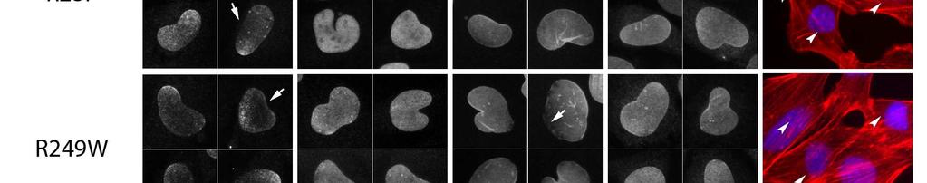


48 Page of xmm (00 x 00 DPI)
49 Page of xmm (00 x 00 DPI)

")


50 Page of xmm (00 x 00 DPI)
THE JOURNAL OF CELL BIOLOGY
 Supplemental Material Mejat et al., http://www.jcb.org/cgi/content/full/jcb.200811035/dc1 THE JOURNAL OF CELL BIOLOGY Figure S1. SUN1, SUN2, and Nesprin-1 localization in muscle fibers. (A) SUN1 and SUN2
L aminopathies represent a heterogeneous group of genetic
 1of5 ONLINE MUTATION REPORT Mutation analysis of the lamin A/C gene (LMNA) among patients with different cardiomuscular phenotypes M Vytopil, S Benedetti, E Ricci, G Galluzzi, A Dello Russo, L Merlini,
HYBRID GENE THERAPY FOR AD-EDMD
 HYBRID GENE THERAPY FOR AD-EDMD Gene Therapy Prof. Isabella Saggio 2017/2018 Bertani Camilla Dezi Clara Difeo Giorgia di Palma Carmen AUTOSOMAL DOMINANT EMERY-DREIFUSS MUSCULAR DYSTROPHY Fig.1 Adapted
THURSDAY, JANUARY
 THURSDAY, JANUARY 15 2015 08.30-09.00 WELCOME COFFEE 09.00-09.15 Welcome and opening comments. A. De Sandre-Giovannoli, N. Lévy Presentation of the French Network on EDMD and other nucleopathies. G. Bonne,
Muscular Dystrophy. Biol 405 Molecular Medicine
 Muscular Dystrophy Biol 405 Molecular Medicine Duchenne muscular dystrophy Duchenne muscular dystrophy is a neuromuscular disease that occurs in ~ 1/3,500 male births. The disease causes developmental
Genotype phenotype correlations in laminopathies: how does fate translate?
 Nuclear Envelope Disease and Chromatin Organization 2009 257 Genotype phenotype correlations in laminopathies: how does fate translate? Juergen Scharner, Viola F. Gnocchi, Juliet A. Ellis 1 and Peter S.
Inner Nuclear Membrane Protein MAN1 and Regulation of R-Smad Signaling
 4th International Melorheostosis Association Conference Inner Nuclear Membrane Protein MAN1 and Regulation of R-Smad Signaling Howard J. Worman Columbia University New York, NY The Nuclear Envelope By
Novel LMNA Mutation in a Taiwanese Family with Autosomal Dominant Emery-Dreifuss Muscular Dystrophy
 ASE REPORT Novel LMNA Mutation in a Taiwanese Family with Autosomal Dominant Emery-Dreifuss Muscular Dystrophy Wen-hen Liang, 1 hung-yee Yuo, 2 hun-ya Liu, 3 hee-siong Lee, 4 Kanako Goto, 5 Yukiko K. Hayashi,
et al.. Rare myopathy associated to MGUS, causing heart failure and responding to chemotherapy.
 Rare myopathy associated to MGUS, causing heart failure and responding to chemotherapy Nicolas Belhomme, Adel Maamar, Thomas Le Gallou, Marie-Christine Minot-Myhié, Antoine Larralde, Nicolas Champtiaux,
Nuclear Envelope and Muscular Dystrophy
 Nationwide Children s Hospital August 25, 2015 Nuclear Envelope and Muscular Dystrophy Howard J. Worman, M.D. Columbia University Columbia University Medical Center The Nuclear Envelope By D. W. Fawcett
The laminopathies: a clinical review
 Clin Genet 2006: 70: 261 274 Printed in Singapore. All rights reserved Review # 2006 The Authors Journal compilation # 2006 Blackwell Munksgaard CLINICAL GENETICS doi: 10.1111/j.1399-0004.2006.00677.x
Early-Onset LMNA-Associated Muscular Dystrophy with Later Involvement of Contracture
 JCN Open Access pissn 1738-6586 / eissn 2005-5013 / J Clin Neurol 2017;13(4):405-410 / https://doi.org/10.3988/jcn.2017.13.4.405 ORIGINAL ARTICLE Early-Onset LMNA-Associated Muscular Dystrophy with Later
LMNA mutations in atypical Werner s syndrome
 Mechanisms of disease LMNA mutations in atypical Werner s syndrome Lishan Chen, Lin Lee, Brian A Kudlow, Heloisa G Dos Santos, Olav Sletvold, Yousef Shafeghati, Eleanor G Botha, Abhimanyu Garg, Nancy B
Genetic diagnosis of limb girdle muscular dystrophy type 2A, A Case Report
 Genetic diagnosis of limb girdle muscular dystrophy type 2A, A Case Report Roshanak Jazayeri, MD, PhD Assistant Professor of Medical Genetics Faculty of Medicine, Alborz University of Medical Sciences
Identification of a novel in-frame de novo mutation in SPTAN1 in intellectual disability and pontocerebellar atrophy
 Hamdan et al., Identification of a novel in-frame de novo mutation in SPTAN1 in intellectual disability and pontocerebellar atrophy Supplementary Information Gene screening and bioinformatics PCR primers
The association of and -related gastroduodenal diseases
 The association of and -related gastroduodenal diseases N. R. Hussein To cite this version: N. R. Hussein. The association of and -related gastroduodenal diseases. European Journal of Clinical Microbiology
Familial DilatedCardiomyopathy Georgios K Efthimiadis, MD
 Familial DilatedCardiomyopathy Georgios K Efthimiadis, MD Dilated Cardiomyopathy Dilated LV/RV, reduced EF, in the absence of CAD valvulopathy pericardial disease Prevalence:40/100.000 persons Natural
Clinical Genetics in Cardiomyopathies
 Clinical Genetics in Cardiomyopathies Γεώργιος Κ Ευθυμιάδης Αναπληρωτής Καθηγητής Καρδιολογίας ΑΠΘ No conflict of interest Genetic terms Proband: The first individual diagnosed in a family Mutation: A
Progeria. Premature human aging: t he progerias. Progerias as models for aging. Hutchinson-Gilford syndrome. Definition:
 Premature human aging: t he progerias Reading: Genetic alterations in accelerated ageing syndromes Do they play a role in natural ageing? Monika Puzianowska- Kuznicka. Jacek Kuznicki. 2005. IJBCB, 37;
The Genes, Proteins, and the Cell Biological Processes Underlying Emery-Dreifuss Muscular Dystrophy
 The Genes, Proteins, and the Cell Biological Processes Underlying Emery-Dreifuss Muscular Dystrophy Megan E. Mastey a and Elise A. Kikis a Emery-Dreifuss Muscular Dystrophy (EDMD) is a type of muscular
Available online at
 Available online at www.sciencedirect.com R Experimental Cell Research 291 (2003) 122 134 www.elsevier.com/locate/yexcr Failure of lamin A/C to functionally assemble in R482L mutated familial partial lipodystrophy
When Lamins Go Bad: Nuclear Structure and Disease
 Leading Edge Review When Lamins Go Bad: Nuclear Structure and Disease Katherine H. Schreiber 1 and Brian K. Kennedy 1,2, * 1 Buck Institute for Research on Aging, 8001 Redwood Boulevard, Novato, CA 94945,
(a) Schematic diagram of the FS mutation of UVRAG in exon 8 containing the highly instable
 Schematic diagram of the FS mutation of UVRAG in exon 8 containing the highly instable") Supplementary Figure 1. Frameshift (FS) mutation in UVRAG. (a) Schematic diagram of the FS mutation of UVRAG in exon 8 containing the highly instable A 10 DNA repeat, generating a premature stop codon
Supplementary Figure 1. Frameshift (FS) mutation in UVRAG. (a) Schematic diagram of the FS mutation of UVRAG in exon 8 containing the highly instable A 10 DNA repeat, generating a premature stop codon
T he LMNA gene encodes two nuclear envelope proteins,
 1of5 ELECTRONIC LETTER A new mutation of the lamin A/C gene leading to autosomal dominant axonal neuropathy, muscular dystrophy, cardiac disease, and leuconychia C Goizet, R Ben aou, L Demay, P Richard,
H utchinson-gilford progeria syndrome (HGPS; MIM
 609 LETTER TO JMG Homozygous missense mutation in the lamin A/C gene causes autosomal recessive Hutchinson-Gilford progeria syndrome M Plasilova*, C Chattopadhyay*, P Pal, N A Schaub, S A Buechner, Hj
Enrichment culture of CSF is of limited value in the diagnosis of neonatal meningitis
 Enrichment culture of CSF is of limited value in the diagnosis of neonatal S. H. Chaudhry, D. Wagstaff, Anupam Gupta, I. C. Bowler, D. P. Webster To cite this version: S. H. Chaudhry, D. Wagstaff, Anupam
A model for calculation of growth and feed intake in broiler chickens on the basis of feed composition and genetic features of broilers
 A model for calculation of growth and feed intake in broiler chickens on the basis of feed composition and genetic features of broilers Bernard Carré To cite this version: Bernard Carré. A model for calculation
Skeletal myopathy in a family with lamin A/C cardiac disease
 Original Article Skeletal myopathy in a family with lamin A/C cardiac disease Subha Ghosh 1, Rahul Renapurkar 1, Subha V. Raman 2 1 Cleveland Clinic Foundation, Cleveland, OH, USA; 2 The Ohio State University
Cardiac effects of the c.1583 C G LMNA mutation in two families with Emery Dreifuss muscular dystrophy
 MOLECULAR MEDICINE REPORTS 12: 5065-5071, 2015 Cardiac effects of the c.1583 C G LMNA mutation in two families with Emery Dreifuss muscular dystrophy LI ZHANG 1, HONGRUI SHEN 2, ZHE ZHAO 2, QI BING 2 and
Single Gene (Monogenic) Disorders. Mendelian Inheritance: Definitions. Mendelian Inheritance: Definitions
 Disorders. Mendelian Inheritance: Definitions. Mendelian Inheritance: Definitions") Single Gene (Monogenic) Disorders Mendelian Inheritance: Definitions A genetic locus is a specific position or location on a chromosome. Frequently, locus is used to refer to a specific gene. Alleles are
Single Gene (Monogenic) Disorders Mendelian Inheritance: Definitions A genetic locus is a specific position or location on a chromosome. Frequently, locus is used to refer to a specific gene. Alleles are
Specific phosphorylation of Ser458 of A-type lamins in LMNA-associated myopathy patients
 Research Article 3893 Specific phosphorylation of Ser458 of A-type lamins in LMNA-associated myopathy patients Hiroaki Mitsuhashi 1, Yukiko K. Hayashi 1, *, Chie Matsuda 2, Satoru Noguchi 1, Shuji Wakatsuki
SALSA MLPA KIT P060-B2 SMA
 SALSA MLPA KIT P6-B2 SMA Lot 111, 511: As compared to the previous version B1 (lot 11), the 88 and 96 nt DNA Denaturation control fragments have been replaced (QDX2). Please note that, in contrast to the
Laminopathies: many diseases, one gene. Report of the first Italian Meeting Course on Laminopathies
 Acta Myologica 2011; XXX: p. 138-143 workshop report Laminopathies: many diseases, one gene. Report of the first Italian Meeting Course on Laminopathies G. Lattanzi, S. Benedetti, E. Bertini, G. Boriani,
Supplementary Figure 1: si-craf but not si-braf sensitizes tumor cells to radiation.
 Supplementary Figure 1: si-craf but not si-braf sensitizes tumor cells to radiation. (a) Embryonic fibroblasts isolated from wildtype (WT), BRAF -/-, or CRAF -/- mice were irradiated (6 Gy) and DNA damage
Iowa Wellstone Center Muscle Tissue and Cell Culture Repository
 Iowa Wellstone Center Muscle Tissue and Cell Culture Repository Steven A. Moore, M.D., Ph.D. The University of Iowa Department of Pathology and Iowa Wellstone Muscular Dystrophy Cooperative Research Center
Properties of lamin A mutants found in Emery-Dreifuss muscular dystrophy, cardiomyopathy and Dunnigantype partial lipodystrophy
 RESEARCH ARTICLE 4435 Properties of lamin A mutants found in Emery-Dreifuss muscular dystrophy, cardiomyopathy and Dunnigantype partial lipodystrophy Cecilia Östlund 1, Gisèle Bonne 2, Ketty Schwartz 2
Reversal of the cellular phenotype in the premature aging disease Hutchinson-Gilford progeria syndrome
 Reversal of the cellular phenotype in the premature aging disease Hutchinson-Gilford progeria syndrome Paola Scaffidi & Tom Misteli Hutchinson-Gilford progeria syndrome (HGPS) is a childhood premature
MRC-Holland MLPA. Description version 19;
 SALSA MLPA probemix P6-B2 SMA Lot B2-712, B2-312, B2-111, B2-511: As compared to the previous version B1 (lot B1-11), the 88 and 96 nt DNA Denaturation control fragments have been replaced (QDX2). SPINAL
Mutations and Disease Mutations in the Myosin Gene
 Biological Sciences Initiative HHMI Mutations and Disease Mutations in the Myosin Gene Goals Explore how mutations can lead to disease using the myosin gene as a model system. Explore how changes in the
Three new cases of dilated cardiomyopathy caused by mutations in LMNA gene
 Acta Myologica 2017; XXXVI: p. 207-212 Three new cases of dilated cardiomyopathy caused by mutations in LMNA gene Larysa N. Sivitskaya 1, Nina G. Danilenko 1, Tatiyana G. Vaikhanskaya 2, Tatsiyana V. Kurushka
p.r623c p.p976l p.d2847fs p.t2671 p.d2847fs p.r2922w p.r2370h p.c1201y p.a868v p.s952* RING_C BP PHD Cbp HAT_KAT11
 ARID2 p.r623c KMT2D p.v650fs p.p976l p.r2922w p.l1212r p.d1400h DNA binding RFX DNA binding Zinc finger KMT2C p.a51s p.d372v p.c1103* p.d2847fs p.t2671 p.d2847fs p.r4586h PHD/ RING DHHC/ PHD PHD FYR N
READ ORPHA.NET WEBSITE ABOUT BETA-SARCOGLYOCANOPATHY LIMB-GIRDLE MUSCULAR DYSTROPHIES
 READ ORPHA.NET WEBSITE ABOUT BETA-SARCOGLYOCANOPATHY LIMB-GIRDLE MUSCULAR DYSTROPHIES (LGMD) Limb-girdle muscular dystrophies (LGMD) are a heterogeneous group of genetically determined disorders with a
Laminopathies and the long strange trip from basic cell biology to therapy
 Review series Laminopathies and the long strange trip from basic cell biology to therapy Howard J. Worman, 1,2 Loren G. Fong, 3 Antoine Muchir, 1,2 and Stephen G. Young 3,4 1 Department of Medicine and
Bio 111 Study Guide Chapter 17 From Gene to Protein
 Bio 111 Study Guide Chapter 17 From Gene to Protein BEFORE CLASS: Reading: Read the introduction on p. 333, skip the beginning of Concept 17.1 from p. 334 to the bottom of the first column on p. 336, and
Lamins, laminopathies and disease mechanisms: Possible role for proteasomal degradation of key regulatory proteins
 Lamins, laminopathies and disease mechanisms: Possible role for proteasomal degradation of key regulatory proteins VEENA KPARNAIK*, PANKAJ CHATURVEDI and BH MURALIKRISHNA Centre for Cellular and Molecular
Cardiac Considerations and Care in Children with Neuromuscular Disorders
 Cardiac Considerations and Care in Children with Neuromuscular Disorders - importance of early and ongoing treatment, management and available able medications. Dr Bo Remenyi Department of Cardiology The
Workshop report. 1. Introduction
 Neuromuscular Disorders 12 (2002) 187 194 Workshop report www.elsevier.com/locate/nmd 82nd ENMC international workshop, 5th international Emery Dreifuss muscular dystrophy (EDMD) workshop, 1st Workshop
Nuclear envelope defects associated with LMNA mutations cause dilated cardiomyopathy and Emery- Dreifuss muscular dystrophy
 RESEARCH ARTICLE 4447 Nuclear envelope defects associated with LMNA mutations cause dilated cardiomyopathy and Emery- Dreifuss muscular dystrophy Wahyu Hendrati Raharjo 1, Paul Enarson 1, Teresa Sullivan
Title:A novel lamin A/C gene mutation causing spinal muscular atrophy-like phenotype with arrhythmia and cardiac hypofunction: report of one case
 Author's response to reviews Title:A novel lamin A/C gene mutation causing spinal muscular atrophy-like phenotype with arrhythmia and cardiac hypofunction: report of one case Authors: Naotoshi Iwahara
Supplemental Materials. STK16 regulates actin dynamics to control Golgi organization and cell cycle
 Supplemental Materials STK16 regulates actin dynamics to control Golgi organization and cell cycle Juanjuan Liu 1,2,3, Xingxing Yang 1,3, Binhua Li 1, Junjun Wang 1,2, Wenchao Wang 1, Jing Liu 1, Qingsong
Mandibuloacral Dysplasia Is Caused by a Mutation in LMNA-Encoding Lamin A/C
 Am. J. Hum. Genet. 71:426 431, 2002 Report Mandibuloacral Dysplasia Is Caused by a Mutation in LMNA-Encoding Lamin A/C Giuseppe Novelli, 1 Antoine Muchir, 6 Federica Sangiuolo, 1 Anne Helbling-Leclerc,
Anesthesia recommendations for patients suffering from Emery-Dreifuss Muscular Dystrophy
 orphananesthesia Anesthesia recommendations for patients suffering from Emery-Dreifuss Muscular Dystrophy Disease name: Emery-Dreifuss Muscular Dystrophy (EDMD) ICD 10: G71.0 Synonyms: Benign Scapuloperoneal
MTC-TT and TPC-1 cell lines were cultured in RPMI medium (Gibco, Breda, The Netherlands)
") Supplemental data Materials and Methods Cell culture MTC-TT and TPC-1 cell lines were cultured in RPMI medium (Gibco, Breda, The Netherlands) supplemented with 15% or 10% (for TPC-1) fetal bovine serum
Supplemental data Materials and Methods Cell culture MTC-TT and TPC-1 cell lines were cultured in RPMI medium (Gibco, Breda, The Netherlands) supplemented with 15% or 10% (for TPC-1) fetal bovine serum
variant led to a premature stop codon p.k316* which resulted in nonsense-mediated mrna decay. Although the exact function of the C19L1 is still
 157 Neurological disorders primarily affect and impair the functioning of the brain and/or neurological system. Structural, electrical or metabolic abnormalities in the brain or neurological system can
SUPPLEMENTARY INFORMATION
 Supplementary Figures Supplementary Figure S1. Binding of full-length OGT and deletion mutants to PIP strips (Echelon Biosciences). Supplementary Figure S2. Binding of the OGT (919-1036) fragments with
Abnormal nuclear shape and impaired mechanotransduction in emerin-deficient cells
 JCB: ARTICLE Abnormal nuclear shape and impaired mechanotransduction in emerin-deficient cells Jan Lammerding, 1 Janet Hsiao, 2 P. Christian Schulze, 1 Serguei Kozlov, 3 Colin L. Stewart, 3 and Richard
In vitro study of the effects of cadmium on the activation of the estrogen response element using the YES screen
 In vitro study of the effects of cadmium on the activation of the estrogen response element using the YES screen Xavier Denier, Jérome Couteau, Magalie Baudrimont, Elisabeth M. Hill, Jeanette Rotchell,
We are IntechOpen, the world s leading publisher of Open Access books Built by scientists, for scientists. International authors and editors
 We are IntechOpen, the world s leading publisher of Open Access books Built by scientists, for scientists 4,100 116,000 120M Open access books available International authors and editors Downloads Our
Cardiac follow-up in patients with hereditary muscular diseases
 Cardiac follow-up in patients with hereditary muscular diseases Tromsø 7. september 2018 Eystein Theodor Ek Skjølsvik, MD Professor Kristina H. Haugaa, MD, PhD Unit for genetic cardiac diseases, OUS Rikshospitalet
Neuromuscular Disorders 13 (2003) Workshop report
 Workshop report") Neuromuscular Disorders 13 (2003) 508 515 Workshop report www.elsevier.com/locate/nmd 108th ENMC International Workshop, 3rd Workshop of the MYO-CLUSTER project: EUROMEN, 7th International Emery-Dreifuss
Neuromuscular Disorders 13 (2003) 508 515 Workshop report www.elsevier.com/locate/nmd 108th ENMC International Workshop, 3rd Workshop of the MYO-CLUSTER project: EUROMEN, 7th International Emery-Dreifuss
Islet viability assay and Glucose Stimulated Insulin Secretion assay RT-PCR and Western Blot
 Islet viability assay and Glucose Stimulated Insulin Secretion assay Islet cell viability was determined by colorimetric (3-(4,5-dimethylthiazol-2-yl)-2,5- diphenyltetrazolium bromide assay using CellTiter
Association of the LMNA gene single nucleotide polymorphism rs4641 with dilated cardiomyopathy
 Association of the LMNA gene single nucleotide polymorphism rs4641 with dilated cardiomyopathy J. Yin 1,2, J. Yang 3, F.X. Ren 3, C.M. Sun 4, L.D. Li 3, L.Y. Han 5, S.L. Cai 1, C.H. Zhang 3, Z.Q. Zhang
TITLE: The Role of hcdc4 as a Tumor Suppressor Gene in Genomic Instability Underlying Prostate Cancer
 AD Award Number: TITLE: The Role of hcdc4 as a Tumor Suppressor Gene in Genomic Instability Underlying Prostate Cancer PRINCIPAL INVESTIGATOR: Audrey van Drogen, Ph.D. CONTRACTING ORGANIZATION: Sidney
Emery-Dreifuss muscular dystrophy: the most recognizable laminopathy
 Review paper Emery-Dreifuss muscular dystrophy: the most recognizable laminopathy Agnieszka Madej-Pilarczyk, Andrzej Kochański Neuromuscular Unit, Mossakowski Medical Research Centre, Polish Academy of
Finding subtle mutations with the Shannon human mrna splicing pipeline
 Finding subtle mutations with the Shannon human mrna splicing pipeline Presentation at the CLC bio Medical Genomics Workshop American Society of Human Genetics Annual Meeting November 9, 2012 Peter K Rogan
EXOME SEQUENCING AS A SECOND-TIER DIAGNOSTIC APPROACH FOR CLINICALLY SUSPECTED DYSFERLINOPATHY PATIENTS
 EXOME SEQUENCING AS A SECOND-TIER DIAGNOSTIC APPROACH FOR CLINICALLY SUSPECTED DYSFERLINOPATHY PATIENTS Marc Bartoli, Jean-Pierre Desvignes, Nicolas Lévy, Martin Krahn To cite this version: Marc Bartoli,
Multi-template approaches for segmenting the hippocampus: the case of the SACHA software
 Multi-template approaches for segmenting the hippocampus: the case of the SACHA software Ludovic Fillon, Olivier Colliot, Dominique Hasboun, Bruno Dubois, Didier Dormont, Louis Lemieux, Marie Chupin To
YES NO UNKNOWN. Stage I: Rule-Out Dashboard Secondary Findings in Adults ACTIONABILITY PENETRANCE SIGNIFICANCE/BURDEN OF DISEASE NEXT STEPS
 Stage I: Rule-Out Dashboard GENE/GENE PANEL: LMNA, EMD, FHL1 DISORDER: Emery-Dreifuss Muscular Dystrophy (AD, XL) HGNC ID: 6636, 3331, 3702 OMIM ID: 181350, 310300, 300696 ACTIONABILITY PENETRANCE 1. Is
Iodide mumps: Sonographic appearance
 Iodide mumps: Sonographic appearance Salvatore Greco, Riccardo Centenaro, Giuseppe Lavecchia, Francesco Rossi To cite this version: Salvatore Greco, Riccardo Centenaro, Giuseppe Lavecchia, Francesco Rossi.
Article. An EDMD Mutation in C. elegans Lamin Blocks Muscle-Specific Gene Relocation and Compromises Muscle Integrity
 Current Biology 21, 163 1614, October 11, 211 ª211 Elsevier Ltd All rights reserved DOI 1.116/j.cub.211.8.3 An EDMD Mutation in C. elegans Lamin Blocks Muscle-Specific Gene Relocation and Compromises Muscle
Supplemental Materials and Methods Plasmids and viruses Quantitative Reverse Transcription PCR Generation of molecular standard for quantitative PCR
 Supplemental Materials and Methods Plasmids and viruses To generate pseudotyped viruses, the previously described recombinant plasmids pnl4-3-δnef-gfp or pnl4-3-δ6-drgfp and a vector expressing HIV-1 X4
Corporate Medical Policy
 Corporate Medical Policy Genetic Testing for Duchenne and Becker Muscular Dystrophy File Name: Origination: Last CAP Review: Next CAP Review: Last Review: genetic_testing_for_duchenne_and_becker_muscular_dystrophy
On the empirical status of the matching law : Comment on McDowell (2013)
") On the empirical status of the matching law : Comment on McDowell (2013) Pier-Olivier Caron To cite this version: Pier-Olivier Caron. On the empirical status of the matching law : Comment on McDowell (2013):
On the empirical status of the matching law : Comment on McDowell (2013) Pier-Olivier Caron To cite this version: Pier-Olivier Caron. On the empirical status of the matching law : Comment on McDowell (2013):
Proteins. Length of protein varies from thousands of amino acids to only a few insulin only 51 amino acids
 Proteins Protein carbon, hydrogen, oxygen, nitrogen and often sulphur Length of protein varies from thousands of amino acids to only a few insulin only 51 amino acids During protein synthesis, amino acids
LYMPHOGRANULOMA VENEREUM PRESENTING AS PERIANAL ULCERATION: AN EMERGING CLINICAL PRESENTATION?
 LYMPHOGRANULOMA VENEREUM PRESENTING AS PERIANAL ULCERATION: AN EMERGING CLINICAL PRESENTATION? Tajinder K Singhrao, Elizabeth Higham, Patrick French To cite this version: Tajinder K Singhrao, Elizabeth
From universal postoperative pain recommendations to procedure-specific pain management
 From universal postoperative pain recommendations to procedure-specific pain management Hélène Beloeil, Francis Bonnet To cite this version: Hélène Beloeil, Francis Bonnet. From universal postoperative
Comments on the article by Tabache F. et al. Acute polyarthritis after influenza A H1N1 immunization,
 Comments on the article by Tabache F. et al. Acute polyarthritis after influenza A H1N1 immunization, Joint Bone Spine, 2011, doi:10.1016/j.jbs.2011.02.007: Primary Sjögren s syndrome occurring after influenza
MRC-Holland MLPA. Description version 29; 31 July 2015
 SALSA MLPA probemix P081-C1/P082-C1 NF1 P081 Lot C1-0114. As compared to the previous B2 version (lot 0813 and 0912), 11 target probes are replaced or added, and 10 new reference probes are included. P082
SUPPLEMENTARY INFORMATION. Supplementary Figures S1-S9. Supplementary Methods
 SUPPLEMENTARY INFORMATION SUMO1 modification of PTEN regulates tumorigenesis by controlling its association with the plasma membrane Jian Huang 1,2#, Jie Yan 1,2#, Jian Zhang 3#, Shiguo Zhu 1, Yanli Wang
Supplemental Data: Detailed Characteristics of Patients with MKRN3. Patient 1 was born after an uneventful pregnancy. She presented in our
 1 2 Supplemental Data: Detailed Characteristics of Patients with MKRN3 Mutations 3 4 5 6 7 8 9 10 11 12 13 14 15 16 17 18 19 20 21 22 23 24 Patient 1 was born after an uneventful pregnancy. She presented
Optimal electrode diameter in relation to volume of the cochlea
 Optimal electrode diameter in relation to volume of the cochlea Dan Gnansia, Thomas Demarcy, Clair Vandersteen, Charles Raffaelli, Nicolas Guevara, Hervé Delingette, Nicholas Ayache To cite this version:
Immunohistochemical Study of Dystrophin Associated Glycoproteins in Limb-girdle Muscular Dystrophies
 Dystrophin Immunohistochemical Study of Dystrophin Associated Glycoproteins in Limb-girdle Muscular Dystrophies NSC 89-2314-B-002-111 88 8 1 89 7 31 ( Peroxidase -AntiPeroxidase Immnofluorescence) Abstract
Moderate alcohol consumption and risk of developing dementia in the elderly: the contribution of prospective studies.
 Moderate alcohol consumption and risk of developing dementia in the elderly: the contribution of prospective studies. Luc Letenneur To cite this version: Luc Letenneur. Moderate alcohol consumption and
Regulators of Cell Cycle Progression
 Regulators of Cell Cycle Progression Studies of Cdk s and cyclins in genetically modified mice reveal a high level of plasticity, allowing different cyclins and Cdk s to compensate for the loss of one
MRC-Holland MLPA. Description version 30; 06 June 2017
 SALSA MLPA probemix P081-C1/P082-C1 NF1 P081 Lot C1-0517, C1-0114. As compared to the previous B2 version (lot B2-0813, B2-0912), 11 target probes are replaced or added, and 10 new reference probes are
Supplemental Information. Otic Mesenchyme Cells Regulate. Spiral Ganglion Axon Fasciculation. through a Pou3f4/EphA4 Signaling Pathway
 Neuron, Volume 73 Supplemental Information Otic Mesenchyme Cells Regulate Spiral Ganglion Axon Fasciculation through a Pou3f4/EphA4 Signaling Pathway Thomas M. Coate, Steven Raft, Xiumei Zhao, Aimee K.
SALSA MLPA KIT P050-B2 CAH
 SALSA MLPA KIT P050-B2 CAH Lot 0510, 0909, 0408: Compared to lot 0107, extra control fragments have been added at 88, 96, 100 and 105 nt. The 274 nt probe gives a higher signal in lot 0510 compared to
Hutchinson-Gilford progeria syndrome accompanied by severe skeletal abnormalities in two Chinese siblings: two case reports
 Xiong et al. Journal of Medical Case Reports 2013, 7:63 JOURNAL OF MEDICAL CASE REPORTS CASE REPORT Open Access Hutchinson-Gilford progeria syndrome accompanied by severe skeletal abnormalities in two
Supplemental Figure 1 ELISA scheme to measure plasma total, mature and furin-cleaved
 1 Supplemental Figure Legends Supplemental Figure 1 ELISA scheme to measure plasma total, mature and furin-cleaved PCSK9 concentrations. 4 Plasma mature and furin-cleaved PCSK9s were measured by a sandwich
CURRENT GENETIC TESTING TOOLS IN NEONATAL MEDICINE. Dr. Bahar Naghavi
 2 CURRENT GENETIC TESTING TOOLS IN NEONATAL MEDICINE Dr. Bahar Naghavi Assistant professor of Basic Science Department, Shahid Beheshti University of Medical Sciences, Tehran,Iran 3 Introduction Over 4000
The toll-like receptor 4 ligands Mrp8 and Mrp14 play a critical role in the development of autoreactive CD8 + T cells
 1 SUPPLEMENTARY INFORMATION The toll-like receptor 4 ligands Mrp8 and Mrp14 play a critical role in the development of autoreactive CD8 + T cells Karin Loser 1,2,6, Thomas Vogl 2,3, Maik Voskort 1, Aloys
Recombinant Protein Expression Retroviral system
 Recombinant Protein Expression Retroviral system Viruses Contains genome DNA or RNA Genome encased in a protein coat or capsid. Some viruses have membrane covering protein coat enveloped virus Ø Essential
WDR62 is associated with the spindle pole and mutated in human microcephaly
 WDR62 is associated with the spindle pole and mutated in human microcephaly Adeline K. Nicholas, Maryam Khurshid, Julie Désir, Ofélia P. Carvalho, James J. Cox, Gemma Thornton, Rizwana Kausar, Muhammad
Supplementary Figure 1: Hsp60 / IEC mice are embryonically lethal (A) Light microscopic pictures show mouse embryos at developmental stage E12.
 Light microscopic pictures show mouse embryos at developmental stage E12.") Supplementary Figure 1: Hsp60 / IEC mice are embryonically lethal (A) Light microscopic pictures show mouse embryos at developmental stage E12.5 and E13.5 prepared from uteri of dams and subsequently genotyped.
Supplementary Figure 1: Hsp60 / IEC mice are embryonically lethal (A) Light microscopic pictures show mouse embryos at developmental stage E12.5 and E13.5 prepared from uteri of dams and subsequently genotyped.
Supplementary Figure 1 Transcription assay of nine ABA-responsive PP2C. Transcription assay of nine ABA-responsive PP2C genes. Total RNA was isolated
 Supplementary Figure 1 Transcription assay of nine ABA-responsive PP2C genes. Transcription assay of nine ABA-responsive PP2C genes. Total RNA was isolated from 7 day-old seedlings treated with or without
Chorea as the presenting manifestation of primary Sjögren s syndrome in a child
 Chorea as the presenting manifestation of primary Sjögren s syndrome in a child Cécile Delorme, Fleur Cohen, Cécile Hubsch, Emmanuel Roze To cite this version: Cécile Delorme, Fleur Cohen, Cécile Hubsch,
Beta Thalassemia Case Study Introduction to Bioinformatics
 Beta Thalassemia Case Study Sami Khuri Department of Computer Science San José State University San José, California, USA sami.khuri@sjsu.edu www.cs.sjsu.edu/faculty/khuri Outline v Hemoglobin v Alpha
Computational Systems Biology: Biology X
 Bud Mishra Room 1002, 715 Broadway, Courant Institute, NYU, New York, USA L#4:(October-0-4-2010) Cancer and Signals 1 2 1 2 Evidence in Favor Somatic mutations, Aneuploidy, Copy-number changes and LOH
Evaluation of noise barriers for soundscape perception through laboratory experiments
 Evaluation of noise barriers for soundscape perception through laboratory experiments Joo Young Hong, Hyung Suk Jang, Jin Yong Jeon To cite this version: Joo Young Hong, Hyung Suk Jang, Jin Yong Jeon.
A rare case of muscular dystrophy with POMT2 and FKRP gene mutation. Present by : Ghasem Khazaei Supervisor :Dr Mina Mohammadi Sarband
 A rare case of muscular dystrophy with POMT2 and FKRP gene mutation Present by : Ghasem Khazaei Supervisor :Dr Mina Mohammadi Sarband Index : Congenital muscular dystrophy (CMD) Dystroglycanopathies Walker-Warburg