Glandular Epithelium. Dr. Hersh Abdul Ham-Karim BVM&S, PG Dip, MSc and PhD
|
|
|
- Vanessa Taylor
- 6 years ago
- Views:
Transcription
1 Glandular Epithelium Dr. Hersh Abdul Ham-Karim BVM&S, PG Dip, MSc and PhD

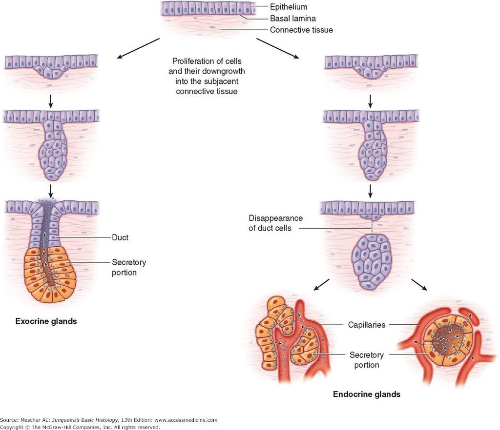
2 Glandular Epithelium Groups of surface cells differentiate, proliferate, and penetrate underlying connective tissue. Their main function is to synthesize and secrete extracellular products. These include hormone, enzyme, etc. Cells specialized to produce secretion. The molecules to be secreted are generally stored in the cells in small membrane-bound vesicles called secretory granules


3 Glands: a specialized cell, group of cells, or organ in the human or animal body which secretes particular chemical substances for use in the body. Glands of the body are classified as either exocrine or endocrine types 1-Exocrine Glands: - maintain connection with the surface epithelium via the tubular ducts through with the secretory product pass to reach the surface (skin, digestive tract).
4 Histologically, exocrine glands composed from two parts: secretory portion: contains the cells responsible for the secretory process Ducts Secretory portion system of ducts: transport the secretion to the exterior of the gland

5 2-Endocrine Glands: are ductless the connection with the surface was obliterated during development and they release their secretory product (hormones) into the bloodstream. * Paracrine Glands: These glands are similar to endocrine glands, but their secretions reach target cells by diffusion through the extracellular space or immediately subjacent connective tissue. Enteroendocrine cells of gastrointestinal tract (GIT).
6
7 What are Hormones? Hormones are special chemical messengers in the body that are created in the endocrine glands. These messengers control most major bodily functions, from simple basic needs like hunger to complex systems like reproduction, and even the emotions and mood. In the human body, hormones are used for two types of communication. The first is for communication between two endocrine glands. For example, the pituitary gland send signals to other endocrine glands to stimulate or inhibit their own hormone production. The second is between an endocrine gland and a target organ, for example when the pancreas releases insulin which causes muscle and fat cells to take up glucose from the bloodstream.
8 What are Enzymes? Enzymes are proteins (complex chains of amino acids) that play a role in all chemical functions in the body including digestion, energy production, and repair of tissues, organs, and cells. Enzymes can be divided into three groups. Metabolic enzymes (enzymes which your body produces that work in blood, tissues, and organs) Digestive enzymes (enzymes that break down food into usable material) Food enzymes (enzymes that are contained in raw food)

9 Classification of Exocrine gland This classification system is based on five different morphological criteria. 1. Number of secretory cells - Unicellular glands Goblet cells are the only example of these single-celled glands in humans. These goblet cells secrete mucus and are easily visualized in slides of the small intestine.


10 - Multicellular glands - These glands have many cells. In addition to the ways that multicellular glands are classified (listed below).


11 2. Nature of secretion: a) Serous A cell-type that produces a thin watery, protein-rich secretion. Serous cells are pyramidal, with round, basally located nuclei. Cytoplasm: granular (stain darkly from pink to dark purple with H&E stain) e.g. pancreas and parotid salivary glands


12 b) Mucous A cell type that produces is a strongly hydrophilic glycoproteins called mucins, viscous secretions that have a lubricating or protective function. Mucous cells are columnar, characterized by numerous large, lightly staining granules with basally located nuclei. Mucous cells are most often organized as tubules, consisting of cylindrical arrays of secretory cells surrounding a lumen. e.g GI tract

13 Differences between serous and mucinous cells Serous Serous cells are mostly pyramidal in shape. They are small in size. When stained with H&E stain, cells take dark stain Nuclei are rounded and placed near the centre but more toward the basal part of cells The apical portion is filled with secretory granules (zymogen granules) The base of serous cell is basophilic (blue) and apical portion acidophilic (pink) Mucous Mucous cells are short columnar to pyramidal in shape they are large in size compared to serous cells Mucous cells stain light; hence, appear empty Nuclei are flat and situated toward basement membrane The apical portion contains large number of secretory granules (mucinogen granules) Cytoplasm takes light pale stain and looks empty or vacuolated. This is because the mucous is lost in preparation of H&E stained slides Serous Mucous

 Sebaceous - Thick, lipid rich secretions of cuboidal cells in")
14 c) Mixed These glands have both serous and mucous cells. The mucous cells are capped by serous cells called serous demilune. e.g. sublingual salivary gland. d) Sebaceous - Thick, lipid rich secretions of cuboidal cells in certain skin regions (e.g. face, nose and axillary regions)

15 3. Mechanism of secretion: on the basis how the secretory product is released a- Merocrine/Eccrine secretion This is the most common type of glandular epithelium secretion where secretory granules within the cytoplasm of the cell gather at the apical region of the cell. Then, the granule s limiting membrane fuses with the apical membrane and the contents of the granule are opened and released. This process of fusion and release are collectively referred to as exocytosis. The secretory granules leave the cell with no loss of other cellular material. (e.g. salivary gland)

16 b- Apocrine secretion A rare type of secretion dependent on sex hormones where secretory granules within the cytoplasm gather at the apical region of the cell. Then, a portion of the cytoplasm of the cell simply pinches off enclosing the granules. Within the lumen, this small secretory vesicle breaks down and releases the gland s products. (e.g. mammary gland)
 or pinched off (apocrine), the whole cell is discharged into the lumen. Once inside the lumen, the cell degenerates and the secretory products are released.")
17 c- Holocrine secretion This secretion consists of disintegrated cells of the gland itself. Granules fill the cell until the entire cell becomes bloated with secretory products. Instead of being released (merocrine) or pinched off (apocrine), the whole cell is discharged into the lumen. Once inside the lumen, the cell degenerates and the secretory products are released. (e.g. sebaceous gland)


 group of secretary cells arranged about a small lumen.")
18 4. Shape of secretory units: Remember exocrine glands have a secretory portion, and ducts. Tubular - An elongated group of secretary cells with a tube-shaped lumen. Mucous cells are most often organized as tubules. Acinar (or alveolar) - A small grape-like (acinus means grape ) or sac-like (alveolus means sac ) group of secretary cells arranged about a small lumen. Tubulo-alveolar Lumen of secretary units have both of the above listed shapes (seen in mixed glands).
19 5. Arrangement (branched or not) and Occurrence of Duct System Simple glands - Glands that have an unbranched duct into which the cells secrete. Branched glands These glands have several secretary units empty into an unbranched excretory duct. Compound glands - These glands have a highly branched duct system. Secretory portions empty into an elaborate branched duct system, which, in turn, drain into larger ducts.


20 Types of Exocrine Glands 1. Simple tubular glands: These glands are epithelial-lined tubules, which open on the apical surface. There are three types. Simple straight tubular glands: The long crypts of Lieberkühn, located runs a straight, unbranched course. within the colon that Simple coiled tubular glands: Within the dermis, sweat glands are located. Simple branched tubular glands: These simple branched tubular glands are found primarily in the stomach


21 2. Simple alveolar (acinar) glands: The paraurethral glands located in the penile urethra or the sebaceous glands located in the skin. 3. Simple branched alveolar glands: Some of the smaller glands of the respiratory tract; minor salivary glands located within the oral cavity are other examples.


22 4. Compound tubular glands: These glands have a highly branched duct system. The secretary cells at the ends of the ducts are in the form of tubules. Brunner s glands of the duodenum are compound tubular glands. 5. Compound alveolar glands: The duct system is similar to the compound tubular and compound tubulo-alveolar glands; however, compound alveolar glands differ from other compound glands in that the ducts end in alveoli with dilated sac-like lumina. The pancreas and parotid gland are the best examples of compound alveolar glands as they are entirely serous.
.")
23 6- Compound tubulo-alveolar glands: These glands also have a highly branched duct system, but some of the ducts end as tubules and others end as alveoli. Two of the major salivary glands, the submandibular and the sublingual glands, are examples of compound tubulo-alveolar glands (as they are mixed glands).
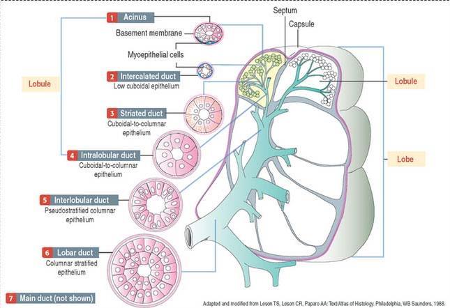
24 Ducts of Exocrine Glands Several different types of ducts exist along the lumen of the glands. Intercalated ducts; receive secretion from acini. They have simple cuboidal epithelium. Striated (intralobular) ducts: cuboidal or low columnar cells with round nuclei. They receive secretion from intercalated ducts. Interlobular (excretory) ducts: are found between lobules, cuboidal to columnar cells. Transmit secretion from striated ducts to interlobar ducts. Interlobar ducts: pseudostratified columnar. Main duct: stratified columnar then stratified squamous at its end.
25

26 Myoepithelial Cells Specialized squamous epithelial cells with power of contraction. Usually found in glandular epithelium as a thin layer above the basement membrane but generally beneath the luminal cells in the secretory portion of glands. They are instrumental in moving the secretions toward the excretory duct.

27 Pancreas The pancreas is a glandular organ in the digestive system and endocrine system of vertebrates. In humans, it is about 6 inches long and sits across the back of the abdomen, behind the stomach. It is surrounded by the stomach, small intestine, liver, spleen and gallbladder.

28 Microscopic structure The pancreas has a thin cover of loose connective tissue from which septa pass into the gland, subdividing it into many small molecule. Each lobule is again composed of several rounded or tubular groups of pancreatic cells called acini. The connective tissue is very little between two acini. Among the acini are the scattered the islets of Langerhans
29 Although it is primarily an exocrine gland, secreting a variety of digestive enzymes, the pancreas has an endocrine function. The bulk of the pancreas is composed of exocrine cells that produce several digestive enzymes like trypsin, amylase, and lipase. These exocrine cells release their enzymes into a series of progressively larger tubes (called ducts) that eventually join together to form the main pancreatic duct. The main pancreatic duct runs the length of the pancreas and drains the fluid produced by the exocrine cells into the duodenum, the first part of the small bowel.
30 The second functional component of the pancreas is the "endocrine" pancreas. Inside the substance of pancreas there are groups of specialized cells surrounded by connective tissue, which form the endocrine part of the gland. These cells are called islets of Langerhans. In standard histological sections of the pancreas, islets are seen as relatively pale-staining groups of cells embedded in a sea of darkerstaining exocrine tissue. These endocrine cells don t release their secretions into the pancreatic ducts, instead they release hormones, into the blood stream.
31 The pancreatic islets each contain four varieties of cells: The alpha cell: secret Glucagon, which plays an important role in regulates blood glucose level. The beta cell: produces the hormone insulin, elevated blood glucose levels stimulate the release of insulin. The delta cell: secretes the peptide hormone somatostatin, inhibiting hormone, pancreatic somatostatin inhibits the release of both glucagon and insulin. The Pancreas Polypeptide cell: secretes the pancreatic polypeptide hormone. It is thought to play a role in appetite. Pancreatic polypeptide released following a meal may reduce further food consumption; however, it is also released in response to fasting.
Glandular Epithelium. Dr. Heba Kalbouneh Associate Professor of Anatomy and Histology
 Glandular Epithelium Dr. Heba Kalbouneh Associate Professor of Anatomy and Histology Glands Glandular epithelia are tissues formed by cells specialized to produce secretion. Secretion: if substances produced
TYPES OF EPITHELIA. Epithelia can be divided into two main groups. A-covering (or lining) epithelia B- Secretory (glandular) epithelia.
 epithelia B- Secretory (glandular) epithelia.") TYPES OF EPITHELIA Epithelia can be divided into two main groups A-covering (or lining) epithelia B- Secretory (glandular) epithelia. Glands Glandular epithelial cells may synthesize, store, and secrete:
TYPES OF EPITHELIA Epithelia can be divided into two main groups A-covering (or lining) epithelia B- Secretory (glandular) epithelia. Glands Glandular epithelial cells may synthesize, store, and secrete:
Dr. Abeer.c.Yousif. Histology -2 nd stage. What is histology?
 What is histology? Histology is the science of microscopic anatomy of cells and tissues, in Greek language Histo= tissue and logos = study and it's tightly bounded to molecular biology, physiology, immunology
Glandular Epithelium. Dr. Heba Kalbouneh Assistant Professor of Anatomy and Histology
 Glandular Epithelium Dr. Heba Kalbouneh Assistant Professor of Anatomy and Histology Glands Gla dular epithelia are tissues for ed y ells spe ialized to produ e se retio. Secretion: if substances produced
Lec.2 Histology Glandular Epithelium A gland 1. Endocrine Glands 2. Exocrine Glands Endocrine Glands Exocrine Glands
 Lec.2 Histology Glandular Epithelium A gland is one or more cells that produce and secrete a specific product. The product is always a water-based fluid (aqueous) and usually contains proteins (the product
5 Dr. Heba Kalbouneh
 5 Dr. Heba Kalbouneh Glandular epithelium Gland: Is a collection of epithelial cells the secrets a certain product, like: proteins, lipids and carbohydrates. Secretion : A certain material that is produced
Dr. Heba Kalbouneh. Dr. Heba Kalbouneh. Dr. Heba Kalbouneh
 Dr. Heba Kalbouneh Dr. Heba Kalbouneh Dr. Heba Kalbouneh Basement membrane: What is the basement membrane? - It is a layer of ECM separating the epithelial cells from the underlying connective tissue Basement
Glands Histology lab 5 Notes by Lojayn Salah
 Glands Histology lab 5 Notes by Lojayn Salah There are two types of glands: - 1) Endocrine gland: collection of epithelial cells with no connection with the epithelial surface, it has no duct, its secretory
Epithelial Tissue. Functions include: 1. Protection 4. Absorption 2. Secretion 5. Filtration 3. Sensory reception
 Tissues There are 4 primary tissue types in the human body: 1. Epithelial (covering/lining) 2. Connective (support) 3. Muscle (movement) 4. Nervous (control) Epithelium Epithelial Tissue Covers the surface
Epithelial Tissue. By the end of this lecture, you should be able to: different types of epithelial membranes.
 Epithelial Tissue Objectives: By the end of this lecture, you should be able to: n Describe general characteristics of epithelial tissue. n Discuss microscopic structure and distribution of different types
Epithelium. Four primary tissue types:
 Epithelium Four primary tissue types: Epithelial (covering) Connective (support) Nervous (control) Muscular (movement) Smooth muscle Cardiac muscle Skeletal muscle 1 Epithelial Tissue Features Epithelial
Epithelia will be discussed according to the following scheme: Type Number of layers Shape Line drawing. Squamous Cuboidal Columnar
 Epithelia Epithelia will be discussed according to the following scheme: Type Number of layers Shape Line drawing Simple Squamous Cuboidal Columnar Covering and Lining epithelium Pseudostratified Stratified
Histology = the study of tissues. Tissue = a complex of cells that have a common function
 { EPITHELIAL TISSUE Histology = the study of tissues Tissue = a complex of cells that have a common function The Four Primary Tissue Types: Epithelium (epithelial tissue) covers body surfaces, lines body
Sheet #6. Dr. Heba Kalbouneh. Dr. Heba Kalbouneh. Dr. Heba Kalbouneh
 Sheet #6 Dr. Heba Kalbouneh Dr. Heba Kalbouneh Dr. Heba Kalbouneh Ducts - In large glands, as you go away from the secretory unit, the duct becomes larger and the lining epithelium becomes thicker (from
Tissue: The Living Fabric: Part A
 PowerPoint Lecture Slides prepared by Janice Meeking, Mount Royal College C H A P T E R 4 Tissue: The Living Fabric: Part A Tissues Groups of cells similar in structure and function Types of tissues Epithelial
Lecture Overview. Chapter 4 Epithelial Tissues Lecture 9. Introduction to Tissues. Epithelial Tissues. Glandular Epithelium
 Visual Anatomy & Physiology First Edition Martini & Ober Chapter 4 Lecture 9 Lecture Overview Introduction to Tissues Location General characteristics Functions Classification Glandular Epithelium 2 Where
Tissue: The Living Fabric
 PowerPoint Lecture Slide Presentation by Vince Austin Human Anatomy & Physiology FIFTH EDITION Elaine N. Marieb Chapter 4 Tissue: The Living Fabric Part A Tissues Groups of cells similar in structure and
Lecture Overview. Marieb s Human Anatomy and Physiology. Chapter 4 Tissues: The Living Fabric Epithelial Tissues Lecture 9. Introduction to Tissues
 Marieb s Human Anatomy and Physiology Marieb Hoehn Chapter 4 Tissues: The Living Fabric Epithelial Tissues Lecture 9 Lecture Overview Introduction to Tissues Epithelial Tissues Location General characteristics
PRACTICAL ROADMAP. GLANDS AFFECTING LIFESTYLE WJ van der Spuy & T Tshabalala
 PRACTICAL ROADMAP GLANDS AFFECTING LIFESTYLE WJ van der Spuy & T Tshabalala GLANDS AFFECTING LIFESTYLE Submandibular gland (salivary gland) Liver Pancreas Hypophysis (pituitary gland) Thyroid Suprarenal
HISTOLOGY VIRTUAL LABORATORY GASTROINTESTINAL SYSTEM
 HISTOLOGY VIRTUAL LABORATORY GASTROINTESTINAL SYSTEM LIP (Slides GI 1, 2) Identify the outer portion lined by stratified squamous (keratinized) epithelium. Note the hair follicles and sebaceous glands
Tissues. Definition. A group of similar cells and their intercellular substances specialized to perform a specific function.
 Chapter 4 - Tissues Tissues Definition A group of similar cells and their intercellular substances specialized to perform a specific function. Tissues Epithelial covers exposed surfaces, lines internal
Tissues. tissue = many cells w/ same structure and function. cell shape aids function tissue shape aids function. Histology = study of tissues
 Tissues tissue = many cells w/ same structure and function cell shape aids function tissue shape aids function Histology = study of tissues 4 types of tissues Epithelial coverings contact openings Connective
Epithelial Tissue. SAC Request. Epithelial Tissue 27/06/12. Linings and? BIOL241
 Epithelial Tissue Linings and? BIOL241 SAC Request From Audrey Rose Cabinet Coordinator Student Administrative Council SAC is looking for dedicated students to apply for the Student Cabinet, Fee Board,
Hole s Human Anatomy and Physiology
 Hole s Human Anatomy and Physiology 1 Chapter 5 Tissues Four major tissue types 1. Epithelial 2. Connective 3. Muscle 4. Nervous 2 Epithelial Tissues General characteristics - cover organs and the body
Anatomy PHL 212. Dr. Dina A. A. Hassan. -
 Anatomy PHL 212 Dr. Dina A. A. Hassan Associate Professor College of Pharmacy (Female Section) Sattam Bin Abdulaziz University Al kharj / Kingdom of Saudi Arabia Email :- da.hassan@psau.edu.sa 1 Anatomy
DIGESTIVE SYSTEM II ACCESSORY DIGESTIVE ORGANS
 DIGESTIVE SYSTEM II ACCESSORY DIGESTIVE ORGANS Dr. Larry Johnson Texas A& M University Objectives Distinguish between the parotid and submandibular salivary glands. Understand and identify the structural
Tissues. tissue = many cells w/ same structure and function. cell shape aids its function tissue shape aids its function
 Tissues tissue = many cells w/ same structure and function cell shape aids its function tissue shape aids its function Histology = study of tissues 4 types of tissues Epithelial coverings contact openings
Cell and Tissue Types. Epithelial, Connective, Muscle, Nerve
 Cell and Tissue Types Epithelial, Connective, Muscle, Nerve Objectives Explain the major stages of the cell cycle and cellular division (mitosis). Describe specific events occurring in each of the phases
Slide 154: Pancreas, H&E
 Slide 154: Pancreas, H&E the pancreas, located adjacent to the duodenum, is a mixed exocrine and endocrine gland; it is usually readily identifiable by the presence of the interspersed endocrine pancreatic
Objectives. Describe the cells of the GI tract and their function. Differentiate between different parts of the GI tract
 GI Histology 1 Objectives Describe the cells of the GI tract and their function Describe the histological features of each part of the GI tract. Differentiate between different parts of the GI tract Appreciate
2. Epithelial Tissues Dr. Manal Othman
 Biology-232 GENERAL HISTOLOGY 2. Epithelial Tissues Dr. Manal Othman Anatomy Department CMMS, AGU HISTOLOGY: w Study of the structure and function of tissues and organs at the microscopic levels. w Tissues
Organs Associated with the Digestive Tract. Dr. Emad I H Shaqoura M.D, M.Sc. Anatomy Faculty of Medicine, IUG March, 2016
 Organs Associated with the Digestive Tract Dr. Emad I H Shaqoura M.D, M.Sc. Anatomy Faculty of Medicine, IUG March, 2016 2 Salivary Glands Salivary Glands Major 90% of saliva Minor 10% of saliva Parotid
Chapter 05. Review. Copyright The McGraw-Hill Companies, Inc. Permission required for reproduction or display.
 Chapter 05 Review 5.1: Introduction Similar cells with a common function are called tissues. The study of tissues is called histology. There are four (4) primary or major tissue types: 1. Epithelial Tissue
Chapter 12 The Digestive Glands
 Chapter 12 The Digestive Glands Lyu Zhengmei Department of Histology and Embryology, Anhui Medical University Components of digestive glands large salivary glands, pancreas, liver, gallbladder. These organs
BIOH111. o Cell Biology Module o Tissue Module o Integumentary system o Skeletal system o Muscle system o Nervous system o Endocrine system
 BIOH111 o Cell Biology Module o Tissue Module o Integumentary system o Skeletal system o Muscle system o Nervous system o Endocrine system Endeavour College of Natural Health endeavour.edu.au 1 Textbook
Chapter 4 - Epithelial Tissues
 Chapter 4 - Epithelial Tissues Tissues Definition A group of closely associated cells that work together to perform a specific function Types Epithelial - covering Connective - support Muscle - movement
GI Histology Lab 1. Prepared by: Zeina Kalaji
 GI Histology Lab 1 Prepared by: Zeina Kalaji Lip ORAL MUCOSA -Arrow shows labial salivary glands in the submucosa. VERMILLION transitional zone. SKIN Stratified Squamous epithelium, keratinized -Arrow
Histology of the Thyroid Gland
 Histology of the Thyroid Gland A Introduction The thyroid hormone is derived The thyroid gland is responsible for the secretion of the from the amino acid tyrosine thyroid hormone that controls the basal
Chapter 5. Tissues. 4 Types of Body Tissues. Tissues
 Chapter 5 Tissues Tissues Tissues - groups of cells that are similar in structure & function RBC, WBC, & platelets are a group of cells working together to form BLOOD tissue Histology Pathohistology study
Histology Notes -Part 1: Epithelial Tissues
 Introduction Group of cells w/ similar structure & function = TISSUE Four Basic Tissue Types 1. Epithelial-covers 2. Connective-supports 3. Muscular*-produces movement (will discuss in the muscular system
Tissues. How do cells form tissues?
 Tissues How do cells form tissues? Using cell junctions Tissues Epithelial tissue Connective tissue Muscle tissue Nervous tissue Epithelial Tissue Closely packed cells in continuous sheets connected by
Tissue Outline (chapter 4) Tissues group of cells that perform structural and roles. List the 4 types:
 Tissues group of cells that perform structural and roles. List the 4 types:") Tissue Outline (chapter 4) Tissues group of cells that perform structural and roles. List the 4 types: 1. 2. 3. 4. I. Epithelial Tissue covers all the surfaces, inside & out. Are the major tissues of,
Tissue Outline (chapter 4) Tissues group of cells that perform structural and roles. List the 4 types: 1. 2. 3. 4. I. Epithelial Tissue covers all the surfaces, inside & out. Are the major tissues of,
Anatomy & Physiology Revealed Instructions. 1. From the Module dropdown menu, chose the 12. Digestive system.
 #10 - Objectives: Examine the histology of selected body organs using Anatomy & Physiology Revealed software and microscope slides. Be able to identify each organ and the specific structures indicated
A adipose cells. B capillary. C epithelium
 EPITHELIA Objective The objective of this class is to observe how different epithelia vary in terms of cell shape, size and number of cell layers enabling them to be well adapted for functions in different
Epithelial Tissues. Types of Epithelial Tissues: Lining of Kidney
 Epithelial Tissues Covers the entire body surface and most of the body s inner cavities Outer epidermis (skin) protects from injury and drying out Inner epidermal tissue (on internal surfaces) often serves
Digestive system L 4. Lecturer Dr. Firdous M. Jaafar Department of Anatomy/Histology section
 Digestive system L 4 Lecturer Dr. Firdous M. Jaafar Department of Anatomy/Histology section objectives 1-Describe the structure of liver. 2-Define liver lobule, and identify its zones. 3-Define portal
PRACTICAL HISTOLOGY LAB
 PRACTICAL HISTOLOGY LAB.1 ----------------------------------------------------------------------------- INTRODUCTION Cells are the smallest units of life, and are named according to their function. Cells
Histology. There are four basic tissue types in the body are :-
 Histology Lab.I There are four basic tissue types in the body are :- 1- Epithelial tissues (Epithelium) 2- Connective tissues 3- Muscular tissues 4- Nervous tissues 1-Epithelial tissues epithelial tissues
Unit II: Tissues and Integumentary System
 Unit II: Tissues and Integumentary System 2.1 - Tissues Chapter 4 Written Response #1 1. What is a tissue? 2. What are four major types of tissues? Tissue Definition: a group or mass of similar cells working
Unit I Problem 9 Histology: Basic Tissues of The Body
 Unit I Problem 9 Histology: Basic Tissues of The Body - What is the difference between cytology and histology? Cytology: it is the study of the structure and functions of cells and their contents. Histology:
Prepared By Student. Dania Abed Al-majeed. Rahma Raad Hanna. Balqees Mohammed Aasim. Dania Hisham. Rasha Rafiee
 Prepared By Student Rahma Raad Hanna Balqees Mohammed Aasim Dania Hisham Dania Abed Al-majeed Rasha Rafiee Epithelia Epithelia can be derived from ectoderm, mesoderm or endoderm -ectoderm gives rise to
Tissues, Glands, and Membranes. Chapter Five Mrs. Hornacek
 Tissues, Glands, and Membranes Chapter Five Mrs. Hornacek Objectives 1. Name the four main groups of tissues and give the location and general characteristics of each. 2. Differentiate between voluntary
DIGESTIVE. CHAPTER 17 Lecture: Part 1 Part 2 BIO 212: ANATOMY & PHYSIOLOGY II
 BIO 212: ANATOMY & PHYSIOLOGY II CHAPTER 17 Lecture: DIGESTIVE Part 1 Part 2 Dr. Lawrence G. Altman www.lawrencegaltman.com Some illustrations are courtesy of McGraw-Hill. SMALL INTESTINE DUODENUM > JEJUNUM
Dr Narmeen S. Ahmad. Lab 1
 Dr Narmeen S. Ahmad Lab 1 1 Tissues are groups of cells with a common structure (form) and function (job). There are (4) types of tissue: 1. Epithelial 2. Connective 3. Muscle 4. Nervous 2 Epithelial cells
Epithelial Tissue lining, covering, glandular tissue > Function protect, absorption, filtration, secretion, excretion
 Chapter 4: TISSUES IX. Tissues Intro Epithelial Tissue lining, covering, glandular tissue > Function protect, absorption, filtration, secretion, excretion Connective Tissue most widespread tissue type
Basic Histology. By Mrs. Bailey
 Basic Histology By Mrs. Bailey Primary Tissues 1. Epithelial Tissue 2. Connective Tissue 3. Muscle Tissue 4. Nervous Tissue Very cellular Supported by underlying connective tissue Epithelial & connective
Histology. Marcello Malpighi ( ) is regarded as Father of Histology.
 is regarded as Father of Histology.") Histology The branch of biology which deals about tissue is called Histology. Marcello Malpighi (1628 1694) is regarded as Father of Histology. Tissue:- Group of identical or, unidentical cells which associate
Histology The branch of biology which deals about tissue is called Histology. Marcello Malpighi (1628 1694) is regarded as Father of Histology. Tissue:- Group of identical or, unidentical cells which associate
Study of Tissues Dr. A. Ebneshahidi
 Study of Tissues Dr. A. Ebneshahidi Tissues Tissues are composed of cells similar in structure and specialized to perform a specific function for the body. The human body is made of four general types
General Structure of Digestive Tract
 Dr. Nabil Khouri General Structure of Digestive Tract Common Characteristics: Hollow tube composed of a lumen whose diameter varies. Surrounded by a wall made up of 4 principal layers: Mucosa Epithelial
d SIMPLE EPITHELIA Top view Side view
 Chapter Two I UPLANd I 23 Cells, Tissues, and Integument me lea SIMPLE EPITHELIA There are four types of tissues in humans and these make up all of the organs and binding material in the body. Epithelial
Tissue = groups of cells that are similar in structure and function
 Tissue = groups of cells that are similar in structure and function Types Epithelial - covering Connective - support Muscle - movement Nervous - control Membranes line body cavities and hold organs together
Outline. Bio 105: Tissues Laboratory. Organization of the Human Body. Tissue - Epithelium. Tissues 3/2/ Copyright 2009 Pearson Education, Inc
 Outline Bio 105: Tissues Laboratory Laboratory 5 Reading: Chapter 4 I. Cell to cell contact II. Body Cavities III. Membranes IV. Homeostasis V. Integumentary System I. Includes skin, hair and nails 1 2
The Tissue Level of Organization
 Tissue The Tissue Level of Organization Chapter 3 Definition an aggregation of cells in which each cooperates with all others in the performance of a given function Examples of general functions Movement
Tissues Review 4 type
 Tissues Review 4 type Tissues Definition: a group of closely associated cells that perform related functions and are similar in structure Between cells: nonliving extracellular material Four basic types
Chapter 1: Cells and Tissues
 Chapter 1: Cells and Tissues Cells and Tissues Carry out all chemical activities needed to sustain life Cells are the building blocks of all living things Tissues are groups of cells that are similar in
B. Classification of epithelium: by number of cell layers present and by shape of the superficial cell layers.
 I. Introduction - tissue: group of cells that are closely associated, similar in structure and function, and perform a common or related function. - four primary tissues: epithelial tissue, connective
The Endocrine System Pituitary
 The Endocrine System Pituitary Look at your slide of the human pituitary with your naked eye. You should see a cellular region and a more fibrous region. Then view each region with your microscope under
Pick a cell that isn t yours!
 Pick a cell that isn t yours! Quiz 1: Introduction and Cells Module 2: Histology The study of tissues This module is very visual! Know these images! Introduction www.quizlet.com is a very useful tool for
Lesson 9A Tissues in Animals
 Lesson 9A Tissues in Animals Levels of Organization in the Human Body Similar types of cells Different types of tissues Different organs Many organ systems cell tissue organ organ system organism Levels
Epithelial tissue definition, classification and histogenesis. Overview of covering and glandular epithelia. Characteristics of glandular cells
 Lecture 7 GenMed_2nd semester Epithelial tissue definition, classification and histogenesis Overview of covering and glandular epithelia. Characteristics of glandular cells Absorptive, respiratory, and
1-It is to prevent back flow of fecal content from colon into small intestine.
 Function of the ileocecal valve: 1-It is to prevent back flow of fecal content from colon into small intestine. 2-The wall of the ileum for several centimeters preceding valve has a thickened muscular
Bio & 241 A&P Unit 1 / Lecture 3
 Bio & 241 A&P Unit 1 / Lecture 3 Tissues All body tissues arise from three fundamental embryonic tissues. Endoderm: forms epithelial tissues lining internal organs such as the GI tract Mesoderm: connective
Tissues (Histology) Ch. 3 Human Anatomy lecture
 Ch. 3 Human Anatomy lecture") I. Histology the study of tissues A. 4 basic tissue types epithelial connective muscle nervous Tissues (Histology) Ch. 3 Human Anatomy lecture B. Usually found in combinations to form organs. C. As you
I. Histology the study of tissues A. 4 basic tissue types epithelial connective muscle nervous Tissues (Histology) Ch. 3 Human Anatomy lecture B. Usually found in combinations to form organs. C. As you
Tissues. groups of cells similar in structure and function 4 types. epithelium connective muscle nervous
 Tissues groups of cells similar in structure and function 4 types epithelium connective muscle nervous Epithelial Tissue lining covering glandular Functions protection absorption filtration secretion Epithelium
Cells and Tissues 3PART C. PowerPoint Lecture Slide Presentation by Patty Bostwick-Taylor, Florence-Darlington Technical College
 PowerPoint Lecture Slide Presentation by Patty Bostwick-Taylor, Florence-Darlington Technical College Cells and Tissues 3PART C Protein Synthesis Gene DNA segment that carries a blueprint for building
VET-113 Animal Anatomy and Physiology 1 Webinar Chapter 4. Tissues
 VET-113 Animal Anatomy and Physiology 1 Webinar Chapter 4 Tissues Tissues: Living Communities Chapter 4 Pages 90-130 Textbook Learning Objectives Chapter 4 Page 90 Describe the functions of epithelial
TISSUES. Dr. Gary Mumaugh
 TISSUES Dr. Gary Mumaugh Tissues Tissues - Groups of cells similar in structure and function and perform a common function Histology The study of tissues The four types of tissues Epithelial Connective
Name: Test Date: Chapter 4- Tissues. Use the choices to identify the major tissue types found below:
 Name: Test Date: Chapter 4- Tissues Use the choices to identify the major tissue types found below: A. Connective B. Epithelium C. Muscle D. Nervous 1. B Lines body cavities and covers the body s external
Study of different tissues Abnormal cells and tissues can be compared to normal tissues to identify disease, such as cancer Being able to know and
 CHAPTER 4 Study of different tissues Abnormal cells and tissues can be compared to normal tissues to identify disease, such as cancer Being able to know and recognize normal tissues under the microscope
Physiology Unit 4 DIGESTIVE PHYSIOLOGY
 Physiology Unit 4 DIGESTIVE PHYSIOLOGY In Physiology Today Functions Motility Ingestion Mastication Deglutition Peristalsis Secretion 7 liters/day! Exocrine/endocrine Digestion Absorption Digestion of
Tongue In the buccal cavity of the digestive system
 Tongue In the buccal cavity of the digestive system same layers as those of tubular organs Mucosa, submucosa, and muscularis muscularis = the muscularis externa no muscularis mucosa 1 Tongue ling = tongue
They cells can not function death.
 Jenna Hellack Jan 2001 Tissues What do you think happens when the cells use up their food and oxygen before there is time to replenish it? They cells can not function death. Blood Cell Cancer cell Plant
THE TISSUE LEVEL OF ORGANIZATION PART I: EPITHELIAL TISSUE
 THE TISSUE LEVEL OF ORGANIZATION PART I: EPITHELIAL TISSUE 4 Main Tissue Types Epithelium Covers surfaces, lines cavities, forms glands Connective Tissue Support and protects body Muscular Tissue Movement
(b) Stomach s function 1. Dilution of food materials 2. Acidification of food (absorption of dietary Fe in small intestine) 3. Partial chemical digest
 Stomach s function 1. Dilution of food materials 2. Acidification of food (absorption of dietary Fe in small intestine) 3. Partial chemical digest") (1) General features a) Stomach is widened portion of gut-tube: between tubular and spherical; Note arranged of smooth muscle tissue in muscularis externa. 1 (b) Stomach s function 1. Dilution of food
(1) General features a) Stomach is widened portion of gut-tube: between tubular and spherical; Note arranged of smooth muscle tissue in muscularis externa. 1 (b) Stomach s function 1. Dilution of food
Connective tissue The Digestive System
 Connective tissue The Digestive System Part 1 Structure of digestive system Functions Basic Structure of the Alimentary Canal Wall Tube is made up of four layers: 1. Mucosa 2. Submucosa 3. Muscularis externa
DO NOW. 10 minutes. Copy the following into your journal on page 5. Open book to page 95. TV/Smart TV/board. Fredericho. Diamond. Shoudeline.
 Period 8 TV/Smart TV/board DO NOW Diamond Victoria Winston Fatoumata Kamilla Shoudeline Jassiem Keniya Damarian Fredericho Donald Copy the following into your journal on page 5. Briana Alexandria Jodi
Histology Lab. looking at microscopic pictures of tissues, for more information use Junqueira book and you can use BlueHistolgy website
 Done By: Aseel Twaijer & Laith Sorour Histology Lab *These notes help in differentiating tissues and you must read them while looking at microscopic pictures of tissues, for more information use Junqueira
A classification of epithelial tissues
 A classification of epithelial tissues Ramray Bhat Molecular Reproduction Development and Genetics ramray@iisc.ac.in Textbooks for my portion Molecular Biology of the Cell (Bruce Alberts) 6 th Edition
Control of Glucose Metabolism
 Glucose Metabolism Control of Glucose Metabolism The pancreas is both an exocrine and endocrine gland. It secretes digestive enzymes into the duodenum (exocrine) and 3 specific hormones into the bloodstream
Anatomy of the liver and pancreas
 Anatomy of the liver and pancreas Prof. Abdulameer Al-Nuaimi E-mail: a.al-nuaimi@sheffield.ac.uk abdulameerh@yahoo.com Liver Aorta Pulm. Trunk Rt. At, Duct. Art. Lt. Ven. Rt. Ven. Internal Posterior
Classification of Tissues
 6 R e v i e w S h e e t Exercise Classification of Tissues NAME LAB TIME/DATE Tissue Structure and Function General Review 1. Define tissue. A group of cells similar to one another in structure that perform
I. Introduction. Unit One. Tendons of the hand. The white glistening appearance results from the collagen of which tendons are composed.
 5 Tendons of the hand tendons The white glistening appearance results from the collagen of which tendons are composed. Chapter 5 Karen Webb Smith Unit One I. Introduction A. Cells are arranged in tissues
Anatomy and Physiology 1 Chapter 4 Outline Tissues and Membranes
 Anatomy and Physiology 1 Chapter 4 Outline Tissues and Membranes 1 Tissue group of cells with similar structure and function o 4 major groups epithelial, connective, muscle, nerve Epithelial tissue (Fig
- Digestion occurs during periods of low activity - Produces more energy than it uses. - Mucosa
 Introduction Digestive System Chapter 29 Provides processes to break down molecules into a state easily used by cells - A disassembly line: Starts at the mouth and ends at the anus Digestive functions
Tissues 10/21/2016. Epithelial Tissue
 Tissues This is a generalized cell diagram. It shows the anatomy of a cell, but most cells do not actually look like this. Cells can have a wide variety of shapes and sizes, depending on their function.
Epithelium Characteristics cont. 2. Apical Surface
 Epithelium Characteristics cont. 2. Apical Surface always has one exposed (apical) surface Some surfaces are smooth & slick, others may have: microvilli fingerlike extensions of the plasma membrane; increase
Digestive System Module 6: Accessory Organs in Digestion: The Liver, Pancreas, and Gallbladder
 Connexions module: m49293 1 Digestive System Module 6: Accessory Organs in Digestion: The Liver, Pancreas, and Gallbladder Donna Browne Based on Accessory Organs in Digestion: The Liver, Pancreas, and
Paneth Cells. Road Map to the Finish. No Review this Friday. Today 11/29 Finish digestion/accessory organs. Wednesday 12/1 Immune System I
 Road Map to the Finish No Review this Friday Today 11/29 Finish digestion/accessory organs Wednesday 12/1 Immune System I Paneth Cells - base of intestinal glands -! large -! intense acidophilic granules
Classification of Tissues
 M06_MARI0000_00_SE_CH06.qxd 3/28/11 4:37 PM Page 35 NAME LAB TIME/DATE R E V I E W S H E E T EXERCISE 6 Classification of Tissues Tissue Structure and Function General Review 1. Define tissue. A group
Body Tissues. Cells are specialized for particular functions Tissues - groups of cells with similar structure. and function Four primary tissue types:
 Chapter 3 Tissues Body Tissues Cells are specialized for particular functions Tissues - groups of cells with similar structure and function Four primary tissue types: Epithelium Connective tissue Nervous