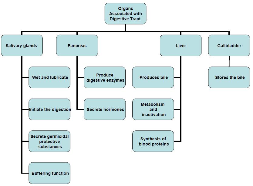
Organs Associated with the Digestive Tract. Dr. Emad I H Shaqoura M.D, M.Sc. Anatomy Faculty of Medicine, IUG March, 2016
|
|
|
- Victoria Poole
- 5 years ago
- Views:
Transcription
1 Organs Associated with the Digestive Tract Dr. Emad I H Shaqoura M.D, M.Sc. Anatomy Faculty of Medicine, IUG March, 2016
2 2
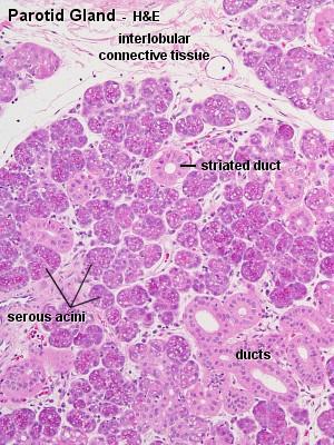
3 Salivary Glands Salivary Glands Major 90% of saliva Minor 10% of saliva Parotid Submandibular Sublingual Scattered in the mucosa & submucosa 3
4 Salivary Glands (cont d) Exocrine glands saliva (ph ) Their secretions may be serous, seromucous or mucous. Each major salivary gland is composed of: 1. Stroma: C.T capsule surrounding the gland & sending septa into its interior. Many lymphocytes & plasma cells also present. 2. Parenchyma: lobules made of secretory units & ducts. 4
5 Medical Application Inadequate saliva production, leading to dry mouth or xerostomia, can be caused by various factors affecting the major salivary glands, as: 1. mumps viral infection, 2. radiation of the glands, or 3. normal side effect of drugs such as antihistamines. Excessive saliva production, or sialorrhea, is associated with the autonomic activity of nausea, inflammation within the oral cavity, and rabies viral infection. 5

6 Salivary Glands (cont d) 6


7 Serous cells Usually pyramidal in shape with rounded nuclei. Broad base resting on the basal lamina. Narrow apical surface with short, irregular microvilli facing the lumen. They exhibit characteristics of polarized protein-secreting cells. Usually form a spherical mass of cells called acinus with very small central lumen. 7

8 Figure 16-3 Copyright McGraw-Hill Companies

9 Mucous cells Usually cuboidal to columnar in shape. Their nuclei are oval and pressed toward the bases of the cells. Secret glycoprotein mucins. Mucous cells are most often organized as tubules. The ends of mucous tubules are capped by serous cells, which constitute the serous demilunes. 9

10 Figure 16-5 Copyright McGraw-Hill Companies


11 Serous & Mucous Cells * Ill-defined cell borders. * Well-defined cell borders. * Narrow lumen. * Wide lumen. 11

12 Figure 16-4 Copyright McGraw-Hill Companies

13 Myoepithelial cells Found between the basal lamina and the basal plasma membrane of the cells forming secretory endpieces and intercalated ducts. Sometimes called basket cells 13
")
14 Myoepithelial cells (cont d) 14
")
15 Myoepithelial cells (cont d) 15
16 Duct System Striated Ducts Excretory Ducts Main Duct Intercalated Ducts Acini 16
 ducts Lining range from simple cuboidal or columnar epithelia to stratified cuboidal or columnar epithelia.")
17 Duct System 1. Intralobular ducts Intercalated ducts Striated ducts 2. Interlobular (excretory) ducts Lining range from simple cuboidal or columnar epithelia to stratified cuboidal or columnar epithelia. Or even pseudostratified type. 3. The main duct Non-keratinized stratified squamous epithelium. 17

18 Striated & Intercalated Ducts 18

19 Striated Ducts, E.M 19

20 Figure 16-6 Copyright McGraw-Hill Companies
21 Salivary Glands (cont d) Salivary Glands Major 90% of saliva Minor 10% of saliva Parotid Submandibular Sublingual Scattered in the mucosa & submucosa 21

22 I. Major Salivary Glands 22
23 1. Parotid Gland Branched acinar gland. Its secretory portion is composed exclusively of serous cells. Secretory granules have abundant prolinerich proteins with antimicrobial & Ca+²binding activities and also have a high α- amylase content. Intercalated and striated ducts are easily observed. 23
24 Figure 16-3 Copyright McGraw-Hill Companies
25 25
26 2. Submandibular Gland Branched tubulo-acinar gland formed of both serous and mucous cells. It produces 2/3 of total saliva. The serous cells are the main component (90%). Serous cells are mostly serous demilunes. Serous cells secrete the enzyme lysozyme in addition to amylase & proline-rich proteins. Striated ducts are easily observed but intercalated ducts are very short. 26
 grouped as tubules in this tubuloacinar gland. Small intralobular ducts (ID) drain each lobule, but these are not composed of columnar cells with welldeveloped striations. X340. H&E.")
27 2. Submandibular Gland (cont d) Submandibular gland is a mixed serous (90%) and mucous (10%) gland and shows: well-stained cells in serous acini (A) and in serous demilunes (S) and pale-staining mucous cells (M) grouped as tubules in this tubuloacinar gland. Small intralobular ducts (ID) drain each lobule, but these are not composed of columnar cells with welldeveloped striations. X340. H&E. 27
")
28 2. Submandibular Gland (cont d) 28
29 3. Sublingual Gland Branched tubuloacinar gland formed of both serous and mucous cells. Mucous cells predominate in this gland. Intralobular ducts are not as well developed as in other major salivary glands. 29
.")
30 3. Sublingual Gland (cont d) Sublingual gland is a mixed but largely mucous gland with a tubuloacinar arrangement of poorly stained mucous cells (M). Small intralobular ducts (ID) are seen in connective tissue, as well as small fascicles of lingual striated muscle (SM(. 30
")
31 3. Sublingual Gland (cont d) 31
")
32 3. Sublingual Gland (cont d) 32
33 II. Minor Salivary Glands Non-encapsulated glands distributed throughout the oral mucosa and submucosa. Saliva is produced by secretory units into short ducts, with little modification of its content. Minor salivary glands are usually mucous except the small serous glands von Ebner's glands. Lymphocyte aggregates are commonly observed and are concerned with IgA secretion. 33
34 Pancreas The pancreas is a mixed exocrineendocrine gland that produces digestive enzymes and hormones. Enzymes are stored and released by cells of the exocrine portion, arranged in acini. The hormones are synthesized in clusters of endocrine epithelial cells known as islets of Langerhans. It has a C.T capsule that sends septa. 34
")
35 Pancreas (cont d) 35
36 Pancreatic Cancer Pancreatic cancer is a carcinoma of duct cells that can arise anywhere in the gland. It occurs most often in the head of the organ near the duodenum. The tumor is usually asymptomatic until growth and metastasis are well advanced, leading to the low rate of early detection and subsequent high rate of mortality. Metastasis may be facilitated by the relatively sparse connective tissue around the ducts and vasculature of the pancreas. 36
")
37 Pancreas (cont d) 37
. 3. No myoepithelial cells. 4. Presence of the islets of Langerhans in the pancreas. N.B.")
38 Pancreas (cont d) Exocrine part (compound acinar) is similar to parotid gland except: 1. Absence of striated ducts. 2. The initial portions of intercalated ducts penetrate the lumens of the acini (centroacinar cells). 3. No myoepithelial cells. 4. Presence of the islets of Langerhans in the pancreas. N.B. Intercalated ducts are tributaries of larger interlobular ducts. 38

39 Pancreas (cont d) The exocrine pancreatic acinus is composed of several serous cells surrounding a lumen. These cells are highly polarized, with a spherical basal nucleus, and are typical proteinsecreting cells with apical zymogen granules. 39
")
40 Pancreas (cont d) 40
")
41 Pancreas (cont d) 41
")
42 Pancreas (cont d) 42

43 Figure Copyright McGraw-Hill Companies
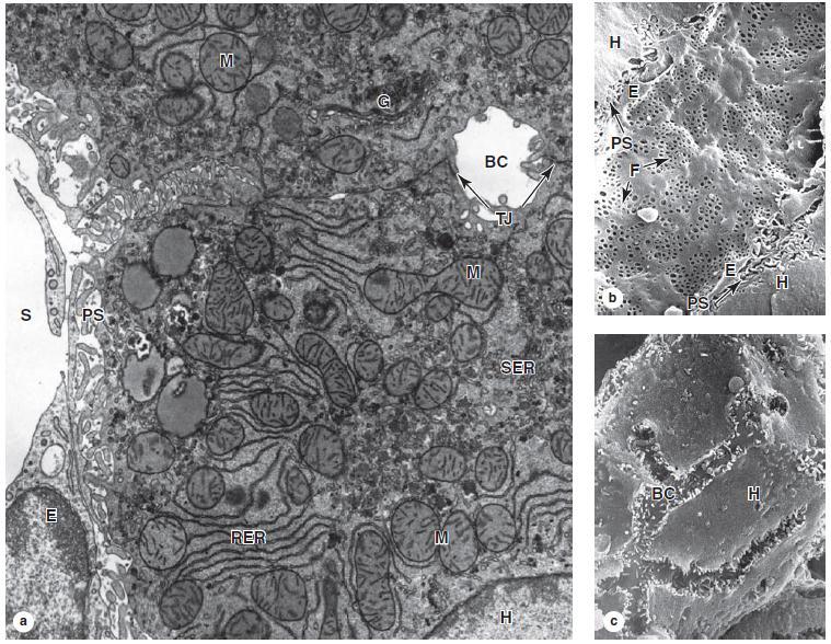
44 Pancreas (cont d) The exocrine pancreas secretes about 1500 ml of alkaline fluid / day containing: - Water - Ions - Proteases (Trypsinogen, chymotrypsinogen, ) - α- amylase - Lipases - Nucleases (DNAase & RNAase) The majority of the enzymes are stored as proenzymes in the secretory granules of acinar cells. 44
45 Pancreas (cont d) Pancreatic tissue is protected against autodigestion by the following: 1. Restricting protease activation to the duodenum. 2. Trypsin inhibitor, which is co-packaged in the secretory granules with trypsinogen. 3. The low ph in the acini and duct system due to HCO3 secreted by the centroacinar and intercalated duct cells, which helps keep all the enzymes inactive. 45
46 Pancreatitis In acute pancreatitis, the proenzymes may be activated and digest pancreatic tissues, leading to very serious complications. Possible causes include infection, gallstones, alcoholism, drugs, and trauma. Chronic pancreatitis can produce progressive fibrosis and loss of pancreatic function. 46
47 Regulation of Pancreatic Secretion Pancreatic secretion is controlled mainly through: 1. Two polypeptide hormones - secretin and cholecystokinin (CCK) - produced by enteroendocrine cells of the intestinal mucosa (duodenum and jejunum). 2. The vagus (parasympathetic) nerve: stimulates both duct and acinar cell secretions. 47
48 Regulation of Pancreatic Secretion Secretin promotes the secretion of an abundant alkaline fluid rich in electrolytes and poor in enzymes by the duct cells. Cholecystokinin promotes the secretion of a less abundant but enzyme-rich fluid acting mainly by extrusion of zymogen granules from acinar cells. 48

49 Liver Rt. Lobe Lt. Lobe Falciform Ligament Spleen Gall Bladder 49
50 Liver (cont d) liver is the second-largest organ of the body and the largest gland. The liver is the organ in which nutrients absorbed in the digestive tract are processed and stored for use by other parts of the body. It has a dual blood supply: 1. 75% comes from the portal vein, (nutrient-rich, O2-poor) 2. 25% is supplied by the hepatic artery (O2-rich). Bile is an exocrine secretion of the liver that is important for lipid digestion and toxic substances elimination. The liver also has the very important function of producing plasma proteins, such as albumin, other carrier proteins, coagulation factors, and growth factors. 50
51 Hepatic Stroma The liver is covered by a thin connective tissue capsule (Glisson's capsule) that becomes thicker at the hilum, where the portal vein and the hepatic artery enter the organ and where the right and left hepatic ducts and lymphatics exit. These vessels and ducts are surrounded by connective tissue all the way to their termination (or origin) in the portal spaces between the liver lobules. At this point, a delicate reticular fiber network that supports the hepatocytes and sinusoidal endothelial cells of the liver lobules is formed. 51

52 Hepatic Stroma Silver stain used to view the reticular fiber network running between hepatocytes. 52
53 Hepatic Parenchyma Is formed of anastomosing plates of hepatocytes radiating from a central vein & arranged in a polyhedral hepatic lobules. Each lobule has 3-6 peripheral portal areas. Portal areas = C.T + Portal triad (AVD). Hepatic sinusoids present between the plates of hepatocytes separated from them by perisinusoidal space (of Disse). 53

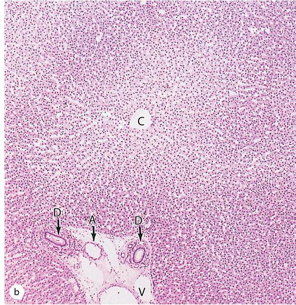
54 Hepatic Lobule 54

55 Hepatic Lobule L.M C: central vein. A: hepatic artery. V: portal vein. D: bile duct. 55

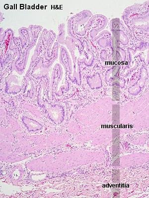
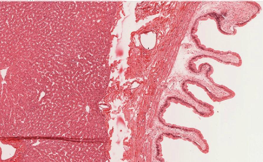
56 Figure Copyright McGraw-Hill Companies

57 Hepatic Lobule 57

58 The Hepatocyte, L.M - Large polyhedral cells. - Central rounded vesicular nucleus. - Cells are frequently binucleated. - Cytoplasm: eosinophilic. 58
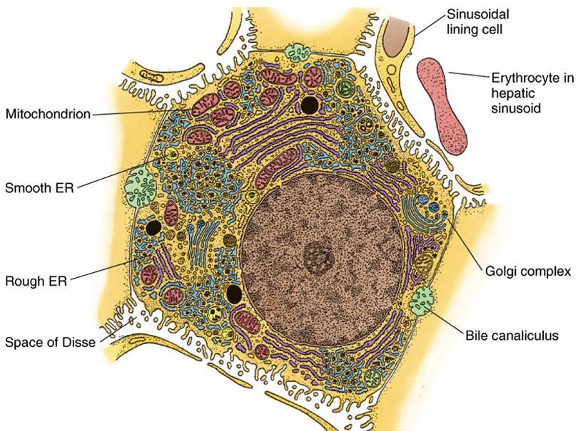
59 The Hepatocyte, E.M 59
60 The Hepatocyte, E.M 60


61 The Hepatocyte Function Protein synthesis & Carbohydrate storage Secretion of bile acids 61
 Secretion of")
62 The Hepatocyte Function (cont d) Secretion of bilirubin 62
 Portal Venules Hepatic")

63 Blood Supply of the Liver Portal Vein 75% (Nutrient-rich, O2- poor) Portal Venules Hepatic Sinusoids Blood Supply Hepatic Artery 25% (O2-rich) Hepatic Arterioles Hepatic Sinusoids Central Venules Hepatic Veins IVC 63

64 Figure Copyright McGraw-Hill Companies

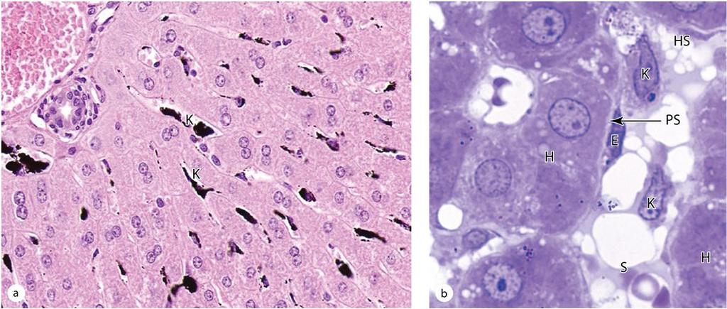
65 Hepatic Sinusoids Have a large diameter. Discontinuous basal lamina. Lined by TWO types of cells: 1. Endothelial cells: having fenestrated cytoplasm not covered by a diaphragm. 2. Kupffer cells: large branching phagocytes that function to metabolize aged RBCs & digest hemoglobin. 65

66 Hepatic Sinusoids (cont d) 66

67 Hepatic Sinusoids (cont d) 67
68 Hepatic Sinusoids (cont d) In the perisinusoidal space are hepatic stellate cells (or Ito cells) with small lipid droplets that store vitamin A and other fatsoluble vitamins. These mesenchymal cells, which are difficult to see in routine preparations, also produce extracellular matrix (ECM) components (becoming myofibroblasts after liver injury) and cytokines that help regulate Kupffer cell activity. 68

69 Figure Copyright McGraw-Hill Companies

70 Internal organization of the liver 70
71 Liver Regeneration Despite its slow rate of cell renewal, the liver has an extraordinary capacity for regeneration. The loss of hepatic tissue by surgical removal or from the action of toxic substances triggers a mechanism by which hepatocytes begin to divide, continuing until the original mass of tissue is restored (compensatory hyperplasia). 71

72 Liver Cirrhosis The regenerated liver tissue is usually well organized, exhibiting the typical lobular arrangement and replacing the functions of the destroyed tissue. However, when there is continuous or repeated damage to hepatocytes over a long period of time, the multiplication of liver cells is followed by a pronounced increase in the amount of connective tissue. 72

73 Clinical features of liver disease 73

74 Bile Duct System Canaliculi Ductules Ducts 74

75 Biliary Tract & Gallbladder 75
76 Biliary Tract & Gallbladder Canaliculi: Lined only by plasma membranes of two adjacent hepatocytes. Ductules: Simple cuboidal epithelium. Ducts: Simple cuboidal or columnar epithelium + C.T. The hepatic, cystic, and common bile ducts are lined with a mucous membrane having a simple columnar epithelium of cholangiocytes + thin L.P & submucosa + thin muscularis. 76

77 Gallbladder The wall of the gallbladder consists of: a mucosa composed of simple columnar epithelium and lamina propria. a layer of smooth muscle. a perimuscular connective tissue layer a serous membrane. 77
78 78
79 79

80 Gallbladder The mucosa has abundant folds that are particularly evident when the gallbladder is empty. The epithelial cells are rich in mitochondria. All these cells are capable of secreting small amounts of mucus. 80
81 Tumors of the Digestive Glands Most 1ry malignant tumors of the liver derive from hepatic parenchyma or epithelial cells of the bile duct. It may be associated with a variety of acquired disorders, such as chronic viral hepatitis (B or C) and liver cirrhosis. In the exocrine pancreas, most tumors arise from ductal epithelial cells. 81
82 Medicine is An Ever-Changing Science 82
Digestive system L 4. Lecturer Dr. Firdous M. Jaafar Department of Anatomy/Histology section
 Digestive system L 4 Lecturer Dr. Firdous M. Jaafar Department of Anatomy/Histology section objectives 1-Describe the structure of liver. 2-Define liver lobule, and identify its zones. 3-Define portal
DIGESTIVE SYSTEM II ACCESSORY DIGESTIVE ORGANS
 DIGESTIVE SYSTEM II ACCESSORY DIGESTIVE ORGANS Dr. Larry Johnson Texas A& M University Objectives Distinguish between the parotid and submandibular salivary glands. Understand and identify the structural
Chapter 12 The Digestive Glands
 Chapter 12 The Digestive Glands Lyu Zhengmei Department of Histology and Embryology, Anhui Medical University Components of digestive glands large salivary glands, pancreas, liver, gallbladder. These organs
Slide 154: Pancreas, H&E
 Slide 154: Pancreas, H&E the pancreas, located adjacent to the duodenum, is a mixed exocrine and endocrine gland; it is usually readily identifiable by the presence of the interspersed endocrine pancreatic
Laboratory exercises for abdominal organs
 Laboratory exercises for abdominal organs Slide #77 (C007- H- 107A). Pancreas, dog. pancreatic islets CENTROACINAR CELLS ARE THE BEGINNING CELLS OF THE INTERCALATED DUCTS THAT DRAIN THE SECRETORY ACINI
Paneth Cells. Road Map to the Finish. No Review this Friday. Today 11/29 Finish digestion/accessory organs. Wednesday 12/1 Immune System I
 Road Map to the Finish No Review this Friday Today 11/29 Finish digestion/accessory organs Wednesday 12/1 Immune System I Paneth Cells - base of intestinal glands -! large -! intense acidophilic granules
Large Intestine. The large intestine consists of a mucosal membrane with no folds except in its distal (rectal) portion
 portion") GI Histology 3 Large Intestine The large intestine consists of a mucosal membrane with no folds except in its distal (rectal) portion No villi are present in this portion of the intestine The intestinal
GI Histology 3 Large Intestine The large intestine consists of a mucosal membrane with no folds except in its distal (rectal) portion No villi are present in this portion of the intestine The intestinal
HISTOLOGY VIRTUAL LABORATORY GASTROINTESTINAL SYSTEM
 HISTOLOGY VIRTUAL LABORATORY GASTROINTESTINAL SYSTEM LIP (Slides GI 1, 2) Identify the outer portion lined by stratified squamous (keratinized) epithelium. Note the hair follicles and sebaceous glands
PRACTICAL ROADMAP. GLANDS AFFECTING LIFESTYLE WJ van der Spuy & T Tshabalala
 PRACTICAL ROADMAP GLANDS AFFECTING LIFESTYLE WJ van der Spuy & T Tshabalala GLANDS AFFECTING LIFESTYLE Submandibular gland (salivary gland) Liver Pancreas Hypophysis (pituitary gland) Thyroid Suprarenal
Glandular Epithelium. Dr. Hersh Abdul Ham-Karim BVM&S, PG Dip, MSc and PhD
 Glandular Epithelium Dr. Hersh Abdul Ham-Karim BVM&S, PG Dip, MSc and PhD Glandular Epithelium Groups of surface cells differentiate, proliferate, and penetrate underlying connective tissue. Their main
Tongue In the buccal cavity of the digestive system
 Tongue In the buccal cavity of the digestive system same layers as those of tubular organs Mucosa, submucosa, and muscularis muscularis = the muscularis externa no muscularis mucosa 1 Tongue ling = tongue
Chapter 18 Liver and Gallbladder
 Chapter 18 Liver and Gallbladder 解剖學科徐淑媛 本堂重點 1. Liver : functions & histology 2. Gallbladder Physiology Liver Produce circulating plasma proteins Vitamin Iron Degradation Metabolism Bile manufacture (exocrine)
Digestive system. Dr. Sami Zaqout. IUG
 Digestive system Digestive system Digestive tract Associated glands Oral cavity Salivary glands Esophagus Liver Stomach Pancreas Small and large intestines Rectum and anus General Structure of the Digestive
Objectives. Describe the cells of the GI tract and their function. Differentiate between different parts of the GI tract
 GI Histology 1 Objectives Describe the cells of the GI tract and their function Describe the histological features of each part of the GI tract. Differentiate between different parts of the GI tract Appreciate
Glandular Epithelium. Dr. Heba Kalbouneh Associate Professor of Anatomy and Histology
 Glandular Epithelium Dr. Heba Kalbouneh Associate Professor of Anatomy and Histology Glands Glandular epithelia are tissues formed by cells specialized to produce secretion. Secretion: if substances produced
DIGESTIVE. CHAPTER 17 Lecture: Part 1 Part 2 BIO 212: ANATOMY & PHYSIOLOGY II
 BIO 212: ANATOMY & PHYSIOLOGY II CHAPTER 17 Lecture: DIGESTIVE Part 1 Part 2 Dr. Lawrence G. Altman www.lawrencegaltman.com Some illustrations are courtesy of McGraw-Hill. SMALL INTESTINE DUODENUM > JEJUNUM
5 Dr. Heba Kalbouneh
 5 Dr. Heba Kalbouneh Glandular epithelium Gland: Is a collection of epithelial cells the secrets a certain product, like: proteins, lipids and carbohydrates. Secretion : A certain material that is produced
Epithelium. Four primary tissue types:
 Epithelium Four primary tissue types: Epithelial (covering) Connective (support) Nervous (control) Muscular (movement) Smooth muscle Cardiac muscle Skeletal muscle 1 Epithelial Tissue Features Epithelial
Dr. Abeer.c.Yousif. Histology -2 nd stage. What is histology?
 What is histology? Histology is the science of microscopic anatomy of cells and tissues, in Greek language Histo= tissue and logos = study and it's tightly bounded to molecular biology, physiology, immunology
Glandular Epithelium. Dr. Heba Kalbouneh Assistant Professor of Anatomy and Histology
 Glandular Epithelium Dr. Heba Kalbouneh Assistant Professor of Anatomy and Histology Glands Gla dular epithelia are tissues for ed y ells spe ialized to produ e se retio. Secretion: if substances produced
Digestive System Module 6: Accessory Organs in Digestion: The Liver, Pancreas, and Gallbladder
 Connexions module: m49293 1 Digestive System Module 6: Accessory Organs in Digestion: The Liver, Pancreas, and Gallbladder Donna Browne Based on Accessory Organs in Digestion: The Liver, Pancreas, and
Chapter 14: The Digestive System
 Chapter 14: The Digestive System Digestive system consists of Muscular tube (digestive tract) alimentary canal Accessory organs teeth, tongue, glandular organs 6 essential activities 1. 2. 3. 4. 5. 6.
Epithelia will be discussed according to the following scheme: Type Number of layers Shape Line drawing. Squamous Cuboidal Columnar
 Epithelia Epithelia will be discussed according to the following scheme: Type Number of layers Shape Line drawing Simple Squamous Cuboidal Columnar Covering and Lining epithelium Pseudostratified Stratified
MICROSCOPIC STRUCTURE OF LIVER, GALLBLADDER, GALL DUCTS, AND PANCREAS OVERVIEW OF DEVELOPMENT OF THE ALIMENTARY CANAL
 Lecture 2 ESS_3rd semester MICROSCOPIC STRUCTURE OF LIVER, GALLBLADDER, GALL DUCTS, AND PANCREAS OVERVIEW OF DEVELOPMENT OF THE ALIMENTARY CANAL MICROSCOPIC STRUCTURE OF LIVER - is the largest gland of
1. Approximately 21 ft. long: duodenum (one ft.), jejunum (eight ft.), and ileum (twelve ft.)
, jejunum (eight ft.), and ileum (twelve ft.)") IV. Small Intestines A. General features and functions 1. Approximately 21 ft. long: duodenum (one ft.), jejunum (eight ft.), and ileum (twelve ft.) 2. Functions: move forward chyme, continue digestion,
IV. Small Intestines A. General features and functions 1. Approximately 21 ft. long: duodenum (one ft.), jejunum (eight ft.), and ileum (twelve ft.) 2. Functions: move forward chyme, continue digestion,
Dr. Heba Kalbouneh. Dr. Heba Kalbouneh. Dr. Heba Kalbouneh
 Dr. Heba Kalbouneh Dr. Heba Kalbouneh Dr. Heba Kalbouneh Basement membrane: What is the basement membrane? - It is a layer of ECM separating the epithelial cells from the underlying connective tissue Basement
Gastric Contrac,le Ac,vity. Regula,on of Gastric Emptying
 Gastric Contrac,le Ac,vity Figure 23.18 Regula,on of Gastric Emptying Gastric emptying is regulated by: Neural enterogastric reflex Hormonal (enterogastrone) mechanisms In the presence of gastric gastrin
Anatomy of the liver and pancreas
 Anatomy of the liver and pancreas Prof. Abdulameer Al-Nuaimi E-mail: a.al-nuaimi@sheffield.ac.uk abdulameerh@yahoo.com Liver Aorta Pulm. Trunk Rt. At, Duct. Art. Lt. Ven. Rt. Ven. Internal Posterior
The Digestive System. Chapter 25
 The Digestive System Chapter 25 Introduction Structure of the digestive system A tube that extends from mouth to anus Accessory organs are attached Functions include Ingestion Movement Digestion Absorption
terminal portion of the main pancreatic duct (like the the common bile duct) will have longitudinally and circularly arranged smooth muscle which is
 will have longitudinally and circularly arranged smooth muscle which is") Chapter 14 Accessory Organs of the Small Intestine 14.1. The Pancreas The pancreas has both an exocrine and an endocrine nature. These two functions are handled by histologically distinct regions of the
Chapter 14 Accessory Organs of the Small Intestine 14.1. The Pancreas The pancreas has both an exocrine and an endocrine nature. These two functions are handled by histologically distinct regions of the
GI Histology Lab 1. Prepared by: Zeina Kalaji
 GI Histology Lab 1 Prepared by: Zeina Kalaji Lip ORAL MUCOSA -Arrow shows labial salivary glands in the submucosa. VERMILLION transitional zone. SKIN Stratified Squamous epithelium, keratinized -Arrow
Small intestine. Small intestine
 General features Tubular organ longest part; 5-6 m most of chemical digestion absorption of nutrients reabsorption of H2O occurs. Two structural features; maximize the lumenal surface area villi microvilli
Gastrointestinal Anatomy and Physiology. Bio 219 Napa Valley College Dr. Adam Ross
 Gastrointestinal Anatomy and Physiology Bio 219 Napa Valley College Dr. Adam Ross Functions of digestive system Digestion Breakdown of food (chemically) using enzymes, acid, and water Absorption Nutrients,
Histology of the liver, and the biliary system
 Histology of the liver, and the biliary system Dr. Zsuzsanna Tóth Semmelweis University Department of Anatomy, Histology and Embryology http://www.mgtowforums.com/forums/lads-night-inn/3969-give-your-liver-art.html
The Digestive System. What is the advantage of a one-way gut? If you swallow something, is it really inside you?
 The Digestive System What is the advantage of a one-way gut?! If you swallow something, is it really inside you? Functions and Processes of the Digestive System: Move nutrients, water, electrolytes from
Tissues. tissue = many cells w/ same structure and function. cell shape aids its function tissue shape aids its function
 Tissues tissue = many cells w/ same structure and function cell shape aids its function tissue shape aids its function Histology = study of tissues 4 types of tissues Epithelial coverings contact openings
Histology 3. We will continue talking about a few things from last lecture, starting with M cells:
 Histology 3 This is the last Histology lecture in the GI system. Enjoy! There are some extra notes listed as footnotes. We will continue talking about a few things from last lecture, starting with M cells:
LIVER & SPLEEN. Color index: Slides.. Important..Notes..Extra..
 LIVER & SPLEEN Color index: Slides.. Important..Notes..Extra.. Objectives: By the end of this lecture, the student should be able to describe: 1. The histological structure of liver with special emphasis
Histology Lab. looking at microscopic pictures of tissues, for more information use Junqueira book and you can use BlueHistolgy website
 Done By: Aseel Twaijer & Laith Sorour Histology Lab *These notes help in differentiating tissues and you must read them while looking at microscopic pictures of tissues, for more information use Junqueira
(b) Stomach s function 1. Dilution of food materials 2. Acidification of food (absorption of dietary Fe in small intestine) 3. Partial chemical digest
 Stomach s function 1. Dilution of food materials 2. Acidification of food (absorption of dietary Fe in small intestine) 3. Partial chemical digest") (1) General features a) Stomach is widened portion of gut-tube: between tubular and spherical; Note arranged of smooth muscle tissue in muscularis externa. 1 (b) Stomach s function 1. Dilution of food
(1) General features a) Stomach is widened portion of gut-tube: between tubular and spherical; Note arranged of smooth muscle tissue in muscularis externa. 1 (b) Stomach s function 1. Dilution of food
Histology Notes -Part 1: Epithelial Tissues
 Introduction Group of cells w/ similar structure & function = TISSUE Four Basic Tissue Types 1. Epithelial-covers 2. Connective-supports 3. Muscular*-produces movement (will discuss in the muscular system
Epithelial Tissue. Functions include: 1. Protection 4. Absorption 2. Secretion 5. Filtration 3. Sensory reception
 Tissues There are 4 primary tissue types in the human body: 1. Epithelial (covering/lining) 2. Connective (support) 3. Muscle (movement) 4. Nervous (control) Epithelium Epithelial Tissue Covers the surface
Midterm 2 is Tuesday 5/28/13
 Business Reminder: No class Monday (Memorial Day) Midterm 2 is Tuesday 5/28/13 Optional review session tomorrow @ 5pm Homework due in Lab 1. PreLab 8 (1pt) 2. Replace a Missing Assignment (4 pts) Homework
Tissue: The Living Fabric: Part A
 PowerPoint Lecture Slides prepared by Janice Meeking, Mount Royal College C H A P T E R 4 Tissue: The Living Fabric: Part A Tissues Groups of cells similar in structure and function Types of tissues Epithelial
General Structure of Digestive Tract
 Dr. Nabil Khouri General Structure of Digestive Tract Common Characteristics: Hollow tube composed of a lumen whose diameter varies. Surrounded by a wall made up of 4 principal layers: Mucosa Epithelial
MICROSTRUCTURES SMALL INTESTIN LARGE INTESTIN PANCREAS LIVER GALLBLADDER SALIVARY GLANDS ADRENALS THYROID AND PARATHYROID GLANDS
 MICROSTRUCTURES SMALL INTESTIN LARGE INTESTIN PANCREAS LIVER GALLBLADDER SALIVARY GLANDS ADRENALS THYROID AND PARATHYROID GLANDS HUMAN ANATOMY: MICROSTRUCTURES CLASSIFICATION: LOCATION AND BOUNDARIES,
Tissues. tissue = many cells w/ same structure and function. cell shape aids function tissue shape aids function. Histology = study of tissues
 Tissues tissue = many cells w/ same structure and function cell shape aids function tissue shape aids function Histology = study of tissues 4 types of tissues Epithelial coverings contact openings Connective
Cell and Tissue Types. Epithelial, Connective, Muscle, Nerve
 Cell and Tissue Types Epithelial, Connective, Muscle, Nerve Objectives Explain the major stages of the cell cycle and cellular division (mitosis). Describe specific events occurring in each of the phases
DIGESTIVE SYSTEM ALIMENTARY CANAL / GI TRACT & ACCESSORY ORGANS. Mar 16 10:34 PM
 DIGESTIVE SYSTEM ALIMENTARY CANAL / GI TRACT & ACCESSORY ORGANS Mar 16 10:34 PM 1 I. Digestive System Functions > Ingestion the taking in of food > Propulsion movement caused by force > Digestion breakdown
Tissues. Definition. A group of similar cells and their intercellular substances specialized to perform a specific function.
 Chapter 4 - Tissues Tissues Definition A group of similar cells and their intercellular substances specialized to perform a specific function. Tissues Epithelial covers exposed surfaces, lines internal
Lymphoid Organs. Dr. Sami Zaqout. Dr. Sami Zaqout IUG Faculty of Medicine
 Lymphoid Organs Dr. Sami Zaqout Cells of the Immune System Lymphocytes Plasma cells Mast cells Neutrophils Eosinophils Cells of the mononuclear phagocyte system Distribution of cells of the immune system
2. Epithelial Tissues Dr. Manal Othman
 Biology-232 GENERAL HISTOLOGY 2. Epithelial Tissues Dr. Manal Othman Anatomy Department CMMS, AGU HISTOLOGY: w Study of the structure and function of tissues and organs at the microscopic levels. w Tissues
Basic Histology. By Mrs. Bailey
 Basic Histology By Mrs. Bailey Primary Tissues 1. Epithelial Tissue 2. Connective Tissue 3. Muscle Tissue 4. Nervous Tissue Very cellular Supported by underlying connective tissue Epithelial & connective
Bio 322 Human Anatomy Objectives for the laboratory exercise Digestive System
 Bio 322 Human Anatomy Objectives for the laboratory exercise Digestive System Required reading before beginning this lab: Saladin, KS: Human Anatomy 5 th ed (2017) Chapter 24 For this lab you will use
BIO 116 Anatomy & Physiology II Practice Assignment 3 - The Lymphatic, Immune and Digestive Systems This is not a required assignment
 BIO 116 Anatomy & Physiology II Practice Assignment 3 - The Lymphatic, Immune and Digestive Systems This is not a required assignment 1. Which are components of the lymphatic system? a: Thyroid gland b:
Histology = the study of tissues. Tissue = a complex of cells that have a common function
 { EPITHELIAL TISSUE Histology = the study of tissues Tissue = a complex of cells that have a common function The Four Primary Tissue Types: Epithelium (epithelial tissue) covers body surfaces, lines body
Epithelium Characteristics cont. 2. Apical Surface
 Epithelium Characteristics cont. 2. Apical Surface always has one exposed (apical) surface Some surfaces are smooth & slick, others may have: microvilli fingerlike extensions of the plasma membrane; increase
Basic Tissue Types and Functions
 Tissues Histology Basic Tissue Types and Functions 1) Epithelial tissue covering 2) Connective tissue support 3) Muscle tissue movement 4) Nervous tissue control Epithelial Tissue 1) Covers a body surface
Unit I Problem 9 Histology: Basic Tissues of The Body
 Unit I Problem 9 Histology: Basic Tissues of The Body - What is the difference between cytology and histology? Cytology: it is the study of the structure and functions of cells and their contents. Histology:
Lab activity manual - Histology of the digestive system. Lab activity 1: esophagus stomach - small intestines
 Lab activity manual - Histology of the digestive system Jeanne Adiwinata Pawitan Prerequisite: Histology of the 4 basic tissues In this module we learn about the histology of the digestive system, from
Includes mouth, pharynx, esophagus, stomach, small intestine, large intestine, rectum, anus. Salivary glands, liver, gallbladder, pancreas
 Chapter 14 The Digestive System and Nutrition Digestive System Brings Nutrients Into the Body The digestive system includes Gastrointestinal (GI) tract (hollow tube) Lumen: space within this tube Includes
HISTOLOGY OF THE RESPIRATORY SYSTEM I. Introduction A. The respiratory system provides for gas exchange between the environment and the blood. B.
 HISTOLOGY OF THE RESPIRATORY SYSTEM I. Introduction A. The respiratory system provides for gas exchange between the environment and the blood. B. The human respiratory system may be subdivided into two
The Digestive System. Chapter 16. Introduction. Overview of Digestive System. Histological Organization. Movement and Mixing of Digestive Materials
 The Digestive System Chapter 16 Introduction Structure of the digestive system A tube that extends from mouth to anus Accessory organs are attached Functions include Ingestion Movement Digestion Absorption
The Digestive system
 The Digestive system The GI tract (gastrointestinal tract) Mouth Pharynx Esophagus Stomach Small intestine Large intestine Anus The accessory digestive organs Supply secretions contributing to the breakdown
Tissue: The Living Fabric
 PowerPoint Lecture Slide Presentation by Vince Austin Human Anatomy & Physiology FIFTH EDITION Elaine N. Marieb Chapter 4 Tissue: The Living Fabric Part A Tissues Groups of cells similar in structure and
Pancreas Fox Chapter 18 part 2 (also Chapter 19.3 & 19.4)
") Vert Phys PCB3743 Pancreas Fox Chapter 18 part 2 (also Chapter 19.3 & 19.4) T. Houpt, Ph.D. Anatomy of Digestive System Peristalsis Stomach and Acid Secretion Liver and Bile Secretion Pancreas and pancreatic
Vert Phys PCB3743 Pancreas Fox Chapter 18 part 2 (also Chapter 19.3 & 19.4) T. Houpt, Ph.D. Anatomy of Digestive System Peristalsis Stomach and Acid Secretion Liver and Bile Secretion Pancreas and pancreatic
NOTES: The Digestive System (Ch 14, part 2)
") NOTES: The Digestive System (Ch 14, part 2) PANCREAS Structure of the pancreas: The pancreas produces PANCREATIC JUICE that is then secreted into a pancreatic duct. The PANCREATIC DUCT leads to the The
NOTES: The Digestive System (Ch 14, part 2) PANCREAS Structure of the pancreas: The pancreas produces PANCREATIC JUICE that is then secreted into a pancreatic duct. The PANCREATIC DUCT leads to the The
Accessory Organs in Digestion: The Liver, Pancreas, and Gallbladder *
 OpenStax-CNX module: m46496 1 Accessory Organs in Digestion: The Liver, Pancreas, and Gallbladder * OpenStax This work is produced by OpenStax-CNX and licensed under the Creative Commons Attribution License
Body Tissues. Cells are specialized for particular functions Tissues - groups of cells with similar structure. and function Four primary tissue types:
 Chapter 3 Tissues Body Tissues Cells are specialized for particular functions Tissues - groups of cells with similar structure and function Four primary tissue types: Epithelium Connective tissue Nervous
Ali Yaghi. Yaseen Fatayer. M.Khatatbeh
 6 Ali Yaghi Yaseen Fatayer M.Khatatbeh P a g e 1 pancreatic secretions note: The pancreas has endocrine (secretions are released toward the blood) and exocrine(secretions are released through the canalicular
Connective tissue The Digestive System
 Connective tissue The Digestive System Part 1 Structure of digestive system Functions Basic Structure of the Alimentary Canal Wall Tube is made up of four layers: 1. Mucosa 2. Submucosa 3. Muscularis externa
The Digestive System
 The Digestive System Identify the Structure and Function. Mesentery of the Large Intestine The mesentery functions to connect the visceral organs to the abdominal wall. Identify the Structure. Nasal Cavity
Chapter 1: Cells and Tissues
 Chapter 1: Cells and Tissues Cells and Tissues Carry out all chemical activities needed to sustain life Cells are the building blocks of all living things Tissues are groups of cells that are similar in
Alimentary Canal (I)
") Alimentary Canal (I) Esophagus and Stomach (Objectives) By the end of this lecture, the student should be able to discuss the microscopic structure in correlation with the function of the following organs:
Alimentary Canal (I) Esophagus and Stomach (Objectives) By the end of this lecture, the student should be able to discuss the microscopic structure in correlation with the function of the following organs:
Gastrointestinal Tract
 CTO Lab #5 GI TRACT & GLANDS; ENDOCRINE SYSTEM Page 1 Gastrointestinal Tract Slide 126 This section through the esophagus shows the characteristic layers of the gastrointestinal tract. Examine the non-keratinized
Chapter 4 - Epithelial Tissues
 Chapter 4 - Epithelial Tissues Tissues Definition A group of closely associated cells that work together to perform a specific function Types Epithelial - covering Connective - support Muscle - movement
Anatomy PHL 212. Dr. Dina A. A. Hassan. -
 Anatomy PHL 212 Dr. Dina A. A. Hassan Associate Professor College of Pharmacy (Female Section) Sattam Bin Abdulaziz University Al kharj / Kingdom of Saudi Arabia Email :- da.hassan@psau.edu.sa 1 Anatomy
Chapter 15 Gastrointestinal System
 Chapter 15 Gastrointestinal System Dr. LL Wang E-mail: wanglinlin@zju.edu.cn Rm 608, Block B, Research Building, School of Medicine, Zijingang Campus Pancreatic Secretion The exocrine cells in the pancreas
Two main groups Alimentary canal continuous coiled hollow tube Accessory digestive organs
 Digestion Breakdown of ingested food Absorption of nutrients into the blood Metabolism Production of cellular energy (ATP) Constructive and degradative cellular activities Two main groups Alimentary canal
Organs Histology D. Sahar AL-Sharqi. Respiratory system
 Respiratory system The respiratory system provides for exchange of O2 and CO2 to and from the blood. Respiratory organs include the lungs and a branching system of bronchial tubes that link the sites of
Epithelial Tissue lining, covering, glandular tissue > Function protect, absorption, filtration, secretion, excretion
 Chapter 4: TISSUES IX. Tissues Intro Epithelial Tissue lining, covering, glandular tissue > Function protect, absorption, filtration, secretion, excretion Connective Tissue most widespread tissue type
A adipose cells. B capillary. C epithelium
 EPITHELIA Objective The objective of this class is to observe how different epithelia vary in terms of cell shape, size and number of cell layers enabling them to be well adapted for functions in different
Anatomy & Physiology Revealed Instructions. 1. From the Module dropdown menu, chose the 12. Digestive system.
 #10 - Objectives: Examine the histology of selected body organs using Anatomy & Physiology Revealed software and microscope slides. Be able to identify each organ and the specific structures indicated
The stomach is formed of three parts: -
 The stomach is formed of three parts: - (a) CARDIAC STOMACH: - It receives the oesophagus through Cardiac aperture guarded by a cardiac sphincter which prevents regurgitation of food. (b) FUNDIC PART:
Tissues Review 4 type
 Tissues Review 4 type Tissues Definition: a group of closely associated cells that perform related functions and are similar in structure Between cells: nonliving extracellular material Four basic types
BIOH122 Human Biological Science 2
 BIOH122 Human Biological Science 2 Session 15 Digestive System 2 Stomach and Accessory Structures Bioscience Department Endeavour College of Natural Health endeavour.edu.au Session Plan o Stomach anatomy
1-It is to prevent back flow of fecal content from colon into small intestine.
 Function of the ileocecal valve: 1-It is to prevent back flow of fecal content from colon into small intestine. 2-The wall of the ileum for several centimeters preceding valve has a thickened muscular
The Digestive System and Body Metabolism Premedical Biology
 The Digestive System and Body Metabolism Premedical Biology Copyright 2003 Pearson Education, Inc. publishing as Benjamin Cummings The Digestive System and Body Digestion Metabolism Breakdown of ingested
Glands Histology lab 5 Notes by Lojayn Salah
 Glands Histology lab 5 Notes by Lojayn Salah There are two types of glands: - 1) Endocrine gland: collection of epithelial cells with no connection with the epithelial surface, it has no duct, its secretory
Lecture Overview. Marieb s Human Anatomy and Physiology. Chapter 4 Tissues: The Living Fabric Epithelial Tissues Lecture 9. Introduction to Tissues
 Marieb s Human Anatomy and Physiology Marieb Hoehn Chapter 4 Tissues: The Living Fabric Epithelial Tissues Lecture 9 Lecture Overview Introduction to Tissues Epithelial Tissues Location General characteristics
Behzad Mobini* Islamic Azad University, Shahrekord Branch, Shahrekord, Iran. Received: 25 October 2010, Accepted: 4 January 2011
 Original Article Veterinary Research Forum Vol: 2, No: 1, March, 2011, 25-29 Histological Studies on Pancreas of Goose (Anser Albifrons) Behzad Mobini* * Department of Anatomical Sciences, Faculty of Veterinary
2. SECRETIONS OF THE DIGESTIVE TRACT
 2. SECRETIONS OF THE DIGESTIVE TRACT SECRETORY GLANDS AND CELLS The alimentary tract produces a large variety and quantity of substances which contribute to digesting the food, and protecting and regulating
Pancreas & Biliary System. Dr. Vohra & Dr. Jamila
 Pancreas & Biliary System Dr. Vohra & Dr. Jamila 1 Objectives At the end of the lecture, the student should be able to describe the: Location, surface anatomy, parts, relations & peritoneal reflection
CHAPTER 05 Histology: EPITHELIUM
 BIO 211: ANATOMY & PHYSIOLOGY I 1 CHAPTER 05 Histology: EPITHELIUM Part 01: Brief Introduction Part 02: Survey of Types Dr. Lawrence G. G. Altman www.lawrencegaltman.com Some illustrations are courtesy
Connective tissue The Digestive System
 Connective tissue The Digestive System Part 1 Structure of digestive system Functions Basic Structure of the Alimentary Canal Wall Tube is made up of four layers: 1. Mucosa 2. Submucosa 3. Muscularis externa
5. March 23, Seminar: Relationship between structure and function of the liver. Practical class: Gastro-intestinal system, part 3.
 Katedra i Zakład Histologii i Embriologii Centrum Biostruktury Warszawski Uniwersytet Medyczny 2014/2015 SECOND SEMESTER HISTOLOGY & EMBRYOLOGY 6 years MD PROGRAM SEMINARS & PRACTICAL CLASSES SPRING SEMESTER
TISSUES TYPES. CHAPTER 05 Histology: EPITHELIUM BIO 211: ANATOMY & PHYSIOLOGY I. HISTOLOGY = the study of tissues
 BIO 211: ANATOMY & PHYSIOLOGY I 1 CHAPTER 05 Histology: EPITHELIUM Part 01: Brief Introduction Part 02: Survey of Types Dr. Lawrence G. G. Altman www.lawrencegaltman.com Some illustrations are courtesy
Anatomy & Histology of The Small intestine
 Anatomy & Histology of The Small intestine Prof. Abdulameer Al-Nuaimi E-mail: a.al-nuaimi@sheffield.ac.uk E. mail: abdulameerh@yahoo.com Jejunum Ileum Histology: Duodenum, jejunum, and ileum
Biology. Dr. Khalida Ibrahim
 Dr. Khalida Ibrahim Biology Histology: Histology: is the study of the tissues of the body. Tissue: group of similar cells combined to perform a common function. The human body is composed of only 4 basic
Chapter 9. The digestive system. Glossary. Louise McErlean
 Chapter 9 The digestive system Louise McErlean Glossary Absorption Process whereby the products of digestion move into the blood or lymph fluid. Acini glands Produce pancreatic juice. Amylase Carbohydrate