Objectives. Describe the cells of the GI tract and their function. Differentiate between different parts of the GI tract
|
|
|
- Audra Griffith
- 6 years ago
- Views:
Transcription
1 GI Histology 1
2 Objectives Describe the cells of the GI tract and their function Describe the histological features of each part of the GI tract. Differentiate between different parts of the GI tract Appreciate the histopathology of the GI tract Describe the histological basis of some clinical problems
3 The digestive system consists of the digestive tract oral cavity, esophagus, stomach, small and large intestines, rectum, and anus and its associated glands salivary glands, liver, and pancreas. Its function is to obtain the molecules necessary for the maintenance, growth, and energy needs of the body from ingested food. Large molecules such as proteins, fats, complex carbohydrates, and nucleic acids are broken down into small molecules that are easily absorbed through the lining of the digestive tract, mostly in the small intestine. Water, vitamins, and minerals are also absorbed from ingested food. In addition, the inner layer of the digestive tract is a protective barrier between the content of the tract's lumen and the internal milieu of the body.
4 General Structure of the Digestive Tract The entire gastrointestinal tract presents certain common structural characteristics is a hollow tube composed of a lumen whose diameter varies, surrounded by a wall made up of four principal layers: the mucosa, submucosa, muscularis, and serosa. The mucosa comprises an epithelial lining; a lamina propria of loose connective tissue rich in blood and lymph vessels and smooth muscle cells, sometimes also containing glands and lymphoid tissue and the muscularis mucosae, usually consisting of a thin inner circular layer and an outer longitudinal layer of smooth muscle cells separating the mucosa from the submucosa. The mucosa is frequently called a mucous membrane.
5 The submucosa is composed of dense connective tissue with many blood and lymph vessels and a submucosal (also called Meissner's) nerve plexus. It may also contain glands and lymphoid tissue. The muscularis contains smooth muscle cells that are spirally oriented and divided into two sublayers according to the main direction the muscle cells follow In the internal sublayer (close to the lumen), the orientation is generally circular; in the external sublayer, it is mostly longitudinal The muscularis also contains the myenteric (or Auerbach's) nerve plexus, which lies between the two muscle sublayers blood and lymph vessels in the connective tissue between the muscle sublayers.
6 The serosa is a thin layer of loose connective tissue, rich in blood and lymph vessels and adipose tissue, and a simple squamous covering epithelium (mesothelium). In the abdominal cavity, the serosa is continuous with the mesenteries and with the peritoneum In places where the digestive organ is bound to other organs or structures, however, the serosa is replaced by a thick adventitia, consisting of connective tissue containing vessels and nerves, without the mesothelium.
7 Basic mucosal forms in the GI tract Protective : stratified squamous epithelium that is found in the oral cavity, pharynx, the esophagus and the anal canal Seceretory : the mucosa consists of a long closely packed tubular glands, found in the stomach Absorptive ; the mucosa is arranged in a fingerlike projections called vili with intervening short glands called crypts, that is typical for the small intestine. In the duodenum some crypts extend from the muscularis mucosa to the submucosa (Brunners Gland) Absorptive/protective ; the mucosa is arranged into closely packed tubular glands specialised for water absorption and mucus secreting goblet cells It lines the whole large intesine
8 The Oral Cavity The oral cavity is lined with stratified squamous epithelium, keratinized or nonkeratinized, depending on the region The keratin layer protects the oral mucosa from damage during masticatory function and is present mostly in the gingiva (gum) and hard palate The lamina propria in these regions has several papillae and rests directly on bony tissue. Nonkeratinized squamous epithelium covers the soft palate, lips, cheeks, and the floor of the mouth. The lamina propria has papillae, similar to those in the dermis of the skin, and is continuous with a submucosa containing diffuse small salivary glands. In the lips, a transition from the oral nonkeratinized epithelium to the keratinized epithelium of the skin can be observed. The soft palate has a core of skeletal muscle, numerous mucous glands, and lymphoid nodules in its submucosa.

9 Tongue The tongue is a mass of striated muscle covered by a mucous membrane whose structure varies according to the region The muscle fibers cross one another in three planes; they are grouped in bundles, usually separated by connective tissue Because the connective tissue of the lamina propria penetrates the spaces between the muscular bundles, the mucous membrane is strongly adherent to the muscle The mucous membrane is smooth on the lower (ventral) surface of the tongue The tongue's dorsal surface is irregular, covered anteriorly by a great number of small eminences called papillae.
 of the")
10 The posterior one-third of the dorsal surface of the tongue is separated from the anterior two-thirds by a V- shaped boundary Behind this boundary, the surface of the tongue shows small bulges composed mainly of two types of small lymphoid aggregations: small collections of lymphoid nodules and the lingual tonsils, where lymphoid nodules aggregate around invaginations (crypts) of the mucous membrane

11 Papillae Papillae are elevations of the oral epithelium and lamina propria that assume various forms and functions. There are four types Filiform Papillae Filiform papillae have an elongated conical shape; they are quite numerous and are present over the entire surface of the tongue Their epithelium, which does not contain taste buds, is keratinized.

12 Fungiform Papillae Fungiform papillae resemble mushrooms in that they have a narrow stalk and a smooth-surfaced, dilated upper part These papillae, which contain scattered taste buds on their upper surfaces, are irregularly interspersed among the filiform papillae. Foliate Papillae Foliate papillae are poorly developed in humans They consist of two or more parallel ridges and furrows on the dorsolateral surface of the tongue and contain many taste buds.

13 Taste buds
 glands drain their contents into the deep groove that encircles the periphery of each papilla This moatlike arrangement provides a continuous flow")
14 Circumvallate Papillae Circumvallate papillae are 7 12 extremely large circular papillae whose flattened surfaces extend above the other papillae They are distributed in the V region in the posterior portion of the tongue Numerous serous (von Ebner's) glands drain their contents into the deep groove that encircles the periphery of each papilla This moatlike arrangement provides a continuous flow of fluid over the great number of taste buds present along the sides of these papillae The glands also secrete a lipase that probably prevents the formation of a hydrophobic layer over the taste buds that would hinder their function. This flow of secretions is important in removing food particles from the vicinity of the taste buds so that they can receive and process new gustatory stimuli
15 Along with this local role, lingual lipase is active in the stomach and can digest up to 30% of dietary triglycerides Other small mucous salivary glands dispersed throughout the lining of the oral cavity act in the same way as the serous glands associated with this type of papilla to prepare the taste buds in other parts of the oral cavity, such as the anterior portion of the tongue, to respond to taste stimuli.
16 Salivary Glands Saliva is a complex fluid that has digestive, lubricating, and protective functions In addition to the small salivary glands scattered throughout the oral cavity, there are three pairs of large salivary glands: the parotid, submandibular (submaxillary), and sublingual glands In humans, the minor salivary glands secrete 10% of the total volume of saliva, but they account for approximately 70% of the mucus secreted.
17 A capsule of connective tissue, rich in collagen fibers, surrounds the large salivary glands. The parenchyma of the glands consists of secretory end pieces and a branching duct system arranged in lobules, separated by septae of connective tissue originating from the capsule The secretory end pieces present two types of secretory cellsâ serous and mucous as well as the nonsecretory myoepithelial cells This secretory portion is followed by a duct system whose components modify and conduct the saliva to the oral cavity.
18 Serous cells are usually pyramidal in shape, with a broad base resting on the basal lamina and a narrow apical surface with short, irregular microvilli facing the lumen They exhibit characteristics of polarized protein-secreting cells. Adjacent secretory cells are joined together by junctional complexes and usually form a spherical mass of cells called acinus, with a small lumen in the center This structure can be thought of as a grape attached to its stem; the stem corresponds to the duct system.
19 Mucous cells are usually cuboidal to columnar in shape; their nuclei are oval and pressed toward the bases of the cells. They exhibit the characteristics of mucus-secreting cells containing glycoproteins important for the moistening and lubricating functions of the saliva Most of these glycoproteins are called mucins and contain 70â 80% carbohydrate moieties in their structure Mucous cells are most often organized as tubules, consisting of cylindrical arrays of secretory cells surrounding a lumen.
20 Myoepithelial cells are found between the basal lamina and the basal plasma membrane of the cells forming secretory end pieces and intercalated ducts (to a lesser extent), which form the initial portion of the duct system Myoepithelial cells surrounding each secretory portion, usually two to three cells per secretory unit, are well developed and branched (and are sometimes called basket cells whereas those associated with intercalated ducts are spindle shaped and lie parallel to the length of the duct These cells show several characteristics that resemble smooth muscle cells, including contractility. However, they also establish intercellular junctions among themselves and with secretory cells, such as desmosomes Although the contraction of myoepithelial cells accelerates the secretion of saliva, their main function seems to be the prevention of end piece distention during secretion due to the increase in intraluminal pressure
21 In the duct system, secretory end pieces empty into the intercalated ducts, lined by cuboidal epithelial cells These cells have the ability to divide and differentiate into secretory or ductal cells Several of these short intercalated ducts join to form striated ducts characterized by radial striations that extend from the bases of the cells to the level of the central nuclei. Intercalated and striated ducts are also called intralobular ducts because of their location within the lobule. When viewed in the electron microscope, the striations are seen to consist of infoldings of the basal plasma membrane with numerous elongated mitochondria that are aligned parallel to the infolded membranes; this structure is characteristic of ion-transporting cells
22 The striated ducts of each lobule converge and drain into ducts located in the connective tissue septae separating the lobules, where they become interlobular, or excretory, ducts They are initially lined with pseudostratified or stratified cuboidal epithelium, but more distal parts of the excretory ducts are lined with stratified columnar epithelium containing a few mucus-secreting cells The main duct of each major salivary gland ultimately empties into the oral cavity and is lined with nonkeratinized-stratified squamous epithelium.
23 Vessels and nerves enter the large salivary glands at the hilum and gradually branch into the lobules. A rich vascular and nerve plexus surrounds the secretory and ductal components of each lobule The capillaries surrounding the secretory end pieces are very important for the secretion of saliva, stimulated by the autonomic nervous system. Parasympathetic stimulation, usually through the smell or taste of food, promotes vasodilation and a copious watery secretion content. Sympathetic stimulation produces small amounts of viscous saliva, rich in organic material

24 Parotid Gland The parotid gland is a branched acinar gland; its secretory portion is composed exclusively of serous cells containing secretory granules that are rich in proteins and have a high amylase activity This activity is responsible for most of the hydrolysis of ingested carbohydrates. The digestion begins in the mouth and continues for a short time in the stomach, before the gastric juice acidifies the food and thus decreases amylase activity considerably Intercalated and striated ducts are easily observed within the lobules, due to their length. As in other large salivary glands, the connective tissue contains many plasma cells and lymphocytes The plasma cells secrete IgA, which forms a complex with a secretory component synthesized by the serous acinar, intercalated duct, and striated duct cells The IgA-rich secretory complex released into the saliva is resistant to enzymatic digestion and constitutes an immunological defense mechanism against pathogens in the oral cavity.

25 Submandibular (Submaxillary) Gland The submandibular gland is a branched tubuloacinar gland its secretory portion contains both mucous and serous cells The serous cells are the main component of this gland and are easily distinguished from mucous cells by their rounded nuclei and basophilic cytoplasm In humans, 90% of the end pieces of the submandibular gland are serous acinar, whereas 10% consist of mucous tubules with serous demilunes Serous cells are responsible for the weak amylolytic activity present in this gland and its saliva The cells that form the demilunes in the submandibular gland secrete the enzyme lysozyme, whose main activity is to hydrolyze the walls of certain bacteria Some acinar and intercalated duct cells in large salivary glands also secrete lactoferrin, which binds iron, a nutrient necessary for bacterial growth Striated ducts are easily observed in the human submandibular gland, but intercalated ducts are very short.

26 Sublingual Gland The sublingual gland, like the submandibular gland, is a branched tubuloacinar gland formed of serous and mucous cells Mucous cells predominate in this gland; serous cells are present almost exclusively on demilunes of mucous tubules As in the submandibular gland, cells that form the demilunes in this gland secrete lysozyme. Intralobular ducts are not as well developed as in other major salivary glands.
27 Minor Salivary Glands These nonencapsulated glands are distributed throughout the oral mucosa and submucosa Saliva is produced by small groups of secretory units and is conducted to the oral cavity by short ducts, with little modification of its content Although variations exist, minor salivary glands are usually mucous The small serous glands present in the posterior region of the tongue (von Ebner's glands) are the only exception Lymphocyte agregates are commonly observed within minor salivary glands, associated with IgA secretion.
Tongue In the buccal cavity of the digestive system
 Tongue In the buccal cavity of the digestive system same layers as those of tubular organs Mucosa, submucosa, and muscularis muscularis = the muscularis externa no muscularis mucosa 1 Tongue ling = tongue
GI Histology Lab 1. Prepared by: Zeina Kalaji
 GI Histology Lab 1 Prepared by: Zeina Kalaji Lip ORAL MUCOSA -Arrow shows labial salivary glands in the submucosa. VERMILLION transitional zone. SKIN Stratified Squamous epithelium, keratinized -Arrow
General Structure of Digestive Tract
 Dr. Nabil Khouri General Structure of Digestive Tract Common Characteristics: Hollow tube composed of a lumen whose diameter varies. Surrounded by a wall made up of 4 principal layers: Mucosa Epithelial
HISTOLOGY VIRTUAL LABORATORY GASTROINTESTINAL SYSTEM
 HISTOLOGY VIRTUAL LABORATORY GASTROINTESTINAL SYSTEM LIP (Slides GI 1, 2) Identify the outer portion lined by stratified squamous (keratinized) epithelium. Note the hair follicles and sebaceous glands
Alimentary Canal (I)
") Alimentary Canal (I) Esophagus and Stomach (Objectives) By the end of this lecture, the student should be able to discuss the microscopic structure in correlation with the function of the following organs:
Alimentary Canal (I) Esophagus and Stomach (Objectives) By the end of this lecture, the student should be able to discuss the microscopic structure in correlation with the function of the following organs:
Epithelium. Four primary tissue types:
 Epithelium Four primary tissue types: Epithelial (covering) Connective (support) Nervous (control) Muscular (movement) Smooth muscle Cardiac muscle Skeletal muscle 1 Epithelial Tissue Features Epithelial
Tissue: The Living Fabric: Part A
 PowerPoint Lecture Slides prepared by Janice Meeking, Mount Royal College C H A P T E R 4 Tissue: The Living Fabric: Part A Tissues Groups of cells similar in structure and function Types of tissues Epithelial
Anatomy & Histology of The Small intestine
 Anatomy & Histology of The Small intestine Prof. Abdulameer Al-Nuaimi E-mail: a.al-nuaimi@sheffield.ac.uk E. mail: abdulameerh@yahoo.com Jejunum Ileum Histology: Duodenum, jejunum, and ileum
Digestive system L 2. Lecturer Dr. Firdous M. Jaafar Department of Anatomy/Histology section
 Digestive system L 2 Lecturer Dr. Firdous M. Jaafar Department of Anatomy/Histology section objectives 1-Describe the general structure of digestive tract: a-mucosa. b-submucosa. c-muscularis externa d-adventitia
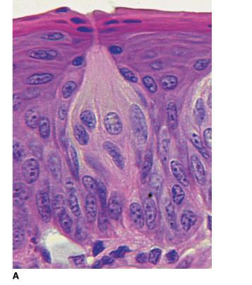
A deep groove encircles the body of the circumvallate papilla. Serous (von Ebner s) glands (serous) drain into the base of this groove.
 glands (serous) drain into the base of this groove.") By Dr. Raja Ali A deep groove encircles the body of the circumvallate papilla. Serous (von Ebner s) glands (serous) drain into the base of this groove. The flow of fluid from these glands serves to wash
By Dr. Raja Ali A deep groove encircles the body of the circumvallate papilla. Serous (von Ebner s) glands (serous) drain into the base of this groove. The flow of fluid from these glands serves to wash
Dorsum of the tongue. Oral Part exhibit lingual papillae of the 4 types. Oral Part of Tongue divided into Left & right halves by shallow median groove
 Histology of TONGUE Figure 22.13 Dorsum of the tongue Oral Part of Tongue divided into Left & right halves by shallow median groove Oral Part exhibit lingual papillae of the 4 types a. filiform papillae,
Cell and Tissue Types. Epithelial, Connective, Muscle, Nerve
 Cell and Tissue Types Epithelial, Connective, Muscle, Nerve Objectives Explain the major stages of the cell cycle and cellular division (mitosis). Describe specific events occurring in each of the phases
MICROSTRUCTURES LIPS TOOTH TONGUE OESOPHAGUS STOMACH, CARDIAC, PYLORIC FUNDIC GLANDS
 MICROSTRUCTURES LIPS TOOTH TONGUE OESOPHAGUS STOMACH, CARDIAC, PYLORIC FUNDIC GLANDS HUMAN ANATOMY: MICROSTRUCTURES CLASSIFICATION: LOCATION AND BOUNDARIES, FORM, FUNCTION, MICROSCOPIC STRUCTURE: A hollow
The Digestive System. Chapter 25
 The Digestive System Chapter 25 Introduction Structure of the digestive system A tube that extends from mouth to anus Accessory organs are attached Functions include Ingestion Movement Digestion Absorption
Histology = the study of tissues. Tissue = a complex of cells that have a common function
 { EPITHELIAL TISSUE Histology = the study of tissues Tissue = a complex of cells that have a common function The Four Primary Tissue Types: Epithelium (epithelial tissue) covers body surfaces, lines body
Dr. Abeer.c.Yousif. Histology -2 nd stage. What is histology?
 What is histology? Histology is the science of microscopic anatomy of cells and tissues, in Greek language Histo= tissue and logos = study and it's tightly bounded to molecular biology, physiology, immunology
Glandular Epithelium. Dr. Hersh Abdul Ham-Karim BVM&S, PG Dip, MSc and PhD
 Glandular Epithelium Dr. Hersh Abdul Ham-Karim BVM&S, PG Dip, MSc and PhD Glandular Epithelium Groups of surface cells differentiate, proliferate, and penetrate underlying connective tissue. Their main
Small intestine. Small intestine
 General features Tubular organ longest part; 5-6 m most of chemical digestion absorption of nutrients reabsorption of H2O occurs. Two structural features; maximize the lumenal surface area villi microvilli
Slide 154: Pancreas, H&E
 Slide 154: Pancreas, H&E the pancreas, located adjacent to the duodenum, is a mixed exocrine and endocrine gland; it is usually readily identifiable by the presence of the interspersed endocrine pancreatic
DIGESTIVE SYSTEM II ACCESSORY DIGESTIVE ORGANS
 DIGESTIVE SYSTEM II ACCESSORY DIGESTIVE ORGANS Dr. Larry Johnson Texas A& M University Objectives Distinguish between the parotid and submandibular salivary glands. Understand and identify the structural
Lecture Overview. Chapter 4 Epithelial Tissues Lecture 9. Introduction to Tissues. Epithelial Tissues. Glandular Epithelium
 Visual Anatomy & Physiology First Edition Martini & Ober Chapter 4 Lecture 9 Lecture Overview Introduction to Tissues Location General characteristics Functions Classification Glandular Epithelium 2 Where
Lecture Overview. Marieb s Human Anatomy and Physiology. Chapter 4 Tissues: The Living Fabric Epithelial Tissues Lecture 9. Introduction to Tissues
 Marieb s Human Anatomy and Physiology Marieb Hoehn Chapter 4 Tissues: The Living Fabric Epithelial Tissues Lecture 9 Lecture Overview Introduction to Tissues Epithelial Tissues Location General characteristics
Tissues. tissue = many cells w/ same structure and function. cell shape aids its function tissue shape aids its function
 Tissues tissue = many cells w/ same structure and function cell shape aids its function tissue shape aids its function Histology = study of tissues 4 types of tissues Epithelial coverings contact openings
Epithelia will be discussed according to the following scheme: Type Number of layers Shape Line drawing. Squamous Cuboidal Columnar
 Epithelia Epithelia will be discussed according to the following scheme: Type Number of layers Shape Line drawing Simple Squamous Cuboidal Columnar Covering and Lining epithelium Pseudostratified Stratified
The Digestive System. Chapter 16. Introduction. Overview of Digestive System. Histological Organization. Movement and Mixing of Digestive Materials
 The Digestive System Chapter 16 Introduction Structure of the digestive system A tube that extends from mouth to anus Accessory organs are attached Functions include Ingestion Movement Digestion Absorption
HISTOLOGY. GIT Block 432 Histology Team. Lecture 1: Alimentary Canal (1) (Esophagus & Stomach) Done by: Ethar Alqarni Reviewed by: Ibrahim Alfuraih
 (Esophagus & Stomach) Done by: Ethar Alqarni Reviewed by: Ibrahim Alfuraih") HISTOLOGY Lecture 1: Alimentary Canal (1) (Esophagus & Stomach) Done by: Ethar Alqarni Reviewed by: Ibrahim Alfuraih Color Guide: Black: Slides. Red: Important. Green: Doctor s notes. Blue: Explanation.
HISTOLOGY Lecture 1: Alimentary Canal (1) (Esophagus & Stomach) Done by: Ethar Alqarni Reviewed by: Ibrahim Alfuraih Color Guide: Black: Slides. Red: Important. Green: Doctor s notes. Blue: Explanation.
Histology Notes -Part 1: Epithelial Tissues
 Introduction Group of cells w/ similar structure & function = TISSUE Four Basic Tissue Types 1. Epithelial-covers 2. Connective-supports 3. Muscular*-produces movement (will discuss in the muscular system
Two main groups Alimentary canal continuous coiled hollow tube Accessory digestive organs
 Digestion Breakdown of ingested food Absorption of nutrients into the blood Metabolism Production of cellular energy (ATP) Constructive and degradative cellular activities Two main groups Alimentary canal
Digestive System 7/15/2015. Outline Digestive System. Digestive System
 Digestive System Biology 105 Lecture 18 Chapter 15 Outline Digestive System I. Functions II. Layers of the GI tract III. Major parts: mouth, pharynx, esophagus, stomach, small intestine, large intestine,
The Digestive System Laboratory
 The Digestive System Laboratory 1 The Digestive Tract The alimentary canal is a continuous tube stretching from the mouth to the anus. Liver Gallbladder Small intestine Anus Parotid, sublingual, and submaxillary
Oral cavity Lab exercises
 Oral cavity Lab exercises Slide #190 (GT-1-32). Oral cavity, goat. large conical buccal papillae stratified squamous epithelium keratinized or non-keratinized no muscularis mucosae connective tissue represents
(b) Stomach s function 1. Dilution of food materials 2. Acidification of food (absorption of dietary Fe in small intestine) 3. Partial chemical digest
 Stomach s function 1. Dilution of food materials 2. Acidification of food (absorption of dietary Fe in small intestine) 3. Partial chemical digest") (1) General features a) Stomach is widened portion of gut-tube: between tubular and spherical; Note arranged of smooth muscle tissue in muscularis externa. 1 (b) Stomach s function 1. Dilution of food
(1) General features a) Stomach is widened portion of gut-tube: between tubular and spherical; Note arranged of smooth muscle tissue in muscularis externa. 1 (b) Stomach s function 1. Dilution of food
Histology Lab. looking at microscopic pictures of tissues, for more information use Junqueira book and you can use BlueHistolgy website
 Done By: Aseel Twaijer & Laith Sorour Histology Lab *These notes help in differentiating tissues and you must read them while looking at microscopic pictures of tissues, for more information use Junqueira
Digestive system. Dr. Sami Zaqout. IUG
 Digestive system Digestive system Digestive tract Associated glands Oral cavity Salivary glands Esophagus Liver Stomach Pancreas Small and large intestines Rectum and anus General Structure of the Digestive
بسم هللا الرحمن الرحيم
 بسم هللا الرحمن الرحيم Today, we will leave all hormones and start with another topic which is GI system This lecture is talking about general histology of Gastrointestinal system.. The gastrointestinal
Connective tissue The Digestive System
 Connective tissue The Digestive System Part 1 Structure of digestive system Functions Basic Structure of the Alimentary Canal Wall Tube is made up of four layers: 1. Mucosa 2. Submucosa 3. Muscularis externa
Glandular Epithelium. Dr. Heba Kalbouneh Associate Professor of Anatomy and Histology
 Glandular Epithelium Dr. Heba Kalbouneh Associate Professor of Anatomy and Histology Glands Glandular epithelia are tissues formed by cells specialized to produce secretion. Secretion: if substances produced
Epithelial Lecture Test Questions
 Epithelial Lecture Test Questions 1. Which of the following free surfaces lack(s) epithelia: a. lung alveoli (air sacs) b. hard palate c. joint cavities d. abdominal cavity e. salivary gland ducts 2. Which
B. Classification of epithelium: by number of cell layers present and by shape of the superficial cell layers.
 I. Introduction - tissue: group of cells that are closely associated, similar in structure and function, and perform a common or related function. - four primary tissues: epithelial tissue, connective
Principles of Anatomy and Physiology
 Principles of Anatomy and Physiology 14 th Edition CHAPTER 24 The Digestive System Introduction The purpose of this chapter is to Identify the anatomical components of the digestive system as well as their
Epithelial Tissue. Functions include: 1. Protection 4. Absorption 2. Secretion 5. Filtration 3. Sensory reception
 Tissues There are 4 primary tissue types in the human body: 1. Epithelial (covering/lining) 2. Connective (support) 3. Muscle (movement) 4. Nervous (control) Epithelium Epithelial Tissue Covers the surface
Connective tissue The Digestive System
 Connective tissue The Digestive System Part 1 Structure of digestive system Functions Basic Structure of the Alimentary Canal Wall Tube is made up of four layers: 1. Mucosa 2. Submucosa 3. Muscularis externa
5 Dr. Heba Kalbouneh
 5 Dr. Heba Kalbouneh Glandular epithelium Gland: Is a collection of epithelial cells the secrets a certain product, like: proteins, lipids and carbohydrates. Secretion : A certain material that is produced
Tissues. Definition. A group of similar cells and their intercellular substances specialized to perform a specific function.
 Chapter 4 - Tissues Tissues Definition A group of similar cells and their intercellular substances specialized to perform a specific function. Tissues Epithelial covers exposed surfaces, lines internal
Unit I Problem 9 Histology: Basic Tissues of The Body
 Unit I Problem 9 Histology: Basic Tissues of The Body - What is the difference between cytology and histology? Cytology: it is the study of the structure and functions of cells and their contents. Histology:
Dana Alrafaiah. Dareen Abu Shalbak. Mohammad Almuhtaseb. 1 P a g e
 2 Dana Alrafaiah Dareen Abu Shalbak Mohammad Almuhtaseb 1 P a g e Esophagus: A muscular tube that is 25 cm long, but if measured from the incisors it would be 45cm long. Extends from C6 of cervical vertebra,
Organs Associated with the Digestive Tract. Dr. Emad I H Shaqoura M.D, M.Sc. Anatomy Faculty of Medicine, IUG March, 2016
 Organs Associated with the Digestive Tract Dr. Emad I H Shaqoura M.D, M.Sc. Anatomy Faculty of Medicine, IUG March, 2016 2 Salivary Glands Salivary Glands Major 90% of saliva Minor 10% of saliva Parotid
The Digestive System and Body Metabolism Premedical Biology
 The Digestive System and Body Metabolism Premedical Biology Copyright 2003 Pearson Education, Inc. publishing as Benjamin Cummings The Digestive System and Body Digestion Metabolism Breakdown of ingested
Glandular Epithelium. Dr. Heba Kalbouneh Assistant Professor of Anatomy and Histology
 Glandular Epithelium Dr. Heba Kalbouneh Assistant Professor of Anatomy and Histology Glands Gla dular epithelia are tissues for ed y ells spe ialized to produ e se retio. Secretion: if substances produced
DIGESTIVE TRACT ESOPHAGUS
 DIGESTIVE TRACT From the lower esophagus to the lower rectum four fundamental layers comprise the wall of the digestive tube: mucosa, submucosa, muscularis propria (externa), and adventitia or serosa (see
Prepared By Student. Dania Abed Al-majeed. Rahma Raad Hanna. Balqees Mohammed Aasim. Dania Hisham. Rasha Rafiee
 Prepared By Student Rahma Raad Hanna Balqees Mohammed Aasim Dania Hisham Dania Abed Al-majeed Rasha Rafiee Epithelia Epithelia can be derived from ectoderm, mesoderm or endoderm -ectoderm gives rise to
(A) Diarrhea. (B) Stomach cramps. (C) Dehydration due to excess fluid loss. (D) A, B, and C are correct. (E) Only answer B is correct.
 Diarrhea. (B) Stomach cramps. (C) Dehydration due to excess fluid loss. (D) A, B, and C are correct. (E) Only answer B is correct.") Human Anatomy - Problem Drill 21: The Digestive System Question No. 1 of 10 1. A 26-year-old male is treated in the emergency department for severe gastrointestinal disturbance. Which of the following
Human Anatomy - Problem Drill 21: The Digestive System Question No. 1 of 10 1. A 26-year-old male is treated in the emergency department for severe gastrointestinal disturbance. Which of the following
Dr Nadine Gravett School of Anatomical Sciences Room 2B10B
 Dr Nadine Gravett School of Anatomical Sciences Room 2B10B Nadine.Gravett@wits.ac.za Oral cavity Mechanical breakdown Formation of bolus Oesophagus Conduit from mouth to stomach Stomach Digestion Temporary
Basic Tissue Types and Functions
 Tissues Histology Basic Tissue Types and Functions 1) Epithelial tissue covering 2) Connective tissue support 3) Muscle tissue movement 4) Nervous tissue control Epithelial Tissue 1) Covers a body surface
HISTOLOGY OF THE RESPIRATORY SYSTEM I. Introduction A. The respiratory system provides for gas exchange between the environment and the blood. B.
 HISTOLOGY OF THE RESPIRATORY SYSTEM I. Introduction A. The respiratory system provides for gas exchange between the environment and the blood. B. The human respiratory system may be subdivided into two
Tissue: The Living Fabric
 PowerPoint Lecture Slide Presentation by Vince Austin Human Anatomy & Physiology FIFTH EDITION Elaine N. Marieb Chapter 4 Tissue: The Living Fabric Part A Tissues Groups of cells similar in structure and
EPITHELIUM 3/12/2018 د. درويش بدران د. ماهر الحديدي د.امجد الشطرات و احسان العمري
 EPITHELIUM 1 2 3 1- SIMPLE SQUAMOUS EPITHELIUM It is a single layer of flat cells that resembles a tiled floor when viewed from apical surface; centrally located nucleus that is flattened and oval or spherical
A. cells that perform related functions and are similar in structure. B. extracellular material - made by cells and secreted into interstitial space
 I. tissue components A. cells that perform related functions and are similar in structure B. extracellular material - made by cells and secreted into interstitial space II. tissue types A. epithelium (e.)
DIGESTIVE. CHAPTER 17 Lecture: Part 1 Part 2 BIO 212: ANATOMY & PHYSIOLOGY II
 BIO 212: ANATOMY & PHYSIOLOGY II 1 CHAPTER 17 Lecture: DIGESTIVE Part 1 Part 2 Dr. Lawrence G. Altman www.lawrencegaltman.com Some illustrations are courtesy of McGraw-Hill. Processes of DIGESTION Mechanical
Organs Histology D. Sahar AL-Sharqi. Digestive System
 Digestive System The digestive system consists of the digestive tract oral cavity, esophagus, stomach, small and large intestines, and anus and its associated glands salivary glands, liver, and pancreas.
Lab activity manual - Histology of the digestive system. Lab activity 1: esophagus stomach - small intestines
 Lab activity manual - Histology of the digestive system Jeanne Adiwinata Pawitan Prerequisite: Histology of the 4 basic tissues In this module we learn about the histology of the digestive system, from
Chapter 9. The digestive system. Glossary. Louise McErlean
 Chapter 9 The digestive system Louise McErlean Glossary Absorption Process whereby the products of digestion move into the blood or lymph fluid. Acini glands Produce pancreatic juice. Amylase Carbohydrate
Alimentary Canal (I) Salivatory Glands. (Esophagus and Stomach) Color index: Slides.. Important..Notes..Extra..
 Salivatory Glands. (Esophagus and Stomach) Color index: Slides.. Important..Notes..Extra..") Alimentary Canal (I) (Esophagus and Stomach) Salivatory Glands Color index: Slides.. Important..Notes..Extra.. Objectives: 1. By the end of this lecture, the student should be able to discuss the microscopic
Alimentary Canal (I) (Esophagus and Stomach) Salivatory Glands Color index: Slides.. Important..Notes..Extra.. Objectives: 1. By the end of this lecture, the student should be able to discuss the microscopic
Chapter 12 The Digestive Glands
 Chapter 12 The Digestive Glands Lyu Zhengmei Department of Histology and Embryology, Anhui Medical University Components of digestive glands large salivary glands, pancreas, liver, gallbladder. These organs
Organs Histology D. Sahar AL-Sharqi. Respiratory system
 Respiratory system The respiratory system provides for exchange of O2 and CO2 to and from the blood. Respiratory organs include the lungs and a branching system of bronchial tubes that link the sites of
Tissues 10/21/2016. Epithelial Tissue
 Tissues This is a generalized cell diagram. It shows the anatomy of a cell, but most cells do not actually look like this. Cells can have a wide variety of shapes and sizes, depending on their function.
Small Intestine, Large Intestine and anal cannel
 Small Intestine, Large Intestine and anal cannel 32409 Small intestine Large intestine Small intestine General Structure of the Digestive Tract rat 32409 Epithelium with goblet cells and absorptive cells
Tissues. tissue = many cells w/ same structure and function. cell shape aids function tissue shape aids function. Histology = study of tissues
 Tissues tissue = many cells w/ same structure and function cell shape aids function tissue shape aids function Histology = study of tissues 4 types of tissues Epithelial coverings contact openings Connective
Esophagus. Transport is achieved by peristaltic contractions and relaxation of the esophageal sphincters (upper and lower)
") GI Histology 2 Esophagus is a muscular tube whose function is to transport foodstuffs from the mouth to the stomach and to prevent the retrograde flow of gastric contents Transport is achieved by peristaltic
GI Histology 2 Esophagus is a muscular tube whose function is to transport foodstuffs from the mouth to the stomach and to prevent the retrograde flow of gastric contents Transport is achieved by peristaltic
Chapter 14: The Digestive System
 Chapter 14: The Digestive System Digestive system consists of Muscular tube (digestive tract) alimentary canal Accessory organs teeth, tongue, glandular organs 6 essential activities 1. 2. 3. 4. 5. 6.
HUMAN ANATOMY II STUDY NOTES. At the end of this chapter the student should be able to answer the following questions:
 HUMAN ANATOMY II STUDY NOTES CHAPTER ONE The Special Senses Learning objectives At the end of this chapter the student should be able to answer the following questions: 1. What is the gross and histological
Lab activity manual Histology of the digestive system
 Lab activity manual Histology of the digestive system Jeanne Adiwinata Pawitan Prerequisite: Histology of the 4 basic tissues In this module we learn about the histology of the digestive system, from the
Basic Histology. By Mrs. Bailey
 Basic Histology By Mrs. Bailey Primary Tissues 1. Epithelial Tissue 2. Connective Tissue 3. Muscle Tissue 4. Nervous Tissue Very cellular Supported by underlying connective tissue Epithelial & connective
Epithelium Characteristics cont. 2. Apical Surface
 Epithelium Characteristics cont. 2. Apical Surface always has one exposed (apical) surface Some surfaces are smooth & slick, others may have: microvilli fingerlike extensions of the plasma membrane; increase
Anatomy PHL 212. Dr. Dina A. A. Hassan. -
 Anatomy PHL 212 Dr. Dina A. A. Hassan Associate Professor College of Pharmacy (Female Section) Sattam Bin Abdulaziz University Al kharj / Kingdom of Saudi Arabia Email :- da.hassan@psau.edu.sa 1 Anatomy
2. Epithelial Tissues Dr. Manal Othman
 Biology-232 GENERAL HISTOLOGY 2. Epithelial Tissues Dr. Manal Othman Anatomy Department CMMS, AGU HISTOLOGY: w Study of the structure and function of tissues and organs at the microscopic levels. w Tissues
Lymphoid Organs. Dr. Sami Zaqout. Dr. Sami Zaqout IUG Faculty of Medicine
 Lymphoid Organs Dr. Sami Zaqout Cells of the Immune System Lymphocytes Plasma cells Mast cells Neutrophils Eosinophils Cells of the mononuclear phagocyte system Distribution of cells of the immune system
Urinary system. Urinary system
 Distal convoluted tubule (DCT) Highly coiled, ~ 5 mm in length Last part of the nephron. Wall; simple cuboidal epithelium Less metabolically active than the PCT no brush border light eosinophilic cytoplasm
The doctor mentioned a few things about the esophagus from the previous lecture:
 السالم عليكم [HISOLOGY 2] April 27, 2014 The doctor mentioned a few things about the esophagus from the previous lecture: Esophagus - It is about 25 cm in length (from the incisor it is 45 cm) Histological
DIGESTIVE SYSTEM CLASS NOTES. tube along with several
 DIGESTIVE SYSTEM CLASS NOTES Digestion Breakdown of food and the of nutrients in the bloodstream. Metabolism Production of for and cellular activities. The digestive system is composed of the canal which
Chapter 4 - Epithelial Tissues
 Chapter 4 - Epithelial Tissues Tissues Definition A group of closely associated cells that work together to perform a specific function Types Epithelial - covering Connective - support Muscle - movement
Lesson 9A Tissues in Animals
 Lesson 9A Tissues in Animals Levels of Organization in the Human Body Similar types of cells Different types of tissues Different organs Many organ systems cell tissue organ organ system organism Levels
Dr. Heba Kalbouneh. Dr. Heba Kalbouneh. Dr. Heba Kalbouneh
 Dr. Heba Kalbouneh Dr. Heba Kalbouneh Dr. Heba Kalbouneh Basement membrane: What is the basement membrane? - It is a layer of ECM separating the epithelial cells from the underlying connective tissue Basement
Overview of the Digestive
 Overview of the Digestive System Bởi: OpenStaxCollege The function of the digestive system is to break down the foods you eat, release their nutrients, and absorb those nutrients into the body. Although
Biology. Dr. Khalida Ibrahim
 Dr. Khalida Ibrahim Biology Histology: Histology: is the study of the tissues of the body. Tissue: group of similar cells combined to perform a common function. The human body is composed of only 4 basic
Histology. There are four basic tissue types in the body are :-
 Histology Lab.I There are four basic tissue types in the body are :- 1- Epithelial tissues (Epithelium) 2- Connective tissues 3- Muscular tissues 4- Nervous tissues 1-Epithelial tissues epithelial tissues
Bio & 241 A&P Unit 1 / Lecture 3
 Bio & 241 A&P Unit 1 / Lecture 3 Tissues All body tissues arise from three fundamental embryonic tissues. Endoderm: forms epithelial tissues lining internal organs such as the GI tract Mesoderm: connective
- Digestion occurs during periods of low activity - Produces more energy than it uses. - Mucosa
 Introduction Digestive System Chapter 29 Provides processes to break down molecules into a state easily used by cells - A disassembly line: Starts at the mouth and ends at the anus Digestive functions
The stomach is formed of three parts: -
 The stomach is formed of three parts: - (a) CARDIAC STOMACH: - It receives the oesophagus through Cardiac aperture guarded by a cardiac sphincter which prevents regurgitation of food. (b) FUNDIC PART:
Section 1.1: What is the function of digestion?
 Section 1.1: What is the function of digestion? When you have completed this section, you should be able to: Describe the overall function of the GI tract. Describe the processes involved in digestion.
Epithelial Tissues. Types of Epithelial Tissues: Lining of Kidney
 Epithelial Tissues Covers the entire body surface and most of the body s inner cavities Outer epidermis (skin) protects from injury and drying out Inner epidermal tissue (on internal surfaces) often serves
DIGESTIVE SYSTEM ALIMENTARY CANAL / GI TRACT & ACCESSORY ORGANS. Mar 16 10:34 PM
 DIGESTIVE SYSTEM ALIMENTARY CANAL / GI TRACT & ACCESSORY ORGANS Mar 16 10:34 PM 1 I. Digestive System Functions > Ingestion the taking in of food > Propulsion movement caused by force > Digestion breakdown
Sunday 29th January. Day 1: The Digestive Tract. From Anatomy to Treatment. A Nutritional Approach MPS.
 Sunday 29th January Day 1: The Digestive Tract From Anatomy to Treatment A Nutritional Approach MPS www.osteopathicea.com mike@osteopathicea.com AKA Digestive Tract, Alimentary Canal, Enteron, or Gut Body
Gastrointestinal Anatomy and Physiology. Bio 219 Napa Valley College Dr. Adam Ross
 Gastrointestinal Anatomy and Physiology Bio 219 Napa Valley College Dr. Adam Ross Functions of digestive system Digestion Breakdown of food (chemically) using enzymes, acid, and water Absorption Nutrients,
Lab 1 ANIMAL TISSUES
 Lab 1 ANIMAL TISSUES Levels of Organization Animals are multicellular heterotrophs whose cells lack cell walls. Most animals exhibit a hierarchical level of organization: Cells are organized into tissues
consists of: Muscular, hollow tube (= digestive tract ) + Various accessory organs
 + Various accessory organs") DIGESTIVE SYSTEM consists of: Muscular, hollow tube (= digestive tract ) + Various accessory organs FUNCTION Individual parts function in: ingestion mechanical digestion chemical and enzymatic digestion
DIGESTIVE SYSTEM consists of: Muscular, hollow tube (= digestive tract ) + Various accessory organs FUNCTION Individual parts function in: ingestion mechanical digestion chemical and enzymatic digestion
Exercise. Digestive System. Digestive system function. 1. Define the following terms: a. Chemical digestionb. Mechanical digestionc.
 Exercise 7 The Digestive System NAME: DATE: INSTRUCTOR: SECTION: Digestive system function 1. Define the following terms: a. Chemical digestionb. Mechanical digestionc. Ingestiond. Digestione. Absorptionf.
Bio 104 Digestive System
 13 Lecture Outline: Digestive System Hole s HAP [Chapters 17 & 18] General Characteristics of the Alimentary Canal A. Functions 1. Ingestion 2. Mechanical digestion 3. Chemical digestion 4. Propulsion
Tissues Review 4 type
 Tissues Review 4 type Tissues Definition: a group of closely associated cells that perform related functions and are similar in structure Between cells: nonliving extracellular material Four basic types
Includes mouth, pharynx, esophagus, stomach, small intestine, large intestine, rectum, anus. Salivary glands, liver, gallbladder, pancreas
 Chapter 14 The Digestive System and Nutrition Digestive System Brings Nutrients Into the Body The digestive system includes Gastrointestinal (GI) tract (hollow tube) Lumen: space within this tube Includes