Recent Advances in Genetics of Retinal Dystrophies and Gene Therapy. Anita Agarwal, MD West Coast Retina San Francisco, CA
|
|
|
- Kevin Cameron Reed
- 5 years ago
- Views:
Transcription
1 Recent Advances in Genetics of Retinal Dystrophies and Gene Therapy Anita Agarwal, MD West Coast Retina San Francisco, CA
2 None Disclosures
3 Retinal Dystrophies Able to identify the disease causing gene(s) Explain the biochemical and physiologic mechanism(s) of the defect Understand the structural changes with imaging Treatment of some of the dystrophies Gene therapy replacing defective genes
4 Dystrophies Best s Vitelliform Dystrophy (VMD2 gene) Other VMD2 Gene Defects North Carolina Macular Dystrophy Malattia leventinese Stargardt s disease (Fundus flavimaculatus) Alport s Disease CRB1 mutation (LCA, Coats type RP, PAPRPE-RP)


5 Bests Vitelliform Dystrophy Brilliant Hyper Autofluorescence

6 Best s: Vitelliform space Egg Yolk


7 Best s: Vitelliform space persists

8 Best s: Atypical Multifocal
9 Bestrophin-1 protein localizes to the basolateral plasma membrane of the RPE and Intracellularly Participates in Ca 2+ activated Cl - channel, a Ca 2+ channel regulator, a volume regulated Cl - channel, and a HCO3 - channel.
10 Bestrophin -1 Protein Electro-oculogram (EOG) reflects ion conductance of the RPE light peak (LP) response of EOG is presumed to be generated by activation of a Ca 2+ -sensitive Cl - conductance LP of EOG is reduced in VMD2 gene defective disorders
11 Best Disease (BVMD) More than 100 mutations Mostly Missense mutations at the N- terminal end of the protein Accumulates vitelliform material & fluid in macula Reduced EOG
12 VMD2 gene defect Accumulation of A2E, which originates from photoreceptor visual pigment - Retinal Altered turnover of photoreceptor outer segments Also affects growth & development of eye Hence ADMRCS microphthalmos, cataract, rod cone dystrophy, staphyloma
13 Newer conditions- Best gene Autosomal Dominant Vitreo Retino Choroidopathy (ADVIRC) Autosomal Recessive Bestrophinopathy (ARB) Autosomal Dominant Microcornea, Rod-cone dystrophy, Cataract, Staphyloma (ADMRCS) Adult onset vitelliform maculopathy (Pattern Dystrophy)


14 Autosomal Dominant Vitreo Retino Choroidopathy (ADVIRC)
15 ADVIRC Normal vision No significant nyctalopia Peripheral pigmentary retinopathy Distinct demarcation between normal and abnormal retina 1 st described in family from Chicago


16 Autosomal Recessive Onset ages 4-40 Recessive Bestrophinopathy Multiple Yellow flecks first described in 2008 Increased fundus AF lipofuscin deposition Islands of RPE loss Intraretinal fluid accumulation Courtesy: Dr. Deeksha


17 Courtesy: Camiel Boon ARB Multiple Yellow flecks Increased fundus AF lipofuscin deposition Intraretinal fluid accumulation
18 Autosomal Recessive Bestrophinopathy Homozygous or double heterozygous Null mutation Hence more severe disorder Vitelliform material Intraretinal fluid Narrow angle Microphthalmos

19 North Carolina Macular Dystrophy 2 main phenotypes Drusen & Staphyloma like


20 North Carolina Macular Dystrophy Drusen are compact Distributed in the fovea mostly Radial arrangement of the drusen occ. Asymptomatic Non progressive Unless CNVM


21 2005 Non Progressive Small et al, Hariprasad et al 2009 Daughter & Father


22 NCMD Caldera or Staphyloma phenotype


23 Choroidal cavitation
24 Pathogenesis of ICC in NCMD 30 days: RPE begins to differentiate 6 weeks: Bruch and Choroid begin to form 12 weeks: Sclera forms in posterior pole Development of RPE, Choroid & Sclera highly interdependent Animal models: absence of normal RPE failure of induction of choroid & Sclera
25 Pathogenesis of NCMD Abnormal development of macular RPE in all phenotypes Staphyloma - more severe and earlier in utero ICC: secondary vitreous derived 9 weeks Since developing inner retina is abnormal in NCMD, it is likely vitreous gets trapped within the choroidal defect.
26 NCMD - associated genes Dysregulation of the Retinal Transcription Factor PRDM13 (MCDR1) on Chromosome 6 IRX1 on Chromosome 5 (MCDR3)
27 North Carolina Macular Dystrophy Likely a congenital defect Occurs in-utero Stable, mostly non progressive Original article Kent Small review of original patients

28 Malattia leventinese Doyne s honey comb dystrophy

29 Classic description Nasal drusen EFEMP1 +ve


30 Not always older sister with 20/40 VA EFEMP1 +ve


31 Nodular thickening of the RPE basement membrane/deposition Malattia leventinese
32 Epidermal growth factorcontaining fibulin-like extracellular matrix protein 1 (EFEMP1) Fibulin 3 member of the fibulin family of seven extracellular matrix proteins Accumulation of abnormal ECM material
33 Stargardt s Disease Fundus flavimaculatus Central bull s eye lesion Flecks- pisciform or fish shaped Vermillion fundus Choroidal vessels not visible


34 Stargardt s Disease Dark or silent Choroid Ralph Eagle

35 Stargardt s disease central vision spared until late

36


37 Alport s Disease Flecks COL4A5 -Type IV collagen - X linked COL4A3 & COL4A4 Autosomal recessive
38 Alport s Disease - Flecks COL4A5 -Type IV collagen - X linked COL4A3 & COL4A4 Autosomal recessive


39 Flecks of Alport s Disease Thickened / nodular ILM - Flecks Temporal thinning of inner retina


40 Alport s Disease


41 Ghost pockets secondary to thinned out inner retina


42 Alport s Disease Other features described Retinoschisis Macular holes multiple or giant holes Courtesy: David Weinberg
43 AD: Abnormal Type IV collagen Likely affects ILM/Posterior hyaloid interaction - flecks - ILM nodules Likely affects ILM/Posterior hyaloid interaction - secondary Thinning of the inner retina & Ghost pockets Effect on Muller cells & other cells causing thinning of the inner retina
44 CRB1(Crumbs homolog 1) Leber s congenital amaurosis Para arterial preserved RPE Retinitis pigmentosa Coats type RP


45 LCA CRB1 mutation 6 months 11 years


46 Very early onset, nystagmus


47 Thick retina, abnormal lamination

48 Para Arterial Preserved RPE RP

49 Para Arterial Preserved RPE RP


50 Thickened retina


51 Abnormal lamination loss of ELM
52 CRB1 gene product Protein involved at the junction between photoreceptors and Muller cells at the outer limiting membrane Abnormality: zonula adherens
53 CRB1 gene products integral for maintenance of zonula adherens Both LCA and PAPRPE RP show similar retinal architecture Thickened retina Abnormal lamination Absence of ELM Thin Ganglion cell layer
54 16 Year old girl: 2011 Strabismus at age 2 Given glasses Medical Hx: healthy Family Hx; Older sister and parents with no similar issues, no other affected close family members VA: 20/50 & 20/40 (Best corrected) IOP: 13 & 17 Anterior segment: normal OU

55

56


57 Coats type RP


58 Thick retina on OCT

59 Coats type RP Early onset Pigmentary retinopathy Nummular pigment lesions Disc drusen Dilated vessels inferiorly Hyperopia

60 Representative Diagram of Genetic heterogeneity
61 Genetic Diagnostics Array-based primer extension (APEX) tech Sanger sequencing Yield 10-20% Next generation sequencing (NGS) Genomic engineering Chromosome microarrays,
62 APEX technology Detect multiple variants in multiple genes simultaneously But only detects a fixed number of mutations from a fixed number of genes.
63 Next Generation Sequencing (NGS) Massively Parallel sequencing (MPS) sequencing of targeted exonic regions, whole exomes & whole genomes fast and relatively cheaper simultaneously sequence regions in parallel allows for hundreds or thousands of sequencing fragments or reads to cover a single region.
64 Whole Exome Sequencing (WES) Whole Genome Sequencing (WGS) all genes are captured bioinformatic filter can be modified to include examination of novel disease genes. capacity in identification of copy number and structural variants
65 Animal models in rodents & larger animals Knock out models Over expression Gene editing techniques
66 Gene Therapy Monogenic Diseases Delivery of functional cdna to the retina via adeno-associated virus (AAV) vectors LCA- RPE 65 Usher type IB Choroideremia Stargardt s disease Leber s Herditary Optic Neuropathy Achromatopsia
67 RPE65 RPE65 gene replacement therapy SPK- RPE65 AAV2-hRPE65v2 Spark Therapeutics University college of London University of Pennsylvania Applied Genetic Technologies Corp
68 Leber hereditary optic neuropathy ND4 gene replacement therapy scaav2- P1ND4v2 - Univ. of Miami Choroideremia Stargardt s Disease Achromatopsia X linked retinoschisis
69 Stem cells Embryonic stem cells (ESCs), Induced pluripotent stem cells (ipscs) derived from child or adult fibroblast Retinal progenitor cells (RPCs) -
70 Clustered regularly interspaced short palindromic repeats (CRISPR)/Cas9 precise genomic regions can be targeted through easily synthesized guide RNA Edits the genome through inducing double stranded or single stranded breaks Precise Medicine use of CRISPR technique on patient s stem cells to precisely modify (edit) the DNA and re implant
71 Stargardt s Disease Pharmacological approach Fenretinide (N-(4-hydroxyphenyl)retinamide, 4-HPR, Sirion Therapeutics) is a synthetic derivative of vitamin A that competes with vitamin A (retinol) in the eye for binding to retinal binding protein (RBP). A1120
72 Retinal Implants Argus II IRIS II (Europe)


73 Argus II Update Second Sight Argus II Retinal Prosthesis System courtesy: Mark Humayun, MD Camera Transmitter Coil Video Processing Unit (VPU) External video processing unit and glasses Provides significant user control over image processing Upgradable camera, processor, and software System performance not affected by ocular opacity

 Electrode")
74 Argus II Update Second Sight Argus II Retinal Prosthesis Retinal tack Dimensions: Equivalent to visual field of 20 Each electrode individually programmable Epiretinal placement using standard vitreoretinal surgical techniques No routine use of silicone oil Surgery can be completed in as little as 2 hours (initially 3 hours) Electrode Array

75 How Does Argus II Work?
76 Summary Imaging & Genetics In Retinal Dystrophies Imaging findings combined with new genetic information Helping understand Pathogenesis & Pathology in some Spearheading genetic & other therapies Still many unanswered questions & findings
Genetics and the Macular Dystrophies. George Anadiotis D.O. Medical Director Clinical and Biochemical Genetics Randall Children s Hospital
 Genetics and the Macular Dystrophies George Anadiotis D.O. Medical Director Clinical and Biochemical Genetics Randall Children s Hospital Stargardt disease Best Vitelliform Macular Dystrophy North Carolina
Fundus Autofluorescence. Jonathan A. Micieli, MD Valérie Biousse, MD
 Fundus Autofluorescence Jonathan A. Micieli, MD Valérie Biousse, MD The retinal pigment epithelium (RPE) has many important functions including phagocytosis of the photoreceptor outer segments Cone Rod
Symptoms, causes and treatment options of different IRDs
 Symptoms, causes and treatment options of different IRDs While all IRDs affect the retina and visual function, the symptoms, onset, progression and cause of each varies. Here, we will give an overview
Patient AB. Born in 1961 PED
 Clinical Atlas Patient AB Born in 1961 PED Autofluorescence Dilated 45 EasyScan Zero-dilation IR 45 Fundus Dilated 45 In the fundus photos (Canon CX1) the PED is not able to be seen. However, the extent
Corporate Medical Policy
 Corporate Medical Policy Voretigene Neparvovec-rzyl (Luxturna) File Name: Origination: Last CAP Review: Next CAP Review: Last Review: voretigene_neparvovec_rzyl_luxturna 1/2018 N/A 6/2018 2/2018 Description
UKGTN Testing Criteria
 UKGTN Testing Criteria Approved name and symbol of disease/condition(s): Retinal Degeneration panel test Approved name and symbol of gene(s): a panel of 105 genes, variants of which have been shown to
Applying structure-function to solve clinical cases
 Applying structure-function to solve clinical cases Professor Michael Kalloniatis Centre for Eye Health, and, School of Optometry and Vision Science Acknowledgements Some material prepared by Nayuta Yoshioka
INHERITED RETINAL DISEASE. The Rod/Cone Dichotomy. Case History/Entrance Skills. Health Assessment 9/4/18. Hereditary Retinal Diseases Epidemiology
 Hereditary Retinal Diseases Epidemiology INHERITED RETINAL DISEASE Blair Lonsberry, MS, OD, MEd., FAAO Professor of Optometry Pacific University College of Optometry blonsberry@pacificu.edu HRDs affect
Advances in assessing and managing vision impairment
 Advances in assessing and managing vision impairment John Grigg Associate Professor and Head Discipline of Ophthalmology Consultant Ophthalmologist Sydney Eye Hospital and The Children s Hospital at Westmead
INDEX. Genetics. French poodle progressive rod-cone degeneration,
 INDEX Acuity in Stargardt's macular dystrophy, 25-34 ADRP (Autosomal dominant retinitis pigmentosa), see Retinitis pigmentosa and Genetics afgf, 294, 296 Age-related maculopathy, see Macular degeneration
OPTIC DISC PIT Pathogenesis and Management OPTIC DISC PIT
 OPTIC DISC PIT Pathogenesis and Management Abdel-Latif Siam Ain Shams University Cairo Egypt OPTIC DISC PIT Congenital pit is an atypical coloboma usually located on the temporal edge of the disc, associated
Optical Coherence Tomography in Diabetic Retinopathy. Mrs Samantha Mann Consultant Ophthalmologist Clinical Lead of SEL-DESP
 Optical Coherence Tomography in Diabetic Retinopathy Mrs Samantha Mann Consultant Ophthalmologist Clinical Lead of SEL-DESP Content OCT imaging Retinal layers OCT features in Diabetes Some NON DR features
Grand Rounds. November 20, SUNY Downstate Medical Center Department of Ophthalmology. ~Boleslav Kotlyar, MD~
 Grand Rounds November 20, 2014 SUNY Downstate Medical Center Department of Ophthalmology ~Boleslav Kotlyar, MD~ Subjective HPI: 28 yo Hispanic F presents for initial eval, c/o gradually worsening vision
Diagnosis in AMD. Managing your AMD Patients
 Managing your AMD Patients Robert W. Dunphy, O.D., F.A.A.O. Diagnosis in AMD Have suspicion Identify relative risk Conduct surveillance Biometry Utilize technology to facilitate detection of change / stability
Unravelling the genetic basis of simplex Retinitis Pigmentosa cases
 SUPPLEMENTARY INFORMATION Unravelling the genetic basis simplex Retinitis Pigmentosa cases Nereida Bravo-Gil 1,2#, María González-del Pozo 1,2#, Marta Martín-Sánchez 1, Cristina Méndez-Vidal 1,2, Enrique
RPE65-associated Leber Congenital Amaurosis
 RPE65-associated Leber Congenital Amaurosis Brian Privett, MD, Edwin M. Stone, MD, PhD February 16, 2010 Chief Complaint: Poor fixation at 4 months of age History of Present Illness: This 7 year old female
Vitreous! Retinal pigment epithelium! and the visual cycle! Retinal degenerations and pigment epithelium!
 Vitreous Bruch s membrane Retinal pigment epithelium and the visual cycle Retinal degenerations and pigment epithelium Basic Science course 2017 Swiss Eye Week, Neuchâtel Ch. E. Remé, Zürich Ch.E. Remé
1.! Yes I do. 2.! No I don t. COPE Approved: COPE # PD! " !! What is electrodiagnostics testing? !! Visual Pathway Basic Understanding !!
 1.! Yes I do 2.! No I don t Nathan Lighthizer, O.D., F.A.A.O Assistant Professor, NSUOCO Chief of Specialty Care Clinics Chief of Electrodiagnostics Clinic COPE Approved: COPE # 3132-PD #$ #$! " 1.! Monthly
8/6/17. Disclosures Aerie Pharmaceuticals Alcon BioTissue Diopsys Optovue Shire
 Nathan Lighthizer, O.D., F.A.A.O. Associate Professor Assistant Dean for Clinical Care Director of Continuing Education Chief of Specialty Care Clinics Oklahoma College of Optometry Tahlequah, OK lighthiz@nsuok.edu
OCT Interpretation. Financial Disclosure. Jay M. Haynie, OD, FAAO. OCT Image Layers 7/21/2014
 OCT Interpretation Jay M. Haynie, OD, FAAO Financial Disclosure I have received honoraria or am on the advisory board for the following companies: Olympia Tacoma Renton Kennewick - Washington Carl Zeiss
af Diagnostic Atlas A Retinal Reference Guide Building The Retina Company
 af Diagnostic Atlas A Retinal Reference Guide Building The Retina Company af Diagnostic Atlas A Retinal Reference Guide Optos core devices produce ultra-widefield (UWF ), high resolution digital images
Histology of the Eye
 Histology of the Eye Objectives By the end of this lecture, the student should be able to describe: The general structure of the eye. The microscopic structure of:»cornea.»retina. EYE BULB Three coats
LUXTURNA (voretigene neparovec-rzyl)
") LUXTURNA (voretigene neparovec-rzyl) Non-Discrimination Statement and Multi-Language Interpreter Services information are located at the end of this document. Coverage for services, procedures, medical
LUXTURNA (voretigene neparovec-rzyl) Non-Discrimination Statement and Multi-Language Interpreter Services information are located at the end of this document. Coverage for services, procedures, medical
Optical Coherence Tomography: Pearls for the Anterior Segment Surgeon Basic Science Michael Stewart, M.D.
 Optical Coherence Tomography: Pearls for the Anterior Segment Surgeon Basic Science Michael Stewart, M.D. Disclosure OCT Optical Coherence Tomography No relevant financial relationships I will refer to
Proposal form for the evaluation of a genetic test for NHS Service Gene Dossier
 Proposal form for the evaluation of a genetic test for NHS Service Gene Dossier Test Disease Population Triad Disease name Leber congenital amaurosis OMIM number for disease 204000 Disease alternative
The Argus II Retinal Prosthesis System: Clinical Trials and Real World Experience
 The Argus II Retinal Prosthesis System: Clinical Trials and Real World Experience Usher Syndrome Coalition Conference Call November 20, 2014 Lisa C. Olmos de Koo, M.D., M.B.A. Mark Humayun, M.D., Ph.D.
Course # Getting to Know Your OCT
 Course # 140 Getting to Know Your OCT Course Title: Lecturer: Getting to Know Your OCT Brad Sutton, OD, FAAO IU School of Optometry Financial Disclosures No financial disclosures Optical Coherence Tomography-OCT
4/19/2018 FUNDUS AUTOFLUORESCENCE. Fluorescence Imaging. Fundus Autofluorescence (FAF) Fluorescence. Fluorescence
 Fluorescence. Fluorescence") I have no financial or proprietary interest in the subject matter of this presentation. FUNDUS AUTOFLUORESCENCE Timothy J. Bennett, CRA, OCT-C, FOPS Penn State Eye Center Hershey, PA Fluorescence Imaging
I have no financial or proprietary interest in the subject matter of this presentation. FUNDUS AUTOFLUORESCENCE Timothy J. Bennett, CRA, OCT-C, FOPS Penn State Eye Center Hershey, PA Fluorescence Imaging
What You Should Know About Angioid Streaks By David J. Browning, MD, PhD
 What You Should Know About Angioid Streaks By David J. Browning, MD, PhD The eye wall has several layers, as shown in figure 1. Proceeding from the inside of the eye to the outside, the layers are as follows:
Optical Coherence Tomography Findings in Highly Myopic Eyes following Cataract Surgery
 Optical Coherence Tomography Findings in Highly Myopic Eyes following Cataract Surgery Fedra Hajizadeh, MD 1 Mohammad Riazi Esfahani, MD 1,2 Hooshang Faghihi, MD 3 Mehdi Khanlari, MD 4 Abstract Purpose:
Funduscopic Interpretation Understanding the Fundus: is that normal?
 Funduscopic Interpretation Understanding the Fundus: is that normal? Gillian McLellan BVMS PhD DVOphthal DECVO DACVO MRCVS With thanks to Christine Heinrich and all who contributed images Fundus Retina
OCT Angiography in Primary Eye Care
 OCT Angiography in Primary Eye Care An Image Interpretation Primer Julie Rodman, OD, MS, FAAO and Nadia Waheed, MD, MPH Table of Contents Diabetic Retinopathy 3-6 Choroidal Neovascularization 7-9 Central
Year 4 Results For a Phase 1 Trial of Voretigene Neparvovec in Biallelic RPE65- Mediated Inherited Retinal Disease
 8:00 AM Year 4 Results For a Phase 1 Trial of Voretigene Neparvovec in Biallelic RPE65- Mediated Inherited Retinal Disease Albert M. Maguire, MD OBJECTIVE Assess maintenance of functional vision/visual
Fundus Autofluorescence
 Brittany Bateman, BS Fundus autofluorescence imaging is used to record fluorescence that may occur naturally in ocular structures or as a byproduct of a disease process. This technique allows the topographic
The College of Optometrists - Learning outcomes for the Professional Certificate in Medical Retina
 Learning outcomes for the Professional Certificate in Medical Retina, incorporating diabetic retinopathy screening and age related macular degeneration The professional certificate is a prerequisite to
vision is our mission
 vision is our mission NASDAQ: OPHT December 2018 Forward-looking Statements Any statements in this presentation about Ophthotech s future expectations, plans and prospects constitute forward-looking statements
Vision I. Steven McLoon Department of Neuroscience University of Minnesota
 Vision I Steven McLoon Department of Neuroscience University of Minnesota 1 Eye Cornea Sclera Conjunctiva 2 Eye The conjunctiva lines the inner surface of the eyelids and outer surface of the sclera. 3
Top Pediatric Retinal Diseases you don t want to miss! Retinopathy of Prematurity (ROP) Aggressive, Posterior ROP (AP ROP)
 Aggressive, Posterior ROP (AP ROP)") Top 10 10 Pediatric Retinal Diseases you don t want to miss! Polly Quiram MD, PhD Vitreoretinal Surgery, PA Retinal Update Jan 26th, 2018 ROP Retinoblastoma Coats disease Persistent fetal vasculature Familial
Top 10 10 Pediatric Retinal Diseases you don t want to miss! Polly Quiram MD, PhD Vitreoretinal Surgery, PA Retinal Update Jan 26th, 2018 ROP Retinoblastoma Coats disease Persistent fetal vasculature Familial
Insertion of an epiretinal prosthesis for retinitis pigmentosa
 NATIONAL INSTITUTE FOR HEALTH AND CARE EXCELLENCE Interventional procedure consultation document Insertion of an epiretinal prosthesis for retinitis pigmentosa Retinitis pigmentosa is a disease that affects
How the eye works. Causes of retinitis pigmentosa
 Retinitis pigmentosa Retinitis pigmentosa (RP) is the name given to a diverse group of inherited eye disorders which affect a part of the eye called the retina. RP causes permanent changes to your vision
Yasser R. Serag, MD Tamer Wasfi, MD El- Saied El-Dessoukey, MD Magdi S. Moussa, MD Anselm Kampik, MD
 Microperimetric Evaluation of Brilliant Blue G- assisted Internal Limiting Membrane Peeling By Yasser R. Serag, MD Tamer Wasfi, MD El- Saied El-Dessoukey, MD Magdi S. Moussa, MD Anselm Kampik, MD The internal
RETINA 2018 OBJECTIVES OCT VERY USEFUL INFORMATION SAFE AND FRIENDLY 1/11/2018 KELLY MITCHELL
 RETINA 2018 KELLY MITCHELL OBJECTIVES HIGHLIGHT NEW DIAGNOSTIC & TREATMENT OPTIONS REVIEW DIAGNOSTIC KEYS OF SELECT RETINAL DISEASES DISCUSS USE OF IMAGING AND REFERRAL RECOURSES FOR PATIENT BENEFIT OCT
The Orbit. The Orbit OCULAR ANATOMY AND DISSECTION 9/25/2014. The eye is a 23 mm organ...how difficult can this be? Openings in the orbit
 The eye is a 23 mm organ...how difficult can this be? OCULAR ANATOMY AND DISSECTION JEFFREY M. GAMBLE, OD COLUMBIA EYE CONSULTANTS OPTOMETRY & UNIVERSITY OF MISSOURI DEPARTMENT OF OPHTHALMOLOGY CLINICAL
The Quick Guide to OCT Mastery 50 Real Cases with Expert Analysis
 OPTICAL COHERENCE TOMOGRAPHY The Quick Guide to OCT Mastery 50 Real Cases with Expert Analysis VOL 1 Sanjay Sharma, MD, FRCS, MSc (Epid), MBA Ophthalmologist, Epidemiologist Queen s University, Canada
OCT Interpretation in Retinal Disease
 OCT Interpretation in Retinal Disease Jay M. Haynie, OD, FAAO Financial Disclosure I have received honoraria or am on the advisory board for the following companies: Carl Zeiss Meditec Advanced Ocular
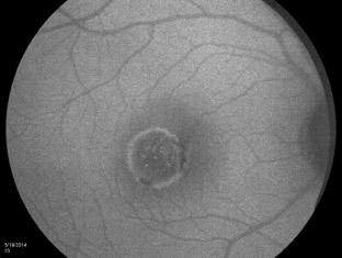
What Is O.C.T. and Why Should I Give A Rip? OCT & Me How Optical Coherence Tomography Changed the Life of a Small Town Optometrist 5/19/2014
 OCT & Me How Optical Coherence Tomography Changed the Life of a Small Town Optometrist Email: myoder@wcoil.com Mark A. Yoder, O.D. 107 N. Main Street PO Box 123 Bluffton, OH 45817 @yoderod 115.02 Histoplasma
High Resolution Imaging in Patients with Retinal Dystrophies
 High Resolution Imaging in Patients with Retinal Dystrophies Ophthalmic Photographers Society Annual Midyear Meeting April 2, 213 Jacque Duncan, M.D. UCSF Department of Ophthalmology How can retinal imaging
Gene therapy for Inherited Retinal Diseases MD(Res) Thesis by Venki Sundaram
 Thesis by Venki Sundaram") Gene therapy for Inherited Retinal Diseases MD(Res) Thesis by Venki Sundaram Division of Molecular Therapy Institute of Ophthalmology University College London Abstract Inherited retinal diseases include
Gene therapy for Inherited Retinal Diseases MD(Res) Thesis by Venki Sundaram Division of Molecular Therapy Institute of Ophthalmology University College London Abstract Inherited retinal diseases include
We are IntechOpen, the world s leading publisher of Open Access books Built by scientists, for scientists. International authors and editors
 We are IntechOpen, the world s leading publisher of Open Access books Built by scientists, for scientists 4,000 116,000 120M Open access books available International authors and editors Downloads Our
Kamron N Khan PhD, FRCOphth [1-3], Keren Carss [5], F. Lucy Raymond [5], Farrah Islam
![Kamron N Khan PhD, FRCOphth [1-3], Keren Carss [5], F. Lucy Raymond [5], Farrah Islam](/thumbs/92/108288836.jpg "Kamron N Khan PhD, FRCOphth [1-3], Keren Carss [5], F. Lucy Raymond [5], Farrah Islam") Title: Vitamin A deficiency - there's more to it than meets the eye. Kamron N Khan PhD, FRCOphth [1-3], Keren Carss [5], F. Lucy Raymond [5], Farrah Islam FCPS, FRCS [2], Anthony T Moore FRCS, FRCOphth
Title: Vitamin A deficiency - there's more to it than meets the eye. Kamron N Khan PhD, FRCOphth [1-3], Keren Carss [5], F. Lucy Raymond [5], Farrah Islam FCPS, FRCS [2], Anthony T Moore FRCS, FRCOphth
Objectives. Myth 1: Weiss ring = PVD. Myths 1/23/2018. To review misconceptions and clinical pearls regarding common vitreoretinal presentations.
 Objectives David RP Almeida MD MBA PhD VitreoRetinal Surgery, PA To review misconceptions and clinical pearls regarding common vitreoretinal presentations. Retina Update Minneapolis MN January 2018 Myths
Corporate Medical Policy
 Corporate Medical Policy Retinal Prosthesis File Name: Origination: Last CAP Review: Next CAP Review: Last Review: retinal_prosthesis 6/2011 6/2018 6/2019 6/2018 Description of Procedure or Service A retinal
A Practical Approach to Pediatric Retinal Diseases
 A Practical Approach to Pediatric Retinal Diseases Diana Shechtman OD FAAO Consultative Optometric Physician RMSM Adjunct Professor, Nova Southeastern University Rachel Stacey Coulter OD MS FAAO Professor,
History/principles of the OCT What does the normal retinal OCT look like Vitreal disorders Retinal/RPE disorders Choroidal disorders
 Nathan Lighthizer, O.D., F.A.A.O. Assistant Professor Assistant Dean for Clinical Care Director of Continuing Education Chief of Specialty Care Clinics Chief of Electrodiagnostics Clinic Oklahoma College
XUE HUI Department of Histology& Embryology, Basic Medicine College of Jilin University
 SENSE ORGAN XUE HUI Department of Histology& Embryology, Basic Medicine College of Jilin University EYE fibrous globe lens photosensitive cells a system of cells and nerves concentric layers the sclera
Cirrus TM HD-OCT. Details defi ne your decisions
 Cirrus TM HD-OCT Details defi ne your decisions 2 With high-defi nition OCT Carl Zeiss Meditec takes you beyond standard spectral domain Built on 10 years experience at the vanguard of innovation, Carl
Doyne honeycomb retinal dystrophy functional improvement following subthreshold nanopulse laser treatment: a case report
 Cusumano et al. Journal of Medical Case Reports (2019) 13:5 https://doi.org/10.1186/s13256-018-1935-1 CASE REPORT Open Access Doyne honeycomb retinal dystrophy functional improvement following subthreshold
EPIRETINAL MEMBRANE & VITREOMACULAR TRACTION
 EPIRETINAL MEMBRANE & VITREOMACULAR TRACTION Management of ERM and VMT K.V.Chalam,MD,PhD,MBA,FACS Professor and Director of Retina Loma Linda Eye Institute Los Angeles, USA REVIEW ANATOMY The vitreous
Glaucoma Glaucoma is a complication which has only recently been confirmed as a feature of
 1.2.4 OPHTHALMOLOGICAL ABNORMALITIES Ocular abnormalities are well documented in patients with NPS 6 62 81 95. 1.2.4.1 Glaucoma Glaucoma is a complication which has only recently been confirmed as a feature
Original Policy Date
 MP 9.03.11 Retinal Prosthesis Medical Policy Section Miscellaneous Policies Issue 12/2013 Original Policy Date 12/2013 Last Review Status/Date Reviewed with literature search/12/2013 Return to Medical
Proposal form for the evaluation of a genetic test for NHS Service Gene Dossier
 Proposal form for the evaluation of a genetic test for NHS Service Gene Dossier Test Disease Population Triad Disease name Choroideremia OMIM number for disease 303100 Disease alternative names please
Amber Priority. Image Library
 Amber Priority Image Library Amber flag Diabetic Maculopathy (M1) Pre-proliferative Diabetic Retinopathy (R2) Old, treated and now inactive DR (R1/M0/P1or R0/M0/P1) Where only partial or incomplete images
ZEISS AngioPlex OCT Angiography. Clinical Case Reports
 Clinical Case Reports Proliferative Diabetic Retinopathy (PDR) Case Report 969 PROLIFERATIVE DIABETIC RETINOPATHY 1 1-year-old diabetic female presents for follow-up of proliferative diabetic retinopathy
The Foundation Fighting Blindness. Stargardt s Disease- Moving to Clinical Trials
 The Foundation Fighting Blindness Stargardt s Disease- Moving to Clinical Trials On Friday, April 6, 2007, a symposium was held at the Coleman Center in New York under the auspices of the Foundation Fighting
Test of visual pathway function
 The visual system Test of visual pathway function Suppose you have a patient who may have some damage to the visual pathways leading to visual cortex, for example from multiple sclerosis. How could you
The Visual System. Retinal Anatomy Dr. Casagrande February 2, Phone: Office: T2302 MCN
 The Visual System Retinal Anatomy Dr. Casagrande February 2, 2004 Phone: 343-4538 Email: vivien.casagrande@mcmail.vanderbilt.edu Office: T2302 MCN Reading assignments and Good Web Sites Chapter 2 in Tovée,
Widefield Retinal Imaging with Auto Fluorescence Technology in the Optometric Practice
 Widefield Retinal Imaging with Auto Fluorescence Technology in the Optometric Practice This course will define ultra-widefield retinal imaging and autofluorescence for the attendee. Will show how it is
ATLAS OF OCT. Retinal Anatomy in Health & Pathology by Neal A. Adams, MD. Provided to you by:
 ATLAS OF OCT Retinal Anatomy in Health & Pathology by Neal A. Adams, MD Provided to you by: Atlas of OCT The OCT Atlas is written by Neal A. Adams, MD, and produced by Heidelberg Engineering, Inc. to help
Stargardt Disease Caused by a Rare Combination of Double Homozygous Mutations
 386 CONTINUING MEDICAL EDUCatION :386-91 Stargardt Disease Caused by a Rare Combination of Double Homozygous Mutations Danielius Serapinas, Viltautė Obrikytė, Raimundas Sakalauskas Department of Pulmonology
Unit VIII Problem 8 Anatomy: Orbit and Eyeball
 Unit VIII Problem 8 Anatomy: Orbit and Eyeball - The bony orbit: it is protecting our eyeball and resembling a pyramid: With a base directed: anterolaterally. And an apex directed: posteromedially. Notes:
Progressive Symptomatic Retinal Detachment Complicating Retinoschisis. Initial Reporting Questionnaire
 Progressive Symptomatic Retinal Detachment Complicating Retinoschisis In association with the British Ophthalmological Surveillance Unit Ethics ref: 13/NW/0037 Initial Reporting Questionnaire Case Definition:
Autosomal recessive bestrophinopathy associated with angle-closure glaucoma
 Doc Ophthalmol (2014) 129:57 63 DOI 10.1007/s10633-014-9444-z CLINICAL CASE REPORT Autosomal recessive bestrophinopathy associated with angle-closure glaucoma C. Crowley R. Paterson T. Lamey T. McLaren
Prevalence and mode of inheritance of major genetic eye diseases in China
 Journal of Medical Genetics 1987, 24, 584-588 Prevalence and mode of inheritance of major genetic eye diseases in China DAN-NING HU From the Zhabei Eye Institute, Shanghai, and Section of Ophthalmic Genetics,
Retro-mode imaging and fundus autofluorescence with scanning laser ophthalmoscope of retinal dystrophies
 Parodi et al. BMC Ophthalmology 2012, 12:8 RESEARCH ARTICLE Retro-mode imaging and fundus autofluorescence with scanning laser ophthalmoscope of retinal dystrophies Battaglia Parodi Maurizio 1, Iacono
Dr/ Marwa Abdellah EOS /16/2018. Dr/ Marwa Abdellah EOS When do you ask Fluorescein angiography for optic disc diseases???
 When do you ask Fluorescein angiography for optic disc diseases??? 1 NORMAL OPTIC DISC The normal optic disc on fluorescein angiography is fluorescent due to filling of vessels arising from the posterior
Subject: Voretigene Neparvovec-rzyl (Luxturna) Injection
 Injection") 09-J2000-96 Original Effective Date: 03/15/18 Reviewed: 02/14/18 Revised: 01/01/19 Subject: Voretigene Neparvovec-rzyl (Luxturna) Injection THIS MEDICAL COVERAGE GUIDELINE IS NOT AN AUTHORIZATION, CERTIFICATION,
09-J2000-96 Original Effective Date: 03/15/18 Reviewed: 02/14/18 Revised: 01/01/19 Subject: Voretigene Neparvovec-rzyl (Luxturna) Injection THIS MEDICAL COVERAGE GUIDELINE IS NOT AN AUTHORIZATION, CERTIFICATION,
Retinal dystrophies, genomic applications in diagnosis and prospects for therapy
 Review Article Retinal dystrophies, genomic applications in diagnosis and prospects for therapy Benjamin M. Nash 1,2,3, Dale C. Wright 2,3, John R. Grigg 1, Bruce Bennetts 2,3, Robyn V. Jamieson 1,3 1
Understanding. Retinitis pigmentosa. and other inherited retinal dystrophies
 Understanding Retinitis pigmentosa and other inherited retinal dystrophies Contact us We re here to answer any questions you have about your eye condition or treatment. If you need further information
Cirrus TM HD-OCT. Details define your decisions
 Cirrus TM HD-OCT Details define your decisions 2 With high-definition OCT Carl Zeiss Meditec takes you beyond standard spectral domain Built on 10 years experience at the vanguard of innovation, Carl Zeiss
Annette Sims, MD, Ophthalmologist next Tuesday! Hooray!!
 BI 358 Lecture 18 Annette Sims, MD, Ophthalmologist next Tuesday! Hooray!! I. Announcements Quiz 5 returned at end of lecture. Eye Dissection & Vision lab next Tuesday > Lecture by Dr. Sims! Final Quiz
Fundus Autofluorescence and its PRACTICAL applications: Retina Beyond the Color. Start to think about this. Disclosure 5/21/2015
 Fundus Autofluorescence and its PRACTICAL applications: Retina Beyond the Color Jeffry D. Gerson, O.D., F.A.A.O Olathe, KS jgerson@hotmail.com Start to think about this. Disclosure I have worked with/consulted
Annette Sims, MD, Ophthalmologist next Tuesday! Hooray!!
 BI 358 Lecture 18 Annette Sims, MD, Ophthalmologist next Tuesday! Hooray!! I. Announcements Quiz 5 returned at end of lecture. Eye Dissection & Vision lab next Tuesday > Lecture by Dr. Sims! Final Quiz
Five Things You re Missing with Your Fundus Camera
 ebook Five Things You re Missing with Your Fundus Camera By Donald J. Siegel, OD, Sun City West Eye Care Sponsored by: Before I began incorporating EIDON true-color imaging into my practice, my retinal
OCT Angiography The Next Frontier
 Choroid Retina avascular 5/13/2017 OCT Angiography The Next Frontier Pierce Kenworthy OD, FAAO June 9, 2017 OCT Angiography (OCTA) 2016 Non-invasive, motion contrast imaging Represents erythrocyte movement
Course C21. Visual Electrophysiology in Children. 12 June, :15-17:45 hrs. Room 118/119 HAND-OUTS
 Course C21 Visual Electrophysiology in Children 12 June, 2017 16:15-17:45 hrs Room 118/119 HAND-OUTS Introducing visual electrophysiology tests and results Ruth Hamilton - A description of paeditaric tests
Fluorescence in Best's vitelliform dystrophy, lipofuscin, and fundus flavimaculatus
 British Journal of Ophthalmology, 1978, 62, 256-260 Fluorescence in Best's vitelliform dystrophy, lipofuscin, and fundus flavimaculatus STEVEN ABBOTT MILLER From the Retina Service, Department of Ophthalmology,
Introduction to Full Field ERGs
 Introduction to Full Field ERGs ISCEV Full Field ERG Standard (Recording protocols and their physiological basis) Laura J. Frishman, PhD University of Houston October 17, 2016 Cellular origins and mechanisms
OCT Assessment of the Vitreoretinal Relationship in CSME
 December 2007 Sonia Rani John et al. - IFIS 375 ORIGINAL ARTICLE OCT Assessment of the Vitreoretinal Relationship in CSME Dr. Manoj S. DNB FRCS, Dr. Unnikrishnan Nair MS DO FRCS, Dr. Gargi Sathish MS Introduction
Retina Conference. Janelle Fassbender, MD, PhD University of Louisville Department of Ophthalmology and Visual Sciences 09/04/2014
 Retina Conference Janelle Fassbender, MD, PhD University of Louisville Department of Ophthalmology and Visual Sciences 09/04/2014 Subjective CC/HPI: 64 year old Caucasian female referred by outside ophthalmologist
SOCT Copernicus REVO. * - Currently import and overlay are avaibale in manual mode only
 SOCT Copernicus REVO Easy Operation (Full auto & Auto mode) Auto alignment (Z-position, C-gate, Focus, Tomogram) Voice guide (support patient through examination) Powerful analysis tools Enhanced tomograms
MRC-Holland MLPA. Description version 12; 13 January 2017
 SALSA MLPA probemix P219-B3 PAX6 Lot B3-0915: Compared to version B2 (lot B2-1111) two reference probes have been replaced and one additional reference probe has been added. In addition, one flanking probe
Moving forward with a different perspective
 Moving forward with a different perspective The Leader In Vision Diagnostics Offers A New Perspective Marco has served the eyecare community by offering exceptional lane products and automated high tech
Local Coverage Determination (LCD): Scanning Computerized Ophthalmic Diagnostic Imaging (SCODI) (L34431)
: Scanning Computerized Ophthalmic Diagnostic Imaging (SCODI) (L34431)") Local Coverage Determination (LCD): Scanning Computerized Ophthalmic Diagnostic Imaging (SCODI) (L34431) Links in PDF documents are not guaranteed to work. To follow a web link, please use the MCD Website.
Local Coverage Determination (LCD): Scanning Computerized Ophthalmic Diagnostic Imaging (SCODI) (L34431) Links in PDF documents are not guaranteed to work. To follow a web link, please use the MCD Website.
Interesting, unusual and eclectic cases from 2017 Robert A. Mittra, MD VitreoRetinal Surgery, P.A. Minneapolis, MN
 Fundus, SG Interesting, unusual and eclectic cases from 2017 Robert A. Mittra, MD VitreoRetinal Surgery, P.A. Minneapolis, MN Which is most likely? A) Age > 65, history of HTN B) Age 40 65, history of
Phenotype Report. Num. Positions Not Called (Missing data) Num. Variants Assessed
 Num. Variants Assessed") Report Date: August 19, 2015 Software Annotation Version: 8 Report Name: NA12144 NW European Genome : NA12144_S1 Sequencing Provider: Illumina Sequencing Type: Exome : Retinitis Pigmentosa Description:
Report Date: August 19, 2015 Software Annotation Version: 8 Report Name: NA12144 NW European Genome : NA12144_S1 Sequencing Provider: Illumina Sequencing Type: Exome : Retinitis Pigmentosa Description:
Interesting, unusual, eclectic cases from 2017 Robert A. Mittra, MD VitreoRetinal Surgery, P.A. Minneapolis, MN
 56 yo female, EW Presented to outside Ophthalmologist Diagnosed with viral conjunctivitis, but viral testing was negative. Also had pain around the eye and on the right side of her face Interesting, unusual,
8 NeuroMeeting Riparare il cervello: nuove frontiere terapeutiche. Napoli 12 e 13 Maggio La terapia genica. Francesca Simonelli
 8 NeuroMeeting Riparare il cervello: nuove frontiere terapeutiche Napoli 12 e 13 Maggio 2016 La terapia genica Francesca Simonelli Direttore clinica oculistica Seconda Universita degli Studi di Napoli
Ocular imaging in acquired retinopathy with multiple myeloma
 Ocular imaging in acquired retinopathy with multiple myeloma ABDELRAHMAN GABER SALMAN MD- FRCS (GLASG)- MRCS (ED) ASSOCIATE PROFESSOR AIN SHAMS UNIVERSITY EVRS 2015 Immunogammopathies Immunogammopathies
Ganglion cell analysis by optical coherence tomography (OCT) Jonathan A. Micieli, MD Valérie Biousse, MD
 Jonathan A. Micieli, MD Valérie Biousse, MD") Ganglion cell analysis by optical coherence tomography (OCT) Jonathan A. Micieli, MD Valérie Biousse, MD Figure 1. Normal OCT of the macula (cross section through the line indicated on the fundus photo)
Ganglion cell analysis by optical coherence tomography (OCT) Jonathan A. Micieli, MD Valérie Biousse, MD Figure 1. Normal OCT of the macula (cross section through the line indicated on the fundus photo)
CLINICALCASE PROVOST J, SEKFALI R, AMOROSO F, ZAMBROWSKI O, MIERE A
 CLINICALCASE PROVOST J, SEKFALI R, AMOROSO F, ZAMBROWSKI O, MIERE A Department of ophthalmology, Souied E. (MD,PhD) Centre Hospitalier Intercommunal de Créteil Université Paris Est HISTORY 13 years old