Objectives. Myth 1: Weiss ring = PVD. Myths 1/23/2018. To review misconceptions and clinical pearls regarding common vitreoretinal presentations.
|
|
|
- Doris Watkins
- 5 years ago
- Views:
Transcription

1 Objectives David RP Almeida MD MBA PhD VitreoRetinal Surgery, PA To review misconceptions and clinical pearls regarding common vitreoretinal presentations. Retina Update Minneapolis MN January 2018 Myths 1. Presence of a Weiss ring indicates a complete posterior vitreous detachment from the retina. 2. All retinal detachments are an emergency. 3. White dots in the posterior segment are due to inflammation. 4. There is no harm from the use of topical corticosteroids for intraocular inflammation. Myth 1: Weiss ring = PVD Usually BUT NOT ALWAYS the presence of a Weiss ring indicates a complete PVD Normally, vitreous detaches out to the retinal break(s) in acute retinal detachment, where it remains adherent and causes traction on the retinal flap 5. Viral vectors for gene therapy are not safe. Not uncommonly, triamcinolone stained residual layer of vitreous over the posterior and peripheral retina in many eyes with retinal detachment Including some with a preexisting Weiss ring 1

2 Not uncommonly, triamcinolone stained residual layer of vitreous over the posterior and peripheral retina in many eyes with retinal detachment (including some with a preexisting Weiss ring) Incomplete PVD? Vitreoschisis? Immature PVR membranes? Eric K Chin, David RP Almeida & James C Folk, Posterior Hyaloid Removal, Ophthalmic Surgery, Lasers & Imaging Retina 2015, 46(4):404 Robin K Kuriakose, Kunyong Xu, Eric K Chin MD & David RP Almeida, Proliferative Vitreoretinopathy (PVR) Update: Current Surgical Techniques and Emerging Medical Management, Journal of VitreoRetinal Diseases 2017, 1(4):261 Kunyong Xu, Eric K Chin, Steven R Bennett, David F Williams, Edwin H Ryan, Sundeep Dev, Robert A Mittra, Polly A Quiram, John B Davies, D. Wilkin Parke III, H Culver Boldt & David RP Almeida, Predictive factors for proliferative vitreoretinopathy formation after uncomplicated primary retinal detachment repair, RETINA 2018, in press Kunyong Xu, Eric K. Chin, D. Wilkin Parke III & David RP Almeida, Epiretinal Membrane and Cystoid Macular Edema as Predictive Factors of Recurrent Proliferative Vitreoretinopathy, Clinical Ophthalmology 2017, 11:1819 Incomplete vitreous separation in some eyes with RRD Partial PVDs in 71 of 786 eyes = 7% (autopsy study) Midperipheral to peripheral vitreous was detached over one large segment of the retina but remained attached in the other segments Incomplete vitreous separation in some eyes with RRD Partial PVDs in 71 of 786 eyes = 7% (autopsy study) Midperipheral to peripheral vitreous was detached over one large segment of the retina but remained attached in the other segments In RRD, posterior hyaloid usually absent from quadrant with retinal breaks In RRD, posterior hyaloid usually absent from quadrant with retinal breaks Dilute intravitreal triamcinolone allows for easy, reliable, and consistent identification of the adherent vitreous layer on the retinal surface Myth 2: All RDs are an emergency Retinal detachments are usually a surgical urgency but this does not apply to all retinal detachments all of the time. 2


3 3
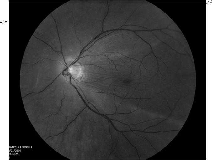
 High watermark above fovea Mac 0n: VA = 20/25 OCT foveal thread is attached")
 Nd:YAG posterior capsulotomy (Pseudophakic Retinal Detachment. Surv Ophthalmol 2003;48:467) RD after Nd:YAG Rhegmatogenous retinal detachment: Where is the break?")
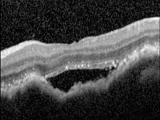
4 Is the macula attached? Macula on or macula off? Acute or chronic? Urgent or emergent? Mac on or mac off? Mac off OCT clearly shows macula is detached in multiple areas Retinal folds present (subretinal fluid) High watermark above fovea Mac 0n: VA = 20/25 OCT foveal thread is attached RD risk factors can help you triage Risk factors Age RD fellow eye (15 20%) High myopia Family history Lattice degeneration Trauma Cataract surgery (0.6% 1.7% annual risk) Nd:YAG posterior capsulotomy (Pseudophakic Retinal Detachment. Surv Ophthalmol 2003;48:467) RD after Nd:YAG Rhegmatogenous retinal detachment: Where is the break? Finding retinal break helps: Rule in rhegmatogenous process Aids triage timing (superior versus inferior) 4


5 Find (historical) breaks Dr. Harvey Lincoff Cryopexy for RD Cryopexy could destroy retinoblastoma Pioneered straight chain perfluorocarbon gases for complicated detachments Lincoff Rules (Lincoff H, Gieser R. Finding the Retinal Hole. Arch Ophthalmol. 1971;85(5):565) RD, but no break found? Chronic RD and retinal break has sealed Myope with small retinal break difficult to find on office dilated depressed exam Retinoschisis Exudative process Case Middle age male with 20/20 OS Superior visual field defect Inferior macula on retinal detachment No break identified 5


6 Case Medium sized malignant melanoma Systemic workup revealed metastatic disease Currently working with oncology team Myth 3: White dots = inflammation White dots in the posterior segment are due to inflammation (white dot syndromes) 6


7 PEARL: Mets have lumpy bumpy surface due to involution in area away from blood supply (vs melanoma with smooth dome due to rich blood supply present). 7


8 1/23/2018 Differential diagnosis Melanoma (amelanotic) Hemangioma Metastasis Osteoma Atypical nevus Posterior scleritis Choroidal detachment Granuloma Old subretinal hemorrhage OD NORMAL FOVEAL DEPRESSION OS NFL GCL IPL INL OPL ONL OLM RPE Choroidal Elevation OS LOSS OF FOVEAL CONTOUR Differential diagnosis Melanoma (amelanotic) Hemangioma Metastasis Osteoma Atypical nevus SRF Posterior scleritis Choroidal detachment Granuloma Old subretinal hemorrhage 8


 Lung (10%) Unknown (10%) Others GI")


9 Choroidal Metastases #1 intraocular tumor in adults Autopsy studies show 10% cancer have choroidal mets Breast cancer choroidal mets almost 40% Hematogenous dissemination Choroid > iris/ciliary body > retina/optic nerve Ciliary body 2% Iris 9% Choroid 89% Primary site Mostly carcinomas 1/3 do not have a known cancer history Presenting symptoms Asymptomatic (11%) Male Lung (40%) Unknown (30%) GI (10%) Kidney Prostate Skin Others Breast Female Breast (70%) Lung (10%) Unknown (10%) Others GI Skin Kidney Blurred vision (70%) Flashes & floaters (12%) Pain (7%) Clinical appearance Relatively flat, ill defined, creamy yellow Overlying RPE changes ( leopard spotting in breast ca) Exudative RD Ultrasonography A scan: Moderate high internal reflectivity B scan: Ill defined, ±lobulated choroidal mass Bilateral/multifocal in 25% (breast ca most likely) 9

10 Fluorescein angiography Early hypofluorescence with late hyperfluorescence Rarely useful to differentiate from other lesions Prognosis & Treatment Prognosis Mean life expectancy 9 10 months Treatment Systemic chemotherapy Hormone therapy External beam, brachytherapy, proton beam Surgery Myth 4: No harm from topical steroids for inflammation When considering intraocular inflammation and a possible uveitic diagnosis, the top priority is to rule out infection. Beware of 5 uveitis red flags First episode in elderly patient Intensive topical corticosteroid therapy can significantly confound clinical picture. 5 uveitis red flags First episode in elderly patient 5 uveitis red flags First episode in elderly patient Significant systemic illness Significant systemic illness Immunosuppressed patients 10
11 5 uveitis red flags First episode in elderly patient Significant systemic illness Immunosuppressed patients HIV/AIDS 5 uveitis red flags First episode in elderly patient Significant systemic illness Immunosuppressed patients HIV/AIDS Drug addicts 5 uveitis red flags First episode in elderly patient Significant systemic illness It s an infection if Hospitalized patients indwelling catheters Post bowel surgery Immunosuppressed patients HIV/AIDS Drug addicts It s an infection if Hospitalized patients with indwelling catheters, bowel surgery HIV/AIDS It s an infection if Hospitalized patients with indwelling catheters, bowel surgery HIV/AIDS Cancer history and on immunosuppression 11


12 It s an infection if Myth 5: Viral vectors not safe Hospitalized patients with indwelling catheters, bowel surgery HIV Cancers on immunosuppression Conditions with low white blood cell counts E.g., Leukemia Leber congenital amaurosis (LCA) Clinical features Group of disorders due to mutation of at least 16 different genes Autosomal recessive in majority of cases Severe visual impairment before age 1, nystagmus, poor pupillary reflexes, normal or mildly abnormal fundus appearance, profoundly abnormal or absent ERG (Graefes Arch Clin Exp Ophthalmol 1869;15:1, Confinia Neurol 1954;14:184, Ophthalmologica 1947;114:332) Clinical features Normal fundus appearance RPE granularity Vessel attenuation Macular coloboma Salt and pepper retinopathy Retinitis punctata albescens Nummular pigmentation LCA: no treatment available! But, you have preservation of all retinal layers Of all causal genes, recessive biallelic RPE 65 is prototype Related to how eyes detect light (Clin Genet 1973;4:270, Am J Ophthalmol 1977;83:27, Int Ophthalmol Clin 1968;8:949) 12


 Low")
 Retinal")


13 RPE65 is enzyme that resets mousetraps 11 cis retinal (Vit A) High Energy State RPE 65 gene replacement supplies HIGH Energy Vitamin A 1 to 1000 All trans retinal (Vit A) Low Energy State } Rod Outer Segment (ROS) Retinal Pigment Epithelium (RPE) 1 : 1000 RPE65 gene (replacement) therapy Use nano injectors to insert DNA into cell Nano injector is adeno associated virus (AAV) Virus has limited space for DNA Small gene small virus class, AAV Large gene large virus 1 month 13


14 Untreated eye Treated eye LCA RPE-65 is a prototypical inherited ocular disease No treatment available Rare but blinding disease for those affected Gene replacement therapy Safe Effective Long-lasting Myths 1. Presence of a Weiss ring indicates a complete posterior vitreous detachment from the retina. Thanks! 2. All retinal detachments are an emergency. 3. White dots in the posterior segment are due to inflammation. 4. There is no harm from the use of topical corticosteroids for intraocular inflammation. 5. Viral vectors for gene therapy are not safe. 14
Moncef Khairallah, MD
 Moncef Khairallah, MD Department of Ophthalmology, Fattouma Bourguiba University Hospital Faculty of Medicine, University of Monastir Monastir, Tunisia INTRODUCTION IU: anatomic form of uveitis involving
Outline. Outline. Vitreous Development & Anatomy OPT - 243
 2010 OPT - 243 Vitreous Disorders & Vitreoretinal Disorders of the Posterior Pole I Leo Semes, OD, FAAO 100% 0% 0% 0% 0% Which of these gives the best resolution for studying vitreoretinal disorders of
Progressive Symptomatic Retinal Detachment Complicating Retinoschisis. Initial Reporting Questionnaire
 Progressive Symptomatic Retinal Detachment Complicating Retinoschisis In association with the British Ophthalmological Surveillance Unit Ethics ref: 13/NW/0037 Initial Reporting Questionnaire Case Definition:
Top Pediatric Retinal Diseases you don t want to miss! Retinopathy of Prematurity (ROP) Aggressive, Posterior ROP (AP ROP)
 Aggressive, Posterior ROP (AP ROP)") Top 10 10 Pediatric Retinal Diseases you don t want to miss! Polly Quiram MD, PhD Vitreoretinal Surgery, PA Retinal Update Jan 26th, 2018 ROP Retinoblastoma Coats disease Persistent fetal vasculature Familial
Top 10 10 Pediatric Retinal Diseases you don t want to miss! Polly Quiram MD, PhD Vitreoretinal Surgery, PA Retinal Update Jan 26th, 2018 ROP Retinoblastoma Coats disease Persistent fetal vasculature Familial
OPTIC DISC PIT Pathogenesis and Management OPTIC DISC PIT
 OPTIC DISC PIT Pathogenesis and Management Abdel-Latif Siam Ain Shams University Cairo Egypt OPTIC DISC PIT Congenital pit is an atypical coloboma usually located on the temporal edge of the disc, associated
Retina Conference. Janelle Fassbender, MD, PhD University of Louisville Department of Ophthalmology and Visual Sciences 09/04/2014
 Retina Conference Janelle Fassbender, MD, PhD University of Louisville Department of Ophthalmology and Visual Sciences 09/04/2014 Subjective CC/HPI: 64 year old Caucasian female referred by outside ophthalmologist
Misdiagnosed Vogt-Koyanagi-Harada (VKH) disease and atypical central serous chorioretinopathy (CSC)
 disease and atypical central serous chorioretinopathy (CSC)") HPTER 12 Misdiagnosed Vogt-Koyanagi-Harada (VKH) disease and atypical central serous chorioretinopathy (S) linical Features VKH disease is a bilateral granulomatous panuveitis often associated with exudative
HPTER 12 Misdiagnosed Vogt-Koyanagi-Harada (VKH) disease and atypical central serous chorioretinopathy (S) linical Features VKH disease is a bilateral granulomatous panuveitis often associated with exudative
ZEISS AngioPlex OCT Angiography. Clinical Case Reports
 Clinical Case Reports Proliferative Diabetic Retinopathy (PDR) Case Report 969 PROLIFERATIVE DIABETIC RETINOPATHY 1 1-year-old diabetic female presents for follow-up of proliferative diabetic retinopathy
evaluation of vitreoretinal adhesions in exudative AMD using optical coherence tomography
 evaluation of vitreoretinal adhesions in exudative AMD using optical coherence tomography Dr. Mahmoud Alaa Abouhusssein, FRCO Lecturer of ophthalmology, Alexandria university Dr. Amir Ramadan Gomaa, MD
Tractional detachments
 Retinal detachment: Surgery and post op care Tractional detachments Causes: diabetes, sickle cell, trauma, von Hippel Lindau disease. Sam S. Dahr, M.D. Retina Center of Oklahoma Key principles Remove the
Clinically Significant Macular Edema (CSME)
") Clinically Significant Macular Edema (CSME) 1 Clinically Significant Macular Edema (CSME) Sadrina T. Shaw OMT I Student July 26, 2014 Advisor: Dr. Uwaydat Clinically Significant Macular Edema (CSME) 2
Clinically Significant Macular Edema (CSME) 1 Clinically Significant Macular Edema (CSME) Sadrina T. Shaw OMT I Student July 26, 2014 Advisor: Dr. Uwaydat Clinically Significant Macular Edema (CSME) 2
Pseudohypopyon in Retinoblastoma. Choroidal Nevus. Masquerade Syndromes. Vision pathways. Flat with uniform color
 Primary Intraocular Tumors Thomas F. Freddo, O.D., Ph.D., F.A.A.O. Professor and Former Director School of Optometry University of Waterloo Masquerade Syndromes
Patient AB. Born in 1961 PED
 Clinical Atlas Patient AB Born in 1961 PED Autofluorescence Dilated 45 EasyScan Zero-dilation IR 45 Fundus Dilated 45 In the fundus photos (Canon CX1) the PED is not able to be seen. However, the extent
Outline. Brief history and principles of ophthalmic ultrasound. Types of ocular ultrasound. Examination techniques. Types of Ultrasound
 Ultrasound and Intraocular Tumors 2015 Ophthalmic Photographers' Society Mid-Year Program Cagri G. Besirli MD, PhD Kellogg Eye Center University of Michigan Outline Brief history and principles of ophthalmic
Royal Berkshire Hospital Dunedin Hospital. Prince Charles Eye Unit Pi Princess Margaret Hospital
 Vitreoretinal Surgery Mr Vaughan Tanner www.tanner-eyes.co.uk eyes Reading Royal Berkshire Hospital Dunedin Hospital Windsor Prince Charles Eye Unit Pi Princess Margaret Hospital Success rates VR surgery
RPE65-associated Leber Congenital Amaurosis
 RPE65-associated Leber Congenital Amaurosis Brian Privett, MD, Edwin M. Stone, MD, PhD February 16, 2010 Chief Complaint: Poor fixation at 4 months of age History of Present Illness: This 7 year old female
ACTIVATED OR NOT? RETINAL CASE PRESENTATION Shorye Payne, MD Medical Retinal Specialist Robley Rex VA Eye Clinic
 ACTIVATED OR NOT? RETINAL CASE PRESENTATION Shorye Payne, MD Medical Retinal Specialist Robley Rex VA Eye Clinic C We anticipate that the future management of posterior uveal melanoma (PUM) will focus
J of Evolution of Med and Dent Sci/ eissn , pissn / Vol. 4/ Issue 55/ July 09, 2015 Page 9665
 RARE PRESENTATION OF BILATERAL CHOROIDAL METASTASIS FROM PRIMARY MUCO-EPIDERMOID CARCINOMA OF THE PAROTID GLAND: A G. Premalatha 1, Ramya Seetamraju 2 HOW TO CITE THIS ARTICLE: G. Premalatha, Ramya Seetamraju.
Optical Coherence Tomography: Pearls for the Anterior Segment Surgeon Basic Science Michael Stewart, M.D.
 Optical Coherence Tomography: Pearls for the Anterior Segment Surgeon Basic Science Michael Stewart, M.D. Disclosure OCT Optical Coherence Tomography No relevant financial relationships I will refer to
Optical Coherence Tomography (OCT) in Uveitis Piergiorgio Neri, BMedSc, MD, PhD Head Ocular Immunology Unit
 in Uveitis Piergiorgio Neri, BMedSc, MD, PhD Head Ocular Immunology Unit") The Eye Clinic Polytechnic University of Marche Head: Prof Alfonso Giovannini November, 1991 Optical Coherence Tomography (OCT) in Uveitis Piergiorgio Neri, BMedSc, MD, PhD Head Ocular Immunology Unit
The Eye Clinic Polytechnic University of Marche Head: Prof Alfonso Giovannini November, 1991 Optical Coherence Tomography (OCT) in Uveitis Piergiorgio Neri, BMedSc, MD, PhD Head Ocular Immunology Unit
8 NeuroMeeting Riparare il cervello: nuove frontiere terapeutiche. Napoli 12 e 13 Maggio La terapia genica. Francesca Simonelli
 8 NeuroMeeting Riparare il cervello: nuove frontiere terapeutiche Napoli 12 e 13 Maggio 2016 La terapia genica Francesca Simonelli Direttore clinica oculistica Seconda Universita degli Studi di Napoli
A Patient s Guide to Diabetic Retinopathy
 Diabetic Retinopathy A Patient s Guide to Diabetic Retinopathy 840 Walnut Street, Philadelphia PA 19107 www.willseye.org Diabetic Retinopathy 1. Definition Diabetic retinopathy is a complication of diabetes
Vitreomacular interface disorders. Ghanbari MD 1393:10:25
 Vitreomacular interface disorders Ghanbari MD 1393:10:25 Human vitreous after dissection of the sclera, choroid, and retina. Lamellar structure of the posterior vitreous cortex (PVC) in the monkey. V =
CLINICAL PEARLS IN OCULAR ONCOLOGY
 CLINICAL PEARLS IN OCULAR ONCOLOGY IRIS NEVUS - Two kinds circumscribed and diffuse - Photodocumentation important to monitor growth - Risk Factors for iris nevus growth to melanoma (ABCDEF) A Age (young),
Interesting, unusual and eclectic cases from 2017 Robert A. Mittra, MD VitreoRetinal Surgery, P.A. Minneapolis, MN
 Fundus, SG Interesting, unusual and eclectic cases from 2017 Robert A. Mittra, MD VitreoRetinal Surgery, P.A. Minneapolis, MN Which is most likely? A) Age > 65, history of HTN B) Age 40 65, history of
PART 1: GENERAL RETINAL ANATOMY
 PART 1: GENERAL RETINAL ANATOMY General Anatomy At Ora Serrata At Optic Nerve Head Fundoscopic View Of Normal Retina What Is So Special About Diabetic Retinopathy? The WHO definition of blindness is
The Human Eye. Cornea Iris. Pupil. Lens. Retina
 The Retina Thin layer of light-sensitive tissue at the back of the eye (the film of the camera). Light rays are focused on the retina then transmitted to the brain. The macula is the very small area in
Retina Center of Oklahoma Sam S. Dahr, M.D. Adult Intraocular Tumors
 Adult Intraocular Tumors Sam S. Dahr, M.D. Retina Center of Oklahoma www.retinacenteroklahoma.com www.rcoklahoma.com Table of Contents Posterior uveal malignant melanoma Uveal metastasis Uveal melanoma
Interesting, unusual, eclectic cases from 2017 Robert A. Mittra, MD VitreoRetinal Surgery, P.A. Minneapolis, MN
 56 yo female, EW Presented to outside Ophthalmologist Diagnosed with viral conjunctivitis, but viral testing was negative. Also had pain around the eye and on the right side of her face Interesting, unusual,
The Foundation. RETINA HEALTH SERIES Facts from the ASRS
 Complex Retinal Detachment: Proliferative Vitreoretinopathy and Giant Retinal Tears Proliferative vitreoretinopathy (PVR) is a condition in which retinal scar tissue, or membranes form; this may occur
TYPES. Full thickness defect in the sensory retina (break) Secondary to Tumour, Inflammation or a Systemic disease
 Secondary to Tumour, Inflammation or a Systemic disease") Dr.A.Divya Introduction Definition : Retinal deatchment is the separation of the neurosensory retina(nsr) from the retinal pigment epithelium(rpe) ; results in the accumulation of subretinal fluid(srf)
Dr.A.Divya Introduction Definition : Retinal deatchment is the separation of the neurosensory retina(nsr) from the retinal pigment epithelium(rpe) ; results in the accumulation of subretinal fluid(srf)
LUXTURNA (voretigene neparovec-rzyl)
") LUXTURNA (voretigene neparovec-rzyl) Non-Discrimination Statement and Multi-Language Interpreter Services information are located at the end of this document. Coverage for services, procedures, medical
LUXTURNA (voretigene neparovec-rzyl) Non-Discrimination Statement and Multi-Language Interpreter Services information are located at the end of this document. Coverage for services, procedures, medical
Yasser R. Serag, MD Tamer Wasfi, MD El- Saied El-Dessoukey, MD Magdi S. Moussa, MD Anselm Kampik, MD
 Microperimetric Evaluation of Brilliant Blue G- assisted Internal Limiting Membrane Peeling By Yasser R. Serag, MD Tamer Wasfi, MD El- Saied El-Dessoukey, MD Magdi S. Moussa, MD Anselm Kampik, MD The internal
OCT Angiography in Primary Eye Care
 OCT Angiography in Primary Eye Care An Image Interpretation Primer Julie Rodman, OD, MS, FAAO and Nadia Waheed, MD, MPH Table of Contents Diabetic Retinopathy 3-6 Choroidal Neovascularization 7-9 Central
Clinical features and surgical management of retinal detachment secondary to round retinal holes
 () 19, 9 & Nature Publishing Group All rights reserved 9-X/ $3. www.nature.com/eye Clinical features and surgical management of retinal detachment secondary to round retinal holes T Ung, MB Comer, AJS
8/6/17. Disclosures Aerie Pharmaceuticals Alcon BioTissue Diopsys Optovue Shire
 Nathan Lighthizer, O.D., F.A.A.O. Associate Professor Assistant Dean for Clinical Care Director of Continuing Education Chief of Specialty Care Clinics Oklahoma College of Optometry Tahlequah, OK lighthiz@nsuok.edu
OCT Interpretation. Financial Disclosure. Jay M. Haynie, OD, FAAO. OCT Image Layers 7/21/2014
 OCT Interpretation Jay M. Haynie, OD, FAAO Financial Disclosure I have received honoraria or am on the advisory board for the following companies: Olympia Tacoma Renton Kennewick - Washington Carl Zeiss
Diabetes & Your Eyes
 Diabetes & Your Eyes Diabetes is a disease that occurs when the pancreas does not secrete enough insulin or the body is unable to process it properly. Insulin is the hormone that regulates the level of
Ultrasound B-Scan for Posterior Segment Evaluation
 Retina Ultrasound B-Scan for Posterior Segment Evaluation Shalini Singh MS Shalini Singh MS, Manisha Agarwal MS, Aditya Bansal DNB Dr. Shroff s Charity Eye Hospital, New Delhi B reproducible investigation
Michael P. Blair, MD Retina Consultants, Ltd Libertyville/Des Plaines, Illinois Clinical Associate University of Chicago 17 October 2015
 Michael P. Blair, MD Retina Consultants, Ltd Libertyville/Des Plaines, Illinois Clinical Associate University of Chicago 17 October 2015 So What Parts of the Eye Retina are Affected by VHL Neural tissue
Diagnosis and treatment of diabetic retinopathy. Blake Cooper MD Ophthalmologist Vitreoretinal Surgeon Retina Associates Kansas City
 Diagnosis and treatment of diabetic retinopathy Blake Cooper MD Ophthalmologist Vitreoretinal Surgeon Retina Associates Kansas City Disclosures Consulted for Novo Nordisk 2017,2018. Will be discussing
CASE PRESENTATION. DR.Sravani 1 st yr PG Dept of Ophthalmology
 CASE PRESENTATION DR.Sravani 1 st yr PG Dept of Ophthalmology Name : X X X X X Age : 50yrs Sex : male Occupation : Farmer Residence : Mothkur CHIEF COMPLAINTS : - Diminision of vision in Right Eye since
The Quick Guide to OCT Mastery 50 Real Cases with Expert Analysis
 OPTICAL COHERENCE TOMOGRAPHY The Quick Guide to OCT Mastery 50 Real Cases with Expert Analysis VOL 1 Sanjay Sharma, MD, FRCS, MSc (Epid), MBA Ophthalmologist, Epidemiologist Queen s University, Canada
R&M Solutions
 Mohamed Hosny El-Bradey, MD., Assistant Professor of Ophthalmology, Tanta University. Wael El Haig, MD., Professor of Ophthalmology. Zagazeeg University. 1 Myopic CNV is considered the most common vision
Fundus Autofluorescence. Jonathan A. Micieli, MD Valérie Biousse, MD
 Fundus Autofluorescence Jonathan A. Micieli, MD Valérie Biousse, MD The retinal pigment epithelium (RPE) has many important functions including phagocytosis of the photoreceptor outer segments Cone Rod
Optical Coherence Tomography in Diabetic Retinopathy. Mrs Samantha Mann Consultant Ophthalmologist Clinical Lead of SEL-DESP
 Optical Coherence Tomography in Diabetic Retinopathy Mrs Samantha Mann Consultant Ophthalmologist Clinical Lead of SEL-DESP Content OCT imaging Retinal layers OCT features in Diabetes Some NON DR features
Pearls, Pitfalls and Advances in Neuro-Ophthalmology
 Pearls, Pitfalls and Advances in Neuro-Ophthalmology Nancy J. Newman, MD Emory University Atlanta, GA Consultant for Gensight Biologics, Santhera Data Safety Monitoring Board for Quark AION Study Medical-legal
NEPTUNE RED BANK BRICK
 NEPTUNE RED BANK BRICK Diabetes & The Eye Diabetics are more likely to develop Cataracts at a younger age. Diabetics are twice as likely to develop Glaucoma when compared to non-diabetics. The primary
EPIRETINAL MEMBRANE & VITREOMACULAR TRACTION
 EPIRETINAL MEMBRANE & VITREOMACULAR TRACTION Management of ERM and VMT K.V.Chalam,MD,PhD,MBA,FACS Professor and Director of Retina Loma Linda Eye Institute Los Angeles, USA REVIEW ANATOMY The vitreous
COEXISTENCE OF OPTIC NERVE HEAD DRUSEN
 COEXISTENCE OF OPTIC NERVE HEAD DRUSEN AND COMBINED HAMARTOMA OF THE RETINA AND RETINAL PIGMENT EPITHELIUM IN A TAIWANESE MALE Yo-Chen Chang 1 and Rong-Kung Tsai 2,3 1 Department of Ophthalmology, Kaohsiung
What Is O.C.T. and Why Should I Give A Rip? OCT & Me How Optical Coherence Tomography Changed the Life of a Small Town Optometrist 5/19/2014
 OCT & Me How Optical Coherence Tomography Changed the Life of a Small Town Optometrist Email: myoder@wcoil.com Mark A. Yoder, O.D. 107 N. Main Street PO Box 123 Bluffton, OH 45817 @yoderod 115.02 Histoplasma
Authors. Introduction. Introduction. Materials and Methods. Objective 10/27/2015
 Idiopathic Polypoidal Choroidal Vasculopathy (IPCV) in Thai Population Presenting with Choroidal Neovascularization (CNV) A multicenter study Authors Yonrawee Piyacomn 1, Chavakij Bhoomibunchoo 1, Yosanan
The effect of intravitreal bevacizumab in a rare case of retinal dystrophy with secondary cystoid macular edema
 Romanian Journal of Ophthalmology, Volume 61, Issue 2, April-June 2017. pp:123-127 CASE REPORT The effect of intravitreal bevacizumab in a rare case of retinal dystrophy with secondary cystoid macular
Clinical Patterns and risk factors for rhegmatogenous retinal detachment at a tertiary eye care centre of northern India
 Original article Clinical Patterns and risk factors for rhegmatogenous retinal detachment at a tertiary eye care centre of northern India Brijesh Takkar 1, Shorya Azad 1, Indrish Bhatia 1, Rajvardhan Azad
Sudden Vision Loss. Brendan Girschek, MD, FRCSC, FACS Vitreoretinal Surgery Cedar Valley Medical Specialists
 Sudden Vision Loss Brendan Girschek, MD, FRCSC, FACS Vitreoretinal Surgery Cedar Valley Medical Specialists My Credentials -Residency in Ophthalmology at the LSU Eye Center in New Orleans, LA -Fellowship
Eyes on Diabetics: How to Avoid Blindness in Diabetic Patient
 Eyes on Diabetics: How to Avoid Blindness in Diabetic Patient Rova Virgana FK Unpad Pusat Mata Nasional RS Mata Cicendo Bandung Eye Center (Hospital and Clinic) PIT IDI Jabar 2018 Keys Facts from WHO
OCT Assessment of the Vitreoretinal Relationship in CSME
 December 2007 Sonia Rani John et al. - IFIS 375 ORIGINAL ARTICLE OCT Assessment of the Vitreoretinal Relationship in CSME Dr. Manoj S. DNB FRCS, Dr. Unnikrishnan Nair MS DO FRCS, Dr. Gargi Sathish MS Introduction
JMSCR Volume 03 Issue 01 Page January 2015
 www.jmscr.igmpublication.org Impact Factor 3.79 ISSN (e)-2347-176x Clinical Evaluation of Patients Presenting With Rhegmatogenous Retinal Detachment Authors Dr Rani Sujatha.M.A 1, Dr Sridevi Prakash T
Course # Getting to Know Your OCT
 Course # 140 Getting to Know Your OCT Course Title: Lecturer: Getting to Know Your OCT Brad Sutton, OD, FAAO IU School of Optometry Financial Disclosures No financial disclosures Optical Coherence Tomography-OCT
VMA at the macula resulting in VMT
 Ocriplasmina for pharmacologic treatment in VMT Teresio Avitabile 1 Introduction PVD is a normal, physiologic process that occurs with aging; however, in some cases, PVD is incomplete Incomplete PVD localized
Disclosures. Objectives. Small gauge vitrectomy POD 1. The routine postoperative course 1/24/2018. None
 Disclosures Retina Surgery: Postoperative Considerations and Complications None D. Wilkin Parke III, M.D. VitreoRetinal Surgery, PA 1 2 Objectives Small gauge vitrectomy To understand the common and serious
Causes of failure of pneumatic retinopexy
 VOL. 9 NO. PHILIPPINE JOURNAL OF Ophthalmology JULY ORIGINAL ARTICLE - SEPTEMBER 00 Roberto E. Flaminiano, MD Robert T. Sy, MD Milagros H. Arroyo, MD Pearl Tamesis-Villalon, MD Department of Ophthalmology
An A to Z guide on Epiretinal Membranes (ERMs) Paris Tranos PhD,ICO,FRCS OPHTHALMICA Vitreoretinal & Uveitis Department
 Paris Tranos PhD,ICO,FRCS OPHTHALMICA Vitreoretinal & Uveitis Department") An A to Z guide on Epiretinal Membranes (ERMs) Paris Tranos PhD,ICO,FRCS OPHTHALMICA Vitreoretinal & Uveitis Department Types of ERM Natural history OCT prognostic factors ERM with co-existing pathology
An A to Z guide on Epiretinal Membranes (ERMs) Paris Tranos PhD,ICO,FRCS OPHTHALMICA Vitreoretinal & Uveitis Department Types of ERM Natural history OCT prognostic factors ERM with co-existing pathology
History/principles of the OCT What does the normal retinal OCT look like Vitreal disorders Retinal/RPE disorders Choroidal disorders
 Nathan Lighthizer, O.D., F.A.A.O. Assistant Professor Assistant Dean for Clinical Care Director of Continuing Education Chief of Specialty Care Clinics Chief of Electrodiagnostics Clinic Oklahoma College
Audit of Macular Hole Surgery, Visual Outcome Prediction on OCT Appearance of Macular Hole
 International Journal of Ophthalmology & Visual Science 2017; 2(4): 93-97 http://www.sciencepublishinggroup.com/j/ijovs doi: 10.11648/j.ijovs.20170204.13 Audit of Macular Hole Surgery, Visual Outcome Prediction
Principle of OCT. Reading Between the Lines: OCT Interpretation. Initial Concept. Advantage: High Resolution Cross Section Images
 Principle of OCT Reading Between the Lines: OCT Interpretation Mohammad Rafieetary, OD, FAAO mrafieetary@charlesretina.com Introduction Optical Biopsy Morphologic Evaluation of Live Tissue Measurements
Corporate Medical Policy
 Corporate Medical Policy Voretigene Neparvovec-rzyl (Luxturna) File Name: Origination: Last CAP Review: Next CAP Review: Last Review: voretigene_neparvovec_rzyl_luxturna 1/2018 N/A 6/2018 2/2018 Description
Fluorescein and Indocyanine Green Videoangiography of Choroidal Melanomas
 luorescein and Indocyanine Green Videoangiography of Choroidal Melanomas Leyla S. Atmaca, igen Batioğlu and Pelin Atmaca Eye Clinic, Ankara University Medical School, Ankara, Turkey Purpose: This study
Characteristic Ultrasonographic Findings of Choroidal Tumors
 Characteristic Ultrasonographic Findings of Choroidal Tumors Tsung-Jen Wang, Chang-Hao Yang, Shu-Lang Liao, Tzyy-Chang Ho, Jen-Shang Huang, Chang-Ping Lin, Chung-May Yang, Muh-Shy Chen and Luke Long-Kuang
Rare Presentation of Ocular Toxoplasmosis
 Case Report Rare Presentation of Ocular Toxoplasmosis Rakhshandeh Alipanahi MD From Department of Ophthalmology, Nikookari Eye Hospital, Tabriz University of Medical Sciences, Tabriz, Iran. Correspondence:
Financial Disclosures. The Eye in Neoplastic Disease. Course Goal. We wish to acknowledge and thank: Tumor Definition
 The Eye in Neoplastic Disease Carlo J. Pelino, OD, FAAO Joseph J. Pizzimenti, OD, FAAO cpelino@salus.edu pizzimen@uiwtx.edu Financial Disclosures! Speakers have no relevant financial relationships to declare.
Convergence in. Introduction. Case Report: Dr. Piyali SenM.B.B.S, Dr. Abhipsha Saha M.B.B.S, Dr. Anuradha Chandra M.S,FAICO
 Convergence in Dr. Piyali SenM.B.B.S, Dr. Abhipsha Saha M.B.B.S, Dr. Anuradha Chandra M.S,FAICO Introduction non-progressive ophthalmoplegia with or without ptosis affecting part or all of the occulomotor
Central serous chorioretinopathy (CSCR) was
 was") Case Report 777 Perfluorocarbon Liquid-Assisted External Drainage in the Management of Central Serous Chorioretinopathy with Bullous Serous Retinal Detachment Hung-Chiao Chen, MD; Jau-Der Ho, MD; San-Ni
Case Report 777 Perfluorocarbon Liquid-Assisted External Drainage in the Management of Central Serous Chorioretinopathy with Bullous Serous Retinal Detachment Hung-Chiao Chen, MD; Jau-Der Ho, MD; San-Ni
Dispelling Rumors about Tumors. Case
 Dispelling Rumors about Tumors Jesse L. Berry, MD Arizona Ophthalmology Society 2017 Associate Director, Ocular Oncology Service Associate Program Director USC/CHLA, Keck School of Medicine Case 65 year
Diabetic Retinopathy
 Diabetic Retinopathy Diabetes mellitus is one of the leading causes of irreversible blindness worldwide. In the United States, it is the most common cause of blindness in people younger than 65 years.
OCT Angiography The Next Frontier
 Choroid Retina avascular 5/13/2017 OCT Angiography The Next Frontier Pierce Kenworthy OD, FAAO June 9, 2017 OCT Angiography (OCTA) 2016 Non-invasive, motion contrast imaging Represents erythrocyte movement
Office Based Practice. Vitreoretinal Disease & Surgery. Coding Fiesta Vitreoretinal Disease & Surgery September 23, 2017 ADULT RETINA
 Vitreoretinal Disease & Surgery Coding Fest 2017 Vitreoretinal Surgery & Disease University of FL College of Medicine ADULT RETINA Medical Retina Surgical Retina Age Related Vascular Disease Vascular Disease
Five Things You re Missing with Your Fundus Camera
 ebook Five Things You re Missing with Your Fundus Camera By Donald J. Siegel, OD, Sun City West Eye Care Sponsored by: Before I began incorporating EIDON true-color imaging into my practice, my retinal
Research Article Scleral Buckling for Rhegmatogenous Retinal Detachment Associated with Pars Planitis
 Ophthalmology Volume 2016, Article ID 4538193, 5 pages http://dx.doi.org/10.1155/2016/4538193 Research Article Scleral Buckling for Rhegmatogenous Retinal Detachment Associated with Pars Planitis Yong-Kyu
Disease-Specific Fluorescein Angiography
 Ruth E. Picchiottino, CRA Disease-Specific Fluorescein Angiography 15 Disease-Specific Fluorescein Angiography Recommendations for tailoring retinal fluorescein angiography to diabetic retinopathy, macular
Vanderbilt Eye Institute Clinical Trials
 April, 2010 Vanderbilt Eye Institute Clinical Trials Ophthalmology Actively Recruiting Studies For information on our clinical trials and other studies, please contact: Sandy Owings, COA, CCRP Clinic Director
Ocular Pathology. I. Congenital and/or developmental. A. Trisomy 21. Hypertelorism (widely spaced eyes) Keratoconus (cone shaped cornea)
 Keratoconus (cone shaped cornea)") I. Congenital and/or developmental Robbins Pathologic Basis of Disease, 6 th Ed. A. Trisomy 21 Hypertelorism (widely spaced eyes) Keratoconus (cone shaped cornea) Focal hypoplasia of iris Cataracts frequently
I. Congenital and/or developmental Robbins Pathologic Basis of Disease, 6 th Ed. A. Trisomy 21 Hypertelorism (widely spaced eyes) Keratoconus (cone shaped cornea) Focal hypoplasia of iris Cataracts frequently
Late retinal reattachment
 :British Journal of Ophthalmology, 1981, 65, 142-146 Late retinal reattachment S. N. KOKOLAKIS, L. BRAVO, AND A. H. CHIGNELL From the Ophthalmic Department, St Thomas's Hospital, London SEI SUMMARY Six
PROFILE OF PATIENTS WITH FLOATERS IN SAIFUL ANWAR HOSPITAL MALANG
 International Journal of Retina (IJRETINA) 2018, Volume 2, Number 1. P-ISSN. 2614-8684, E-ISSN.2614-8536 PROFILE OF PATIENTS WITH FLOATERS IN SAIFUL ANWAR HOSPITAL MALANG Fenti Kusumawardhani Hidayah,
The Foundation WHAT IS THE RETINA? continued next page. RETINA HEALTH SERIES Facts from the ASRS
 The Foundation American Society of Retina Specialists Committed to improving the quality of life of all people with retinal disease. Vitreomacular Traction Syndrome The vitreous humor is a transparent,
Update on management of Anterior Uveitis
 Update on management of Anterior Uveitis Parthopratim Dutta Majumder Senior Consultant, Department of Uvea & Intraocular Inflammation Medical Research Foundation, Sankara Nethralaya ABCD of Treating a
Acute posterior vitreous detachment: the predictive value of vitreous pigment and symptomatology
 1264 Vitreo-Retinal Unit, Department of Ophthalmology, St Thomas s Hospital, Lambeth Palace Road, London SE1 7EH, UK V Tanner D Harle JTan T H Williamson A H Chignell Audit Department, Royal College of
Optical Coherence Tomograpic Features in Idiopathic Retinitis, Vasculitis, Aneurysms and Neuroretinitis (IRVAN)
") Columbia International Publishing Journal of Ophthalmic Research (2014) Research Article Optical Coherence Tomograpic Features in Idiopathic Retinitis, Vasculitis, Aneurysms and Neuroretinitis (IRVAN)
Columbia International Publishing Journal of Ophthalmic Research (2014) Research Article Optical Coherence Tomograpic Features in Idiopathic Retinitis, Vasculitis, Aneurysms and Neuroretinitis (IRVAN)
Optical Coherence Tomography Findings in Highly Myopic Eyes following Cataract Surgery
 Optical Coherence Tomography Findings in Highly Myopic Eyes following Cataract Surgery Fedra Hajizadeh, MD 1 Mohammad Riazi Esfahani, MD 1,2 Hooshang Faghihi, MD 3 Mehdi Khanlari, MD 4 Abstract Purpose:
When to Refer to RETINA. Joseph M. Coney, MD February 17, 2017 Memphis, TN
 When to Refer to RETINA Joseph M. Coney, MD February 17, 2017 Memphis, TN Financial Disclosure Commercial Interest What was received For what role Aerpio Grant Support Contracted Research Alcon Laboratories
Factors influencing anatomic and visual results in primary scleral buckling
 European Journal of Ophthalmology / Vol. 10 no. 2, 2000 / pp. 153-159 Factors influencing anatomic and visual results in primary scleral buckling H. AHMADIEH, M. ENTEZARI, M. SOHEILIAN, M. AZARMINA, M.H.
Dr/ Marwa Abdellah EOS /16/2018. Dr/ Marwa Abdellah EOS When do you ask Fluorescein angiography for optic disc diseases???
 When do you ask Fluorescein angiography for optic disc diseases??? 1 NORMAL OPTIC DISC The normal optic disc on fluorescein angiography is fluorescent due to filling of vessels arising from the posterior
Why Is Imaging Critical in My Uveitis Practice?
 Why Is Imaging Critical in My Uveitis Practice? Dilraj S. Grewal, MD Developed in collaboration Imaging Is the Backbone of Uveitis Workup and Monitoring Treatment Response FP FAF B- scan Multimodal Imaging
Tuberous sclerosis presenting as atypical aggressive retinal astrocytoma with proliferative retinopathy and vitreous haemorrhage
 Case Report Brunei Int Med J. 2015; 11 (1): 49-53 Tuberous sclerosis presenting as atypical aggressive retinal astrocytoma with proliferative retinopathy and vitreous haemorrhage Pui Ling TANG and Mae-Lynn
Brampton Hurontario Street Brampton, ON L6Y 0P6
 Diabetic Retinopathy What is Diabetic Retinopathy Diabetic retinopathy is one of the leading causes of blindness world-wide. Diabetes damages blood vessels in many organs of the body including the eyes.
Spontaneous Large Serous Retinal Pigment Epithelial Tear
 This is an Open Access article licensed under the terms of the Creative Commons Attribution-NonCommercial-NoDerivs 3.0 License (www.karger.com/oa-license), applicable to the online version of the article
When Retina is not detached anymore. Alexandra Mouallem, Agnès Glacet-Bernard Service du Professeur Souied Le 19/03/2014
 When Retina is not detached anymore, Agnès Glacet-Bernard Service du Professeur Souied Le 19/03/2014 Medical History Mr V. 57 yo man December 2013 : Bullous superior retinal detachment caused by 2 retinal
Retinal Tears and Detachments
 Retinal Tears and Detachments Understanding Retinal Problems When your eyes are working well, it s easy to take them for granted. But a tear or detachment of your eye s retina (the light-sensing lining
Late-onset Retinal Detachment Associated with Regressed Retinopathy of Prematurity
 Late-onset Retinal Detachment Associated with Regressed Retinopathy of Prematurity Hiroko Terasaki*, and Tatsuo Hirose* *Schepens Retina Associates, Schepens Eye Research Institute, Harvard Medical School,
Note: This is an outcome measure and can be calculated solely using registry data.
 Measure #191 (NQF 0565): Cataracts: 20/40 or Better Visual Acuity within 90 Days Following Cataract Surgery -- National Quality Strategy Domain: Effective Clinical Care DESCRIPTION: Percentage of patients