In-vivo Cerebral Aneurysm Wall Imaging
|
|
|
- Coral Oliver
- 5 years ago
- Views:
Transcription

1 In-vivo Cerebral Aneurysm Wall Imaging David M. Hasan, MD Associate Professor Chief of Vascular Neurosurgery University of Iowa Hospitals and Clinics
2 Disclosures NIH funding Founder and CEO of Advanced Endovascular Therapeutics, INC
3 Agenda for the Presentation Introduction Enzyme/ Cytokine imaging Myeloperoxidase (MPO) Macrophage Imaging Nanoparticles (Ferumoxytol) [ 11 C]-PK11195 PET 18-FDG PET/CT Ferric iron imaging Non-specific Inflammation imaging Conclusion
4 Learning Objectives Inflammation play a critical role in aneurysm formation, growth, and rupture It is possible to image in-vivo inflammatory cytokines/enzymes and/or cells in aneurysm wall Imaging inflammation in the aneurysm wall could stratify aneurysms to stable and unstable These techinques are in their infancy (case reports) and to be extrapolated into clinical practice, larger prospective observational studies or clinical trial are needed.

5 Inflammation and Cerebral Aneurysm Formation, growth, and Rupture Chalouhi N, Hoh BL, Hasan D: Review of cerebral aneurysm formation, growth, and rupture. Stroke. 2013;44:
6 Enzyme/Cytokine Imaging

7

8

9
10

11
12

13
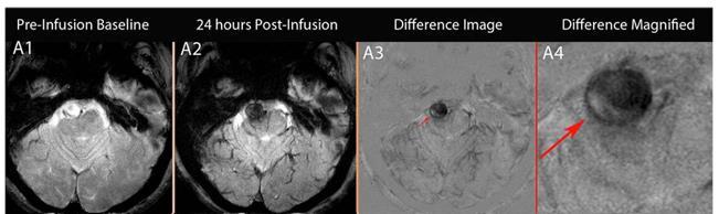
14 MPO activity-specific paramagnetic imaging agent (DTPA(Gd) bishydroxyindolamide [bis-5ht-dtpagd] Using 8-element SENSE receive-only and RF transmit coils we applied the following sequences: black blood double inversion recovery (DIR)19, small field-of-view quadrature inversion recovery (SF-QIR)20, and motion-sensitized driven equilibrium (MSDE).21 All images were acquired with 2 mm slices along the longaxis of the aneurysm in a 50 mm field-of-view giving a measured and reconstructed in-plane resolution of 0.3 and 0.2 mm, respectively. The DIR, SF-QIR, and MSDE scan parameters included TR/TE of 456/10, 750/11 and 698/9 ms; flip angle of 90, 90, and 70, respectively. The number of signal averages for all sequences was 2. The flow velocity encoded gradient echo imaging parameter (VENC) had values between 1 to 8 cm/s.

15

16
17 Macrophage-Imaging

18
19

20 Iron oxide nanoparticles

21 Nanoparticle-Enhanced MRI

22


23

24


25 Control Aspirin

26


27


28

29
30

31
32
33

34
35

36

37

38
39
40

41

42
43
44 Ferric Iron Imaging
45 We hypothesize that QSM will act as an imaging marker for aneurysm micro-bleed Insight on anatomical and functional parameters of the aneurysm Delineation of the aneurysm wall and hemosiderin accumulation around the aneurysm Compare multiple methods of detection: QSM Susceptibility weighted imaging (SWAN) Cushing Uiowa 2015
46


47 Anatomy SWAN QSM Cushing Uiowa 2015

48

49
50

51

52

53

54
55 Non-Specific Inflammation Imaging

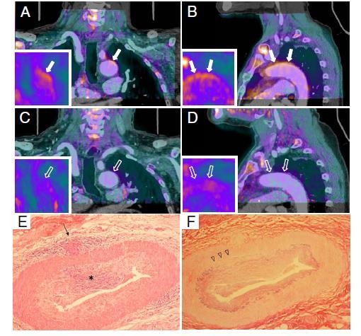
56 Contrast-Enhanced MRI Vessel wall magnetic resonance imaging identifies the site of rupture in patients with multiple intracranial aneurysms: proof of principle. Matouk CC, et al. Neurosurgery 2013 Vascular Wall Imaging of Unruptured Cerebral Aneurysms with a Hybrid of Opposite-Contrast MR Angiography. Matsushige T, et al. AJNR 2015 No in-vivo visualization of wall microstructure with current methods.
 Mural contrast uptake is a function of the density of vasa vasorum Histopathologic")

57 Contrast-Enhanced MRI 87% of unstable ANs showed CE (+) 28% of stable ANs showed CE (+) Mural contrast uptake is a function of the density of vasa vasorum Histopathologic studies in unstable ANs revealed thickened wall with inflammatory process Matouk CC, et al. Neurosurg 2013 Edjlali M, et al. Stroke 2014 Nagahata S, et al. Clin Neuroradiol 2014

58

59
60

61

62

63

64 Copyright 2017 Topics in Magnetic Resonance Imaging. Published by Lippincott Williams & Wilkins. 65

65 Copyright 2017 Topics in Magnetic Resonance Imaging. Published by Lippincott Williams & Wilkins. 66


66 Dynamic-Enhanced MRI Vakil P, et al. AJNR 2015 Increased contrast agent permeability across the aneurysm wall correlated significantly with both aneurysm size and size-independent anatomic risk factors, predicting high-risk aneurysms.


67 Atherosclerosis detected by HOP-MRA IC Ach An. AcoA An. The hybrid of opposite-contrast MRA can detect atherosclerotic plaques in the unruptured aneurysm wall Matsushige T et al. AJNR 2015
68 Summary Inflammation play a critical part in aneurysm formation, growth, and rupture It is possible to image in-vivo inflammatory cytokines/enzymes and/or cells in aneurysm wall Imaging inflammation in the aneurysm wall could stratify aneurysms to stable and unstable These techinques are in its infancy (case reports) and to be extrapolated into clinical practice, larger prospective observational studies or clinical trial are needed.


69 Questions??? Unruptured Aneurysm Ruptured Aneurysm


70 A B

71 B A
72
73 A B


74 A B C D A B
75 Video
Intracranial aneurysms are common vascular lesions, often consisting
 ORIGINAL RESEARCH INTERVENTIONAL Vascular Wall Imaging of Unruptured Cerebral Aneurysms with a Hybrid of Opposite-Contrast MR Angiography T. Matsushige, Y. Akiyama, T. Okazaki, K. Shinagawa, N. Ichinose,
1Pulse sequences for non CE MRA
 MRI: Principles and Applications, Friday, 8.30 9.20 am Pulse sequences for non CE MRA S. I. Gonçalves, PhD Radiology Department University Hospital Coimbra Autumn Semester, 2011 1 Magnetic resonance angiography
MR Advance Techniques. Vascular Imaging. Class II
 MR Advance Techniques Vascular Imaging Class II 1 Vascular Imaging There are several methods that can be used to evaluate the cardiovascular systems with the use of MRI. MRI will aloud to evaluate morphology
Vessel Wall Imaging of Intracranial Arterial Disease Commercial Interests
 Vessel Wall Imaging of Intracranial Arterial Disease Commercial Interests Disclosures No relevant commercial interests Off Label / Investigational Use No off label / investigational use Daniel Mandell,
NIH Public Access Author Manuscript J Am Coll Radiol. Author manuscript; available in PMC 2013 June 24.
 NIH Public Access Author Manuscript Published in final edited form as: J Am Coll Radiol. 2010 January ; 7(1): 73 76. doi:10.1016/j.jacr.2009.06.015. Cerebral Aneurysms Janet C. Miller, DPhil, Joshua A.
Magnetic Resonance Angiography
 Magnetic Resonance Angiography 1 Magnetic Resonance Angiography exploits flow enhancement of GR sequences saturation of venous flow allows arterial visualization saturation of arterial flow allows venous
Magnetic Resonance Imaging. Basics of MRI in practice. Generation of MR signal. Generation of MR signal. Spin echo imaging. Generation of MR signal
 Magnetic Resonance Imaging Protons aligned with B0 magnetic filed Longitudinal magnetization - T1 relaxation Transverse magnetization - T2 relaxation Signal measured in the transverse plane Basics of MRI
MR Imaging of Atherosclerotic Plaques
 MR Imaging of Atherosclerotic Plaques Yeon Hyeon Choe, MD Department of Radiology, Samsung Medical Center, Sungkyunkwan University, Seoul MRI for Carotid Atheroma Excellent tissue contrast (fat, fibrous
Essentials of Clinical MR, 2 nd edition. 99. MRA Principles and Carotid MRA
 99. MRA Principles and Carotid MRA As described in Chapter 12, time of flight (TOF) magnetic resonance angiography (MRA) is commonly utilized in the evaluation of the circle of Willis. TOF MRA allows depiction
Brain AVM with Accompanying Venous Aneurysm with Intracerebral and Intraventricular Hemorrhage
 Cronicon OPEN ACCESS EC PAEDIATRICS Case Report Brain AVM with Accompanying Venous Aneurysm with Intracerebral and Intraventricular Hemorrhage Dimitrios Panagopoulos* Neurosurgical Department, University
Cardiac Imaging Tests
 Cardiac Imaging Tests http://www.medpagetoday.com/upload/2010/11/15/23347.jpg Standard imaging tests include echocardiography, chest x-ray, CT, MRI, and various radionuclide techniques. Standard CT and
CARDIAC MRI. Cardiovascular Disease. Cardiovascular Disease. Cardiovascular Disease. Overview
 CARDIAC MRI Dr Yang Faridah A. Aziz Department of Biomedical Imaging University of Malaya Medical Centre Cardiovascular Disease Diseases of the circulatory system, also called cardiovascular disease (CVD),
Surface Appearance of the Vertebrobasilar Artery Revealed on Basiparallel Anatomic Scanning (BPAS) MR Imaging: Its Role for Brain MR Examination
 MR Imaging: Its Role for Brain MR Examination") AJNR Am J Neuroradiol 26:2508 2513, November/December 2005 Surface Appearance of the Vertebrobasilar Artery Revealed on Basiparallel Anatomic Scanning (BPAS) MR Imaging: Its Role for Brain MR Examination
AJNR Am J Neuroradiol 26:2508 2513, November/December 2005 Surface Appearance of the Vertebrobasilar Artery Revealed on Basiparallel Anatomic Scanning (BPAS) MR Imaging: Its Role for Brain MR Examination
Enterprise Stent-assisted Cerebral Aneurysm Coiling: Can Antiplatelet Therapy be Terminated after Neointima Formation with the Enterprise Stent?
 Journal of Neuroendovascular Therapy 2016; 10: 201 205 Online September 9, 2016 DOI: 10.5797/jnet.oa.2016-0052 Enterprise Stent-assisted Cerebral Aneurysm Coiling: Can Antiplatelet Therapy be Terminated
Willem J.M. Mulder, Ph.D. Assistant Professor of Radiology Assistant Professor of Gene and Cell Medicine Director Nanomedicine Laboratory
 Willem J.M. Mulder, Ph.D. Assistant Professor of Radiology Assistant Professor of Gene and Cell Medicine Director Nanomedicine Laboratory Noordwijkerhout, The Netherlands 17-18 March 2011 Nanometer (10-9
Time-resolved Magnetic Resonance Angiography for assessment of recanalization after coil embolization of visceral artery aneurysms
 Signature: Pol J Radiol, 2013; 78(1): 64-68 DOI: 10.12659/PJR.883769 CASE REPORT Received: 2012.09.29 Accepted: 2013.01.15 Time-resolved Magnetic Resonance Angiography for assessment of recanalization
Η Πυρηνική Καρδιολογία Το 2017 ΟΜΑΔΑ ΕΡΓΑΣΙΑΣ ΑΠΕΙΚΟΝΙΣΤΙΚΩΝ ΤΕΧΝΙΚΩΝ
 Η Πυρηνική Καρδιολογία Το 2017 ΟΜΑΔΑ ΕΡΓΑΣΙΑΣ ΑΠΕΙΚΟΝΙΣΤΙΚΩΝ ΤΕΧΝΙΚΩΝ huma human n Setting diagnosis of the early stages of chronic diseases (i.e cancer, neuropsychiatric, cardiovascular disorders), in
Imaging veins, oxygen extraction fraction, arteries and vessel wall using susceptibility weighted imaging (SWI) and susceptibility mapping (SWIM)
 and susceptibility mapping (SWIM)") Imaging veins, oxygen extraction fraction, arteries and vessel wall using susceptibility weighted imaging (SWI) and susceptibility mapping (SWIM) SWI E. Mark Haacke Department of Radiology, Wayne State
Imaging veins, oxygen extraction fraction, arteries and vessel wall using susceptibility weighted imaging (SWI) and susceptibility mapping (SWIM) SWI E. Mark Haacke Department of Radiology, Wayne State
Index. average stress 146. see ACIS
 Index ACIS (autonomous catheter insertion system) 156, 237 39, 241 49 acute stroke treatment 59, 69, 71 anatomical model 88 aneurismal clipping treatment 106, 110 aneurysm 2 3, 26, 47 50, 52 55, 67 68,
Speed, Comfort and Quality with NeuroDrive
 Speed, Comfort and Quality with NeuroDrive Echelon Oval provides a broad range of capabilities supporting fast, accurate diagnosis of brain conditions and injuries. From anatomical depiction to vascular
Non Contrast MRA. Mayil Krishnam. Director, Cardiovascular and Thoracic Imaging University of California, Irvine
 Non Contrast MRA Mayil Krishnam Director, Cardiovascular and Thoracic Imaging University of California, Irvine No disclosures Non contrast MRA-Why? Limitations of CTA Radiation exposure Iodinated contrast
IE with cerebral hemorrhage
 IE with cerebral hemorrhage Gilbert Habib / Patrizio Lancellotti La Timone Hospital Marseille - France Palermo, 26 April 2018 Case report: aortic bioprosthetic IE History of the disease 75 year-old man
Innovation Interlude: Molecular Imaging in Cardiology. Celestial Doppelgangers and Relativity
 Innovation Interlude: Molecular Imaging in Cardiology Jonathan R. Lindner, M.D. M. Lowell Edwards Professor of Cardiology Knight Cardiovascular Institute Oregon National Primate Research Center Oregon
Disclosure. Acknowledgement. What is the Best Workup for Rectal Cancer Staging: US/MRI/PET? Rectal cancer imaging. None
 What is the Best Workup for Rectal Cancer Staging: US/MRI/PET? Zhen Jane Wang, MD Assistant Professor in Residence UC SF Department of Radiology Disclosure None Acknowledgement Hueylan Chern, MD, Department
VasoRx: Atherosclerotic Plaque Regression
 VasoRx: Atherosclerotic Plaque Regression A progressive disease characterized by plaque build-up in large arteries, leading to heart attacks, stroke, and peripheral vascular disease. Induced by a combination
ARTICLE IN PRESS. Available online at Magnetic Resonance Imaging xx (2008) xxx xxx
 xxx xxx") Available online at www.sciencedirect.com Magnetic Resonance Imaging xx (2008) xxx xxx An optimized 3D inversion recovery prepared fast spoiled gradient recalled sequence for carotid plaque hemorrhage
Available online at www.sciencedirect.com Magnetic Resonance Imaging xx (2008) xxx xxx An optimized 3D inversion recovery prepared fast spoiled gradient recalled sequence for carotid plaque hemorrhage
NIH Public Access Author Manuscript J Vasc Interv Radiol. Author manuscript; available in PMC 2012 July 1.
 NIH Public Access Author Manuscript Published in final edited form as: J Vasc Interv Radiol. 2011 July ; 22(7): 1007 1011. doi:10.1016/j.jvir.2011.01.425. Temporal Stability of Dysmorphic, Fusiform Aneurysms
Treatment of Unruptured Vertebral Artery Dissecting Aneurysms
 33 Treatment of Unruptured Vertebral Artery Dissecting Aneurysms Isao NAITO, M.D., Shin TAKATAMA, M.D., Naoko MIYAMOTO, M.D., Hidetoshi SHIMAGUCHI, M.D., and Tomoyuki IWAI, M.D. Department of Neurosurgery,
Table 1. Summary of PET and fmri Methods. What is imaged PET fmri BOLD (T2*) Regional brain activation. Blood flow ( 15 O) Arterial spin tagging (AST)
 Regional brain activation. Blood flow ( 15 O) Arterial spin tagging (AST)") Table 1 Summary of PET and fmri Methods What is imaged PET fmri Brain structure Regional brain activation Anatomical connectivity Receptor binding and regional chemical distribution Blood flow ( 15 O)
Table 1 Summary of PET and fmri Methods What is imaged PET fmri Brain structure Regional brain activation Anatomical connectivity Receptor binding and regional chemical distribution Blood flow ( 15 O)
Spontaneous intracranial dissecting aneurysms are
 CLINICAL ARTICLE J Neurosurg 128:747 755, 2018 Aneurysm wall enhancement on magnetic resonance imaging as a risk factor for progression of unruptured vertebrobasilar dissecting aneurysms after reconstructive
Department of Radiology University of California San Diego. MR Angiography. Techniques & Applications. John R. Hesselink, M.D.
 Department of Radiology University of California San Diego MR Angiography Techniques & Applications John R. Hesselink, M.D. Vascular Imaging Arterial flow void Flow enhancement Gadolinium enhancement Vascular
MODERN METHODS FOR TREATING ABDOMINAL ANEURYSMS AND THORACIC AORTIC DISEASE
 MODERN METHODS FOR TREATING ABDOMINAL ANEURYSMS AND THORACIC AORTIC DISEASE AAA FACTS 200,000 New Cases Each Year Ruptured AAA = 15,000 Deaths per Year in U.S. 13th Leading Cause of Death 80% Chance of
Neuroradiology MR Protocols
 Neuroradiology MR Protocols Brain protocols N 1: Brain MRI without contrast N 2: Pre- and post-contrast brain MRI N 3 is deleted N 4: Brain MRI without or pre-/post-contrast (seizure protocol) N 5: Pre-
Cerebral Aneurysms. U.S. DEPARTMENT OF HEALTH AND HUMAN SERVICES Public Health Service National Institutes of Health
 Cerebral Aneurysms U.S. DEPARTMENT OF HEALTH AND HUMAN SERVICES Public Health Service National Institutes of Health Cerebral Aneurysms What is a cerebral aneurysm? cerebral aneurysm (also known as an
Small UIAs, <7 mm in diameter, uncommonly cause aneurysmal symptoms and are the most frequently detected incidentally.
 Research grant from Stryker Neurovascular Research grant from Covidien/ Medtronic Consultant and proctor for Stryker Neurovascular Consultant and proctor for Covidien/ Medtronic Consultant for Codman Neurovascular
Plaque Imaging: What It Can Tell Us. Kenneth Snyder, MD, PhD L Nelson Hopkins MD FACS Elad Levy MD MBA FAHA FACS Adnan Siddiqui MD PhD
 Plaque Imaging: What It Can Tell Us Kenneth Snyder, MD, PhD L Nelson Hopkins MD FACS Elad Levy MD MBA FAHA FACS Adnan Siddiqui MD PhD Buffalo Disclosure Information FINANCIAL DISCLOSURE: Research and consultant
Anatomic Evaluation of the Circle of Willis: MR Angiography versus Intraarterial Digital Subtraction Angiography
 Anatomic Evaluation of the Circle of Willis: MR Angiography versus Intraarterial Digital Subtraction Angiography K. W. Stock, S. Wetzel, E. Kirsch, G. Bongartz, W. Steinbrich, and E. W. Radue PURPOSE:
Magnetization Preparation Sequences
 Magnetization Preparation Sequences Acquisition method may not give desired contrast Prep block adds contrast (and/or encoding) MP-RAGE = Magnetization prepared rapid acquisition with gradient echo (Mugler,
Assessment of Cardio- & Neurovascular Hemodynamics in the Human Circulatory System using 4D flow MRI
 Assessment of Cardio- & Neurovascular Hemodynamics in the Human Circulatory System using 4D flow MRI Michael Markl, Ph.D. Departments of Radiology & Biomedical Engineering Northwestern University, Chicago,
Management of Cerebral Aneurysms in Polycystic Kidney Disease. Dr H Stockley Consultant Neuroradiologist Greater Manchester Neuroscience Centre
 Management of Cerebral Aneurysms in Polycystic Kidney Disease Dr H Stockley Consultant Neuroradiologist Greater Manchester Neuroscience Centre What is a cerebral aneurysm? Developmental degenerative arterial
PHYSICS OF MRI ACQUISITION. Alternatives to BOLD for fmri
 PHYSICS OF MRI ACQUISITION Quick Review for fmri HST-583, Fall 2002 HST.583: Functional Magnetic Resonance Imaging: Data Acquisition and Analysis Harvard-MIT Division of Health Sciences and Technology
MRI and CT of the CNS
 MRI and CT of the CNS Dr.Maha ELBeltagy Assistant Professor of Anatomy Faculty of Medicine The University of Jordan 2018 Computed Tomography CT is used for the detection of intracranial lesions. CT relies
Medical imaging X-ray, CT, MRI, scintigraphy, SPECT, PET Györgyi Műzes
 Medical imaging X-ray, CT, MRI, scintigraphy, SPECT, PET Györgyi Műzes Semmelweis University, 2nd Dept. of Medicine Medical imaging: definition technical process of creating visual representations about
Methods. Yahya Paksoy, Bülent Oğuz Genç, and Emine Genç. AJNR Am J Neuroradiol 24: , August 2003
 AJNR Am J Neuroradiol 24:1364 1368, August 2003 Retrograde Flow in the Left Inferior Petrosal Sinus and Blood Steal of the Cavernous Sinus Associated with Central Vein Stenosis: MR Angiographic Findings
MRI of the Brain: A Primer on What, How, Why, and When. September Amit Malhotra, Harvard Medical School, Year- IV. Gillian Lieberman, MD
 September 2000 MRI of the Brain: A Primer on What, How, Why, and When Hornak, J.P. The Basics of MRI. 1996-2000 Amit Malhotra, Harvard Medical School, Year- IV Magnetic Resonance Imaging A Brief History
 Current Status and Future Trends of MRI Technology for Carotid Plaque Imaging LI Rui*, CHEN Hui-Jun, YUAN Chun. *Biomedical Engineering Department, School of Medicine, Tsinghua University, Beijing 100084,
Epidemiology And Treatment Of Cerebral Aneurysms At An Australian Tertiary Level Hospital
 ISPUB.COM The Internet Journal of Neurosurgery Volume 9 Number 2 Epidemiology And Treatment Of Cerebral Aneurysms At An Australian Tertiary Level Hospital A Granger, R Laherty Citation A Granger, R Laherty.
DOWNLOAD OR READ : MRI CLINICAL MAGNETIC RESONANCE IMAGING PDF EBOOK EPUB MOBI
 DOWNLOAD OR READ : MRI CLINICAL MAGNETIC RESONANCE IMAGING PDF EBOOK EPUB MOBI Page 1 Page 2 mri clinical magnetic resonance imaging mri clinical magnetic resonance pdf mri clinical magnetic resonance
Long-term Observation of Lateral Medullary Infarction due to Vertebral Artery Dissection Assessed with Multimodal Neuroimaging
 Case Reports Long-term Observation of Lateral Medullary Infarction due to Vertebral Artery Dissection Assessed with Multimodal Neuroimaging Koichi Nomura 1, Masahiro Mishina 1,SeijiOkubo 1, Satoshi Suda
Non-invasive Imaging of Carotid Artery Atherosclerosis
 Non-invasive Imaging of Carotid Artery Atherosclerosis 최연현 성균관의대삼성서울병원영상의학과 Noninvasive Techniques US with Doppler CT MRI Ultrasonography Techniques of Carotid US US Anatomy (ICA vs ECA) Gray scale and
Postoperative Assessment of Extracranial Intracranial Bypass by Time- Resolved 3D Contrast-Enhanced MR Angiography Using Parallel Imaging
 AJNR Am J Neuroradiol 26:2243 2247, October 2005 Postoperative Assessment of Extracranial Intracranial Bypass by Time- Resolved 3D Contrast-Enhanced MR Angiography Using Parallel Imaging Kazuhiro Tsuchiya,
Spinal dural AV fistula: One stop shop imaging with MR?
 Spinal dural AV fistula: One stop shop imaging with MR? Poster No.: C-3378 Congress: ECR 2010 Type: Educational Exhibit Topic: Neuro Authors: J. C. Röper-Kelmayr 1, C. Ginthör 1, D. Flöry 1, R. Chapot
Susceptibility-weighted MRI ups contrast, offers minute detail 9/15/04 By: Shalmali Pal
 Susceptibility-weighted MRI ups contrast, offers minute detail 9/15/04 By: Shalmali Pal With its flashy sequences and high-speed protocols, there's no shortage of razzle-dazzle in MRI. But learning to
FOR CMS (MEDICARE) MEMBERS ONLY NATIONAL COVERAGE DETERMINATION (NCD) FOR MAGNETIC RESONANCE IMAGING:
 MEMBERS ONLY NATIONAL COVERAGE DETERMINATION (NCD) FOR MAGNETIC RESONANCE IMAGING:") National Imaging Associates, Inc. Clinical guidelines SINUS MRI Original Date: November 2007 Page 1 of 5 CPT Codes: 70540, 70542, 70543 Last Review Date: July 2014 NCD 220.2 MRI Last Effective Date: July
National Imaging Associates, Inc. Clinical guidelines SINUS MRI Original Date: November 2007 Page 1 of 5 CPT Codes: 70540, 70542, 70543 Last Review Date: July 2014 NCD 220.2 MRI Last Effective Date: July
Advanced Vascular Imaging: Pulsatile Tinnitus. Disclosures. Pulsatile Tinnitus: Differential Diagnosis. Pulsatile Tinnitus
 Advanced Vascular Imaging: Pulsatile Tinnitus Patrick Turski MD, Zach Clark MD, Tabby Kennedy MD The Objectives of this presentation are to: Review the differential diagnosis of pulsatile tinnitus Discuss
Computed tomography. Department of Radiology, University Medical School, Szeged
 Computed tomography Department of Radiology, University Medical School, Szeged voxel +1-4 +2 +5 +3 +1 0-2 pixel -2 0 +1-4 -6 +5 +2 +1 Department of Radiology, University Medical School, Szeged
The aorta is an integral part of the cardiovascular system and should not be considered as just a conduit for blood supply from the heart to the
 The aorta is an integral part of the cardiovascular system and should not be considered as just a conduit for blood supply from the heart to the limbs and major organs. A range of important pathologies
MAGNETIC RESONANCE LATE GADOLINIUM ENHANCEMENT DETECTS ACUTE AORTIC INTRAMURAL HEMATOMA
 MAGNETIC RESONANCE LATE GADOLINIUM ENHANCEMENT DETECTS ACUTE AORTIC INTRAMURAL HEMATOMA Diego Perez de Arenaza, Santiago del Castillo, Marcelo Pietrani, Mariano Falconi, Juan Benger, Ezequiel Levy Yeyati,
Localization and Treatment of Unruptured Paraclinoid Aneurysms: A Proton Density MRI-based Study
 Journal of Cerebrovascular and Endovascular Neurosurgery pissn 2234-8565, eissn 2287-3139, http://dx.doi.org/10.7461/jcen.2015.17.3.180 Original Article Localization and Treatment of Unruptured Paraclinoid
Stress analysis of cerebral aneurysms
 Stress analysis of cerebral aneurysms Madhavan Lakshmi Raghavan, Ph.D. Biomedical Engineering BioMOST division, Center for Computer-Aided Design University of Iowa Aneurysm size and shape metrics Shape
What You Should Know About Cerebral Aneurysms
 American Society of Neuroradiology American Society of Interventional & Therapeutic Neuroradiology What You Should Know About Cerebral Aneurysms From the Cerebrovascular Imaging and Intervention Committee
Carotid Artery Wall Imaging: Perspective and. Guidelines from the ASNR Vessel Wall. Imaging Study Group and Expert Consensus
 Carotid Artery Wall Imaging: Perspective and Guidelines from the ASNR Vessel Wall Imaging Study Group and Expert Consensus Recommendations of the American Society of Neuroradiology Type of manuscript:
Procedia - Social and Behavioral Sciences 159 ( 2014 ) WCPCG 2014
 WCPCG 2014") Available online at www.sciencedirect.com ScienceDirect Procedia - Social and Behavioral Sciences 159 ( 2014 ) 743 748 WCPCG 2014 Differences in Visuospatial Cognition Performance and Regional Brain Activation
Available online at www.sciencedirect.com ScienceDirect Procedia - Social and Behavioral Sciences 159 ( 2014 ) 743 748 WCPCG 2014 Differences in Visuospatial Cognition Performance and Regional Brain Activation
Outline. Neuroradiology. Diffusion Imaging in. Clinical Applications of. Basics of Diffusion Imaging. Basics of Diffusion Imaging
 Clinical Applications of Diffusion Imaging in Neuroradiology No disclosures Stephen F. Kralik Assistant Professor of Radiology Indiana University School of Medicine Department of Radiology and Imaging
Vessel wall characterization using quantitative MRI: what s in a number?
 Magn Reson Mater Phy (2018) 31:201 222 https://doi.org/10.1007/s10334-017-0644-x REVIEW ARTICLE Vessel wall characterization using quantitative MRI: what s in a number? Bram F. Coolen 1,2 Claudia Calcagno
Imaging in the Evaluation of Coronary Artery Disease and Abdominal Aortic Aneurysm
 Imaging in the Evaluation of Coronary Artery Disease and Abdominal Aortic Aneurysm Mark J. Sands, MD Vice Chairman, Imaging Institute Clinical Operations and Quality Objectives Review of available radiologic
AEROMEDICAL DECISION MAKING IN CEREBRAL ANEURYSMS. Pooshan Navāthé Michael Drane Peter Clem David Fitzgerald
 AEROMEDICAL DECISION MAKING IN CEREBRAL ANEURYSMS Pooshan Navāthé Michael Drane Peter Clem David Fitzgerald Disclaimer I receive a salary from the Commonwealth of Australia. I have no financial relationships
Cerebral Bypass and Advanced Neuroendovascular Course 2018
 Mayo Clinic Cerebral Bypass and Advanced Neuroendovascular Course 2018 Co-directed by: Giuseppe Lanzino MD and Leonardo Rangel-Castilla MD Honored Guest: Luca Regli MD, Zurich, Switzerland Thursday, November
Cerebral cavernous malformation (CCM) comprise
 comprise") ORIGINAL RESEARCH J.M. de Souza R.C. Domingues L.C.H. Cruz, Jr. F.S. Domingues T. Iasbeck E.L. Gasparetto Susceptibility-Weighted Imaging for the Evaluation of Patients with Familial Cerebral Cavernous
ORIGINAL RESEARCH J.M. de Souza R.C. Domingues L.C.H. Cruz, Jr. F.S. Domingues T. Iasbeck E.L. Gasparetto Susceptibility-Weighted Imaging for the Evaluation of Patients with Familial Cerebral Cavernous
MR Advance Techniques. Cardiac Imaging. Class III
 MR Advance Techniques Cardiac Imaging Class III Black Blood Imaging & IR Blue= O2 poor blood Red=O2 rich blood Inversion pulses can produce black blood imaging in GRE pulse sequences. Specially on the
Coil Embolization of Cerebral Tiny Aneurysms
 Journal of Neuroendovascular Therapy 2016; 10: 243 248 Online November 9, 2016 DOI: 10.5797/jnet.oa.2016-0035 Coil Embolization of Cerebral Tiny Aneurysms Terumasa Kuroiwa, 1 Fuminori Shimizu, 2 Taro Yamashita,
Non-Invasive Follow-up Evaluation of Post-Embolized AVM with Time-Resolved MRA: A Case Report
 Non-Invasive Follow-up Evaluation of Post-Embolized AVM with Time-Resolved MRA: A Case Report Yong Woon Shim, MD 1 Tae-Sub Chung, MD 1 Won-Suk Kang, MD 1 Jin-Yang Joo, MD 2 Ralph Strecker, MD 3 Juergen
Cardiac Imaging. Kimberly Delcour, DO, FACC. Mahi Ashwath, MD, FACC, FASE. Director, Cardiac CT. Director, Cardiac MRI
 Cardiac Imaging Kimberly Delcour, DO, FACC Director, Cardiac CT Mahi Ashwath, MD, FACC, FASE Director, Cardiac MRI Cardiac Imaging Discuss the clinical applications of and indications for: Cardiac CT Nuclear
Cerebral MR Venography: Normal Anatomy and Potential Diagnostic Pitfalls
 AJNR Am J Neuroradiol 21:74 78, January 2000 Cerebral MR Venography: Normal Anatomy and Potential Diagnostic Pitfalls R. H. Ayanzen, C. R. Bird, P. J. Keller, F. J. McCully, M. R. Theobald, and J. E. Heiserman
Imaging and Hemodynamics in Aneurysm Evolution
 Disclosures Imaging and Hemodynamics in Aneurysm Evolution David Saloner, PhD There are no financial conflicts of interest to report Department of Radiology and Biomedical Imaging VA Medical Center San
Alessandro Della Puppa
 Intraoperative measurement of arterial blood flow in complex cerebral aneurysms surgery Studio flussimetrico intra-operatorio nel clipping degli aneurismi complessi Alessandro Della Puppa NEUROSURGERY
Paul Gigante HMS IV Gillian Lieberman, MD. Sept Mr. T s T s Headache. Paul Gigante,, Harvard Medical School Year IV Gillian Lieberman, MD
 Sept 2005 Mr. T s T s Headache Paul Gigante,, Harvard Medical School Year IV Mr. T s T s Presentation 45 year-old welder complains of sudden severe headache and witnessed seizure with loss of consciousness
Carotid Artery Disease and What s Pertinent JOSEPH A PAULISIN DO
 Carotid Artery Disease and What s Pertinent JOSEPH A PAULISIN DO Goal of treatment of carotid disease Identify those at risk of developing symptoms Prevent patients at risk from developing symptoms Prevent
Clinical Applications
 C H A P T E R 16 Clinical Applications In selecting pulse sequences and measurement parameters for a specific application, MRI allows the user tremendous flexibility to produce variations in contrast between
From the Cerebrovascular Imaging and Intervention Committee of the American Heart Association Cardiovascular Council
 American Society of Neuroradiology What Is a Stroke? From the Cerebrovascular Imaging and Intervention Committee of the American Heart Association Cardiovascular Council Randall T. Higashida, M.D., Chair
Bilateral blunt carotid artery injury: A case report and review of the literature
 CASE REPORT Bilateral blunt carotid artery injury: A case report and review of the literature S Cheddie, 1 MMed (Surg), FCS (SA); B Pillay, 2 FCS (SA), Cert Vascular Surgery; R Goga, 2 FCS (SA) 1 Department
非對比劑與對比劑增強 MRA. 血管攝影與對比劑 A Course of MRI. 本週課程內容 -MR Angiography (MRA) Unenhanced MRA
 Unenhanced MRA") 本週課程內容 -MR Angiography (MRA) 血管攝影與對比劑 A Course of MRI 盧家鋒助理教授國立陽明大學物理治療暨輔助科技學系 alvin4016@ym.edu.tw 非對比劑增強 MRA(Unenhanced MRA) Time-of-flight (TOF) angiography Phase-contrast (PC) angiography 對比劑增強 MRA(Contrast-enhanced
本週課程內容 -MR Angiography (MRA) 血管攝影與對比劑 A Course of MRI 盧家鋒助理教授國立陽明大學物理治療暨輔助科技學系 alvin4016@ym.edu.tw 非對比劑增強 MRA(Unenhanced MRA) Time-of-flight (TOF) angiography Phase-contrast (PC) angiography 對比劑增強 MRA(Contrast-enhanced
Field Strength. Regional Perfusion Imaging (RPI) matches cerebral arteries to flow territories
 matches cerebral arteries to flow territories") Field Strength Changing how the world looks at MR. Regional Perfusion Imaging (RPI) matches cerebral arteries to flow territories Research groups in Utrecht, Baltimore and Singapore collaborate on this
Field Strength Changing how the world looks at MR. Regional Perfusion Imaging (RPI) matches cerebral arteries to flow territories Research groups in Utrecht, Baltimore and Singapore collaborate on this
The Low Sensitivity of Fluid-Attenuated Inversion-Recovery MR in the Detection of Multiple Sclerosis of the Spinal Cord
 The Low Sensitivity of Fluid-Attenuated Inversion-Recovery MR in the Detection of Multiple Sclerosis of the Spinal Cord Mark D. Keiper, Robert I. Grossman, John C. Brunson, and Mitchell D. Schnall PURPOSE:
Supplementary Online Content
 Supplementary Online Content Gregg NM, Kim AE, Gurol ME, et al. Incidental cerebral microbleeds and cerebral blood flow in elderly individuals. JAMA Neurol. Published online July 13, 2015. doi:10.1001/jamaneurol.2015.1359.
The role of Imaging (scans of the kidneys) in the management of Autosomal Dominant Polycystic Kidney Disease
 in the management of Autosomal Dominant Polycystic Kidney Disease") The role of Imaging (scans of the kidneys) in the management of Autosomal Dominant Polycystic Kidney Disease Dr Roslyn Simms NIHR Clinical Lecturer in Nephrology Saturday 17 th September 2016 Role of Imaging
The role of Imaging (scans of the kidneys) in the management of Autosomal Dominant Polycystic Kidney Disease Dr Roslyn Simms NIHR Clinical Lecturer in Nephrology Saturday 17 th September 2016 Role of Imaging
MR Flow Imaging in Vascular Malformations Using Gradient Recalled Acquisition
 637 MR Flow Imaging in Vascular Malformations Using Gradient Recalled Acquisition William M. Needell 1 Kenneth R. Maravilla Twenty patients with known or suspected intracranial vascular lesions were evaluated
Quality Metrics. Stroke Related Procedure Outcomes
 Quality Metrics Stroke Related Procedure Outcomes Below is a description of some of the stroke-related procedures performed at St. Dominic Hospital in Jackson, with quality information on the complication
Preliminary Experience with 3-Tesla MRI and Cushing s Disease
 TECHNICAL NOTE Preliminary Experience with 3-Tesla MRI and Cushing s Disease LouisJ.Kim,M.D., 1 Gregory P. Lekovic, M.D., Ph.D., J.D., 1 William L.White, M.D., 1 and John Karis, M.D. 2 ABSTRACT Because
FOR CMS (MEDICARE) MEMBERS ONLY NATIONAL COVERAGE DETERMINATION (NCD) FOR MAGNETIC RESONANCE IMAGING:
 MEMBERS ONLY NATIONAL COVERAGE DETERMINATION (NCD) FOR MAGNETIC RESONANCE IMAGING:") National Imaging Associates, Inc. Clinical guidelines BONE MARROW MRI Original Date: July 2008 Page 1 of 5 CPT Codes: 77084 Last Review Date: September 2014 NCD 220.2 MRI Last Effective Date: July 2011
National Imaging Associates, Inc. Clinical guidelines BONE MARROW MRI Original Date: July 2008 Page 1 of 5 CPT Codes: 77084 Last Review Date: September 2014 NCD 220.2 MRI Last Effective Date: July 2011
Introducing a New Treatment Method for Brain Aneurysms
 Pipeline Embolization Device Introducing a New Treatment Method for Brain Aneurysms UNDERSTANDING ANEURYSMS What is a brain aneurysm? An aneurysm is an outpouching in an artery caused by weakness in the
Supratentorial multiple little meningiomas with transitory stroke symptoms like. MRI case presentation
 114 Romanian Neurosurgery (2010) XVII 1: 114-121 Supratentorial multiple little meningiomas with transitory stroke symptoms like. MRI case presentation E. Moldovanu 1,2, Adriana Moldovanu 1,2, Carmen Gherman
Intracranial vessel wall imaging: current applications and clinical implications
 Kontzialis and Wasserman Neurovascular Imaging (2016) 2:4 DOI 10.1186/s40809-016-0014-5 REVIEW Intracranial vessel wall imaging: current applications and clinical implications Marinos Kontzialis 1 and
What Is the Significance of a Large Number of Ruptured Aneurysms Smaller than 7 mm in Diameter?
 online ML Comm www.jkns.or.kr 10.3340/jkns.2009.45.2.85 J Korean Neurosurg Soc 45 : 85-89, 2009 Print ISSN 2005-3711 On-line ISSN 1598-7876 Copyright 2009 The Korean Neurosurgical Society Clinical Article
The ejournal of the European Society of Minimally Invasive Neurological Therapy
 The ejournal of the European Society of Minimally Invasive Neurological Therapy Performance of Contrast Enhanced Magnetic Resonance Angiography and CTA in the assessment of intracranial aneurysm coilability
Deborah K. Mann & Jennifer Bash. Coding Documentation and Education Managers
 Deborah K. Mann & Jennifer Bash Coding Documentation and Education Managers OBJECTIVES Review the basics of Diagnostic, CT, & MRI documentation Risk areas in radiology associated with Diagnostic, CT, &
Computer-Aided Diagnosis of Cerebral Aneurysm Based on Fuzzy Expert System: MR Angiography Study
 Computer-Aided Diagnosis of Cerebral Aneurysm Based on Fuzzy Expert System: MR Angiography Study Mika Ninomiya, Syoji Kobashi, Katsuya Kondo, Yutaka Hata, and Tomoharu Nakano Graduate School of Engineering,
CT or PET/CT for coronary artery disease
 CT or PET/CT for coronary artery disease Rotterdam 2012 Juhani Knuuti, MD, PhD, FESC Turku PET Centre University of Turku Turku, Finland Juhani.knuuti@utu.fi Turku PET Centre University of Turku Åbo Akademi
ROLE OF CONTRAST ENHANCED MR ANGIOGRAPHY IN AORTIC COARCTATION
 ROLE OF CONTRAST ENHANCED MR ANGIOGRAPHY IN AORTIC COARCTATION By Adel El Badrawy, Ahmed Abdel Razek, Nermin Soliman, Hala El Marsafawy *, Sameh Amer** From Radiodiagnosis, Pediatric Cardiology* & Cardiothoracic
Neurovascular elements of the PCF
 Level I: Neurovascular elements of the PCF Trigeminal and Abducent Superior cerebellar artery and vein Dandy s vein Level II: Facial and Cochleovestibular AICA and internal auditory artery, veins Level