Assessment of Cardio- & Neurovascular Hemodynamics in the Human Circulatory System using 4D flow MRI
|
|
|
- Marcus McDaniel
- 5 years ago
- Views:
Transcription


1 Assessment of Cardio- & Neurovascular Hemodynamics in the Human Circulatory System using 4D flow MRI Michael Markl, Ph.D. Departments of Radiology & Biomedical Engineering Northwestern University, Chicago, IL, USA Ensight UGM, Tokyo, October 2014
2 Outline Methodology: Measurement of 3D blood in the human body based on 4D flow MRI. Use of Ensight: 3D visualization & quantification of cardiovascular hemodynamics. Applications: Examples of clinical relevant questions. Potential of 4D flow & Ensight to improve diagnostics. Demonstration: Use of Ensight for analysis of 3D blood flow for a clinical case of congenital heart disease.
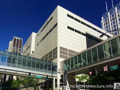
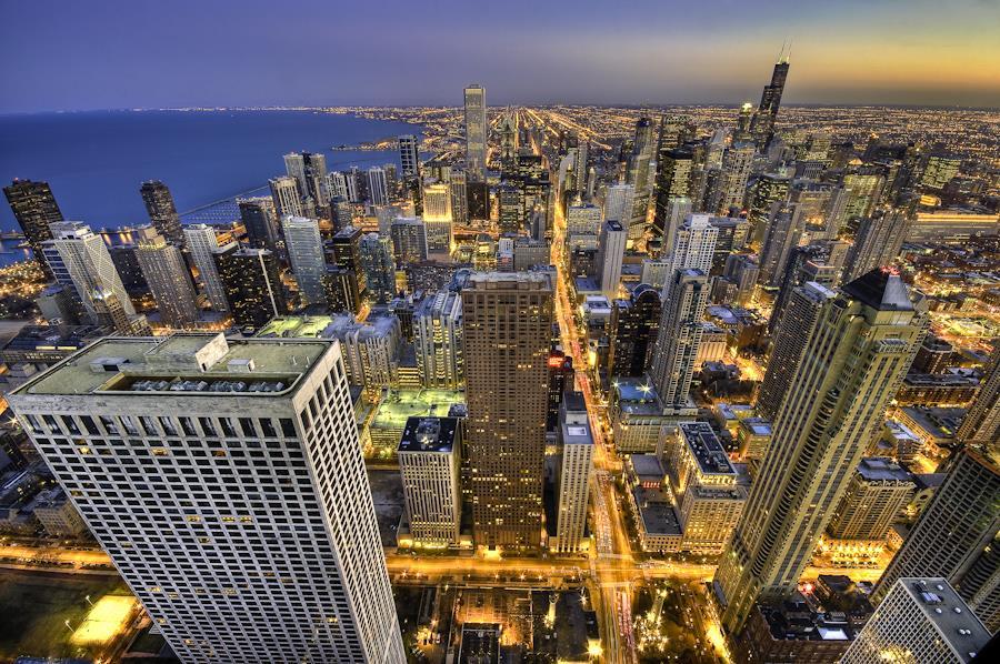

3 Cardiovascular Imaging Northwestern 737 N. Michigan Ave


")
 Small")

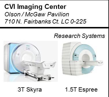
4 Cardiovascular Imaging Northwestern Imaging Core 'Center for Translational Imaging' Director: Andrew Larson, Ph.D. Neuroimaging fmri (Todd Parrish, PhD) Cardiovascular Imaging (Michael Markl, PhD) Small Animal Molecular Imaging (Daniel Procissi, PhD)


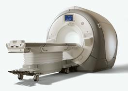

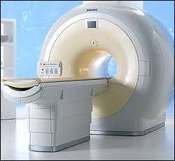
5 Magnetic Resonance Imaging Background MRI of the Human Body external magnetic fields no harmful radiation high soft tissue contrast flexible image orientation


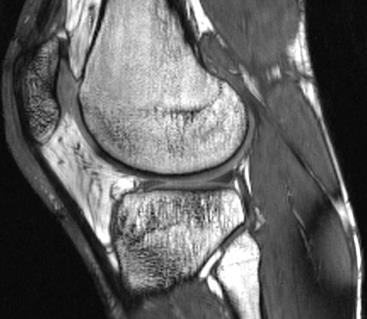


6 MRI How to generate image contrast? Image Contrast Many mechanisms 9 Angiography 6 3 Tissue perfusion Neuronal activity Source Water Fat

7 Cardiovascular MRI Imaging of Blood Flow MR Signal = Vector Signal Phase ~ Flow Measurement of blood flow

8 Cardiovascular MRI Imaging of Blood Flow 1960: Detection of Flow / Motion with MRI, E. Hahn E. Hahn : Direct Measurements of flow / tissue motion Initial clinical applications Moran PR. Magn Reson Imaging, 1: , 1982 O Donnell M. Med Phys, 12:59 64, 1985, Nayler GL et al. J Comput Assist Tomogr, 10: , 1986 Pelc NJ et al. J Magn Reson Imaging, 1: , 1991


9 Cardiovascular MRI Imaging of Blood Flow 2D Phase-Contrast (PC) MRI ECG synchronized Anatomy and pulsatile blood flow Aorta, 3D CE-MRA Anatomy Flow y x Flow, z

10 Cardiovascular MRI Imaging of Blood Flow Flow Quantification Net flow, cardiac output Regurgitation, valve function, peak velocity, etc.
 3 T Res ~ 30-60ms T Acq ~ 5-15min respiration control 4D (3D volume + time)")
11 MR Imaging 4D Flow Data Methods Res. ~ ( mm) 3 T Res ~ 30-60ms T Acq ~ 5-15min respiration control 4D (3D volume + time) flow (3-dir. velocity encoding) MRI
 flow (3-dir.")
12 Flow sensitive 4D Data 3D Visualization Methods Ensight, CEI - pathlines Adaptive navigator respiration control Res. ~ 2mm 3, T Res ~ 40ms, T Acq ~ 10-15min 4D (3D volume + time) flow (3-dir. velocity encoding) MRI
:457-466 Mahadevia R, et al.")
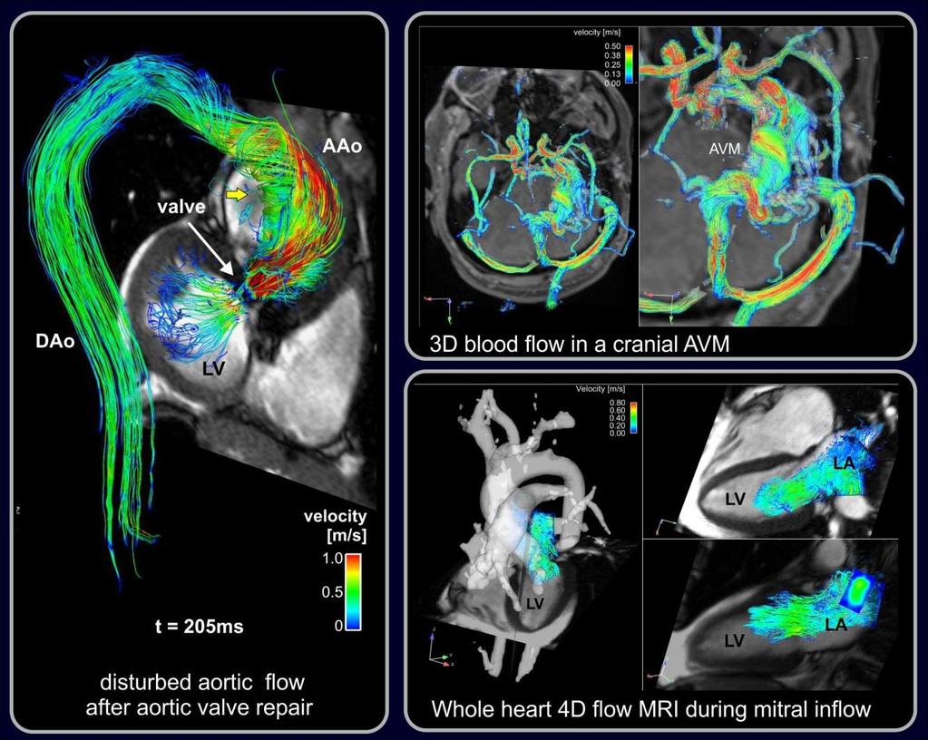
13 Applications Ensight, CEI streamlines Normal volunteer: Coherent systolic streamlines Bicuspid Aortic Valve (BAV): Flow jet & wall impingement Barker AJ, et al. Circ Cardiovasc Imaging 2012;5(4): Mahadevia R, et al. Circulation 2014;129:673-82


14 Applications Carotid arteries Portal vein

15 Congenital Heart Disease normal heart congenital heart disease


16 Congenital Heart Disease Single Ventricle Physiology Fontan Circulation complete reworking of cardiovascular system Normal cardiovascular physiology: After surgical Fontan correction: Gaca AM et al. Radiology 2008; 247:


17 Congenital Heart Disease Single Ventricle Physiology: Fontan Circulation AAo PA IVC SVC RRV LPV Markl M, et al. J Magn Reson Imaging. 2012;35:

18 Congenital Heart Disease

19 Congenital Heart Disease Single Ventricle Physiology single ventricle extracardiac TCPC
 PAVF Shah MJ, et al. Ann Thorac Surg. 1997;63:960-3. Hiramatsu et al.")
20 Congenital Heart Disease Background Fontan Circulation Some patients develop Failing Fontan physiology Caval blood distribution to PAs Suspected to influence patient outcome through delivery of protein-rich venous return Pulmonary arteriovenous malformations, fistulas (PAVF) PAVF Shah MJ, et al. Ann Thorac Surg. 1997;63: Hiramatsu et al. Ann Thorac Cardiovasc Surg, 2008

21 Congenital Heart Disease Fontan Circulation: Flow Distribution Quantification

22 Congenital Heart Disease Flow Asymmetry: Relationship with Fontan Geometry

23 Congenital Heart Disease Congenital Heart Disease Significance - Long scan times & general anesthesia in young patients - Complex CHD: standard parameters provide incomplete risk stratification

24 Congenital Heart Disease Congenital Heart Disease Innovation - Reduced scan time & exposure to anesthesia - 4D flow: New imaging biomarkers for improved evaluation / risk stratification of CHD

25 Arteriovenous Malformation AVM Complex vascular network No intervening capillary bed hemorrhagic stroke Risk Stratification & Treatment Spetzler-Martin grade (SMG) X-ray angio & staged embolization 4D flow MRI AVM treatment monitoring pre-intervention assessment Arteriovenous malformations of the brain in adults. N Engl J Med 1999;340: Spetzler RF, Martin NA. J Neurosurg 1986;65: Stapf C, et al. Neurology 2006;66(9): Batjer HH, et al. Neurosurgery 1988;23(3): Kader A, et al.. Neurosurgery 1994;34(5):
![Peak Velocity [m/s] Blood Flow [ml/s] 4D Flow](/docs-images/82/85716979/images/26-0.jpg "MRI Arteriovenous Malformation mag 4D flow Raw")


26 Peak Velocity [m/s] Blood Flow [ml/s] 4D Flow MRI Arteriovenous Malformation mag 4D flow Raw Data Pre-Processing 3D PC-MRA v z mag v z v x v y v x v y denoise velocity anti-aliasing eddy-current correction Flow Quantification 1.0 Peak Velocities 8 Blood Flow Time 400 [ms] Time [ms] D Flow Visualization

27 Arteriovenous Malformation 3D PC-MRA & Time-Resolved Pathlines vessel anatomy & 3D blood flow over cardiac cycle large unruptured AVM SMG = 4

28 Arteriovenous Malformation Time-Integrated Pathlines vessel anatomy & cumulative flow over cardiac cycle large unruptured AVM SMG = 4

29 Arteriovenous Malformation Heterogeneity in 3D flow despite identical SMG

30 AVM-11 Arteriovenous Malformation Vascular Network Diagram SMG-A: low-grade AVM SMG = 2 Analysis planes: feeding arteries, contralateral arteries, draining veins, straight sinus

31 AVM-14 Arteriovenous Malformation Vascular Network Diagram SMG-B: high-grade AVM SMG = 4 Analysis planes: feeding arteries, contralateral arteries, draining veins, straight sinus

32 Arteriovenous Malformation Follow-up during Embolization large AVM SMG = 4 compacting of AVM redistribution of peak velocities Ansari S, et al. AJNR 2013

33 Arteriovenous Malformation 3D Flow Connectivity Mapping Tag individual vessels Color coding = origin of blood flow Complex feeding & draining patterns Ansari S, et al. AJNR 2013

34 Arteriovenous Malformation 3D Flow Connectivity Mapping: Visualization of complex feeding & draining patterns Ansari S, et al. AJNR 2013
35 Arteriovenous Malformation Intracranial 4D Flow MRI Visualization of AVM vascular network Complex & individually different flow despite similar SMG AVM Treatment Monitoring Staged Embolization: Marked changes in velocities in all territories including sagittal sinus & contralateral arteries Systemic impact of embolization on cerebral flow Pre-Operative Assessment Complex feeding & draining patterns treatment planning?


36 Pre- Processing Data Analysis 3D Flow Visualization 4D Flow Data Corrections Maxwell Eddy currents Anti-aliasing Data Preparation Noise masking 3D-PC-MRA Demo Ensight, CEI

37 Congenital Heart Disease Patient : 39-y hockey player, aortic bypass & hypoplastic DAo neurological symptoms at rest # - resolved with exercise # upper extremity weakness, dizziness, visual changes, facial tingling

38 Congenital Heart Disease Patient Aortic bypass & hypoplastic DAo Repeat 4D Flow after 1.5mg Atropine Mimic effect of exercise on flow Increase in heart rate bpm Decrease in retrograde fraction Gupta S, et al. Circulation. 2012;125:e
:1218-1231. 2. Oshinski JN, et al.")
:1529-1536 5. Frydrychowicz A et al. J Magn Reson Imaging. 2009;30(1):77-84 6. Ebbers T, et al.")
:1079-1088. 9. Dyverfeldt P, et al. Magn Reson Med 2006;56(4):850-858. 10. Stalder A.F. et al. J Magn Reson Imaging 2011; 33: 839 846 11.")
:1303-1310. 14. Vulliemoz S, et al. Magn Reson Med 2002;47(4):649-654. 15. Hardy CJ, et al.")
39 Evolving Technology Quantification & Biomechanical Modeling Wall shear stress 1-5 Pressure difference mapping 6-8 Turbulence & turbulent kinetic energy 9,10 Pulse wave velocity & vessel elasticity Stalder AF, et al. Magn Reson Med 2008;60(5): Oshinski JN, et al. J Magn Reson Imaging 1995;5(6): Oyre S, et al. Eur J Vasc Endovasc Surg 1998;16(6): Harloff A, et al. Magn Reson Med. 2010;63(6): Frydrychowicz A et al. J Magn Reson Imaging. 2009;30(1): Ebbers T, et al. Biomech Eng 2002;124(3): Tyszka JM, et al. J Magn Reson Imaging 2000;12(2): Bock J, et al. Magn Reson Med 2011;66(4): Dyverfeldt P, et al. Magn Reson Med 2006;56(4): Stalder A.F. et al. J Magn Reson Imaging 2011; 33: Laffon E, et al. J Magn Reson Imaging 2005;21(1): Markl M, et al. Magn Reson Med 2010;63(6): Peng HH, et al. J Magn Reson Imaging 2006;24(6): Vulliemoz S, et al. Magn Reson Med 2002;47(4): Hardy CJ, et al. Magn Reson Med 1994;31(5):

40 Evolving Technology Patient - re-stenosis & post-stenotic dilatation
41 Summary 4D flow MRI can measurement in-vivo 3D blood in the human body Ensight can be employed for o 3D visualization of complex blood flow characteristics o Quantification of flow parameters Applications: Improved understanding of the impact of cardiovascular disease on hemodynamics
Who Needs Hemodynamic Cath When We Have MR 4D Flow?
 Who Needs Hemodynamic Cath When We Have MR 4D Flow? Michael Markl, Ph.D. Departments of Radiology & Biomedical Engineering Northwestern University, Chicago, IL, USA October 2016 Disclosures Research Support
Functional Cardiovascular MRI
 Functional Cardiovascular MRI Assessment, & Quantification of 3D Blood Flow Characteristics Cardiovascular MRI Morphology: heart and vessels Cardiac & valve function Blood Flow flow sensitive 4D MRI Outline
Go With The Flow: Role of 4D Flow Imaging
 4D Flow Go With The Flow: Role of 4D Flow Imaging Niti R. Aggarwal, MD Associate Director of Cardiac MRI Assistant Professor of Medicine & Radiology University of Wisconsin Madison Disclosures GE Healthcare
1Pulse sequences for non CE MRA
 MRI: Principles and Applications, Friday, 8.30 9.20 am Pulse sequences for non CE MRA S. I. Gonçalves, PhD Radiology Department University Hospital Coimbra Autumn Semester, 2011 1 Magnetic resonance angiography
Cardiac MRI in ACHD What We. ACHD Patients
 Cardiac MRI in ACHD What We Have Learned to Apply to ACHD Patients Faris Al Mousily, MBChB, FAAC, FACC Consultant, Pediatric Cardiology, KFSH&RC/Jeddah Adjunct Faculty, Division of Pediatric Cardiology
What effects will proximal or distal disease have on a waveform?
 Spectral Doppler Interpretation Director of Ultrasound Education & Quality Assurance Baylor College of Medicine Division of Maternal-Fetal Medicine Maternal Fetal Center Imaging Manager Texas Children
The natural history of the bicuspid aortic valve and bicuspid aortopathy
 The natural history of the bicuspid aortic valve and bicuspid aortopathy Paul W.M. Fedak, MD PhD FRCSC FAHA Associate Professor, University of Calgary Libin Cardiovascular Institute of Alberta Adjunct
What effects will proximal or distal disease have on an waveform?
 Spectral Doppler Interpretation Director Director of of Ultrasound Ultrasound Education Education & & Quality Quality Assurance Assurance Baylor Baylor College College of of Medicine Medicine Division
Computed Tomography of the Coronary Arteries
 Cardiology Update DAVOS 2011 Computed Tomography of the Coronary Arteries Anders Persson M.D., Ph.D Director, Assoc. Professor Center for Medical Image Science and Visualization Linköping University SWEDEN
7/5/2016. Neonatal high-output cardiac failure. Case 1 POSTNATAL STRATEGIES FOR CEREBRAL ATERIOVENOUS MALFORMATIONS
 John Deveikis, M.D. POSTNATAL STRATEGIES FOR CEREBRAL ATERIOVENOUS MALFORMATIONS JULY, 2016 Neonatal high-output cardiac failure Tachypnea, tachycardia, hypotension, failure to thrive When congenital heart
Objectives and Outline
 Development and Clinical Applications of Time- Resolved Magnetic Resonance Angiography Thomas M. Grist, MD, FACR ICRU Gray Symposium AAPM 2017 Denver, CO Objectives and Outline Objectives: Share some key
Objectives. CMR Volumetric Analysis 8/25/11. CMR Volumetric Analysis Technique. Cardiac imaging plane acquisition. CMR Volumetric Analysis
 Objectives Cynthia K. Rigsby Children s Memorial Hospital Chicago, IL CMR volumetric analysis Techniques Normalized data Sources of error CMR phase contrast flow analysis Techniques What we can do with
METHODS Phase Contrast MRI: Basic Principle and Standard Techniques
 CME JOURNAL OF MAGNETIC RESONANCE IMAGING 36:1015 1036 (2012) Review 4D Flow MRI Michael Markl, PhD, 1,2 * Alex Frydrychowicz, MD, 3,4 Sebastian Kozerke, PhD, 5 Mike Hope, MD, 6 and Oliver Wieben, PhD
Cerebral arteriovenous malformations are associated with an
 ORIGINAL RESEARCH INTERVENTIONAL Evaluation of 4D Vascular Flow and Tissue Perfusion in Cerebral Arteriovenous Malformations: Influence of Spetzler-Martin Grade, Clinical Presentation, and AVM Risk Factors
MRI and ce-mra in patients who have undergone a Fontan operation or a Bidirectional Cavo-Pulmonary Connection for Single Ventricle physiology
 MRI and ce-mra in patients who have undergone a Fontan operation or a Bidirectional Cavo-Pulmonary Connection for Single Ventricle physiology R.Crepaz, J.Stuefer*, C.Romeo, C.Pedron **, O.Milanesi ^, G.
The study of local hemodynamics within anatomically complex
 TECHNICAL NOTE S. Wetzel S. Meckel A. Frydrychowicz L. Bonati E.-W. Radue K. Scheffler J. Hennig M. Markl In Vivo Assessment and Visualization of Intracranial Arterial Hemodynamics with Flow- Sensitized
Neurosurgical decision making in structural lesions causing stroke. Dr Rakesh Ranjan MS, MCh, Dip NB (Neurosurgery)
") Neurosurgical decision making in structural lesions causing stroke Dr Rakesh Ranjan MS, MCh, Dip NB (Neurosurgery) Subarachnoid Hemorrhage Every year, an estimated 30,000 people in the United States experience
Neurosurgical decision making in structural lesions causing stroke Dr Rakesh Ranjan MS, MCh, Dip NB (Neurosurgery) Subarachnoid Hemorrhage Every year, an estimated 30,000 people in the United States experience
Carotid Imaging. Dr Andrew Farrall. Consultant Neuroradiologist
 20121123 SSCA http://www.neuroimage.co.uk/network Andrew Farrall Carotid Imaging Dr Andrew Farrall Consultant Neuroradiologist SFC Brain Imaging Research Centre (www.sbirc.ed.ac.uk), SINAPSE Collaboration
MR Advance Techniques. Vascular Imaging. Class II
 MR Advance Techniques Vascular Imaging Class II 1 Vascular Imaging There are several methods that can be used to evaluate the cardiovascular systems with the use of MRI. MRI will aloud to evaluate morphology
La dissection aortique de type B : Dépistage et Suivi
 La dissection aortique de type B : Dépistage et Suivi Philippe Cluzel Département d Imagerie Cardiovasculaire et de Radiologie Interventionnelle et Thoracique (DICVRIT) Sorbonne Université Médecine, UMR
PREGNANCY AND CONGENITAL HEART DISEASE
 PREGNANCY AND CONGENITAL HEART DISEASE SIDDHARTH JADHAV M.D. Assistant Professor of Radiology E.B. Singleton Department of Pediatric Radiology Texas Children's Hospital COMMERCIAL DISCLOSURE - None Objectives
Dynamic 3D MR Angiography of Intra- and Extracranial Vascular Malformations at 3T: A Technical Note
 AJNR Am J Neuroradiol 26:630 634, March 2005 Technical Note Dynamic 3D MR Angiography of Intra- and Extracranial Vascular Malformations at 3T: A Technical Note S. Ziyeh, R. Strecker, A. Berlis, J. Weber,
Functional Chest MRI in Children Hyun Woo Goo
 Functional Chest MRI in Children Hyun Woo Goo Department of Radiology and Research Institute of Radiology Asan Medical Center, University of Ulsan College of Medicine, Seoul, Korea No ionizing radiation
S. Bruce Greenberg, MD FNASCI and President, NASCI Professor of Radiology and Pediatrics University of Arkansas for Medical Sciences
 S. Bruce Greenberg, MD FNASCI and President, NASCI Professor of Radiology and Pediatrics University of Arkansas for Medical Sciences No financial disclosures Aorta Congenital aortic stenosis/insufficiency
What Is an Arteriovenous malformation (AVM)?
?") American Society of Neuroradiology What Is an Arteriovenous malformation (AVM)? From the Cerebrovascular Imaging and Intervention Committee of the American Heart Association Cardiovascular Council Randall
American Society of Neuroradiology What Is an Arteriovenous malformation (AVM)? From the Cerebrovascular Imaging and Intervention Committee of the American Heart Association Cardiovascular Council Randall
Non Contrast MRA. Mayil Krishnam. Director, Cardiovascular and Thoracic Imaging University of California, Irvine
 Non Contrast MRA Mayil Krishnam Director, Cardiovascular and Thoracic Imaging University of California, Irvine No disclosures Non contrast MRA-Why? Limitations of CTA Radiation exposure Iodinated contrast
Rotation: Imaging 2. Nuclear Cardiology (in Imaging 1 and 2)
") Rotation: Imaging 2 Imaging 2 provides addition nuclear cardiology experience and COCATS Level 1 cardiac MRI experience. Fellows administer, process, and read VHVI cardiac nuclear studies with cardiology
Rotation: Imaging 2 Imaging 2 provides addition nuclear cardiology experience and COCATS Level 1 cardiac MRI experience. Fellows administer, process, and read VHVI cardiac nuclear studies with cardiology
chap ter 01 General introduction
 chap ter 01 General introduction General introduction General introduction The aorta is not simply a tube or conduit, but a highly complex part of the vascular tree, originating from the left ventricular
MRI MEASUREMENTS OF CRANIOSPINAL AND INTRACRANIAL VOLUME CHANGE IN HEALTHY AND HEAD TRAUMA CASES
 1of 4 MRI MEASUREMENTS OF CRANIOSPINAL AND INTRACRANIAL VOLUME CHANGE IN HEALTHY AND HEAD TRAUMA CASES N. Alperin, Y. Kadkhodayan, B. Varadarajalu, C. Fisher, B. Roitberg Department of Radiology and Neurosurgery,
MR Advance Techniques. Cardiac Imaging. Class IV
 MR Advance Techniques Cardiac Imaging Class IV Heart The heart is a muscular organ responsible for pumping blood through the blood vessels by repeated, rhythmic contractions. Layers of the heart Endocardium
CVS Hemodynamics. Faisal I. Mohammed, MD,PhD.
 CVS Hemodynamics Faisal I. Mohammed, MD,PhD. Objectives point out the physical characteristics of the circulation: distribution of blood volume total cross sectional area velocity blood pressure List the
How to Integrate Imaging and Biochemistry Into Risk Stratification of Bicuspid Aortopathy
 How to Integrate Imaging and Biochemistry Into Risk Stratification of Bicuspid Aortopathy David G. Guzzardi, MD/PhD (candidate) on behalf of: Paul W.M. Fedak, MD PhD FRCSC FAHA Associate Professor, University
Glenn Shunts Revisited
 Glenn Shunts Revisited What is a Super Glenn Patricia O Brien, MSN, CPNP-AC Nurse Practitioner, Pediatric Cardiology No Disclosures Single Ventricle Anatomy Glenn Shunt Cavopulmonary Anastomosis Anastomosis
CMR for Congenital Heart Disease
 CMR for Congenital Heart Disease * Second-line tool after TTE * Strengths of CMR : tissue characterisation, comprehensive access and coverage, relatively accurate measurements of biventricular function/
The Cardiac Cycle Clive M. Baumgarten, Ph.D.
 The Cardiac Cycle Clive M. Baumgarten, Ph.D. OBJECTIVES: 1. Describe periods comprising cardiac cycle and events within each period 2. Describe the temporal relationships between pressure, blood flow,
Cardiac Imaging Tests
 Cardiac Imaging Tests http://www.medpagetoday.com/upload/2010/11/15/23347.jpg Standard imaging tests include echocardiography, chest x-ray, CT, MRI, and various radionuclide techniques. Standard CT and
Pre-and Post Procedure Non-Invasive Evaluation of the Patient with Carotid Disease
 Pre-and Post Procedure Non-Invasive Evaluation of the Patient with Carotid Disease Michael R. Jaff, D.O., F.A.C.P., F.A.C.C. Assistant Professor of Medicine Harvard Medical School Director, Vascular Medicine
Doppler Basic & Hemodynamic Calculations
 Doppler Basic & Hemodynamic Calculations August 19, 2017 Smonporn Boonyaratavej MD Division of Cardiology, Department of Medicine Chulalongkorn University Cardiac Center, King Chulalongkorn Memorial Hospital
Brain AVM with Accompanying Venous Aneurysm with Intracerebral and Intraventricular Hemorrhage
 Cronicon OPEN ACCESS EC PAEDIATRICS Case Report Brain AVM with Accompanying Venous Aneurysm with Intracerebral and Intraventricular Hemorrhage Dimitrios Panagopoulos* Neurosurgical Department, University
(D) (E) (F) 6. The extrasystolic beat would produce (A) increased pulse pressure because contractility. is increased. increased
 (E) (F) 6. The extrasystolic beat would produce (A) increased pulse pressure because contractility. is increased. increased") Review Test 1. A 53-year-old woman is found, by arteriography, to have 5% narrowing of her left renal artery. What is the expected change in blood flow through the stenotic artery? Decrease to 1 2 Decrease
Review Test 1. A 53-year-old woman is found, by arteriography, to have 5% narrowing of her left renal artery. What is the expected change in blood flow through the stenotic artery? Decrease to 1 2 Decrease
Research Presentation June 23, Nimish Muni Resident Internal Medicine
 Research Presentation June 23, 2009 Nimish Muni Resident Internal Medicine Research Question In adult patients with repaired Tetralogy of Fallot, how does Echocardiography compare to MRI in evaluating
Numerical Computational Modeling using Electrical Networks for Cerebral Arteriovenous Malformation
 Numerical Computational Modeling using Electrical Networks for Cerebral Arteriovenous Malformation By Y.Kiran Kumar Philips Electronics India Ltd. Bangalore. 2012 The MathWorks, Inc. 1 Agenda Problem Statement
Scientific Exhibit Authors: M. Sugiyama, Y. Takehara, T. Saito, N. Ooishi, M. Alley,
 Abnormal flow dynamics within the ascending aorta of the patients with aortic valve stenosis. Assessments with phase resolved three dimensional phase contrast MR image (4DFlow). Poster No.: C-2504 Congress:
What is the mechanism of the audible carotid bruit? How does one calculate the velocity of blood flow?
 CASE 8 A 65-year-old man with a history of hypertension and coronary artery disease presents to the emergency center with complaints of left-sided facial numbness and weakness. His blood pressure is normal,
Policy #: 222 Latest Review Date: March 2009
 Name of Policy: MRI Phase-Contrast Flow Measurement Policy #: 222 Latest Review Date: March 2009 Category: Radiology Policy Grade: Active Policy but no longer scheduled for regular literature reviews and
Coronary Artery Anomalies from Birth to Adulthood; the Role of CT Coronary Angiography in Sudden Cardiac Death Screening
 Coronary Artery Anomalies from Birth to Adulthood; the Role of CT Coronary Angiography in Sudden Cardiac Death Screening E O Dwyer 1, C O Brien 1, B Loo 1, A Snow Hogan 1, O Buckley1 2, B 1. Department
Karthik Gadabanahalli 1, Venkatraman Bhat 1, Pradeep Kumar 2, Murali Mohan 2. Introduction
 Letter to the Editor Implication of pulmonary-systemic flow information in the management of complex presentation of pulmonary arterial hypertension: exploring role of phase contrast MRI technique Karthik
Objectives 8/17/2011. Challenges in Cardiac Imaging. Challenges in Cardiac Imaging. Basic Cardiac MRI Sequences
 8/17/2011 Traditional Protocol Model for Tomographic Imaging Cardiac MRI Sequences and Protocols Frandics Chan, M.D., Ph.D. Stanford University Medical Center Interpretation Lucile Packard Children s Hospital
Cardiac Computed Tomography
 Cardiac Computed Tomography Authored and approved by Koen Nieman Stephan Achenbach Francesca Pugliese Bernard Cosyns Patrizio Lancellotti Anastasia Kitsiou Contents CARDIAC COMPUTED TOMOGRAPHY Page 1.
DECLARATION OF CONFLICT OF INTEREST
 DECLARATION OF CONFLICT OF INTEREST Cardiovascular magnetic resonance for timing pulmonary valve replacement E.Valsangiacomo Buechel University Children s Hospital Zurich Outline Introduction Pulmonary
COMPUTER SIMULATION OF BLOOD FLOW IN ARTERIES AFFECTED BY MULTIPLE ANEURYSM
 COMPUTER SIMULATION OF BLOOD FLOW IN ARTERIES AFFECTED BY MULTIPLE ANEURYSM H. GIRIJA BAI 1 and K.B. NAIDU 2 Department of Mathematics, Sathyabama University, Chennai-600 119, Tamil Nadu, India 1 girijanameprakash@gmail.com
Modelling of Cerebral Complex Vessels Cerebral ArterioVenous Malformation
 Modelling of Cerebral Complex Vessels Cerebral ArterioVenous Malformation Y.Kiran Kumar, Dr. Shashi Mehta, Dr. Manjunath Ramachandra Philips Healthcare July 10, 2014 Agenda Problem Statement Introduction
Field Strength. Regional Perfusion Imaging (RPI) matches cerebral arteries to flow territories
 matches cerebral arteries to flow territories") Field Strength Changing how the world looks at MR. Regional Perfusion Imaging (RPI) matches cerebral arteries to flow territories Research groups in Utrecht, Baltimore and Singapore collaborate on this
Field Strength Changing how the world looks at MR. Regional Perfusion Imaging (RPI) matches cerebral arteries to flow territories Research groups in Utrecht, Baltimore and Singapore collaborate on this
Adult Echocardiography Examination Content Outline
 Adult Echocardiography Examination Content Outline (Outline Summary) # Domain Subdomain Percentage 1 2 3 4 5 Anatomy and Physiology Pathology Clinical Care and Safety Measurement Techniques, Maneuvers,
Chapter 01. General introduction and outline
 Chapter 01 General introduction and outline General introduction and outline Introduction Cardiovascular disease is the main cause of death in patients with hypertension and in patients with type-1 diabetes
Key personnel: Principal investigator: Francis Loth, Ph.D., Department of Mechanical Engineering, University of Akron, Akron, OH
 American Syringomyelia Alliance Project - Project Report Project Title: Importance of the Mechanical Forces in the Pathogenesis of Syringomyelia Key personnel: Principal investigator: Francis Loth, Ph.D.,
10. Thick deposits of lipids on the walls of blood vessels, called, can lead to serious circulatory issues. A. aneurysm B. atherosclerosis C.
 Heart Student: 1. carry blood away from the heart. A. Arteries B. Veins C. Capillaries 2. What is the leading cause of heart attack and stroke in North America? A. alcohol B. smoking C. arteriosclerosis
Assessment of wall shear stress in patients without aortic disease and with aortic dissection using velocity encoding 4D MRI
 Assessment of wall shear stress in patients without aortic disease and with aortic dissection using velocity encoding 4D MRI Poster No.: C-0841 Congress: ECR 2015 Type: Scientific Exhibit Authors: J. P.
Aortic Coarctation Imaging and Management in Adults. Michael D. Hope, MD
 Aortic Coarctation Imaging and Management in Adults Michael D. Hope, MD 1 Background 2 Imaging - Morphology 3 Imaging - Hemodynamics 4 Associations and Complications Campbell M. British Heart Journal 1970
Digital subtraction angiography is the current reference
 ORIGINAL RESEARCH EXTRACRANIAL VASCULAR Comparison of Blood Flow Velocity Quantification by 4D Flow MR Imaging with Ultrasound at the Carotid Bifurcation A. Harloff, T. Zech, F. Wegent, C. Strecker, C.
Evaluation of the Right Ventricle and Risk Stratification for Sudden Cardiac Death
 Evaluation of the Right Ventricle and Risk Stratification for Sudden Cardiac Death Presenters: Sabrina Phillips, MD FACC FASE Director, Adult Congenital Heart Disease Services The University of Oklahoma
Material characterization of HeartPrint models and comparison with arterial tissue properties
 Material characterization of HeartPrint models and comparison with arterial tissue properties Over the years, catheter-based interventions have gained popularity for the treatment of cardiovascular diseases
MOLINA HEALTHCARE OF MICHIGAN PRIOR AUTHORIZATION / PRE-SERVICE REVIEW GUIDE IMAGING CODES REQUIRING PRIOR AUTHORIZATION EFFECTIVE 1/1/2014
 70336 MRI MRI, temporomandibular joint(s) 70450 CT/CTA CT, head or brain; without contrast material 70460 CT/CTA CT, head or brain; with contrast material(s) 70470 CT/CTA CT, head or brain; without contrast
Magnetic Resonance Imaging. Basics of MRI in practice. Generation of MR signal. Generation of MR signal. Spin echo imaging. Generation of MR signal
 Magnetic Resonance Imaging Protons aligned with B0 magnetic filed Longitudinal magnetization - T1 relaxation Transverse magnetization - T2 relaxation Signal measured in the transverse plane Basics of MRI
Spontaneous occlusion of a cerebral arteriovenous malformation after subtotal endovascular embolisation
 206 Chiriac et al Spontaneous occlusion of a cerebral arteriovenous malformation Spontaneous occlusion of a cerebral arteriovenous malformation after subtotal endovascular embolisation A. Chiriac, N. Dobrin*,
Cardiovascular System
 Cardiovascular System BELLWORK: Define using technology angio hemo/hema cardio brady as in bradycardia tachy as in tachycardia Standards 8) Outline basic concepts of normal structure and function of all
DISCLOSURE OBJECTIVES PULMONARY VEIN STENOSIS DIAGNOSTIC TOOLS. Echo with Doppler Catheterization with angiography CT angiography MRI
 1 2 ND INTERNATIONAL CONFERENCE: NEONATAL AND CHILDHOOD PULMONARY VASCULAR DISEASE, MARCH 13-14, 2009, SAN FRANCISCO, USA PATHOPHYSIOLOGY OF PULMONARY VEIN FLOW: IMAGING NORMAL AND ABNORMAL PULMONARY VEIN
Can SCMR CMR protocol recommendations
 Can SCMR CMR protocol recommendations V1.3 - April 2009 CanSCMR CMR Protocol and SOP Recommendation 2009 (15 minutes) 2 Planning of LV fct. real time multiple axes Realtime 3 cine long axis 6 long axes
Flow Measurement Techniques from Medical Modalities for Computational Fluid Dynamics. Fukasaku K, Negoro M*
 Flow Measurement Techniques from Medical Modalities for Computational Fluid Dynamics # ## Fukasaku K, Negoro M* Riken, Department of Neurosurgery Motojima General Hospital Department of Neurosurgery Fujita
Introduction. Cardiac Imaging Modalities MRI. Overview. MRI (Continued) MRI (Continued) Arnaud Bistoquet 12/19/03
 MRI (Continued) Arnaud Bistoquet 12/19/03") Introduction Cardiac Imaging Modalities Arnaud Bistoquet 12/19/03 Coronary heart disease: the vessels that supply oxygen-carrying blood to the heart, become narrowed and unable to carry a normal amount
Introduction Cardiac Imaging Modalities Arnaud Bistoquet 12/19/03 Coronary heart disease: the vessels that supply oxygen-carrying blood to the heart, become narrowed and unable to carry a normal amount
Index. cardiology.theclinics.com. Note: Page numbers of article titles are in boldface type.
 Index Note: Page numbers of article titles are in boldface type. A ACHD. See Adult congenital heart disease (ACHD) Adult congenital heart disease (ACHD), 503 512 across life span prevalence of, 504 506
가천의대길병원소아심장과최덕영 PA C IVS THE EVALUATION AND PRINCIPLES OF TREATMENT STRATEGY
 가천의대길병원소아심장과최덕영 PA C IVS THE EVALUATION AND PRINCIPLES OF TREATMENT STRATEGY PA c IVS (not only pulmonary valve disease) Edwards JE. Pathologic Alteration of the right heart. In: Konstam MA, Isner M, eds.
Chapter 14. The Cardiovascular System
 Chapter 14 The Cardiovascular System Introduction Cardiovascular system - heart, blood and blood vessels Cardiac muscle makes up bulk of heart provides force to pump blood Function - transports blood 2
ViosWorks: A Paradigm Shift in Cardiac MR Imaging
 Figure 1. ViosWorks image of a patient with shunted pulmonary venous return. Image courtesy of Dr. Shreyas Vasanawala, Stanford University. ViosWorks: A Paradigm Shift in Cardiac MR Imaging The value of
Multimodality Imaging of Anomalous Left Coronary Artery from the Pulmonary
 1 IMAGES IN CARDIOVASCULAR ULTRASOUND 2 3 4 Multimodality Imaging of Anomalous Left Coronary Artery from the Pulmonary Artery 5 6 7 Byung Gyu Kim, MD 1, Sung Woo Cho, MD 1, Dae Hyun Hwang, MD 2 and Jong
The Fontan circulation. Folkert Meijboom
 The Fontan circulation Folkert Meijboom What to expect? Why a Fontan-circulation Indications How does it work Types of Fontan circulation Historical overview Role of echocardiography What to expect? Why
The Role of MR Imaging in the Diagnosis of CCSVI and in Pre-Treatment Planning and Monitoring Patient Outcomes. E.
 The Role of MR Imaging in the Diagnosis of CCSVI and in Pre-Treatment Planning and Monitoring Patient Outcomes E. Mark Haacke, PhD The MRI Institute for Biomedical Research Detroit, Michigan 48202 Wayne
Anatomy Review: The Heart Graphics are used with permission of A.D.A.M. Software, Inc. and Benjamin/Cummings Publishing Co.
 Anatomy Review: The Heart Graphics are used with permission of A.D.A.M. Software, Inc. and Benjamin/Cummings Publishing Co. Anatomy Views Label the diagrams of the heart below: Interactive Physiology Study
Lung Perfusion Analysis New Pathways in Lung Imaging. Case Study Brochure PLA 309 Hospital
 Lung Perfusion Analysis New Pathways in Lung Imaging Case Study Brochure PLA 309 Hospital http://www.toshibamedicalsystems.com Toshiba Medical Systems Corporation 2012 all rights reserved. Design and specifications
cardiac imaging planes planning basic cardiac & aortic views for MR
 cardiac imaging planes planning basic cardiac & aortic views for MR Dianna M. E. Bardo, M. D. Assistant Professor of Radiology & Cardiovascular Medicine Director of Cardiac Imaging cardiac imaging planes
CFD Challenge: Simulation of Hemodynamics in a Patient-Specific Aortic Coarctation Model
 CFD Challenge: Simulation of Hemodynamics in a Patient-Specific Aortic Coarctation Model Background Coarctation of the aorta (CoA) accounts for 8%-11% of congenital heart defects, affecting tens of thousands
CVS Hemodynamics. Change in blood pressure:
 CVS Hemodynamics -The distribution of blood inside the circulation: The major part of blood volume is found in the venous system 60% (2/3), that s why veins are called the capacitance vessels. -Arteries
Cardiovascular MRI of Adult Congenital Heart Disease
 Cardiovascular MRI of Adult Congenital Heart Disease Anil K. Attili, MD Cardiovascular Magnetic Resonance imaging of Adult Congenital Heart Disease Anil Attili, M.D. Assistant Professor of Radiology /Cardiology
Cardiovascular Physiology
 Cardiovascular Physiology Lecture 1 objectives Explain the basic anatomy of the heart and its arrangement into 4 chambers. Appreciate that blood flows in series through the systemic and pulmonary circulations.
To To Advance Clinical Performance
 To Flexible Solutions The ANGIO Mentor family of products exemplifies Simbionix s commitment to provide educators and clinicians with flexible, cost-effective solutions suitable for a wide range of settings.
Management of Heart Failure in Adult with Congenital Heart Disease
 Management of Heart Failure in Adult with Congenital Heart Disease Ahmed Krimly Interventional and ACHD consultant King Faisal Cardiac Center National Guard Jeddah Background 0.4% of adults have some form
MRI Assessment of the Right Ventricle and Pulmonary Blood Flow, Perfusion and Ventilation
 MRI Assessment of the Right Ventricle and Pulmonary Blood Flow, Perfusion and Ventilation Dr. Richard Thompson Department of Biomedical Engineering University of Alberta Heart and Lung Imaging Many Constantly
Contents 1 Computational Haemodynamics An Introduction 2 The Human Cardiovascular System
 Contents 1 Computational Haemodynamics An Introduction... 1 1.1 What is Computational Haemodynamics (CHD)... 1 1.2 Advantages of CHD... 3 1.3 Applications in the Cardiovascular System... 4 1.3.1 CHD as
BICUSPID AORTIC VALVE DISEASE. Northwestern s Bluhm Cardiovascular Institute Center for Heart Valve Disease
 BICUSPID AORTIC VALVE DISEASE Northwestern s Bluhm Cardiovascular Institute Center for Heart Valve Disease Martha and Richard Melman Family Bicuspid Aortic Valve Program BICUSPID AORTIC VALVE What is Bicuspid
Cardiac MR -Complimentary -Competitor -Conqueror?
 Cardiac MR -Complimentary -Competitor -Conqueror? Dr Girish Dwivedi MRCP (UK), PhD (UK), FASE Staff Cardiologist, Assistant Professor in Medicine University of Ottawa Heart Institute University of Ottawa,
Normal Three-Dimensional Pulmonary Artery Flow Determined by Phase Contrast Magnetic Resonance Imaging
 Annals of Biomedical Engineering, Vol. 26, pp. 557 566, 1998 0090-6964/98 $10.50.00 Printed in the USA. All rights reserved. Copyright 1998 Biomedical Engineering Society Normal Three-Dimensional Pulmonary
Explaining All of the Options for AVM: Cerebral Arteriovenous Malformation
 Explaining All of the Options for AVM: Cerebral Arteriovenous Malformation Recorded on: November 19, 2012 Bernard Bendok, M.D. Director of the Neurointerventional Program Northwestern Memorial Hospital
Advanced Vascular Imaging: Pulsatile Tinnitus. Disclosures. Pulsatile Tinnitus: Differential Diagnosis. Pulsatile Tinnitus
 Advanced Vascular Imaging: Pulsatile Tinnitus Patrick Turski MD, Zach Clark MD, Tabby Kennedy MD The Objectives of this presentation are to: Review the differential diagnosis of pulsatile tinnitus Discuss
Occlusive hyperemia: a theory for the hemodynamic complications following resection of intracerebral arteriovenous malformations
 J Neurosurg 78: 167-175, 1993 Occlusive hyperemia: a theory for the hemodynamic complications following resection of intracerebral arteriovenous malformations NAYEF R. F. AL-RODHAN, M.D., PH.D., THORALF
Low-dose prospective ECG-triggering dual-source CT angiography in infants and children with complex congenital heart disease: first experience
 Low-dose prospective ECG-triggering dual-source CT angiography in infants and children with complex congenital heart disease: first experience Ximing Wang, M.D., Zhaoping Cheng, M.D., Dawei Wu, M.D., Lebin
September 28-30, 2018
 September 28-30, 2018 Course Director Optimizing Detection of Congenital Heart Disease: Important Anatomic Cardiac Regions The Top 5 Critical Anatomic Regions in Fetal Cardiac Imaging Alfred Abuhamad,
Works in LUMC
 Works in Progress @ LUMC 13th SPR Symposium on Advanced Pediatric Cardiovascular Imaging Arno AW Roest Pediatric Cardiology WILLEM-ALEXANDER CHILDREN S HOSPITAL No financial disclosures 2 Insert > Header
Blood Flow, Blood Pressure, Cardiac Output. Blood Vessels
 Blood Flow, Blood Pressure, Cardiac Output Blood Vessels Blood Vessels Made of smooth muscle, elastic and fibrous connective tissue Cells are not electrically coupled Blood Vessels Arteries arterioles
Introduction. Aims. Keywords Aortic diseases Haemodynamics Magnetic resonance imaging
 European Heart Journal Cardiovascular Imaging (2016) 17, 877 884 doi:10.1093/ehjci/jev228 Four-dimensional flow magnetic resonance imaging-based characterization of aortic morphometry and haemodynamics:
CY2015 Hospital Outpatient: Endovascular Procedure APCs and Complexity Adjustments
 CY2015 Hospital Outpatient: Endovascular Procedure APCs Complexity Adjustments Comprehensive Ambulatory Payment Classifications (c-apcs) CMS finalized the implementation of 25 Comprehensive APC to further