Hemodynamics: Cardiac and Vascular Jeff Davis, RRT, RCIS
|
|
|
- Mariah Matthews
- 6 years ago
- Views:
Transcription
1 Hemodynamics: Cardiac and Vascular Jeff Davis, RRT, RCIS Program Director, Cardiovascular Technology Florida SouthWestern State College Fort Myers, FL
2 Disclosures Speaker s Bureau: None Stockholder: None Honorarium: None Grant/Research Support: None Consultant: None Medical/Scientific Boards: None
3 Hemodynamics: Cardiac and Vascular Thank you to: Sindee Karpel, RRT Darren Powell, RCIS Wes Todd, RCIS Cardiac Self Assessment 3
4 Objectives Define cardiac output and stroke volume Identify the determinants of cardiac output and stroke volume Discuss the hemodynamic monitoring system Identify normal and abnormal hemodynamic values Identify the components of normal atrial, ventricular, and arterial hemodynamic waveforms Demonstrate understanding of the relationship between the cardiovascular pathophysiology and hemodynamics 4
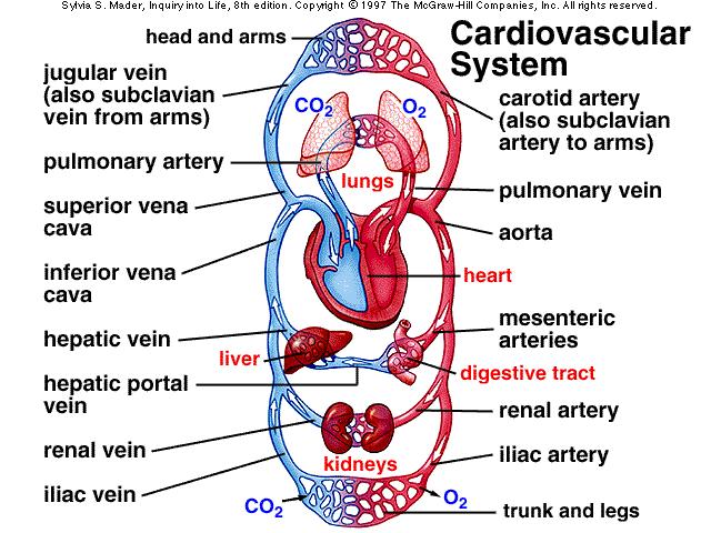
5 Cardiovascular System 5

6 Normal 02 Saturations SVC 65%-72% AO 95-98% PA 75% LA 95-98% PCW 99% RA 65%-75% LV 95-98% IVC 68%-75% RV 65%-75% 6
7 Normal Cardiac Pressures AO 120/80/93 PA 15-30/8-12/14 PCWP 4-12 LA 2-12 RA 2-6 RV 15-30/0/2-6 LV 120/0/4-12 7
8 8
9 Components of Hemodynamic Pressure Monitoring Invasive catheter High pressure tubing Manifold system Transducer Flush system Monitor 9

10 Wheatstone Bridge Circuit Copyright Lawrence Educational Media Consists of 4 resistors arranged in a diamond orientation. An input DC voltage, or excitation voltage, is applied between the top and bottom of the diamond and the output voltage is measured across the middle. When the output voltage is zero, the bridge is said to be balanced. 10
11 LEVEL TRANSDUCER SYSTEM Angles Re-level the transducer with any change in the patient s position Referencing the system 1 cm above the left atrium decreases the pressure by 0.73 mm Hg (1 ~2mmHg) 0 AACN PAP Measurement Practice Alert Referencing the system 1 cm below the left atrium increases the pressure by 0.73 mm Hg (1 ~2mmHg) 11

12 Top of transducer that connects to the line to the patient This brand is 4.5 cms distance Open Air reference point that is to be at the phlebostatic axis. Turn stopcock up, tell monitor to zero, and when zero, turn it back to this position. Transducer meat that includes the Wheatstone bridge This is generally the direction we use the transducer because of the way the cable hangs, but it can be used in either orientation Flush exhaust port, always turned off except to flush 12

13 UNDER DAMPED SYSTEM 13

14 Over Damped System AO Pressure Damped AO Pressure 14


15 Respiration effect PA RA Quiet Passive expiration Active Inspiration 15
16 16
17 17
18 Stroke Volume Stroke volume is the volume of blood ejected from the ventricle with each beat and is influenced by three factors: The amount of blood in the ventricles at the end of diastole just prior to systole (Preload) The amount of pressure that the ventricle has to pump against (Afterload) The degree of myocardial contractility 18
19 Stroke Volume Preload-PCWP/LVEDP (4-12 mmhg) and CVP/RVEDP (2-6 mmhg) Stretch, volume, pressure Starlings law Afterload- SVR and PVR Contractility-Dp/Dt, EF% Definition and effects of each on SV & CO 19


20 Vascular Resistance SVR = AOmean RAmean CO X 80 PVR = PAmean PWmean CO Normal SVR: 900 and 1440 dynes/sec/cm -5 Normal PVR: X dynes/sec/cm -5
21 21
22 Cardiac Output Cardiac output = heart rate X stroke volume CO = 70 beats/min X 65 mls CO = 4550 mls or 4.55 liters/min Normal cardiac output ranges from 4-8 liters per minute. Variances in CO are caused by either changes in HR or SV 22

23 Cardiac Index (CI) CI = CO/BSA CI = 4.55/2.0 CI = Liters/min/M 2 23
24 Normal Cardiac Pressures RA mean=2-6 RV=15-30/0/2-6 AO 120/80/93 PA 15-30/8-12/14 PCWP 4-12 PA=15-30/10-15 LA 2-12 LA mean=2-12 LV= /0/4-12 AO= /60-80 RA 2-6 RV 15-30/0/2-6 LV 120/0/
25 3 Types of Waveforms Atrial CVP, RA, LA, PCWP Sine wave Ventricular RV, LV Rectangle up on end Arterial AO,BA, RA, FA Triangle 25

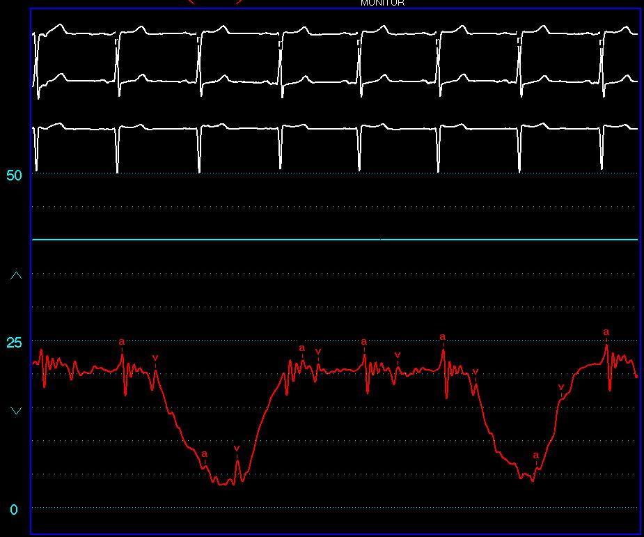
26 Atrial Waveforms 26
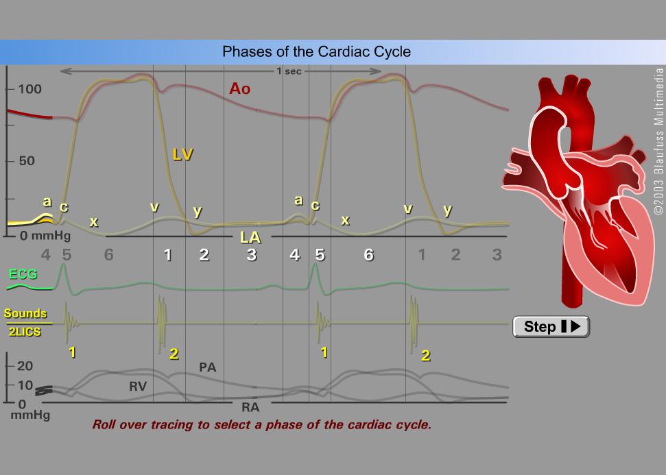
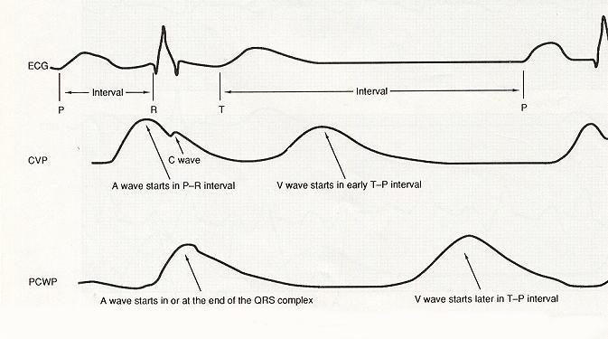
27 RA and LA Hemodynamics The atrial waveform portions: a wave x descent c wave v wave y descent 27
28 Atrial Waveforms a wave atrial contraction--pr interval active vent filling (30%) x decent atrial relaxation c wave AV valve closure--after QRS v wave atrial filling--t wave y decent Atrial empting--passive ventricular filling (70%) 28

29 Atrial - A V,mean 29
30 30

31 PCWP Hemodynamics The PCWP or PAW is a reflection of the LA hemodynamics 31
32 PCWP 32
33 PCW Analysis 33
34 CVP/RA and PCWP/LA Increased A wave Stenotic AV valves Non compliant stiff ventricle Inc. resistance to ventricular filling Constrictive pericarditis Hypervolemia No A wave Atrial fibrillation 34
35 Increased V wave Regurgitant AV valves VSD with L-R shunt Constrictive pericarditis Cardiac tamponade Hypervolemia CVP/RA and PCWP/LA 35
36 Ventricular Waveforms Systole Isovolumetric contraction Rapid ejection Slow ejection Diastole Isovolumetric relaxation Passive/rapid filling Atrial kick End diastolic pressure (R wave of EKG) 36

37 Ventricular Waveforms (S/D/EDP) 37
38 RV LV 38

39 Systole After QRS Arterial Waveforms Dicrotic notch Semi-lunar valve closure Diastole Runoff to the periphery Invasive vs: Non-Invasive monitoring 39
40 AO PA 40

41 Alterations in Arterial Pressure Elevated Pressure Arteriosclerosis Renal Failure Aortic regurgitation Hypervolemia Reduced Pressure Low cardiac output Aortic stenosis Dysrhythmias Vasodilator therapy Decreased LV function Mitral stenosis Cardiac tamponade Dilated cardiomyopathy Abnormal Pressure Reduction Vascular stenosis 41


42 Venous Return Skeletal muscle pump-muscles press against thin-walled veins
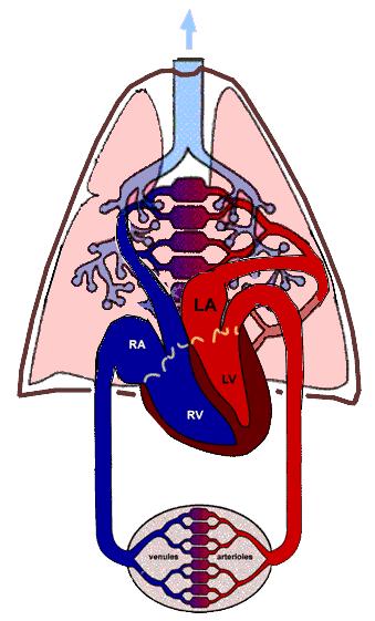
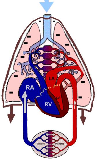
43 Circulation Schematic Pulmonary Vein Left Side of Heart Aorta A V Lungs Mitral Valve Pulmonary Valve Aortic Valve Tricuspid Valve Tissues V A Pulmonary Artery Right Side of Heart Sup. & Inf. Vena Cava 43
44 Alterations in CVP Elevations in CVP Increased blood volume Right ventricular failure Tricuspid or pulmonic value disease Pulmonary hypertension/copd Cardiac tamponade Positive pressure ventilation Vasoactive drugs venoconstriction Chronic left heart failure 44
45 Alterations in CVP Reductions in CVP Decreased blood volume Damped waveforms Beta adrenergic stimulation (causing venodilatation) 45
46 Pulmonary Artery Pressure Monitoring Normal 15-30/10-15, mean mmhg Increased in anything that increases PVR, pulmonary volume or LV failure LV MI, aortic/mitral valve diz, HTN, tamponade, hypervolemia Increased PVR/lung disease (COPD) Increased pulmonary blood flow, shunt Decreased in decreased right heart cardiac output, hypovolemia, damped waveform 46
47 PCWP Monitoring Normally PCWP=PAd=LA=LVEDP (preload) Does not equilibrate with Mitral stenosis, stiff LV, cor-triatrium, PE, Left atrial myxoma, pulmonary venous obstruction, catheter in nondependent zone of the lung 47
48 PCWP Monitoring Normal PCWP 4-12 mmhg Increased in anything that causes LV failure LV MI, aortic/mitral valve diz, HTN, cardiac tamponade, hypervolemia NOT with increased PVR or lung disease Decreased in hypovolemia, damped waveform 48
49 PA/PCWP Monitoring Lung disease- increased PA, normal PCWP Left heart disease- increased PA and PCWP 49


50 Identify Catheter Location on Pullback LV-AO PAW-PA AO knob PA LA PA-RV RA LV RV-RA After sach.com Know the normal pullback sequence through cardiac chambers 50
51 Identify Normal and Abnormal Pressures Right Heart Pressures in mm Hg Left Heart Pressure in mm Hg PAW = 12,12,10 (a,v,m) LA = 12,12,10 (a,v,m) PA = 25/10, 15 (S/D, m) AO = 130/80, 100 (s/d, m) RV = 25/0, 4 (s/bd, ed) LV = 130/0, 8 (s/bd, ed) RA = 5,5,4 (a,v,m) These are the only 4 numbers you need to remember: 4mmHg, 10mmHg, 25mmHg, & 130 mmhg 51
52 52
53 LV to AO Pullback 53

54 The End For Now But there is much more to come!!!!!! 54
55 Hemodynamics: Cardiac and Vascular Jeff Davis, RRT, RCIS Program Director, Cardiovascular Technology Florida SouthWestern State College Fort Myers, FL
CATCH A WAVE.. INTRODUCTION NONINVASIVE HEMODYNAMIC MONITORING 4/12/2018
 WAVES CATCH A WAVE.. W I S C O N S I N P A R A M E D I C S E M I N A R A P R I L 2 0 1 8 K E R I W Y D N E R K R A U S E R N, C C R N, E M T - P Have you considered that if you don't make waves, nobody
Cath Lab Essentials: Basic Hemodynamics for the Cath Lab and ICU
 Cath Lab Essentials: Basic Hemodynamics for the Cath Lab and ICU Ailin Barseghian El-Farra, MD, FACC Assistant Professor, Interventional Cardiology University of California, Irvine Department of Cardiology
Calculations the Cardiac Cath Lab. Thank You to: Lynn Jones RN, RCIS, FSICP Jeff Davis RCIS, FSICP Wes Todd, RCIS CardioVillage.
 Calculations the Cardiac Cath Lab Thank You to: Lynn Jones RN, RCIS, FSICP Jeff Davis RCIS, FSICP Wes Todd, RCIS CardioVillage.com Disclosure Information Calculations the Cardiac Cath Lab Darren Powell,
Topics to be Covered. Cardiac Measurements. Distribution of Blood Volume. Distribution of Pulmonary Ventilation & Blood Flow
 Topics to be Covered MODULE F HEMODYNAMIC MONITORING Cardiac Output Determinants of Stroke Volume Hemodynamic Measurements Pulmonary Artery Catheterization Control of Blood Pressure Heart Failure Cardiac
Principles of Biomedical Systems & Devices. Lecture 8: Cardiovascular Dynamics Dr. Maria Tahamont
 Principles of Biomedical Systems & Devices Lecture 8: Cardiovascular Dynamics Dr. Maria Tahamont Review of Cardiac Anatomy Four chambers Two atria-receive blood from the vena cave and pulmonary veins Two
Relax and Learn At the Farm 2012
 Relax and Learn At the Farm Session 9: Invasive Hemodynamic Assessment and What to Do with the Data Carol Jacobson RN, MN Cardiovascular Nursing Education Associates Function of CV system is to deliver
Cardiovascular Physiology. Heart Physiology. Introduction. The heart. Electrophysiology of the heart
 Cardiovascular Physiology Heart Physiology Introduction The cardiovascular system consists of the heart and two vascular systems, the systemic and pulmonary circulations. The heart pumps blood through
Cardiac Cycle MCQ. Professor of Cardiovascular Physiology. Cairo University 2007
 Cardiac Cycle MCQ Abdel Moniem Ibrahim Ahmed, MD Professor of Cardiovascular Physiology Cairo University 2007 1- Regarding the length of systole and diastole: a- At heart rate 75 b/min, the duration of
Hemodynamic Monitoring
 Perform Procedure And Interpret Results Hemodynamic Monitoring Tracheal Tube Cuff Pressure Dean R. Hess PhD RRT FAARC Hemodynamic Monitoring Cardiac Rate and Rhythm Arterial Blood Pressure Central Venous
FUNDAMENTALS OF HEMODYNAMICS, VASOACTIVE DRUGS AND IABP IN THE FAILING HEART
 FUNDAMENTALS OF HEMODYNAMICS, VASOACTIVE DRUGS AND IABP IN THE FAILING HEART CINDY BITHER, MSN, ANP, ANP, AACC, CHFN CHIEF NP, ADV HF PROGRAM MEDSTAR WASHINGTON HOSPITAL CENTER CONFLICTS OF INTEREST NONE
(D) (E) (F) 6. The extrasystolic beat would produce (A) increased pulse pressure because contractility. is increased. increased
 (E) (F) 6. The extrasystolic beat would produce (A) increased pulse pressure because contractility. is increased. increased") Review Test 1. A 53-year-old woman is found, by arteriography, to have 5% narrowing of her left renal artery. What is the expected change in blood flow through the stenotic artery? Decrease to 1 2 Decrease
Review Test 1. A 53-year-old woman is found, by arteriography, to have 5% narrowing of her left renal artery. What is the expected change in blood flow through the stenotic artery? Decrease to 1 2 Decrease
Heart Pump and Cardiac Cycle. Faisal I. Mohammed, MD, PhD
 Heart Pump and Cardiac Cycle Faisal I. Mohammed, MD, PhD 1 Objectives To understand the volume, mechanical, pressure and electrical changes during the cardiac cycle To understand the inter-relationship
Chapter 9, Part 2. Cardiocirculatory Adjustments to Exercise
 Chapter 9, Part 2 Cardiocirculatory Adjustments to Exercise Electrical Activity of the Heart Contraction of the heart depends on electrical stimulation of the myocardium Impulse is initiated in the right
The Cardiac Cycle Clive M. Baumgarten, Ph.D.
 The Cardiac Cycle Clive M. Baumgarten, Ph.D. OBJECTIVES: 1. Describe periods comprising cardiac cycle and events within each period 2. Describe the temporal relationships between pressure, blood flow,
Right Heart Catheterization. Franz R. Eberli MD Chief of Cardiology Stadtspital Triemli, Zurich
 Right Heart Catheterization Franz R. Eberli MD Chief of Cardiology Stadtspital Triemli, Zurich Right Heart Catheterization Pressure measurements Oxygen saturation measurements Cardiac output, Vascular
THE CARDIOVASCULAR SYSTEM. Heart 2
 THE CARDIOVASCULAR SYSTEM Heart 2 PROPERTIES OF CARDIAC MUSCLE Cardiac muscle Striated Short Wide Branched Interconnected Skeletal muscle Striated Long Narrow Cylindrical PROPERTIES OF CARDIAC MUSCLE Intercalated
Georgios C. Bompotis Cardiologist, Director of Cardiological Department, Papageorgiou Hospital,
 Georgios C. Bompotis Cardiologist, Director of Cardiological Department, Papageorgiou Hospital, Disclosure Statement of Financial Interest I, Georgios Bompotis DO NOT have a financial interest/arrangement
BUSINESS. Articles? Grades Midterm Review session
 BUSINESS Articles? Grades Midterm Review session REVIEW Cardiac cells Myogenic cells Properties of contractile cells CONDUCTION SYSTEM OF THE HEART Conduction pathway SA node (pacemaker) atrial depolarization
Ejection across stenotic aortic valve requires a systolic pressure gradient between the LV and aorta. This places a pressure load on the LV.
 Valvular Heart Disease Etiology General Principles Cellular and molecular mechanism of valve damage Structural pathology Functional pathology - stenosis/regurgitation Loading conditions - pressure/volume
Outline. Echocardiographic Assessment of Pericardial Effusion/Tamponade: The Essentials
 Echocardiographic Assessment of Pericardial Effusion/Tamponade: The Essentials John R Schairer DO FACC Henry Ford Heart and Vascular Institute No Disclosures Outline Normal Anatomy and Physiology Pathophysiology
Circulation: Chapter 25. Cardiac Output. The Mammalian Heart Fig Right side of the heart
 Circulation: Chapter 25 1. Limits of Diffusion A. Small organisms use diffusion B. rapid over small distances 2. Most animals have circulatory systems A. Blood B. Pump (Heart) or propulsive structures
Hemodynamic Monitoring and Circulatory Assist Devices
 Hemodynamic Monitoring and Circulatory Assist Devices Speaker: Jana Ogden Learning Unit 2: Hemodynamic Monitoring and Circulatory Assist Devices Hemodynamic monitoring refers to the measurement of pressure,
Chapter 18 - Heart. I. Heart Anatomy: size of your fist; located in mediastinum (medial cavity)
") Chapter 18 - Heart I. Heart Anatomy: size of your fist; located in mediastinum (medial cavity) A. Coverings: heart enclosed in double walled sac called the pericardium 1. Fibrous pericardium: dense connective
Chapter 18 - Heart I. Heart Anatomy: size of your fist; located in mediastinum (medial cavity) A. Coverings: heart enclosed in double walled sac called the pericardium 1. Fibrous pericardium: dense connective
CARDIAC CYCLE CONTENTS. Divisions of cardiac cycle 11/13/13. Definition. Badri Paudel GMC
 CARDIAC CYCLE Badri Paudel GMC CONTENTS Ø DEFINATION Ø DIVISION OF CARDIAC CYCLE Ø SUB DIVISION AND DURATION OF CARDIAC CYCLE Ø SYSTOLE Ø DIASTOLE Ø DESCRIPTION OF EVENTS OF CARDIAC CYCLE Ø SUMMARY Ø ELECTROCARDIOGRAPHY
SIKLUS JANTUNG. Rahmatina B. Herman
 SIKLUS JANTUNG Rahmatina B. Herman The Cardiac Cycle Definition: The cardiac events that occur from the beginning of one heartbeat to the beginning of the next The cardiac cycle consists of: - Diastole
Ejection across stenotic aortic valve requires a systolic pressure gradient between the LV and aorta. This places a pressure load on the LV.
 Valvular Heart Disease General Principles Etiology Cellular and molecular mechanism of valve damage Structural pathology Functional pathology - stenosis/regurgitation Loading conditions - pressure/volume
The circulatory system
 Introduction to Physiology (Course # 72336) 1 הלב עקרונות בסיסיים (הכנה למעבדת לב) Adi Mizrahi mizrahia@cc.huji.ac.il Textbook Chapter 12 2 The circulatory system To the heart Away from the heart 3 L 2.5
The Mammalian Circulatory System
 The Mammalian Heart The Mammalian Circulatory System Recall: What are the 3 cycles of the mammalian circulatory system? What are their functions? What are the three main vessel types in the mammalian circulatory
Introduction to Physiology (Course # 72336) 1. Adi Mizrahi Textbook Chapter 12
 1. Adi Mizrahi Textbook Chapter 12") Introduction to Physiology (Course # 72336) 1 עקרונות בסיסיים (הכנה למעבדת לב) הלב Adi Mizrahi mizrahia@cc.huji.ac.il Textbook Chapter 12 2 The circulatory system To the heart Away from the heart 3 L 2.5
Introduction to Physiology (Course # 72336) 1 עקרונות בסיסיים (הכנה למעבדת לב) הלב Adi Mizrahi mizrahia@cc.huji.ac.il Textbook Chapter 12 2 The circulatory system To the heart Away from the heart 3 L 2.5
Technique. Technique. Technique. Monitoring 1. Local anesthetic? Aseptic technique Hyper-extend (if radial)
") Critical Care Monitoring Hemodynamic Monitoring Arterial Blood Pressure Cannulate artery Uses 2 Technique Sites Locate artery, prep 3 1 Technique Local anesthetic? Aseptic technique Hyper-extend (if radial)
Critical Care Monitoring Hemodynamic Monitoring Arterial Blood Pressure Cannulate artery Uses 2 Technique Sites Locate artery, prep 3 1 Technique Local anesthetic? Aseptic technique Hyper-extend (if radial)
Καθετηριασμός δεξιάς κοιλίας. Σ. Χατζημιλτιάδης Καθηγητής Καρδιολογίας ΑΠΘ
 Καθετηριασμός δεξιάς κοιλίας Σ. Χατζημιλτιάδης Καθηγητής Καρδιολογίας ΑΠΘ The increasing interest in pulmonary arterial hypertension (PAH), the increasing interest in implantation of LVADs, and the evolution
Examen MMD Which of the following causes increased inotropy and chronotropy? A Ca gluconate B Glucagon C Atropine D Digoxin E Phenylephrine
 1-Which of the following causes an increase in HR in a denervated heart? A Pancuronium B Atropine C Propranolol D Norepinephrine E Neostigmine 2-Which of the following causes increased inotropy and chronotropy?
The Cardiovascular System
 The Cardiovascular System The Cardiovascular System A closed system of the heart and blood vessels The heart pumps blood Blood vessels allow blood to circulate to all parts of the body The function of
Basic Hemodynamics. July 19, 2006 Joe M. Moody, Jr, MD UTHSCSA and STVHCS
 Basic Hemodynamics July 19, 2006 Joe M. Moody, Jr, MD UTHSCSA and STVHCS The Cardiac Cycle Types of Measures Pressure Outline Flow (Calculated from Temperature, O 2 Saturation, Indicator Concentration,
Cardiac output and Venous Return. Faisal I. Mohammed, MD, PhD
 Cardiac output and Venous Return Faisal I. Mohammed, MD, PhD 1 Objectives Define cardiac output and venous return Describe the methods of measurement of CO Outline the factors that regulate cardiac output
IABP Timing & Fidelity. Pocket Reference Guide
 IABP Timing & Fidelity Pocket Reference Guide Correct IABP Timing A = One complete cardiac cycle R B = Unassisted aortic end diastolic pressure P T C = Unassisted systolic pressure D = Diastolic augmentation
Brief View of Calculation and Measurement of Cardiac Hemodynamics
 Cronicon OPEN ACCESS EC CARDIOLOGY Review Article Brief View of Calculation and Measurement of Cardiac Hemodynamics Samah Alasrawi* Pediatric Cardiologist, Al Jalila Children Heart Center, Dubai, UAE *
Cardiovascular Physiology
 Cardiovascular Physiology Lecture 1 objectives Explain the basic anatomy of the heart and its arrangement into 4 chambers. Appreciate that blood flows in series through the systemic and pulmonary circulations.
Cardiovascular System
 Cardiovascular System The Heart Cardiovascular System The Heart Overview What does the heart do? By timed muscular contractions creates pressure gradients blood moves then from high pressure to low pressure
Appendix II: ECHOCARDIOGRAPHY ANALYSIS
 Appendix II: ECHOCARDIOGRAPHY ANALYSIS Two-Dimensional (2D) imaging was performed using the Vivid 7 Advantage cardiovascular ultrasound system (GE Medical Systems, Milwaukee) with a frame rate of 400 frames
Heart. Structure Physiology of blood pressure and heartbeat
 Heart Structure Physiology of blood pressure and heartbeat Location and Anatomy Location and Anatomy Pericardial cavity: surrounds, isolates, and anchors heart Parietal pericardium lined with serous membrane
Lecture 13: The Cardiovascular System ref: Cardiovascular Physiology, D. Mohrman and L. Heller, 4th ed. McGraw-Hill (1997)
") Lecture 13: The Cardiovascular System ref: Cardiovascular Physiology, D. Mohrman and L. Heller, 4th ed. McGraw-Hill (1997) Blood Heart Blood Vessels Arteries - capillaries - Veins Ventilation-Perfusion
Lecture 13: The Cardiovascular System ref: Cardiovascular Physiology, D. Mohrman and L. Heller, 4th ed. McGraw-Hill (1997) Blood Heart Blood Vessels Arteries - capillaries - Veins Ventilation-Perfusion
DOPPLER HEMODYNAMICS (1) QUANTIFICATION OF PRESSURE GRADIENTS and INTRACARDIAC PRESSURES
 QUANTIFICATION OF PRESSURE GRADIENTS and INTRACARDIAC PRESSURES") THORAXCENTRE DOPPLER HEMODYNAMICS (1) QUANTIFICATION OF PRESSURE GRADIENTS and INTRACARDIAC PRESSURES J. Roelandt DOPPLER HEMODYNAMICS Intracardiac pressures and pressure gradients Volumetric measurement
THORAXCENTRE DOPPLER HEMODYNAMICS (1) QUANTIFICATION OF PRESSURE GRADIENTS and INTRACARDIAC PRESSURES J. Roelandt DOPPLER HEMODYNAMICS Intracardiac pressures and pressure gradients Volumetric measurement
LV geometric and functional changes in VHD: How to assess? Mi-Seung Shin M.D., Ph.D. Gachon University Gil Hospital
 LV geometric and functional changes in VHD: How to assess? Mi-Seung Shin M.D., Ph.D. Gachon University Gil Hospital LV inflow across MV LV LV outflow across AV LV LV geometric changes Pressure overload
HISTORY. Question: What category of heart disease is suggested by this history? CHIEF COMPLAINT: Heart murmur present since early infancy.
 HISTORY 18-year-old man. CHIEF COMPLAINT: Heart murmur present since early infancy. PRESENT ILLNESS: Although normal at birth, a heart murmur was heard at the six week check-up and has persisted since
For more information about how to cite these materials visit
 Author(s): Louis D Alecy, D.M.D., Ph.D., 2009 License: Unless otherwise noted, this material is made available under the terms of the Creative Commons Attribution Non-commercial Share Alike 3.0 License:
SECTION I FUNDAMENTALS AND CLINICAL APPLICATIONS OF HEMODYNAMICS: COPYRIGHTED MATERIAL
 SECTION I FUNDAMENTALS AND CLINICAL APPLICATIONS OF HEMODYNAMICS: COPYRIGHTED MATERIAL UNDERSTANDING THE PRESSURE WAVES IN THE HEART: THE WIGGER S DIAGRAM Everything you want to know about hemodynamics
THE CARDIOVASCULAR SYSTEM
 THE CARDIOVASCULAR SYSTEM AND RESPONSES TO EXERCISE Mr. S. Kelly PSK 4U North Grenville DHS THE HEART: A REVIEW Cardiac muscle = myocardium Heart divided into two sides, 4 chambers (L & R) RS: pulmonary
SymBioSys Exercise 2 Cardiac Function Revised and reformatted by C. S. Tritt, Ph.D. Last updated March 20, 2006
 SymBioSys Exercise 2 Cardiac Function Revised and reformatted by C. S. Tritt, Ph.D. Last updated March 20, 2006 The goal of this exercise to explore the behavior of the heart as a mechanical pump. For
Cardiovascular Physiology
 Cardiovascular Physiology Introduction The cardiovascular system consists of the heart and two vascular systems, the systemic and pulmonary circulations. The heart pumps blood through two vascular systems
*Generating blood pressure *Routing blood: separates. *Ensuring one-way blood. *Regulating blood supply *Changes in contraction
 *Generating blood pressure *Routing blood: separates pulmonary and systemic circulations *Ensuring one-way blood flow: valves *Regulating blood supply *Changes in contraction rate and force match blood
Collin County Community College
 Collin County Community College BIOL. 2402 Anatomy & Physiology WEEK 5 The Heart 1 The Heart Beat and the EKG 2 1 The Heart Beat and the EKG P-wave = Atrial depolarization QRS-wave = Ventricular depolarization
IP: Regulation of Cardiac Output
 ANP 1105D Winter 2013 Assignment 9: The Heart, part 2: Chap... Assignment 9: The Heart, part 2: Chapter 18 Signed in as Alex Sokolowski Help Close Resources Due: 11:59pm on Monday, March 25, 2013 Note:
BIOL 219 Spring Chapters 14&15 Cardiovascular System
 1 BIOL 219 Spring 2013 Chapters 14&15 Cardiovascular System Outline: Components of the CV system Heart anatomy Layers of the heart wall Pericardium Heart chambers, valves, blood vessels, septum Atrioventricular
Adult Echocardiography Examination Content Outline
 Adult Echocardiography Examination Content Outline (Outline Summary) # Domain Subdomain Percentage 1 2 3 4 5 Anatomy and Physiology Pathology Clinical Care and Safety Measurement Techniques, Maneuvers,
Cardiology. the Sounds: #7 HCM. LV Outflow Obstruction: Aortic Stenosis. (Coming Soon - HCM)
") A Cardiology HCM LV Outflow Obstruction: Aortic Stenosis (Coming Soon - HCM) the Sounds: #7 Howard J. Sachs, MD www.12daysinmarch.com E-mail: Howard@12daysinmarch.com Aortic Valve Disorders Stenosis Regurgitation
A Cardiology HCM LV Outflow Obstruction: Aortic Stenosis (Coming Soon - HCM) the Sounds: #7 Howard J. Sachs, MD www.12daysinmarch.com E-mail: Howard@12daysinmarch.com Aortic Valve Disorders Stenosis Regurgitation
Disclosures. Objectives 6/16/2016. A Look at the Other Side: Focus on the Right Ventricle and Pulmonary Hypertension
 A Look at the Other Side: Focus on the Right Ventricle and Pulmonary Hypertension Susan P. D Anna MSN, APN-BC, CHFN June 24, 2016 Disclosures Objectives Differentiate structure and function of RV and LV
Coronary Anomalies & Hemodynamic Identification
 Coronary Anomalies & Hemodynamic Identification David Stultz, MD Cardiology Fellow, PGY 6 May 2, 2006 Anomaly #1 Anomaly #2 Anomaly #3 Figure 18-27 Anomalous origin of the left circumflex artery.
Approximately the size of your fist Location. Pericardial physiology
 Heart Anatomy Approximately the size of your fist Location Superior surface of diaphragm Left of the midline Anterior to the vertebral column, posterior to the sternum Wednesday, March 28, 2012 Muscle
11/10/2014. Muscular pump Two atria Two ventricles. In mediastinum of thoracic cavity 2/3 of heart's mass lies left of midline of sternum
 It beats over 100,000 times a day to pump over 1,800 gallons of blood per day through over 60,000 miles of blood vessels. During the average lifetime, the heart pumps nearly 3 billion times, delivering
Electrical Conduction
 Sinoatrial (SA) node Electrical Conduction Sets the pace of the heartbeat at 70 bpm AV node (50 bpm) and Purkinje fibers (25 40 bpm) can act as pacemakers under some conditions Internodal pathway from
PHYSIOLOGY MeQ'S (Morgan) All the following statements related to blood volume are correct except for: 5 A. Blood volume is about 5 litres. B.
 All the following statements related to blood volume are correct except for: 5 A. Blood volume is about 5 litres. B.") PHYSIOLOGY MeQ'S (Morgan) Chapter 5 All the following statements related to capillary Starling's forces are correct except for: 1 A. Hydrostatic pressure at arterial end is greater than at venous end.
PHYSIOLOGY MeQ'S (Morgan) Chapter 5 All the following statements related to capillary Starling's forces are correct except for: 1 A. Hydrostatic pressure at arterial end is greater than at venous end.
-12. -Ensherah Mokheemer - ABDULLAH ZREQAT. -Faisal Mohammad. 1 P a g e
 -12 -Ensherah Mokheemer - ABDULLAH ZREQAT -Faisal Mohammad 1 P a g e In the previous lecture we talked about: - cardiac index: we use the cardiac index to compare the cardiac output between different individuals,
Cardiovascular System
 Cardiovascular System Chapter 8 1 Cardiovascular System Functions: pump saturated oxygenated blood into arterial system cells pump desaturated deoxygenated blood to lungs via veins for reoxygenation Heart
McHenry Western Lake County EMS System Paramedic, EMT-B and PHRN Optional Continuing Education 2018 #12 Understanding Preload and Afterload
 McHenry Western Lake County EMS System Paramedic, EMT-B and PHRN Optional Continuing Education 2018 #12 Understanding Preload and Afterload Cardiac output (CO) represents the volume of blood that is delivered
See below for descriptions of the waveform
 The internal jugular vein (IJV) connects to the right atrium without any intervening valves. The pulsation of the right atrium therefore causes the column of blood in the IJV to rise and fall this is called
The Heart. Size, Form, and Location of the Heart. 1. Blunt, rounded point; most inferior part of the heart.
 12 The Heart FOCUS: The heart is composed of cardiac muscle cells, which are elongated, branching cells that appear striated. Cardiac muscle cells behave as a single electrical unit, and the highly coordinated
HAEMODYNAMIC IN THE CATH LABORATORY INTRO TO BASICS
 HAEMODYNAMIC IN THE CATH LABORATORY INTRO TO BASICS BY NOOR FADZLY ALIAS CARDIOVASCULAR TECHNOLOGIST NCL Department National Heart Institute Kuala Lumpur INTRODUCTION ROLES OF HAEMODYNAMIC MONITORING PURPOSE
CARDIOVASCULAR PHYSIOLOGY
 CARDIOVASCULAR PHYSIOLOGY LECTURE 4 Cardiac cycle Polygram - analysis of cardiac activity Ana-Maria Zagrean MD, PhD The Cardiac Cycle - definitions: the sequence of electrical and mechanical events that
Lab #3: Electrocardiogram (ECG / EKG)
") Lab #3: Electrocardiogram (ECG / EKG) An introduction to the recording and analysis of cardiac activity Introduction The beating of the heart is triggered by an electrical signal from the pacemaker. The
Lab #3: Electrocardiogram (ECG / EKG) An introduction to the recording and analysis of cardiac activity Introduction The beating of the heart is triggered by an electrical signal from the pacemaker. The
Chapter 13 The Cardiovascular System: Cardiac Function
 Chapter 13 The Cardiovascular System: Cardiac Function Overview of the Cardiovascular System The Path of Blood Flow through the Heart and Vasculature Anatomy of the Heart Electrical Activity of the Heart
The Doppler Examination. Katie Twomley, MD Wake Forest Baptist Health - Lexington
 The Doppler Examination Katie Twomley, MD Wake Forest Baptist Health - Lexington OUTLINE Principles/Physics Use in valvular assessment Aortic stenosis (continuity equation) Aortic regurgitation (pressure
Practice Exercises for the Cardiovascular System
 Practice Exercises for the Cardiovascular System On the diagram below, color the oxygen-rich blood red and the oxygen-poor blood blue. Label the parts: Continued on the next page... Label the parts on
Cardiovascular System- Heart. Miss Wheeler Unit 8
 Cardiovascular System- Heart Miss Wheeler Unit 8 Overview CARDIOVASCULAR SYSTEM heart vessels Made up of heart, blood vessels, and blood Functions Heart- pump blood Vessels- (veins, arteries, capillaries)
Heart Failure. Cardiac Anatomy. Functions of the Heart. Cardiac Cycle/Hemodynamics. Determinants of Cardiac Output. Cardiac Output
 Cardiac Anatomy Heart Failure Professor Qing ZHANG Department of Cardiology, West China Hospital www.blaufuss.org Cardiac Cycle/Hemodynamics Functions of the Heart Essential functions of the heart to cover
10. Thick deposits of lipids on the walls of blood vessels, called, can lead to serious circulatory issues. A. aneurysm B. atherosclerosis C.
 Heart Student: 1. carry blood away from the heart. A. Arteries B. Veins C. Capillaries 2. What is the leading cause of heart attack and stroke in North America? A. alcohol B. smoking C. arteriosclerosis
Arterial Line Waveform Interpretation
 Arterial Line Waveform Interpretation PICU CPM October 2014 1. Introduction The document is primarily aimed at education to provide some basic understanding of arterial line wave form 2. Scope The interpretation
Hemodynamic Assessment. Assessment of Systolic Function Doppler Hemodynamics
 Hemodynamic Assessment Matt M. Umland, RDCS, FASE Aurora Medical Group Milwaukee, WI Assessment of Systolic Function Doppler Hemodynamics Stroke Volume Cardiac Output Cardiac Index Tei Index/Index of myocardial
AS Level OCR Cardiovascular System
 AS Level OCR Cardiovascular System Learning Objectives The link between the Cardiac Cycle and the Conduction system of the heart. The relationship between Stroke volume, Heart rate and Cardiac Output.
BME 5742 Bio-Systems Modeling and Control. Lecture 41 Heart & Blood Circulation Heart Function Basics
 BME 5742 Bio-Systems Modeling and Control Lecture 41 Heart & Blood Circulation Heart Function Basics Dr. Zvi Roth (FAU) 1 Pumps A pump is a device that accepts fluid at a low pressure P 1 and outputs the
Impedance Cardiography (ICG) Method, Technology and Validity
 Method, Technology and Validity") Method, Technology and Validity Hemodynamic Basics Cardiovascular System Cardiac Output (CO) Mean arterial pressure (MAP) Variable resistance (SVR) Aortic valve Left ventricle Elastic arteries / Aorta
Method, Technology and Validity Hemodynamic Basics Cardiovascular System Cardiac Output (CO) Mean arterial pressure (MAP) Variable resistance (SVR) Aortic valve Left ventricle Elastic arteries / Aorta
CVS Hemodynamics. Faisal I. Mohammed, MD,PhD.
 CVS Hemodynamics Faisal I. Mohammed, MD,PhD. Objectives point out the physical characteristics of the circulation: distribution of blood volume total cross sectional area velocity blood pressure List the
CARDIOVASCULAR SYSTEM
 CARDIOVASCULAR SYSTEM Overview Heart and Vessels 2 Major Divisions Pulmonary Circuit Systemic Circuit Closed and Continuous Loop Location Aorta Superior vena cava Right lung Pulmonary trunk Base of heart
The Cardiovascular System
 Chapter 18 Part A The Cardiovascular System 1/19/16 1 Annie Leibovitz/Contact Press Images Similarities of Cardiac and Skeletal Muscle RMP Ion concentration Deploarization Action Potential Repolarization
P = 4V 2. IVC Dimensions 10/20/2014. Comprehensive Hemodynamic Evaluation by Doppler Echocardiography. The Simplified Bernoulli Equation
 Comprehensive Hemodynamic Evaluation by Doppler Echocardiography Itzhak Kronzon, MD North Shore LIJ/ Lenox Hill Hospital New York, NY Disclosure: Philips Healthcare St. Jude Medical The Simplified Bernoulli
Echo Doppler Assessment of Right and Left Ventricular Hemodynamics.
 Echo Doppler Assessment of Right and Left Ventricular Hemodynamics. Itzhak Kronzon, MD, FASE, FACC, FESC, FAHA, FACP, FCCP Northwell, Lenox Hill Hospital, New York Professor of Cardiology Hofstra University
Cardiovascular Nursing Practice: A Comprehensive Resource Manual and Study Guide for Clinical Nurses 2 nd Edition
 Cardiovascular Nursing Practice: A Comprehensive Resource Manual and Study Guide for Clinical Nurses 2 nd Edition Table of Contents Volume 1 Chapter 1: Cardiovascular Anatomy and Physiology Basic Cardiac
Hemodynamic Monitoring in the CCU. Edward G. Hamaty Jr., D.O. FACCP, FACOI
 Hemodynamic Monitoring in the CCU Edward G. Hamaty Jr., D.O. FACCP, FACOI Waveform Review Left Ventricular Pressure Normal left ventricular pressures are: Systolic 100 to 140 mm of mercury End-diastolic
GIGA - In Silico Medicine, University of Liege, Belgium, 2
 S. Kosta 1,*, A. Pironet 1, J.A. Negroni 2, E.C. Lascano 2, P.C. Dauby 1 1 GIGA - In Silico Medicine, University of Liege, Belgium, 2 Department of Comparative Cellular and Molecular Biology, Favaloro
The cardiovascular system is composed of a pump the heart and blood
 5 E X E R C I S E Cardiovascular Dynamics O B J E C T I V E S 1. To understand the relationships among blood flow, pressure gradient, and resistance 2. To define resistance and describe the main factors
Review of Cardiac Mechanics & Pharmacology 10/23/2016. Brent Dunworth, CRNA, MSN, MBA 1. Learning Objectives
 Brent Dunworth, CRNA, MSN, MBA Associate Director of Advanced Practice Division Chief, Nurse Anesthesia Vanderbilt University Medical Center Nashville, Tennessee Learning Objectives Review the principles
The Cardiovascular System
 The Cardiovascular System The Manila Times College of Subic Prepared by: Stevens B. Badar, RN, MANc THE HEART Anatomy of the Heart Location and Size approx. the size of a person s fist, hollow and cone-shaped,
Evalua&on)of)Le-)Ventricular)Diastolic) Dysfunc&on)by)Echocardiography:) Role)of)Ejec&on)Frac&on)
of)Le-)Ventricular)Diastolic) Dysfunc&on)by)Echocardiography:) Role)of)Ejec&on)Frac&on)") Evalua&on)of)Le-)Ventricular)Diastolic) Dysfunc&on)by)Echocardiography:) Role)of)Ejec&on)Frac&on) N.Koutsogiannis) Department)of)Cardiology) University)Hospital)of)Patras)! I have no conflicts of interest
Evalua&on)of)Le-)Ventricular)Diastolic) Dysfunc&on)by)Echocardiography:) Role)of)Ejec&on)Frac&on) N.Koutsogiannis) Department)of)Cardiology) University)Hospital)of)Patras)! I have no conflicts of interest
Outline. Electrical Activity of the Human Heart. What is the Heart? The Heart as a Pump. Anatomy of the Heart. The Hard Work
 Electrical Activity of the Human Heart Oguz Poroy, PhD Assistant Professor Department of Biomedical Engineering The University of Iowa Outline Basic Facts about the Heart Heart Chambers and Heart s The
IB TOPIC 6.2 THE BLOOD SYSTEM
 IB TOPIC 6.2 THE BLOOD SYSTEM THE BLOOD SYSTEM TERMS TO KNOW circulation ventricle artery vein 6.2.U1 - Arteries convey blood at high pressure from the ventricles to the tissues of the body Circulation
CARDIOVASCULAR MONITORING. Prof. Yasser Mostafa Kadah
 CARDIOVASCULAR MONITORING Prof. Yasser Mostafa Kadah Introduction Cardiovascular monitoring covers monitoring of heart and circulatory functions It makes it possible to commence interventions quickly in
Cardiovascular System Notes: Physiology of the Heart
 Cardiovascular System Notes: Physiology of the Heart Interesting Heart Fact Capillaries are so small it takes ten of them to equal the thickness of a human hair. Review What are the 3 parts of the cardiovascular