Point of Care Ultrasound in the ICU
|
|
|
- Charles Hubbard
- 6 years ago
- Views:
Transcription
1 Point of Care Ultrasound in the ICU JENNIFER P. KANAAN, M.D. ASSISTANT PROFESSOR OF MEDICINE UNIVERSITY OF CONNECTICUT I have no disclosures 1
2 Ultrasound Ultrasound imaging is among the fastest, safest and most universal diagnostic methods ever invented. It provides much of the information that can be obtained by expensive technologies, such as X-ray, computed tomography, or magnetic resonance imagery, and it is the only method to produce a real-time or live image that can be interpreted and/or transmitted at the same time. In the right hands, ultrasound instantly answers many clinical questions, shortening the assessment time and improving outcome. NASA website 2015 Ultrasound Observation of bat sonar Beginning in WWI to detect ships sunk by German subs Developed in the 1950s 2


3 Life Magazine 9/20/54 Early Ultrasound 1950 s by radiologists 1960 s by cardiologists 1970 s by OB/GYN 1980 s ultrasound use transitioned to other fields and for use in battlefields, EMS and military 3


4 4


5 Bovine Ultrasound Specialist Critical Care Ultrasound Development of small and portable machines Decreased costs Improved resolution of images Aka POCUS, WBU, FOCUS 5
6 Why CCUS Portable Lack of Radiation Ease of Use Rapid Results Repeatable exam Inexpensive Brings the physician back to the bedside CCUS Elements Vascular Thoracic Abdominal-pelvic Cardiac- basic and Advanced 6
7 Vascular Ultrasound Catheter placement TLC A-lines Deep Venous Thrombosis Peripheral Vein Access Ultrasound Use in Vascular Access Systemic review and meta analysis of RCT 18 Trials (1646 participants) Ultrasound resulted in a significantly lower failure rate cannulating the internal jugular- relative risk of 0.14 Lower failure rate on the first attempt Fewer complications with placement Hind, D et al BMJ 2003; 327 7
8 Ultrasound Use for Vascular Access Meta-Analysis comparing ultrasound guidance to landmark techniques Decreases need for multiple attempts by 40% Decreases complications by 78% Decreases placement failure by 64% Randolph et al Crit Care Med 1996 Dec 24(12) Agency for Healthcare Research and Quality listed real time ultrasound guidance as 1 of the 12 most highly rated patient safety practices designed to decrease medical errors Ultrasound Use for Vascular Access Ultrasound improved vascular access even in seasoned providers Decrease in infectious complications May be related to decrease number of attempts More attempts may also lead to break down in aseptic technique Kirakitsos et al Critical Care 2006; 10(6); 1-8 8


9 Peripheral Venous Access Rule Veins should be 3cm straight Veins should be <1 cm deep Veins should be at least 3 mm in diameter Medscape 03/09/2015 Abdominopelvic Ultrasound Possible source of sepsis Acute undifferentiated abdominal pain Abdominal aortic anerysm Detection of urinary tract obstruction Hydronephorsis Bladder Distension Ascites 9
10 Thoracic Ultrasound Presence or absence of pneumothorax Post procedure Pleural effusion Presence Thoracentesis Normal aeration versus alveolar/interstitial abnormality Evaluation of respiratory failure Advantages Study of 404 patients presenting with dyspnea to the ED Ultrasound completed within minutes CXR and interpretation in 1 hour 35 minutes Better accuracy with ultrasound for pleural effusions Zanobetti, M CHEST 2011;139; Observational study in the critical care unit of 301 consecutive patients 90.5% accuracy when compared to standard methods including history, physical, CXR and CT when indicated Test performed within 20 min of presentation to the ICU and lasting <3min Lichtenstein, D CHEST 2008; 134;

11 Advantages Lung Ultrasound Consolidation 95% diagnostic accuracy Interstitial syndrome 94% diagnostic accuracy Pneumothorax 92% diagnostic accuracy Pleural Effusion 100% diagnostic accuracy Chest Xray Consolidation 49% diagnostic accuracy Interstitial syndrome 58% diagnostic accuracy Pneumothorax 89% diagnostic accuracy Pleural Effusion 69% diagnostic accuracy Xirouchaki, N et. Al. Intensive Care Med 2011; 37: 1488 Normal Lung 11

12 Pneumothorax Pleural Effusion 12

13 Complex Pleural Effusion B lines 3 or more indicate an alveolar interstitial process 13


14 Etiology of B lines Smooth pleural line and profuse B lines= CHF Irregular pleural line and focal B lines= Pulmonary process such as ARDS Copetti et al Cardiovasc Ultrasound 2008 Apr 29:6:16 14

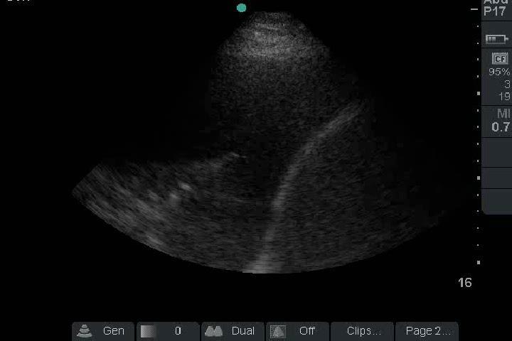
15 Advantages of Thoracic Ultrasound Reduction in number of images during a hospital stay Reduced time to diagnosis Improved identification of pleural effusions and pneumothorax compared to chest xray Atelectatic Lung with Small Effusion 15


16 Pneumonia Watch the next four slides in succession- Recruitment Maneuver 16

17 17
18 Critical Care Echocardiography Distinct from complete cardiac echo Used to narrow the DDx Confirm a diagnosis Prompt changes in management Follow response to therapy 18
19 Critical Care Echo 5 Views Parasternal long Parasternal short Apical 4 chamber Subxiphoid view IVC view Complete Echo Table 1 Recommended images for complete adult 2D transthoracic echocardiography with Doppler* Parasternal long axis 2D image M mode of left ventricle and left atrium/aorta (if lab standard) Color flow Doppler of valves RV inflow view Color and spectral Doppler Parasternal short axis Short-axis view at the aortic valve level and RVOT Color flow Doppler should be used to evaluate pulmonic, aortic and tricuspid valves Spectral Doppler of RVOT and pulmonic valve Left ventricle at MV level Left ventricle at mid level M mode if lab standard Left ventricle at apex Apical four chamber 2D imaging of the four chambers (maximizing length of left ventricle) Color flow Doppler of valvular inflow and regurgitation should be assessed at the valves Pulsed-wave Doppler of all valves should be assessed Pulsed-wave Doppler of pulmonary veins (for diastolic function) Doppler tissue imaging (for diastolic function) Strain and strain rate are optional CW Doppler to evaluate valves Multiple views should be used to get highest velocity of abnormal flows. Transmitral color M mode is optional Color Doppler of interatrial septum Apical five chamber 2D imaging Color flow Doppler of LVOT Pulsed-wave Doppler of LVOT if aortic stenosis or insufficiency is present or suspected or for calculation of stroke volume/cardiac output CW Doppler of aortic valve if aortic stenosis is present or suspected Apical two chamber 2D imaging Color flow Doppler of MV Apical long axis 2D imaging Color flow Doppler to visualize aortic and mitral forward and regurgitant flow Pulsed-wave Doppler of LVOT if aortic stenosis or insufficiency is present or suspected or for calculation of stroke volume/cardiac output CW Doppler of aortic valve if aortic stenosis is present or suspected Subcostal views Four chamber 2D imaging, including assessment of interatrial septum Color flow at interatrial septum to assess for shunt Short axis Complementary to parasternal views IVC assessment IVC images to evaluate size and dynamics Doppler of hepatic veins, when appropriate (Continued)Table 1 Recommended images for complete adult 2D transthoracic echocardiography with Doppler* Parasternal long axis 2D image M mode of left ventricle and left atrium/aorta (if lab standard) Color flow Doppler of valves RV inflow view Color and spectral Doppler Parasternal short axis Short-axis view at the aortic valve level and RVOT Color flow Doppler should be used to evaluate pulmonic, aortic and tricuspid valves Spectral Doppler of RVOT and pulmonic valve Left ventricle at MV level Left ventricle at mid level M mode if lab standard Left ventricle at apex Apical four chamber 2D imaging of the four chambers (maximizing length of left ventricle) Color flow Doppler of valvular inflow and regurgitation should be assessed at the valves Pulsed-wave Doppler of all valves should be assessed Pulsed-wave Doppler of pulmonary veins (for diastolic function) Doppler tissue imaging (for diastolic function) Strain and strain rate are optional CW Doppler to evaluate valves Multiple views should be used to get highest velocity of abnormal flows. Transmitral color M mode is optional Color Doppler of interatrial septum Apical five chamber 2D imaging Color flow Doppler of LVOT Pulsed-wave Doppler of LVOT if aortic stenosis or insufficiency is present or suspected or for calculation of stroke volume/cardiac output CW Doppler of aortic valve if aortic stenosis is present or suspected Apical two chamber 2D imaging Color flow Doppler of MV Apical long axis 2D imaging Color flow Doppler to visualize aortic and mitral forward and regurgitant flow Pulsed-wave Doppler of LVOT if aortic stenosis or insufficiency is present or suspected or for calculation of stroke volume/cardiac output CW Doppler of aortic valve if aortic stenosis is present or suspected Subcostal views Four chamber 2D imaging, including assessment of interatrial septum Color flow at interatrial septum to assess for shunt Short axis Complementary to parasternal views IVC assessment IVC images to evaluate size and dynamics Doppler of hepatic veins, when appropriate (Continued) 19
20 Goals of Limited Echo Is the heart working well? Is there a pericardial effusion? Identification of right or left ventricular enlargement Intravascular volume assessment Goals of Limited Echo Rapid evaluation of hemodynamics Characterization of shock state Guide management Follow evolution and response to therapy Used in CPR resuscitation 20
21 Complete Echo Time delay in performing Time delay in interpretation Not repeated in time TREMENDOUS VALUE IN SERIAL ECHOS Clinical disassociation Goal directed echo is a supplement not a replacement Can Intensivists Perform Echocardiograms? Mandavia DP, Hoffner RJ, Mahaney K, Henderson SO: Bedside echocardiography by emergency physicians. Ann Emerg Med 2001, 383: Moore CL, Rose GA, Tayal VS, et al: Determination of left ventricular function by emergency physician echocardiography of hypotensive patients. Acad Emerg Med 2002, 9: Vignon P, Chastagner C, François B, Martaillé JF, Normand S, Bonnivard M, Gastinne H: Diagnostic ability of hand-held echocardiography in ventilated critically ill patients. Critical Care 2003, 7:R84-R Randazzo MR, Snoey ER, Levitt MA, Binder K: Accuracy of emergency physician assessment of left ventricular ejection fraction and central venous pressure using echocardiography. Acad Emerg Med 2003, 10: Lemola K, Yamada E, Jagasia D, Kerber RE: A hand-carried personal ultrasound device for rapid evaluation of left ventricular function: use after limited echo training. Echocardiography 2003, 20: DeCara JM, Lang RM, Koch R, Bala R, Penzotti J, Spencer KT: The use of small personal ultrasound devices by internists without formal training in echocardiography. Eur J Echocardiogr 2003, 4: Pershad J, Myers S, Plouman C, Rosson C, Elam K, Wan J, Chin T: Bedside limited echocardiography by the emergency physician is accurate during evaluation of the critically ill patient. Pediatrics 2004, 114: e667-e671. Jones AE, Tayal VS, Sullivan DM, Kline JA: Randomized, controlled trial of 21
22 Feasibility and Utility of Goal Directed Echo Prospective, observational study Intensivists trained with 10 one hour tutorials Performed a limited goal directed echo, interpreted images and determined if echo added any information Cardiologist repeated exam and gave an opinion of technical adequacy and accuracy of interpretation Manasia et.al. Journal of Cardiothoracic and Vascular Anesthesisa, Vol 19, No2, 2005; Feasibility and Utility of Goal Directed Echo Successfully performed diagnostic study 94% Correct interpretation 84% Acquisition time 10.5 minutes + 4.2minutes Change in management 37% New information but no change in management 47% of patients Manasia et.al. Journal of Cardiothoracic and Vascular Anesthesisa, Vol 19, No2, 2005;
23 Goals of Limited Echo Rapid evaluation of hemodynamics Characterization of shock state Guide management Follow evolution and response to therapy Used in CPR resuscitation Etiology of Shock Physician performed goal directed ultrasound protocol for diagnosis and management of hypotension Early goal directed ultrasound led to a more focused differential diagnosis Median number of diagnoses of 4 vs. 9 More accurate physician impression of final diagnosis Jones et al Crit Care Med 2004 Vol 32 No


24 Normal Cardiac Function Cardiogenic Shock 24
25 Goals of Limited Echo Rapid evaluation of hemodynamics Characterization of shock state Guide management Follow evolution and response to therapy Used in CPR resuscitation Volume Status IVC diameter < 1cm in hypotensive patient indicates preload responsiveness Intubated patients who are passively breathing measurement of IVC, respiratory variation in IVC size and small hyperdynamic LV indicate preload sensitivity Kaplan A et al CHEST 2009; 135: In a patient with sepsis who is passive on mechanical ventilation and in regular cardiac rhythm, IVC variability >12% indicates fluid responsiveness. IVC variability is calculated as follows: maximum IVC diameter - minimum IVC diameter mean IVC diameter Barber et al Intensive Care Med 2004; 30:
 IVC diameter >2.")
26 IVC CVP=3 (0-5 mmhg) IVC diameter <2.1cm, >50% collapsibility Hypovolemic and distributive shock CVP=15 (10-20mmHg) IVC diameter >2.1cm, <50% collapsibility Cardiogenic and obstructive shock American Society of Echocardiography 2010 IVC Collapse 26

27 Plump IVC Goals of Limited Echo Rapid evaluation of hemodynamics Characterization of shock state Guide management Follow evolution and response to therapy Used in CPR resuscitation 27

28 CPR Resuscitation Aide in diagnosis Pericardial Tamponade Profound hypovolemia Access for cardiac contractility following a reasonable period of CPR Cardiac standstill 28

29 Cardiac Tamponade Future of Point of Care Ultrasound Transcranial dopplers Transesophageal echocardiograms by intensivists Ultrasound machines that trend VTI, LVOT allowing providers to trend cardiac output 29
30 Conclusion Point of Care Ultrasound is fast, inexpensive and portable Helpful in narrowing differential diagnoses Useful in following patient response to therapeutic maneuvers Necessary in bedside procedures such as central line placement, thoracentesis and paracentesis Conclusion Point of Care Ultrasound is valuable in the evaluation of respiratory failure Worthwhile in evaluation of the etiology of shock and during CPR resuscitation 30
31 Practical Concerns Billing CPT with 26 modifier Must have image and report Independent of critical care time Image storage Training and Education Certification and accreditation Quality assurance 31
Echocardiography as a diagnostic and management tool in medical emergencies
 Echocardiography as a diagnostic and management tool in medical emergencies Frank van der Heusen MD Department of Anesthesia and perioperative Care UCSF Medical Center Objective of this presentation Indications
Ultrasound in the ICU
 Ultrasound in the ICU Kristine E. W. Breyer, MD Assistant Professor Anesthesia & Critical Care Medicine UCSF DISCLOSURES: NONE Definition The Ultrasound Exam Types & Uses Training Clinical Examples Objectives
Diagnostic Bedside Ultrasound for the Hospitalist
 Diagnostic Bedside Ultrasound for the Hospitalist Trevor Jensen MD MS Assistant Professor, UCSF Nima Afshar MD Associate Professor, UCSF Diagnostic Bedside Ultrasound AKA Point-of-Care Ultrasound (POCUS)
Transthoracic Echocardiography:
 Transthoracic Echocardiography: An essential tool for the obstetric anaesthetist? Brendan Carvalho MBBCh, FRCA Department of Anesthesiology Stanford University, California Focused TTE Stethoscope of the
Index. K Knobology, TTE artifact, image resolution, ultrasound, 14
 A Acute aortic regurgitation (AR), 124 128 Acute aortic syndrome (AAS) classic aortic dissection diagnosis, 251 263 evolutive patterns, 253 255 pathology, 250 251 classifications, 247 248 incomplete aortic
Intro Case. Outline What We ll Cover. What we won t cover. Cardiac Ultrasound and The RUSH Exam: Bedside Ultrasound in Resuscitation and Shock
 Cardiac Ultrasound and The RUSH Exam: Bedside Ultrasound in Resuscitation and Shock Justin Davis, MD, MPH, RDMS Associate Physician Subchief for Emergency Ultrasound Services Kaiser Oakland Medical Center
TAVR: Echo Measurements Pre, Post And Intra Procedure
 2017 ASE Florida, Orlando, FL October 10, 2017 8:00 8:25 AM 25 min TAVR: Echo Measurements Pre, Post And Intra Procedure Muhamed Sarić MD, PhD, MPA Director of Noninvasive Cardiology Echo Lab Associate
A Practical Approach to Ultrasound Assessment of Respiratory Distress
 A Practical Approach to Ultrasound Assessment of Respiratory Distress Yanick Beaulieu, MD, FRCPC Director, Bedside Ultrasound Curriculum Division of Cardiology and Critical Care Hôpital du Sacré-Coeur
PART II ECHOCARDIOGRAPHY LABORATORY OPERATIONS ADULT TRANSTHORACIC ECHOCARDIOGRAPHY TESTING
 PART II ECHOCARDIOGRAPHY LABORATORY OPERATIONS ADULT TRANSTHORACIC ECHOCARDIOGRAPHY TESTING STANDARD - Primary Instrumentation 1.1 Cardiac Ultrasound Systems SECTION 1 Instrumentation Ultrasound instruments
Certificate in Clinician Performed Ultrasound (CCPU) Syllabus. Basic Echocardiography in Life Support
 Syllabus. Basic Echocardiography in Life Support") Certificate in Clinician Performed Ultrasound (CCPU) Syllabus Basic Echocardiography in Life Support Page 1 of 7 05/18 ACN 001 679 161 ABN 64 001 679 Basic Echocardiography in Life Support (BELS) Syllabus
Certificate in Clinician Performed Ultrasound (CCPU) Syllabus Basic Echocardiography in Life Support Page 1 of 7 05/18 ACN 001 679 161 ABN 64 001 679 Basic Echocardiography in Life Support (BELS) Syllabus
Emergency department bedside echocardiography diagnosis of massive pulmonary embolism with direct visualization of thrombus in the pulmonary artery
 Crit Ultrasound J (2011) 3:155 160 DOI 10.1007/s13089-011-0081-4 CASE REPORT Emergency department bedside echocardiography diagnosis of massive pulmonary embolism with direct visualization of thrombus
Adult Echocardiography Examination Content Outline
 Adult Echocardiography Examination Content Outline (Outline Summary) # Domain Subdomain Percentage 1 2 3 4 5 Anatomy and Physiology Pathology Clinical Care and Safety Measurement Techniques, Maneuvers,
Certificate in Clinician Performed Ultrasound (CCPU) Syllabus. Rapid Cardiac Echo (RCE)
 Syllabus. Rapid Cardiac Echo (RCE)") Certificate in Clinician Performed Ultrasound (CCPU) Syllabus Rapid Cardiac Echo (RCE) Purpose: Rapid Cardiac Echocardiography (RCE) This unit is designed to cover the theoretical and practical curriculum
Certificate in Clinician Performed Ultrasound (CCPU) Syllabus Rapid Cardiac Echo (RCE) Purpose: Rapid Cardiac Echocardiography (RCE) This unit is designed to cover the theoretical and practical curriculum
JFICMI Basic Critical Care Echocardiography (BCCE)
") JFICMI Basic Critical Care Echocardiography (BCCE) 2017 Introduction The International expert statement on training standards for critical care ultrasonography position paper published in Intensive Care
JFICMI Basic Critical Care Echocardiography (BCCE) 2017 Introduction The International expert statement on training standards for critical care ultrasonography position paper published in Intensive Care
British Society of Echocardiography
 British Society of Echocardiography Affiliated to the British Cardiac Society A Minimum Dataset for a Standard Adult Transthoracic Echocardiogram From the British Society of Echocardiography Education
The Doppler Examination. Katie Twomley, MD Wake Forest Baptist Health - Lexington
 The Doppler Examination Katie Twomley, MD Wake Forest Baptist Health - Lexington OUTLINE Principles/Physics Use in valvular assessment Aortic stenosis (continuity equation) Aortic regurgitation (pressure
Disclosures. Cardiac Ultrasound. Introductory Case. 80 y/o male Syncope at home Emesis x 3 in ambulance Looks sick. No pain.
 Disclosures Cardiac Ultrasound Justin A Davis, MD MPH RDMS Subchief for Emergency Ultrasound Kaiser Permanente East Bay Medical Center I have nothing to disclose. Introductory Case HR 118 BP 65/43 RR 27
Patrick C. Cullinan, DO, NBPNS, FCCM, FACOEP, FACOI Associate Clinical Professor, UIWSOM, San Antonio, Texas Adjunct Assistant Professor, University
 Patrick C. Cullinan, DO, NBPNS, FCCM, FACOEP, FACOI Associate Clinical Professor, UIWSOM, San Antonio, Texas Adjunct Assistant Professor, University of Texas Health Science Center, Department of Emergency
Pericardial Disease: Case Examples. Echo Fiesta 2017
 Pericardial Disease: Case Examples Echo Fiesta 2017 2014 2014 MFMER MFMER 3346252-1 slide-1 Objectives Have a systematic approach to evaluation of constriction 2014 MFMER 3346252-2 CASE 1 2013 MFMER 3248567-3
POCUS is the future of the physical exam
 Diagnostic Point of Care Ultrasound For Hospitalists Nima Afshar MD Associate Professor Trevor Jensen MD MS Assistant Professor Department of Medicine, UCSF Oct 2018 POCUS is the future of the physical
COMPREHENSIVE EVALUATION OF FETAL HEART R. GOWDAMARAJAN MD
 COMPREHENSIVE EVALUATION OF FETAL HEART R. GOWDAMARAJAN MD Disclosure No Relevant Financial Relationships with Commercial Interests Fetal Echo: How to do it? Timing of Study -optimally between 22-24 weeks
Echocardiography: Guidelines for Valve Quantification
 Echocardiography: Guidelines for Echocardiography: Guidelines for Chamber Quantification British Society of Echocardiography Education Committee Richard Steeds (Chair), Gill Wharton (Lead Author), Jane
Index. Note: Page numbers of article titles are in boldface type.
 Index Note: Page numbers of article titles are in boldface type. A Acute coronary syndrome(s), anticoagulant therapy in, 706, 707 antiplatelet therapy in, 702 ß-blockers in, 703 cardiac biomarkers in,
Shock, Monitoring Invasive Vs. Non Invasive
 Shock, Monitoring Invasive Vs. Non Invasive Paula Ferrada MD Assistant Professor Trauma, Critical Care and Emergency Surgery Virginia Commonwealth University Shock Fluid Pressors Ionotrope Intervention
The role of bedside ultrasound in the diagnosis of pericardial effusion and cardiac tamponade
 Symposium The role of bedside ultrasound in the diagnosis of pericardial effusion and cardiac tamponade Adam Goodman, Phillips Perera, Thomas Mailhot, Diku Mandavia Department of Emergency Medicine, Los
Point of Care Ultrasound (PoCUS)
") Point of Care Ultrasound (PoCUS) Competency Assessment Forms AORTA Competency A Focussed Assessment of the Aorta (AAA) Guidance Please follow this guidance as closely as possible to ensure consistency
Point of Care Ultrasound (PoCUS) Competency Assessment Forms AORTA Competency A Focussed Assessment of the Aorta (AAA) Guidance Please follow this guidance as closely as possible to ensure consistency
Doppler Basic & Hemodynamic Calculations
 Doppler Basic & Hemodynamic Calculations August 19, 2017 Smonporn Boonyaratavej MD Division of Cardiology, Department of Medicine Chulalongkorn University Cardiac Center, King Chulalongkorn Memorial Hospital
P = 4V 2. IVC Dimensions 10/20/2014. Comprehensive Hemodynamic Evaluation by Doppler Echocardiography. The Simplified Bernoulli Equation
 Comprehensive Hemodynamic Evaluation by Doppler Echocardiography Itzhak Kronzon, MD North Shore LIJ/ Lenox Hill Hospital New York, NY Disclosure: Philips Healthcare St. Jude Medical The Simplified Bernoulli
Adel Hasanin Ahmed 1
 Adel Hasanin Ahmed 1 PERICARDIAL DISEASE The pericardial effusion ends anteriorly to the descending aorta and is best visualised in the PLAX. PSAX is actually very useful sometimes for looking at posterior
cardiac imaging planes planning basic cardiac & aortic views for MR
 cardiac imaging planes planning basic cardiac & aortic views for MR Dianna M. E. Bardo, M. D. Assistant Professor of Radiology & Cardiovascular Medicine Director of Cardiac Imaging cardiac imaging planes
ORIGINAL ARTICLE. Role of Ultrasound in Evaluation of Undifferentiated Shock in ICU Settings
 Journal of The Association of Physicians of India Vol. 66 August 2018 13 Role of Ultrasound in Evaluation of Undifferentiated Shock in ICU Settings Tanvi Vaidya 1*, Pradeep D costa 2, Satish Pande 3 ORIGINAL
Emergency department diagnosis of atrial and ventricular septal defects, bicuspid aortic valve and pulmonary hypertension
 Crit Ultrasound J (2011) 3:35 39 DOI 10.1007/s13089-011-0061-8 CASE REPORT Emergency department diagnosis of atrial and ventricular septal defects, bicuspid aortic valve and pulmonary hypertension David
ASCeXAM / ReASCE. Practice Board Exam Questions Monday Morning
 ASCeXAM / ReASCE Practice Board Exam Questions Monday Morning Ultrasound Physics Artifacts Doppler Physics Imaging, Knobology, and Artifacts Echocardiographic Evaluation of the RV Tricuspid and Pulmonary
FLUID RESUSCITATION AND MONITORING IN SEPSIS PROTOCOLIZED VS USUAL CARE DEEPA BANGALORE GOTUR MD, FCCP ASSISTANT PROFESSOR, WEILL CORNELL MEDICAL
 FLUID RESUSCITATION AND MONITORING IN SEPSIS PROTOCOLIZED VS USUAL CARE DEEPA BANGALORE GOTUR MD, FCCP ASSISTANT PROFESSOR, WEILL CORNELL MEDICAL COLLEGE NOVEMBER 10 TH 2017 TEXAS SCCM SYMPOSIUM Disclosures
Comprehensive Hemodynamics By Doppler Echocardiography. The Echocardiographic Swan-Ganz Catheter.
 Comprehensive Hemodynamics By Doppler Echocardiography. The Echocardiographic Swan-Ganz Catheter. Itzhak Kronzon, MD, FASE, FACC, FESC, FAHA, FACP, FCCP North Shore HS, LIJ/Lenox Hill Hospital, New York
2019 Qualified Clinical Data Registry (QCDR) Performance Measures
 Performance Measures") 2019 Qualified Clinical Data Registry (QCDR) Performance Measures Description: This document contains the 18 performance measures approved by CMS for inclusion in the 2019 Qualified Clinical Data Registry
2019 Qualified Clinical Data Registry (QCDR) Performance Measures Description: This document contains the 18 performance measures approved by CMS for inclusion in the 2019 Qualified Clinical Data Registry
Echocardiography Volume assessment. Justin Mandeville 2014
 Echocardiography Volume assessment Justin Mandeville 2014 Volume assessment and the intensivist Hypovolaemic shock Fluid tolerance Optimising cardiac output Avoiding overloading Guided fluid removal Add
2/4/2011. Nathan Kerner, M.D.
 Nathan Kerner, M.D. Definition Elevated pressures - cut off usually >40 mmhg pulmonary artery systolic pressure (PASP) Usually associated with elevated pulmonary vascular resistance (PVR) measured in dynessec/cm
Echocardiographic Cardiovascular Risk Stratification: Beyond Ejection Fraction
 Echocardiographic Cardiovascular Risk Stratification: Beyond Ejection Fraction October 4, 2014 James S. Lee, M.D., F.A.C.C. Associates in Cardiology, P.A. Silver Spring, M.D. Disclosures Financial none
AAENP US WORKSHOP 2/25/17
 Know the components of the Rapid Ultrasound for Shock & Hypotension & Extended Focused Assessment Sonography in Trauma & how they can help quickly determine diagnosis. Be comfortable obtaining and interpreting
Pediatric Echocardiography Examination Content Outline
 Pediatric Echocardiography Examination Content Outline (Outline Summary) # Domain Subdomain Percentage 1 Anatomy and Physiology Normal Anatomy and Physiology 10% 2 Abnormal Pathology and Pathophysiology
Atrial Septal Defects
 Supplementary ACHD Echo Acquisition Protocol for Atrial Septal Defects The following protocol for echo in adult patients with atrial septal defects (ASDs) is a guide for performing a comprehensive assessment
Practical Echocardiography and Ultrasound in Critical Care
 WINFOCUS BASIC ECHO (WBE) Practical Echocardiography and Ultrasound in Critical Care Colin K. Grissom, MD Critical Care Medicine, Shock Trauma ICU Intermountain Medical Center, Murray, Utah Professor of
The background of the Cardiac Sonographer Network News masthead is a diagnostic image:
 Number 5 Welcome Number 5 Welcome to the newsletter created just for you: sonographers who perform pediatric echocardiograms in primarily adult echo labs. Each issue features tips on echocardiography of
Value of echocardiography in chronic dyspnea
 Value of echocardiography in chronic dyspnea Jahrestagung Schweizerische Gesellschaft für /Schweizerische Gesellschaft für Pneumologie B. Kaufmann 16.06.2016 Chronic dyspnea Shortness of breath lasting
Breakout Session: Transesophageal Echocardiography
 Breakout Session: Transesophageal Echocardiography Doris Ockert, MD Andrew Schroeder, MD University of Wisconsin School of Medicine and Public Health Jutta Novalija, MD, PhD Medical College of Wisconsin
New murmur: acute valvular regurgitations. A.Pasquet, MD,PhD. UCL -Cliniques Saint Luc
 New murmur: acute valvular regurgitations. A.Pasquet, MD,PhD UCL -Cliniques Saint Luc Acute valvular regurgitation Clinical case Mr Dupont, a 53 y old men, without any particular medical history On Thursday
Echo Doppler Assessment of Right and Left Ventricular Hemodynamics.
 Echo Doppler Assessment of Right and Left Ventricular Hemodynamics. Itzhak Kronzon, MD, FASE, FACC, FESC, FAHA, FACP, FCCP Northwell, Lenox Hill Hospital, New York Professor of Cardiology Hofstra University
Hemodynamic Assessment. Assessment of Systolic Function Doppler Hemodynamics
 Hemodynamic Assessment Matt M. Umland, RDCS, FASE Aurora Medical Group Milwaukee, WI Assessment of Systolic Function Doppler Hemodynamics Stroke Volume Cardiac Output Cardiac Index Tei Index/Index of myocardial
Bedside Ultrasound. US Guided Fluid Resuscitation. Michiel J. van Veelen, Emergency Physician, DTM&H
 Bedside Ultrasound US Guided Fluid Resuscitation Michiel J. van Veelen, Emergency Physician, DTM&H Outline Shock and Fluid Resuscitation in ICU Ultrasound in Shock Ultrasound Guided Fluid Resuscitation
Background: Bedside ultrasound is emerging as a useful tool in the assessment of
 Abstract: Background: Bedside ultrasound is emerging as a useful tool in the assessment of intravascular volume status by examining measurements of the inferior vena cava (IVC). Many previous studies do
DOPPLER HEMODYNAMICS (1) QUANTIFICATION OF PRESSURE GRADIENTS and INTRACARDIAC PRESSURES
 QUANTIFICATION OF PRESSURE GRADIENTS and INTRACARDIAC PRESSURES") THORAXCENTRE DOPPLER HEMODYNAMICS (1) QUANTIFICATION OF PRESSURE GRADIENTS and INTRACARDIAC PRESSURES J. Roelandt DOPPLER HEMODYNAMICS Intracardiac pressures and pressure gradients Volumetric measurement
THORAXCENTRE DOPPLER HEMODYNAMICS (1) QUANTIFICATION OF PRESSURE GRADIENTS and INTRACARDIAC PRESSURES J. Roelandt DOPPLER HEMODYNAMICS Intracardiac pressures and pressure gradients Volumetric measurement
HAND held echocardiography a dangerous toy?
 HAND held echocardiography a dangerous toy? Eva Gerdts Professor of Medicine University of Bergen Visiting Professor of Medicine Federico II University, Napoli Diagnostic accuracy Vscan vs high end echocardiograph
Response to questions at ACCA Webinar on Echocardiography in Critical Care
 Response to questions at ACCA Webinar on Echocardiography in Critical Care The participants in the Webinar were Bernard Cosyns [Professor of Cardiology at the Free University, Brussels, Belgium, and Chairman
Assessment of LV systolic function
 Tutorial 5 - Assessment of LV systolic function Assessment of LV systolic function A knowledge of the LV systolic function is crucial in the undertanding of and management of unstable hemodynamics or a
Patient Management Code Blue in the CT Suite
 Patient Management Code Blue in the CT Suite David Stultz, MD November 28, 2001 Case Presentation A 53-year-old woman experienced acute respiratory distress during an IV contrast enhanced CT scan of the
Left atrial function. Aliakbar Arvandi MD
 In the clinic Left atrial function Abstract The left atrium (LA) is a left posterior cardiac chamber which is located adjacent to the esophagus. It is separated from the right atrium by the inter-atrial
Appendix II: ECHOCARDIOGRAPHY ANALYSIS
 Appendix II: ECHOCARDIOGRAPHY ANALYSIS Two-Dimensional (2D) imaging was performed using the Vivid 7 Advantage cardiovascular ultrasound system (GE Medical Systems, Milwaukee) with a frame rate of 400 frames
Echo in Pulmonary HTN
 Echo in Pulmonary HTN Steven A. Goldstein MD FACC FASE Professor of Medicine Georgetown University Medical Center MedStar Heart Institute Washington Hospital Center Monday, October 10, 2017 Pulmonary Artery
Echocardiography Conference
 Echocardiography Conference David Stultz, MD Cardiology Fellow, PGY-6 September 20, 2005 Atrial Septal Aneurysm Bulging of Fossa Ovalis Associated commonly with Atrial septal defect or small perforations
Echocardiography. Guidelines for Valve and Chamber Quantification. In partnership with
 Echocardiography Guidelines for Valve and Chamber Quantification In partnership with Explanatory note & references These guidelines have been developed by the Education Committee of the British Society
CHAPTER 13. Fluid Responsiveness
 CHAPTER 13 Fluid Responsiveness SECTION 1 Introduction Administration of an intravenous fluid challenge is a common medical intervention in the hypotensive or hypovolemic patient. Ideally, a fluid challenge
POCUS for the Internist: Lungs & Pericardial Effusions
 POCUS for the Internist: Lungs & Pericardial Effusions Jeremy S. Boyd, MD, FACEP Asst. Professor of Emergency Medicine Vanderbilt University Medical Illustrations courtesy of Robinson Ferre, MD, FACEP
PERICARDIAL DIAESE. Kaijun Cui Associated professor Sichuan University
 PERICARDIAL DIAESE Kaijun Cui Associated professor Sichuan University CLASSIFICATION acute pericarditis pericardial effusion cardiac tamponade constrictive pericarditis congenitally absent pericardium
Fig.1 Normal appearance of RV in SAX:
 Tutorial 7 - Assessment of the right heart Assessment of the Right heart The right heart assessment clinically and echocardiographically is not a very important part of mainstream cardiology. In the ICU,
Copyright 2017 American College of Emergency Physicians. All rights reserved.
 POLICY Approved April 2017 Guidelines for the Use of Transesophageal Echocardiography (TEE) in the ED for Cardiac Arrest Approved by the ACEP Board of Directors April 2017 1. Introduction The American
AIMI-HF PROCEDURE MANUAL TECHNICAL GUIDE FOR ECHOCARDIOGRAPHY. MHI Core Laboratory E. O Meara - J.C. Tardif J. Vincent, G. Grenier, C.
 AIMI-HF PROCEDURE MANUAL TECHNICAL GUIDE FOR ECHOCARDIOGRAPHY MHI Core Laboratory E. O Meara - J.C. Tardif J. Vincent, G. Grenier, C. Roy February 2016 Montreal Heart Institute HF Research Aude Turgeon,
Introduction. Cardiac Imaging Modalities MRI. Overview. MRI (Continued) MRI (Continued) Arnaud Bistoquet 12/19/03
 MRI (Continued) Arnaud Bistoquet 12/19/03") Introduction Cardiac Imaging Modalities Arnaud Bistoquet 12/19/03 Coronary heart disease: the vessels that supply oxygen-carrying blood to the heart, become narrowed and unable to carry a normal amount
Introduction Cardiac Imaging Modalities Arnaud Bistoquet 12/19/03 Coronary heart disease: the vessels that supply oxygen-carrying blood to the heart, become narrowed and unable to carry a normal amount
JOINT MEETING 2 Tricuspid club Chairpersons: G. Athanassopoulos, A. Avgeropoulou, M. Khoury, G. Stavridis
 JOINT MEETING 2 Tricuspid club Chairpersons: G. Athanassopoulos, A. Avgeropoulou, M. Khoury, G. Stavridis Similarities and differences in Tricuspid vs. Mitral Valve Anatomy and Imaging. Echo evaluation
HISTORY. Question: How do you interpret the patient s history? CHIEF COMPLAINT: Dyspnea of two days duration. PRESENT ILLNESS: 45-year-old man.
 HISTORY 45-year-old man. CHIEF COMPLAINT: Dyspnea of two days duration. PRESENT ILLNESS: His dyspnea began suddenly and has been associated with orthopnea, but no chest pain. For two months he has felt
Session 2: Ultrasonography for Primary Care Clinicians Learning Objectives
 Session 2: Ultrasonography for Primary Care Clinicians Learning Objectives 1. Assess the main components and functions of a portable ultrasound unit. 2. Identify three clinical applications of portable
MITRAL STENOSIS. Joanne Cusack
 MITRAL STENOSIS Joanne Cusack BSE Breakdown Recognition of rheumatic mitral stenosis Qualitative description of valve and sub-valve calcification and fibrosis Measurement of orifice area by planimetry
HEMODYNAMIC ASSESSMENT
 HEMODYNAMIC ASSESSMENT INTRODUCTION Conventionally hemodynamics were obtained by cardiac catheterization. It is possible to determine the same by echocardiography. Methods M-mode & 2D echo alone can provide
University of Cape Town
 The copyright of this thesis vests in the author. No quotation from it or information derived from it is to be published without full acknowledgement of the source. The thesis is to be used for private
Quantitation of right ventricular dimensions and function
 SCCS Basics of cardiac assessment Quantitation of right ventricular dimensions and function Tomasz Kukulski, MD PhD Dept of Cardiology, Congenital Heart Disease and Electrotherapy Silesian Medical University
Pericardial Diseases. Smonporn Boonyaratavej, MD. Division of Cardiology, Department of Medicine Chulalongkorn University
 Pericardial Diseases Smonporn Boonyaratavej, MD Division of Cardiology, Department of Medicine Chulalongkorn University Cardiac Center, King Chulalongkorn Memorial Hospital 21 AUGUST 2016 Pericardial
Perioperative Ultrasonography Ehab Farag, MD, FRCA Hesham Elsharkawy David G. Anthony, M.D.
 Perioperative Ultrasonography Ehab Farag, MD, FRCA Hesham Elsharkawy David G. Anthony, M.D. Cleveland Clinic, Cleveland OH 1 Complications during central venous catheterization (CVC) occur 2% -15% of the
Echo in the Emergency Room: Who Does It and To Whom?
 Echo in the Emergency Room: Who Does It and To Whom? Vera H. Rigolin, MD Professor of Medicine Northwestern University Feinberg School of Medicine Medical Director, Echocardiography Laboratory Northwestern
Prof. Dr. Iman Riad Mohamed Abdel Aal
 The Use of New Ultrasound Indices to Evaluate Volume Status and Fluid Responsiveness in Septic Shock Patients Thesis Submitted for partial fulfillment of MD degree in Anesthesiology, Surgical Intensive
Evaluation of the Right Ventricle in Candidates for Right Ventricular Assist Device Implantation.
 Evaluation of the Right Ventricle in Candidates for Right Ventricular Assist Device Implantation. Evaluation of RVAD Function. Ioannis A Paraskevaidis Attikon University Hospital Historical Perspective
POLICY ON CREDENTIALING FOR FOCUSSED ECHOCARDIOGRAPHY IN LIFE SUPPORT
 POLICY Document No: P61 Approved: Jul 2000 Last Revised: Feb 2016 Version No: 03 POLICY ON CREDENTIALING FOR FOCUSSED ECHOCARDIOGRAPHY IN LIFE SUPPORT 1. PURPOSE AND SCOPE This document is a policy of
Cardiac ultrasound protocols
 Cardiac ultrasound protocols IDEXX Telemedicine Consultants Two-dimensional and M-mode imaging planes Right parasternal long axis four chamber Obtained from the right side Displays the relative proportions
Constrictive Pericarditis Pitfalls in MR Diagnosis Cylen Javidan-Nejad Associate Professor Mallinckrodt Institute of Radiology Washington University
 Constrictive Pericarditis Pitfalls in MR Diagnosis Cylen Javidan-Nejad Associate Professor Mallinckrodt Institute of Radiology Washington University in St. Louis Goal o To review the imaging criteria of
Brief View of Calculation and Measurement of Cardiac Hemodynamics
 Cronicon OPEN ACCESS EC CARDIOLOGY Review Article Brief View of Calculation and Measurement of Cardiac Hemodynamics Samah Alasrawi* Pediatric Cardiologist, Al Jalila Children Heart Center, Dubai, UAE *
Giovanni Di Salvo MD, PhD, FESC Second University of Naples Monaldi Hospital
 Giovanni Di Salvo MD, PhD, FESC Second University of Naples Monaldi Hospital VSD is one of the most common congenital cardiac abnormalities in the newborn. It can occur as an isolated finding or in combination
Swan Song: Echocardiography as a Pulmonary Artery Catheter? Interdepartmental Division of Critical Care Medicine
 Swan Song: Echocardiography as a Pulmonary Artery Catheter? The swan is without spot, and it sings sweetly as it dies, that song ending its life Leonardo Da Vinci Curr Opin Anesthesiol 2016, 29:36 45 Circulation.
LV Function Cardiac Output EPSS
 LV Function Cardiac Output EPSS Mike Mallin, MD Why is LV function important? Systolic Dysfunction is bad... Is it worse? Is it the cause of my patients dyspnea? Does my patient need a inotrope? Why is
Data Collected: June 17, Reported: June 30, Survey Dates 05/24/ /07/2010
 Job Task Analysis for ARDMS Pediatric Echocardiography Data Collected: June 17, 2010 Reported: Analysis Summary For: Pediatric Echocardiography Exam Survey Dates 05/24/2010-06/07/2010 Invited Respondents
Point-of-Care Ultrasound Closer look at the Inferior Vena Cavae &
 Point-of-Care Ultrasound Closer look at the Inferior Vena Cavae & Brief Introduction to Gross Systolic Function Omar S. Darwish, MS, DO Certified in Point-of-Care Ultrasound Hospitalist University of California,
B-Mode measurements protocols:
 Application Note How to Perform the Most Commonly Used Measurements from the Cardiac Measurements Package associated with Calculations of Cardiac Function using the Vevo Lab Objective The Vevo LAB offline
Emergency Echo, Emergency Setting, ABCD Approach
 ECHO Alex Conference 2010 Emergency Echo, Emergency Setting, ABCD Approach Aleksandar N. Nešković Clinical Hospital Center Zemun Belgrade University School of Medicine Emergency Echocardiography Why ECHO
가천의대길병원소아심장과최덕영 PA C IVS THE EVALUATION AND PRINCIPLES OF TREATMENT STRATEGY
 가천의대길병원소아심장과최덕영 PA C IVS THE EVALUATION AND PRINCIPLES OF TREATMENT STRATEGY PA c IVS (not only pulmonary valve disease) Edwards JE. Pathologic Alteration of the right heart. In: Konstam MA, Isner M, eds.
Sepsis Wave II Webinar Series. Sepsis Reassessment
 Sepsis Wave II Webinar Series Sepsis Reassessment Presenters Nova Panebianco, MD Todd Slesinger, MD Fluid Reassessment in Sepsis Todd L. Slesinger, MD, FACEP, FCCM, FCCP, FAAEM Residency Program Director
Emergency Ultrasound. Case ECG. Potential diagnoses. An 80-year-old man in shock. ED patient with non traumatic undifferentiated hypotension
 Emergency Ultrasound An 80-year-old man in shock Case 80/M Present with chest pain Found low BP 62/44 at triage Resus. Room Pulse 86 Temp 35.6, SpO2 95% (RA) No history of drug overdose Past health PAF,
Normal TTE/TEE Examinations
 Normal TTE/TEE Examinations Geoffrey A. Rose, MD FACC FASE Sanger Heart & Vascular Institute Before you begin imaging... Obtain the patient s Height Weight BP PLAX View PLAX View Is apex @ 9-10 o clock?
ECHOCARDIOGRAPHY DATA REPORT FORM
 Patient ID Patient Study ID AVM - - Date of form completion / / 20 Initials of person completing the form mm dd yyyy Study period Preoperative Postoperative Operative 6-month f/u 1-year f/u 2-year f/u
TAMPONADE CARDIAQUE. Dr Cédrick Zaouter TUSAR 15 décembre 2015
 TAMPONADE CARDIAQUE Dr Cédrick Zaouter TUSAR 15 décembre 2015 OUTLINE History Incidence Definition Pathophysiology Aetiologies Investigations - Echocardiography Treatment of cardiac tamponade Pericardial
ΚΑΡΔΙΟΛΟΓΟΣ EUROPEAN ACCREDITATION IN TRANSTHORACIC AND TRANSESOPHAGEAL ECHOCARDIOGRAPHY
 1 ΚΑΡΔΙΟΛΟΓΟΣ EUROPEAN ACCREDITATION IN TRANSTHORACIC AND TRANSESOPHAGEAL ECHOCARDIOGRAPHY 2 Constrictive pericarditis (CP) is characterized by impaired ventricular filling due to a stiffened or noncompliant
Case # 1. Page: 8. DUKE: Adams
 Case # 1 Page: 8 1. The cardiac output in this patient is reduced because of: O a) tamponade physiology O b) restrictive physiology O c) coronary artery disease O d) left bundle branch block Page: 8 1.
10/1/2016. Constrictive Pericarditis Unique Hemodynamics. What s New in Pericardial Disease? Case-based Discussion
 Mayo Clinic Department of Cardiovascular Diseases Mayo Clinic Echocardiography Review Course for Boards and Recertification What s New in Pericardial Disease? Case-based Discussion Jae K. Oh, MD Samsung