MISTIE Catheter Placement Analysis. Qian Qian Liu
|
|
|
- Barrie Henderson
- 6 years ago
- Views:
Transcription
1 MISTIE Catheter Placement Analysis Qian Qian Liu
2 Objectives Determine various factors that may affect catheter placement score and percentage clot volume reduction Location of the clots ICH designation of the clots ICH stability volume Age of Patient Surgeon Experience ICH longest length How many sides of the catheter port is touching the clot at the EOT scan? Would an additional dose be beneficial?
 Entry point at the superficial area")
3 ICH Designation: Location and Trajectory Type A Anterior 1/3 of the basal ganglia Entry point in the anterior frontal area Catheter trajectory along the longitudinal axis of clot Type B Posterior 1/3 of the basal ganglia Entry point in the posterior parietal-occipital area Catheter trajectory along the longitudinal axis of clot Type C Superficial (lobar) Entry point at the superficial area closest to clot at the widest equatorial point of a spherical-shaped clot
 (5 points) Catheter Placement Score")

4 Description of Catheter Placement Score (5 points) (5 points) Catheter Placement Score (5 points) % Clot Reduction Coeffficient Std Err. t P> t Catheter Placement Score


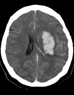


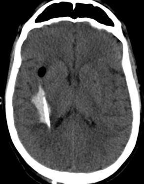
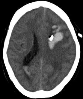
5 Type A Clot: Type A Trajectory Stability CT 3/1/1 12:2:21 PM CP Score: 135 % Clot volume reduction: 89.18% Catheter CT 3/12/1 12:1:46 AM EOT CT 3/15/1 5:3 AM


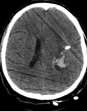


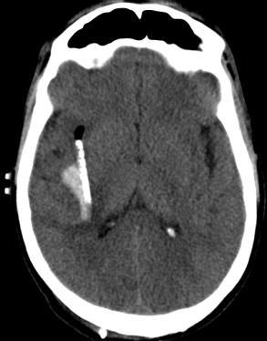
6 Type A Clot: Type A Trajectory Stability CT 2/1/9 6:2:29 AM CP Score: 45 % Clot volume reduction: 24.99% Catheter CT 2/11/9 7:15:2 PM EOT CT 2/14/9 1:2: AM


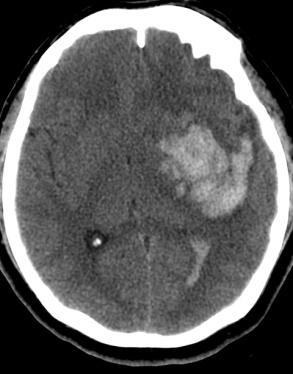
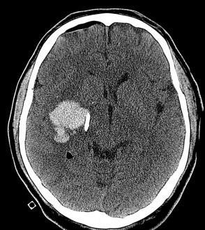
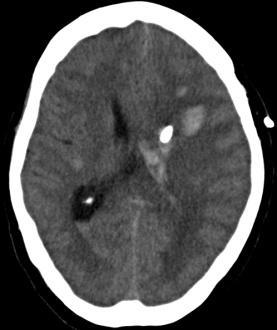
7 Type B Clot: Type B Trajectory CP Score: 126 % Clot volume reduction: 74.14% Stability CT 9/14/1 12:2:35 PM Catheter CT 9/14/1 9::28 PM EOT CT 9/16/1 3:59:2 PM


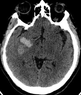
8 Type B Clot: Type B Trajectory Stability CT 2/28/11 4:52: AM CP Score: 28 % Clot volume reduction: 14.7% Catheter CT 3/2/11 2:38 PM EOT CT 3/3/11 8:52: AM


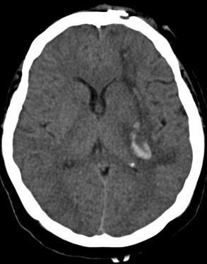
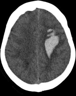
9 Type C Clot: Type C Trajectory Stability CT 2/6/1 3:24:33 AM CP Score: 118 % Clot volume reduction: 74.67% Catheter CT 2/7/1 9:2:14 PM EOT CT 2/9/1 2:57:21 PM


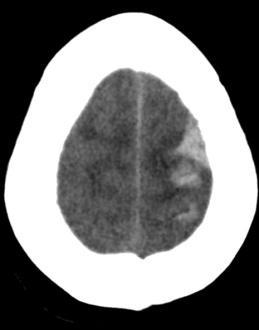


10 Type C Clot: Type C Trajectory Stability CT 1/4/1 11:53:33 AM CP Score: 29 % Clot volume reduction: 44.83% Catheter CT 1/4/1 6:51:27 PM EOT CT 1/6/1 5:19:7 PM

11 Number of Patients Number of Patients ICH Designation for Clot Locations: Surgical and Medical Patients 5 1% 45 9% 4 8% 35 7% 3 6% 25 5% 2 4% 15 3% 1 2% 5 1% Caudate Thalamus Globus Pallidus Putamen Lobar % Caudate Thalamus Globus Pallidus Putamen Lobar A A B B C C Most clot locations were designated as Type A, except for thalamus and lobar clots. As can be expected, the majority of lobar clots were designated as type C.


12 Number of Patients Number of Patients ICH Designation for Clot Locations: Surgical Patients 35 1% % 8% 7% 2 6% % 4% 3% 5 2% Caudate Thalamus Glo Pal Putamen Lobar A B C % % Caudate Thalamus Glo Pal Putamen Lobar A B C Most clot locations were designated as Type A, except for thalamus and lobar clots. As can be expected, the majority of lobar clots were designated as type C.
13 Effects of Age and Stability Clot Volume Catheter Placement Score Coeffficient Std Err. t P> t Age Stability volume % Clot Reduction Coeffficient Std Err. t P> t Age Stability volume


14 Average Catheter Placement Score Average catheter placement score higher for Type A ICH designation and basal ganglia clots Average Catheter Placement Score vs. Clot Location Stage 1 Avg CP Score Stage 2 Avg CP Score 2 N Mean N Mean Catheter Placement Score Coeffficient Std Err. t P> t ICH designation Type B to Type A ICH designation Type C to Type A Clot Location Average catheter placement score is higher for Type A ICH designation compared to those of type B and C designations Average catheter placement score is higher when the clot is located in the basal ganglia compared to when is lobar

15 Multivariate Regression of Catheter Placement Score Catheter Placement Score Coeffficient Std Err. t P> t Patient Number ICH Designation Patient Number ICH Designation ICH Stability clot volume Type B-A Type C-A Type B-A Type C-A When regress catheter placement score with patient number and ICH designation, do see significance in the placement score between type B compared to type A and between type C and type A.


16 Average % Clot Volume Reduction No Significant Difference in Average % Clot Volume Reduction Average % Clot Volume Reduction vs. Clot Location Stage 1 Stage 2-1 % Clot Volume Reduction Coeffficient Std Err. t P> t ICH designation Type B to Type A ICH designation Type C to Type A Clot Location


17 Age distribution of clot location and ICH designation BG = 8 patients Lobar = 37 patients Age Coeff Std Err. t P> t ICH Designation Type B compared to Type A ICH Designation Type C compared to Type A ICH Designation Type C compared to Type B Clot Location


18 Number of Patients Catheter Placement Score by Age and Clot Location <65 >= 65 Age N= 36 N= 19 N= 3 N= 22 Lobar Basal Ganglia Catheter Placement Score Coeff Std Err. t P> t Age
19 Evaluating Surgeon Experience Effects of training, location, # subjects


20 Average Catheter Placement Score by Surgeon Experience
21 Number of Patients Location of Clots (Deep or Lobar) per Surgeon Experience Basal Ganglia Lobar Patient Number for Surgeon (Surgeon Experience)
22 % Clot Volume Reduction % Clot Volume Reduction % Clot Volume Reduction Catheter Placement Score Catheter Placement Score Catheter Placement Score Surgeon s catheter placement score by individual patients Dr. Aldrich Dr. Awad Dr. Broaddus Stage 1 Stage Patient Number Patient Number Patient Number Dr. Aldrich Dr. Awad Dr. Broaddus Stage 1 Stage Patient Number Patient Number Patient Number
23 % Clot Volume Reduction Catheter Placement SCore Surgeon s catheter placement score by individual patients Dr. Camarata Dr. Dodd Dr. Huang Dr. Zucarello Stage 1 Stage Dr. Camarata Patient Number Dr. Dodd Patient Number Dr. Huang Patient Number Dr. Zucarello 5 1 Patient Number Stage 1 Stage 2 Catheter placement score increased on average in stage 2 for each neurosurgeon Don t see a general increase in placement score after each subsequent patient
24 Average Catheter Placement Score % Clot Volume Reduction 16 Average Catheter Placement Score and % Clot Reduction for each Surgeon Average Catheter Placement Score for Each Surgeon Separated by ICH Designation 1. Average % Clot Volume Reduction for Each Surgeon Separated by ICH Designation Type A Type B Type C Type A Type B Type C Average Catheter Placement Score for each type for each surgeon Average % clot volume reduction for each type for each surgeon Aldrich Awad Broaddus Camarata Dodd Huang Zuccarello Aldrich Awad Broaddus Camarata Dodd Huang Zuccarello
25 Number of Patients Number of Patients Effects of ICH length on Catheter Placement Basal Ganglia Clot A-C Length Distribution A-C length (cm) 4 Lobar Clot A-C Length Distribution A-C length (cm) Basal Ganglia Lobar Less than Greater than 4 13
26 A-C Length Distribution by ICH Designation Basal Ganglia Clot A-C Length Distribution by ICH Designation Type C Type B Type A Lobar Clot A-C Length Distribution by ICH Designation Type C Type B Type A
27 Number of Patients Number of Patients A-C Length Distribution by Actual Catheter Trajectory Basal Ganglia Clot A-C Length Distribution by Actual Trajectory Freq of Type C- Vertical Freq of Type C- Horizontal Freq of Type B Freq of Type A A-C Length (cm) Lobar Clot A-C Length Distribution by Actual Trajectory Freq of Type C- Vertical Freq of Type C- Horizontal Freq of Type B Freq of Type A A-C Length (cm)
28 Summary Most basal ganglia clots were designated as Type A, while most lobar clots were designated as Type C; however, the longest axis of the clots may not always follow the catheter track axis. The average catheter placement score is higher for type A designation and for basal ganglia clots, although the difference is not significant. However, when we account for surgeon experience, the ICH designation does significantly impact the catheter placement score (Type C to Type A P>.23; Type B to Type A P>.32). Type C designation tends to occur in older patients. Catheter placement score did not increase with surgeon experience. Catheter placement score did increase in stage 2 compared to stage 1. When planning trajectories, the surgeon may want to look at the longest axis to inform the trajectory designation.

29 Number of catheter side ports touching clot on the EOT CT scan Problem Will an additional dose of r-tpa at the EOT CT scan be beneficial in reducing the ICH volume? Methods: Determined number of sides of the catheter ports touching the clot by looking at anterior, posterior, right, and left sides of the catheter. Looked at the EOT scans and the scans after catheter placement. Maximum of 4 sides touching = clot surrounds the catheter side ports Minimum of sides touching = clot does not touch any of the side ports


30 Observations CP Score: 58 Sides touching: CP Score: 112 Sides touching: CP Score: 45 Sides touching: CP Score: 134 Sides touching: 3
31 Number of Patients Number of Patients Number of Patients Number of Patients Catheter Position in Relation to the Clot Scans after Catheter Placement EOT Catheter Position in relation to the clot Stage 1: Number of catheter sides touching clot for 3 categories of catheter placement scores Stage 1: Number of catheter sides touching clot for 3 categories of catheter placement scores Sides 1 Side 2 Sides 3 Sides 4 Sides < = 7 >7 and < =1 > Sides 1 Side 2 Sides 3 Sides 4 Sides <= 7 >7 and < =1 >1 Stage 2: Number of catheter sides touching clot for 3 categories of catheter placement scores Stage 2: Number of catheter sides touching clot for 3 categories of catheter placement scores < =7 >7 and < =1 > <= 7 >7 and < =1 >1 Sides 1 Side 2 Sides 3 Sides 4 Sides Sides 1 Side 2 Sides 3 Sides 4 Sides
32 Number of catheter side ports touching clot on the EOT CT scan Sides after Catheter Placement Coeffficient Std Err. t P> t Catheter Placement Score Sides at EOT Coeffficient Std Err. t P> t Catheter Placement Score Total Doses Given Catheter Placement Score Total Doses Given Sides Reduced Coeffficient Std Err. t P> t Catheter Placement Score Total Doses Given Sides touching after catheter placement
33 EOT Catheter Position in relation to the clot Observations of scans A number of the patients who had good catheter placements still had clot surrounding the catheter side ports and would appear to benefit from an additional dose before catheter removal. Patients with poor catheter placements had catheter side ports that were already located outside the clot, so by the EOT scan, not as much clot was touching the catheter ports. Analysis sides touching does not correlate with catheter placement score because a lower score would indicate that the catheter is placed more to the side of the clot. More sides touching could indicate that the catheter was placed well, and more doses could be given before catheter removal. When regress sides touching to catheter placement score and total doses given, find a significance between both variables in determining the number of sides touching. Future Direction: Look at the Superior and Inferior Sides of the Catheter Give different weights to different sides of the catheter touching the clot

34 Acknowledgements Natalie Ullman Andrew Mould Katie Smith Sam Nekoovaght-Tak Carol Thompson Shane Thorp Ryan Fisico Dr. Dan Hanley
Develop the different patterns of collapse. Help better understand the EVD catheter and it s effect on the ICH
 Ryan Noel Fisico Develop the different patterns of collapse Help better understand the EVD catheter and it s effect on the ICH End of Treatment Patient CT Post- Surgery CT Reposition Timeline Stability
New Clinical Trials For ICH: MISTIE III Minimally invasive techniques for hemorrhagic stroke
 New Clinical Trials For ICH: MISTIE III Minimally invasive techniques for hemorrhagic stroke 1 Wendy Ziai, MD, MPH Daniel F. Hanley, MD Johns Hopkins Medical Institutions Dept. of Neurology Division of
Pediatric MS MRI Study Methodology
 General Pediatric MS MRI Study Methodology SCAN PREPARATION axial T2-weighted scans and/or axial FLAIR scans were obtained for all subjects when available, both T2 and FLAIR scans were scored. In order
Biological Bases of Behavior. 3: Structure of the Nervous System
 Biological Bases of Behavior 3: Structure of the Nervous System Neuroanatomy Terms The neuraxis is an imaginary line drawn through the spinal cord up to the front of the brain Anatomical directions are
Medical Neuroscience Tutorial Notes
 Medical Neuroscience Tutorial Notes Blood Supply to the Brain MAP TO NEUROSCIENCE CORE CONCEPTS 1 NCC1. The brain is the body's most complex organ. LEARNING OBJECTIVES After study of the assigned learning
Announcement. Danny to schedule a time if you are interested.
 Announcement If you need more experiments to participate in, contact Danny Sanchez (dsanchez@ucsd.edu) make sure to tell him that you are from LIGN171, so he will let me know about your credit (1 point).
PSY 302: CHAPTER 3 NOTES THE BRAIN (PART II) - 9/5/17. By: Joseline
 - 9/5/17. By: Joseline") PSY 302: CHAPTER 3 NOTES THE BRAIN (PART II) - 9/5/17 By: Joseline Left 3 MAJOR FISSURES : 2HEMISPHERES Right Lateral Ventricle Central Fissure Third Ventricle Sulcus Lateral Fissure Gyros Fissure- Fissures
PSY 302: CHAPTER 3 NOTES THE BRAIN (PART II) - 9/5/17 By: Joseline Left 3 MAJOR FISSURES : 2HEMISPHERES Right Lateral Ventricle Central Fissure Third Ventricle Sulcus Lateral Fissure Gyros Fissure- Fissures
BASAL GANGLIA. Dr JAMILA EL MEDANY
 BASAL GANGLIA Dr JAMILA EL MEDANY OBJECTIVES At the end of the lecture, the student should be able to: Define basal ganglia and enumerate its components. Enumerate parts of Corpus Striatum and their important
Blood Supply of the CNS
 Blood Supply of the CNS Lecture Objectives Describe the four arteries supplying the CNS. Follow up each artery to its destination. Describe the circle of Willis and its branches. Discuss the principle
Supplementary Online Material Supplementary Table S1 to S5 Supplementary Figure S1 to S4
 Supplementary Online Material Supplementary Table S1 to S5 Supplementary Figure S1 to S4 Table S1: Brain regions involved in the adapted classification learning task Brain Regions x y z Z Anterior Cingulate
CEREBRUM Dr. Jamila Elmedany Dr. Essam Eldin Salama
 CEREBRUM Dr. Jamila Elmedany Dr. Essam Eldin Salama Objectives At the end of the lecture, the student should be able to: List the parts of the cerebral hemisphere (cortex, medulla, basal nuclei, lateral
Systems Neuroscience Dan Kiper. Today: Wolfger von der Behrens
 Systems Neuroscience Dan Kiper Today: Wolfger von der Behrens wolfger@ini.ethz.ch 18.9.2018 Neurons Pyramidal neuron by Santiago Ramón y Cajal (1852-1934, Nobel prize with Camillo Golgi in 1906) Neurons
Prospective Memory as a Specific Form of Task Switching. Intention and Executive Control
 Prospective Memory as a Specific Form of Task Switching Intention and Executive Control Yehene E, Meiran N, Soroker N Taskalternationcostwithoutask alternation:measuringintentionality. A behavioral dissociation
Essentials of Clinical MR, 2 nd edition. 14. Ischemia and Infarction II
 14. Ischemia and Infarction II Lacunar infarcts are small deep parenchymal lesions involving the basal ganglia, internal capsule, thalamus, and brainstem. The vascular supply of these areas includes the
Tyler Carson D.O., Vladamir Cortez D.O., Dan E. Miulli D.O.
 Bedside Intracranial Hematoma Evacuation and Intraparenchymal Drain Placement for Spontaneous Intracranial Hematoma Larger than 30 cc in Volume: Institutional Experience and Patient Outcomes Tyler Carson
Chapter 3. Structure and Function of the Nervous System. Copyright (c) Allyn and Bacon 2004
 Allyn and Bacon 2004") Chapter 3 Structure and Function of the Nervous System 1 Basic Features of the Nervous System Neuraxis: An imaginary line drawn through the center of the length of the central nervous system, from the
Chapter 3 Structure and Function of the Nervous System 1 Basic Features of the Nervous System Neuraxis: An imaginary line drawn through the center of the length of the central nervous system, from the
Blood Supply. Allen Chung, class of 2013
 Blood Supply Allen Chung, class of 2013 Objectives Understand the importance of the cerebral circulation. Understand stroke and the types of vascular problems that cause it. Understand ischemic penumbra
DISSECTION OF THE SHEEP'S BRAIN
 Sheep Brain Dissection Guide Page 1 DISSECTION OF THE SHEEP'S BRAIN Introduction The purpose of the sheep brain dissection is to familiarize you with the threedimensional structure of the brain and teach
Cerebrum-Cerebral Hemispheres. Cuneyt Mirzanli Istanbul Gelisim University
 Cerebrum-Cerebral Hemispheres Cuneyt Mirzanli Istanbul Gelisim University The largest part of the brain. Ovoid shape. Two incompletely separated cerebral hemispheres. The outer surface of the cerebral
PROPERTY OF ELSEVIER SAMPLE CONTENT - NOT FINAL. Gross Anatomy and General Organization of the Central Nervous System
 3 Gross Anatomy and General Organization of the Central Nervous System C h a p t e r O u t l i n e The Long Axis of the CNS Bends at the Cephalic Flexure Hemisecting a Brain Reveals Parts of the Diencephalon,
Anatomy of the basal ganglia. Dana Cohen Gonda Brain Research Center, room 410
 Anatomy of the basal ganglia Dana Cohen Gonda Brain Research Center, room 410 danacoh@gmail.com The basal ganglia The nuclei form a small minority of the brain s neuronal population. Little is known about
Telencephalon (Cerebral Hemisphere)
") Telencephalon (Cerebral Hemisphere) OUTLINE The Cortex - Lobes, Sulci & Gyri - Functional Subdivisions - Limbic Lobe & Limbic System The Subcortex - Basal Ganglia - White Matter (Internal Capsule) - Relations
Telencephalon (Cerebral Hemisphere) OUTLINE The Cortex - Lobes, Sulci & Gyri - Functional Subdivisions - Limbic Lobe & Limbic System The Subcortex - Basal Ganglia - White Matter (Internal Capsule) - Relations
Ex. 1 :Language of Anatomy
 Collin College BIOL 2401 : Human Anatomy & Physiology Ex. 1 :Language of Anatomy The Anatomical Position Used as a reference point when referring to specific areas of the human body Body erect Head and
Deep Brain Stimulation Surgery for Parkinson s Disease
 Deep Brain Stimulation Surgery for Parkinson s Disease Demystifying Medicine 24 January 2012 Kareem A. Zaghloul, MD, PhD Staff Physician, Surgical Neurology Branch NINDS Surgery for Parkinson s Disease
[(PHY-3a) Initials of MD reviewing films] [(PHY-3b) Initials of 2 nd opinion MD]
![[(PHY-3a) Initials of MD reviewing films] [(PHY-3b) Initials of 2 nd opinion MD]](/thumbs/89/98619893.jpg "[(PHY-3a) Initials of MD reviewing films] [(PHY-3b) Initials of 2 nd opinion MD]") 2015 PHYSICIAN SIGN-OFF (1) STUDY NO (PHY-1) CASE, PER PHYSICIAN REVIEW 1=yes 2=no [strictly meets case definition] (PHY-1a) CASE, IN PHYSICIAN S OPINION 1=yes 2=no (PHY-2) (PHY-3) [based on all available
2015 PHYSICIAN SIGN-OFF (1) STUDY NO (PHY-1) CASE, PER PHYSICIAN REVIEW 1=yes 2=no [strictly meets case definition] (PHY-1a) CASE, IN PHYSICIAN S OPINION 1=yes 2=no (PHY-2) (PHY-3) [based on all available
Introduction to the Central Nervous System: Internal Structure
 Introduction to the Central Nervous System: Internal Structure Objective To understand, in general terms, the internal organization of the brain and spinal cord. To understand the 3-dimensional organization
Regional and Lobe Parcellation Rhesus Monkey Brain Atlas. Manual Tracing for Parcellation Template
 Regional and Lobe Parcellation Rhesus Monkey Brain Atlas Manual Tracing for Parcellation Template Overview of Tracing Guidelines A) Traces are performed in a systematic order they, allowing the more easily
Sectional Anatomy Head Practice Problems
 1. Which of the following is illustrated by #3? (Fig. 5-42) A) maxillary sinus B) vomer C) septal cartilage D) perpendicular plate of ethmoid bone 2. What number illustrates the cornea? (Fig. 5-42) A)
The Central Nervous System I. Chapter 12
 The Central Nervous System I Chapter 12 The Central Nervous System The Brain and Spinal Cord Contained within the Axial Skeleton Brain Regions and Organization Medical Scheme (4 regions) 1. Cerebral Hemispheres
The Nervous System: Sensory and Motor Tracts of the Spinal Cord
 15 The Nervous System: Sensory and Motor Tracts of the Spinal Cord PowerPoint Lecture Presentations prepared by Steven Bassett Southeast Community College Lincoln, Nebraska Introduction Millions of sensory
The neurvous system senses, interprets, and responds to changes in the environment. Two types of cells makes this possible:
 NERVOUS SYSTEM The neurvous system senses, interprets, and responds to changes in the environment. Two types of cells makes this possible: the neuron and the supporting cells ("glial cells"). Neuron Neurons
TRANSVERSE SECTION PLANE Scalp 2. Cranium. 13. Superior sagittal sinus
 TRANSVERSE SECTION PLANE 1 1. Scalp 2. Cranium 3. Superior sagittal sinus 4. Dura mater 5. Falx cerebri 6. Frontal lobes of the cerebrum 7. Middle meningeal artery 8. Cortex, grey matter 9. Cerebral vessels
ANATOMY & PHYSIOLOGY DISSECTION OF THE SHEEP BRAIN LAB GROUP:
 ANATOMY & PHYSIOLOGY DISSECTION OF THE SHEEP BRAIN LAB GROUP: Introduction The purpose of the sheep brain dissection is to familiarize you with the three dimensional structure of the brain and teach you
Biomedical Technology Research Center 2011 Workshop San Francisco, CA
 Diffusion Tensor Imaging: Parkinson s Disease and Atypical Parkinsonism David E. Vaillancourt court1@uic.edu Associate Professor at UIC Departments t of Kinesiology i and Nutrition, Bioengineering, and
Outlook for intracerebral haemorrhage after a MISTIE spell
 Outlook for intracerebral haemorrhage after a MISTIE spell David J Werring PhD FRCP Stroke Research Centre, Department of Brain Repair and Rehabilitation, UCL Institute of Neurology, National Hospital
NACC Vascular Consortium. NACC Vascular Consortium. NACC Vascular Consortium
 NACC Vascular Consortium NACC Vascular Consortium Participating centers: Oregon Health and Science University ADC Rush University ADC Mount Sinai School of Medicine ADC Boston University ADC In consultation
CEREBRUM. Dr. Jamila EL Medany
 CEREBRUM Dr. Jamila EL Medany Objectives At the end of the lecture, the student should be able to: List the parts of the cerebral hemisphere (cortex, medulla, basal nuclei, lateral ventricle). Describe
 Passport control a bit carried away. appreciated the advice forgot to talk to the manager, next thing I know my fmri thankfully, when aroused things back to normal Inattentive impaired children and adolescents:
For more information about how to cite these materials visit
 Author(s): Peter Hitchcock, PH.D., 2009 License: Unless otherwise noted, this material is made available under the terms of the Creative Commons Attribution Non-commercial Share Alike 3.0 License: http://creativecommons.org/licenses/by-nc-sa/3.0/
2017, Joule Inc. or its licensors Online appendices are unedited and posted as supplied by the authors.
 Results Validation: Reproducibility Figure S1. Reproducibility of the results of small-world parameters. Differences in topological properties of functional brain networks between bulimia nervosa (BN)
Pearls and Pitfalls in Neuroradiology of Cerebrovascular Disease The Essentials with MR and CT
 Pearls and Pitfalls in Neuroradiology of Cerebrovascular Disease The Essentials with MR and CT Val M. Runge, MD Wendy R. K. Smoker, MD Anton Valavanis, MD Control # 823 Purpose The focus of this educational
Human Paleoneurology and the Evolution of the Parietal Cortex
 PARIETAL LOBE The Parietal Lobes develop at about the age of 5 years. They function to give the individual perspective and to help them understand space, touch, and volume. The location of the parietal
Introduction to Anatomical Terms. Packet #3
 Introduction to Anatomical Terms Packet #3 Directional Terms Directional terms describe the positions of structures relative to other structures or locations in the body. Introduction Superior vs. Inferior
CNS Imaging. Dr Amir Monir, MD. Lecturer of radiodiagnosis.
 CNS Imaging Dr Amir Monir, MD Lecturer of radiodiagnosis www.dramir.net Types of radiological examinations you know Plain X ray X ray with contrast GIT : barium (swallow, meal, follow through, enema) ERCP
Central nervous system (CNS): brain and spinal cord Collections of cell body and dendrites (grey matter) are called nuclei/nucleus Nucleus can also
: brain and spinal cord Collections of cell body and dendrites (grey matter) are called nuclei/nucleus Nucleus can also") Chapter 3 Part 1 Orientation Directions in the nervous system are described relatively to the neuraxis An imaginary line drawn through the center of the length of the central nervous system, from the bottom
Chapter 3 Part 1 Orientation Directions in the nervous system are described relatively to the neuraxis An imaginary line drawn through the center of the length of the central nervous system, from the bottom
The management of ICH when to operate when not to?
 The management of ICH when to operate when not to? Intracranial Hemorrhage High Incidence o Accounts for 10-15% of all strokes 1,2,5 o 80,000 cases in US; 2 million WW 2,5 o Incidence doubles for African-
Unit Three. The brain includes: cerebrum, diencephalon, brain stem, & cerebellum. The brain lies within the cranial cavity of the skull.
 Human Anatomy & Physiology 11 Divisions of the Nervous System Karen W. Smith, Instructor Unit Three BRAIN & SPINAL CORD Refer to the following URLs. Be sure to study these along with your book. http://www.sirinet.net/~jgjohnso/nervous.html
Welcome to our MISTIE III Safety Forum September 12, 2016
 Welcome to our MISTIE III Safety Forum September 12, 2016 Agenda: Update from our Surgical Centers: Where we stand surgically Mario Zuccarello, MD, University of Cincinnati Revisiting the Importance of
CT Fluoroscopy-guided Aspiration of Intracerebral Hematomas: Technique and Outcomes
 Journal of Cerebrovascular and Endovascular Neurosurgery pissn 2234-8565, eissn 2287-3139, http://dx.doi.org/10.7461/jcen.2015.17.1.7 Original Article CT Fluoroscopy-guided Aspiration of Intracerebral
On-line Table 1: Dementia diagnoses and related ICD codes for the diagnostic groups a
 On-line Table 1: diagnoses and related ICD codes for the diagnostic groups a Diagnosis (N = 1504) ICD Code Patients Scanned with 3T; SWI (%) Subjective cognitive impairment (n 385) Z03.2A, Z03.3, and R41.8A
CT and MR Imaging in Young Stroke Patients
 CT and MR Imaging in Young Stroke Patients Ashfaq A. Razzaq,Behram A. Khan,Shahid Baig ( Department of Neurology, Aga Khan University Hospital, Karachi. ) Abstract Pages with reference to book, From 66
Use of Multimodal Neuroimaging Techniques to Examine Age, Sex, and Alcohol-Related Changes in Brain Structure Through Adolescence and Young Adulthood
 American Psychiatric Association San Diego, CA 24 May 2017 Use of Multimodal Neuroimaging Techniques to Examine Age, Sex, and Alcohol-Related Changes in Brain Structure Through Adolescence and Young Adulthood
OBJECTIVES. At the end of the lecture, students should be able to: List the cerebral arteries.
 DR JAMILA EL MEDANY OBJECTIVES At the end of the lecture, students should be able to: List the cerebral arteries. Describe the cerebral arterial supply regarding the origin, distribution and branches.
The management of ICH when to operate when not to?
 The management of ICH when to operate when not to? ICH is a Bad Disease High Incidence o Accounts for 10-15% of all strokes 1,2,5 o 80,000 cases in US; 2 million WW 2,5 o Incidence doubles for African-
Chapter 18: The Brain & Cranial Nerves. Origin of the Brain
 Chapter 18: The Brain & Cranial Nerves BIO 218 Fall 2015 Origin of the Brain The brain originates from a structure called the neural tube, which arises during a developmental stage called neurulation.
Supplementary Material S3 Further Seed Regions
 Supplementary Material S3 Further Seed Regions Figure I. Changes in connectivity with the right anterior insular cortex. (A) wake > mild sedation, showing a reduction in connectivity between the anterior
Supplementary Digital Content
 Supplementary Digital Content Contextual modulation of pain in masochists: involvement of the parietal operculum and insula Sandra Kamping a, Jamila Andoh a, Isabelle C. Bomba a, Martin Diers a,b, Eugen
Stroke Awareness. Presented by: Duane Anderson, MD Snoqualmie Valley Hospital Emergency Department Medical Director
 Stroke Awareness Presented by: Duane Anderson, MD Snoqualmie Valley Hospital Emergency Department Medical Director What is a stroke? Stroke can happen to anyone. Stroke is the fourth leading cause of death
Stroke Imaging Basics. Jeremy Hopkin M.D.
 Stroke Imaging Basics Jeremy Hopkin M.D. Goals Introduce the basic physical properties of imaging used in stroke. Understand why each modality is used in the setting of stroke. Understand some strengths
Demonstrate the bony features of Cl and C2 vertebrae evident on this Xray
 SUBJECT: ANATOMY 7 September 2007 am. TOPIC: X-ray: Lateral C spine NUMBER: JL Demonstrate the bony features of Cl and C2 vertebrae evident on this Xray 1 Odontoid peg (dens) 2 Bodies of Cl andc2 3 anterior
For Emergency Doctors. Dr Suzanne Smallbane November 2011
 For Emergency Doctors Dr Suzanne Smallbane November 2011 A: Orbit B: Sphenoid Sinus C: Temporal Lobe D: EAC E: Mastoid air cells F: Cerebellar hemisphere A: Frontal lobe B: Frontal bone C: Dorsum sellae
Methods to examine brain activity associated with emotional states and traits
 Methods to examine brain activity associated with emotional states and traits Brain electrical activity methods description and explanation of method state effects trait effects Positron emission tomography
Morbidity of Stereotactic Biopsy for Intracranial Lesions
 Kobe J. Med. Sci., Vol. 56, No. 4, pp. E148-E153, 2010 Morbidity of Stereotactic Biopsy for Intracranial Lesions MASAMITSU NISHIHARA 1 *, TAKASHI SASAYAMA 2, HIROSHI KUDO 3, and EIJI KOHMURA 2 1 Department
SUPPLEMENTAL DIGITAL CONTENT
 SUPPLEMENTAL DIGITAL CONTENT FIGURE 1. Unilateral subthalamic nucleus (STN) deep brain stimulation (DBS) electrode and internal pulse generator. Copyright 2010 Oregon Health & Science University. Used
Supplementary Information
 Supplementary Information The neural correlates of subjective value during intertemporal choice Joseph W. Kable and Paul W. Glimcher a 10 0 b 10 0 10 1 10 1 Discount rate k 10 2 Discount rate k 10 2 10
Exam 2 PSYC Fall (2 points) Match a brain structure that is located closest to the following portions of the ventricular system
 Match a brain structure that is located closest to the following portions of the ventricular system") Exam 2 PSYC 2022 Fall 1998 (2 points) What 2 nuclei are collectively called the striatum? (2 points) Match a brain structure that is located closest to the following portions of the ventricular system
Exam 2 PSYC 2022 Fall 1998 (2 points) What 2 nuclei are collectively called the striatum? (2 points) Match a brain structure that is located closest to the following portions of the ventricular system
The Language of Anatomy. (Anatomical Terminology)
") The Language of Anatomy (Anatomical Terminology) Terms of Position The anatomical position is a fixed position of the body (cadaver) taken as if the body is standing (erect) looking forward with the upper
The Language of Anatomy (Anatomical Terminology) Terms of Position The anatomical position is a fixed position of the body (cadaver) taken as if the body is standing (erect) looking forward with the upper
Cerebro-vascular stroke
 Cerebro-vascular stroke CT Terminology Hypodense lesion = lesion of lower density than the normal brain tissue Hyperdense lesion = lesion of higher density than normal brain tissue Isodense lesion = lesion
Stroke - Intracranial hemorrhage. Dr. Amitesh Aggarwal Associate Professor Department of Medicine
 Stroke - Intracranial hemorrhage Dr. Amitesh Aggarwal Associate Professor Department of Medicine Etiology and pathogenesis ICH accounts for ~10% of all strokes 30 day mortality - 35 45% Incidence rates
Assessing the Stroke Patient. Arlene Boudreaux, MSN, RN, CCRN, CNRN
 Assessing the Stroke Patient Arlene Boudreaux, MSN, RN, CCRN, CNRN Cincinnati Pre-Hospital Stroke Scale May be done by EMS o One of many o F facial droop on one side o A arm drift (hold a pizza box, close
The Hydrocephalus Clinical Research Network
 J Neurosurg Pediatrics 14:173 178, 2014 AA, 2014 Factors associated with ventricular catheter movement and inaccurate catheter location: post hoc analysis of the Hydrocephalus Clinical Research Network
BESA Research Quick Guide
 BESA Research Quick Guide BESA 3D Maps Quick Guide An introduction how to interpret 3D voltage and phase maps in the scalp EEG Copyright and Trademarks The BESA products and their documentation are copyrighted
Background & Indications Probe Selection
 Teresa S. Wu, MD, FACEP Director, EM Ultrasound Program & Fellowship Co-Director, Simulation Based Training Program & Fellowship Associate Program Director, EM Residency Program Maricopa Medical Center
The human brain. of cognition need to make sense gives the structure of the brain (duh). ! What is the basic physiology of this organ?
. ! What is the basic physiology of this organ?") The human brain The human brain! What is the basic physiology of this organ?! Understanding the parts of this organ provides a hypothesis space for its function perhaps different parts perform different
The human brain The human brain! What is the basic physiology of this organ?! Understanding the parts of this organ provides a hypothesis space for its function perhaps different parts perform different
Radiological anatomy of frontal sinus By drtbalu
 2009 Radiological anatomy of frontal sinus By drtbalu Anatomy of frontal sinus is highly variable. Precise understanding of these variables will help a surgeon to avoid unnecessary complications during
Biology 210 Chapter 8: Skeletal Tissues Supplement 1
 Biology 210 Chapter 8: Skeletal Tissues Supplement 1 By John McGill Material contributed by Beth Wyatt & Jack Bagwell DIVISIONS OF THE SKELETAL SYSTEM AXIAL SKELETON (80 BONES) Bones of the Head, Neck,
Stroke School for Internists Part 1
 Stroke School for Internists Part 1 November 4, 2017 Dr. Albert Jin Dr. Gurpreet Jaswal Disclosures I receive a stipend for my role as Medical Director of the Stroke Network of SEO I have no commercial
Extrapyramidal Motor System. Basal Ganglia or Striatum. Basal Ganglia or Striatum 3/3/2010
 Extrapyramidal Motor System Basal Ganglia or Striatum Descending extrapyramidal paths receive input from other parts of motor system: From the cerebellum From the basal ganglia or corpus striatum Caudate
1/2/2019. Basal Ganglia & Cerebellum a quick overview. Outcomes you want to accomplish. MHD-Neuroanatomy Neuroscience Block. Basal ganglia review
 This power point is made available as an educational resource or study aid for your use only. This presentation may not be duplicated for others and should not be redistributed or posted anywhere on the
Dr. Farah Nabil Abbas. MBChB, MSc, PhD
 Dr. Farah Nabil Abbas MBChB, MSc, PhD The Basal Ganglia *Functions in association with motor cortex and corticospinal pathways. *Regarded as accessory motor system besides cerebellum. *Receive most of
Introduction to The Human Body
 1 Introduction to The Human Body FOCUS: The human organism is often examined at seven structural levels: chemical, organelle, cell, tissue, organ, organ system, and the organism. Anatomy examines the structure
Slide 1. Slide 2. Slide 3. Tomography vs Topography. Computed Tomography (CT): A simplified Topographical review of the Brain. Learning Objective
: A simplified Topographical review of the Brain. Learning Objective") Slide 1 Computed Tomography (CT): A simplified Topographical review of the Brain Jon Wheiler, ACNP-BC Slide 2 Tomography vs Topography Tomography: A technique for displaying a representation of a cross
Slide 1 Computed Tomography (CT): A simplified Topographical review of the Brain Jon Wheiler, ACNP-BC Slide 2 Tomography vs Topography Tomography: A technique for displaying a representation of a cross
PETER PAZMANY CATHOLIC UNIVERSITY Consortium members SEMMELWEIS UNIVERSITY, DIALOG CAMPUS PUBLISHER
 PETER PAZMANY CATHOLIC UNIVERSITY SEMMELWEIS UNIVERSITY Development of Complex Curricula for Molecular Bionics and Infobionics Programs within a consortial* framework** Consortium leader PETER PAZMANY
SHOULDER JOINT ANATOMY AND KINESIOLOGY
 SHOULDER JOINT ANATOMY AND KINESIOLOGY SHOULDER JOINT ANATOMY AND KINESIOLOGY The shoulder joint, also called the glenohumeral joint, consists of the scapula and humerus. The motions of the shoulder joint
NIH Public Access Author Manuscript Proc SPIE. Author manuscript; available in PMC 2014 February 07.
 NIH Public Access Author Manuscript Published in final edited form as: Proc SPIE. 2007 March 5; 6512: 651236. doi:10.1117/12.708950. Semi-Automatic Parcellation of the Corpus Striatum Ramsey Al-Hakim a,
Gross Morphology of the Brain
 Gross Morphology of the Brain Done by : Marah Marahleh & Razan Krishan *slides in bold Principal Parts of the Brain Cerebrum : largest part of the brain Diencephalon Thalamus & hypothalamus Cerebellum
Brain Arteriovenous Malformations Endovascular Therapy and Associated Therapeutic Protocols Jorge Guedes Cabral de Campos
 Endovascular Therapy and Associated Therapeutic Protocols Jorge Guedes Cabral de Campos Neuroradiology Department Hospital de Santa Maria University of Lisbon CEREBRAL AVM CLINICAL / EPIDEMIOLOGY Brain
Overview of the Nervous System (some basic concepts) Steven McLoon Department of Neuroscience University of Minnesota
 Steven McLoon Department of Neuroscience University of Minnesota") Overview of the Nervous System (some basic concepts) Steven McLoon Department of Neuroscience University of Minnesota 1 Coffee Hour Tuesday (Sept 11) 10:00-11:00am Friday (Sept 14) 8:30-9:30am Surdyk s
Overview of the Nervous System (some basic concepts) Steven McLoon Department of Neuroscience University of Minnesota 1 Coffee Hour Tuesday (Sept 11) 10:00-11:00am Friday (Sept 14) 8:30-9:30am Surdyk s
Lecture XIII. Brain Diseases I - Parkinsonism! Brain Diseases I!
 Lecture XIII. Brain Diseases I - Parkinsonism! Bio 3411! Wednesday!! Lecture XIII. Brain Diseases - I.! 1! Brain Diseases I! NEUROSCIENCE 5 th ed! Page!!Figure!!Feature! 408 18.9 A!!Substantia Nigra in
Making Things Happen 2: Motor Disorders
 Making Things Happen 2: Motor Disorders How Your Brain Works Prof. Jan Schnupp wschnupp@cityu.edu.hk HowYourBrainWorks.net On the Menu in This Lecture In the previous lecture we saw how motor cortex and
Leah Militello, class of 2018
 Leah Militello, class of 2018 Objectives 1. Describe the general organization of cerebral hemispheres. 2. Describe the locations and features of the different functional areas of cortex. 3. Understand
COGNITIVE SCIENCE 107A. Motor Systems: Basal Ganglia. Jaime A. Pineda, Ph.D.
 COGNITIVE SCIENCE 107A Motor Systems: Basal Ganglia Jaime A. Pineda, Ph.D. Two major descending s Pyramidal vs. extrapyramidal Motor cortex Pyramidal system Pathway for voluntary movement Most fibers originate
Geography of the Forehead
 5. Brain Areas Geography of the Forehead Everyone thinks the brain is so complicated, but let s look at the facts. The frontal lobe, for example, is located in the front! And the temporal lobe is where
Basal Ganglia. Introduction. Basal Ganglia at a Glance. Role of the BG
 Basal Ganglia Shepherd (2004) Chapter 9 Charles J. Wilson Instructor: Yoonsuck Choe; CPSC 644 Cortical Networks Introduction A set of nuclei in the forebrain and midbrain area in mammals, birds, and reptiles.
I T IS well known that aneurysms occur at
 The Lateral Perforating Branches of the Anterior and Middle Cerebral Arteries* HARRY A. KAPLAN, M.D. Division of Neurosurgery, Seton Hall College of Medicine, and Jersey City Medical Center, Jersey City,
FDG-PET e parkinsonismi
 Parkinsonismi FDG-PET e parkinsonismi Valentina Berti Dipartimento di Scienze Biomediche, Sperimentali e Cliniche Sez. Medicina Nucleare Università degli Studi di Firenze History 140 PubMed: FDG AND parkinsonism
Prof. Saeed Abuel Makarem & Dr.Sanaa Alshaarawy
 Prof. Saeed Abuel Makarem & Dr.Sanaa Alshaarawy 1 Objectives By the end of the lecture, you should be able to: Describe the anatomy and main functions of the thalamus. Name and identify different nuclei
Parenchyma-sparing lung resections are a potential therapeutic
 Lung Segmentectomy for Patients with Peripheral T1 Lesions Bryan A. Whitson, MD, Rafael S. Andrade, MD, and Michael A. Maddaus, MD Parenchyma-sparing lung resections are a potential therapeutic option
Attenuation value in HU From -500 To HU From -10 To HU From 60 To 90 HU. From 200 HU and above
 Brain Imaging Common CT attenuation values Structure Air Fat Water Brain tissue Recent hematoma Calcifications Bone Brain edema and infarction Normal liver parenchyma Attenuation value in HU From -500
Objectives. Pelvic Anatomy: Staying Out of Trouble. Disclosures. Anatomy 101. Anterior Abdominal Wall. Arcuate Line. Abheha Satkunaratnam MD, FRCS(C)
") Objectives Pelvic Anatomy: Staying Out of Trouble Abheha Satkunaratnam MD, FRCS(C) To focus on key anatomy for the gynaecologic surgeon advancing their minimally invasive gynaecologic skills To provide
Objectives Pelvic Anatomy: Staying Out of Trouble Abheha Satkunaratnam MD, FRCS(C) To focus on key anatomy for the gynaecologic surgeon advancing their minimally invasive gynaecologic skills To provide