Murmurs and the Cardiac Physical Exam. Carolyn A. Altman Texas Children s Hospital Advanced Practice Provider Conference Houston, TX April 6, 2018
|
|
|
- Laura George
- 6 years ago
- Views:
Transcription

1 Murmurs and the Cardiac Physical Exam Carolyn A. Altman Texas Children s Hospital Advanced Practice Provider Conference Houston, TX April 6, 2018

2 The Cardiac Physical Exam Before applying a stethoscope.. Some pearls on General appearance Physical exam beyond the heart 2


3 Jugular Venous Distention Pallor 3 Cyanosis


4 4 Work of Breathing Normal infant breathing Quiet Tachypnea Increased Rate, Work of Breathing


5 5 Beyond the Chest Clubbing Observed in children older than 6 mos with chronic cyanosis Loss of the normal angle of the nail plate with the axis of the finger Abnormal sponginess of the base of the nail bed Increasing convexity of the nail Etiology:? sludging

6 6 Chest Chest wall development and symmetry Long standing cardiomegaly can lead to hemihypertrophy and flared rib edge: Harrison s groove or sulcus
7 7 Ready to Examine the Heart Palpation Auscultation General overview Defects Innocent versus pathologic
8 8 Cardiac Palpation Consistent approach: palm of your hand, hypothenar eminence, or finger tips Precordium, suprasternal notch PMI? RV impulse? Thrills? Heart Sounds?
9 9 Cardiac Auscultation Where to listen: 4 main positions Inching Ancillary sites: don t forget the head in infants
10 Cardiac Auscultation Focus separately on v Heart sounds: S2 normal splitting and intensity? Abnormal sounds? Clicks, gallops v Murmurs 10 v Rubs
11 11 Cardiac Auscultation Etiology of heart sounds: Aortic and pulmonic valves actually close silently Heart sounds reflect vibrations of the cardiac structures after valve closure Sudden deceleration of retrograde flow of the column of blood in the aorta and pulmonary artery when the elastic limits of the tensed valve leaflets are met
12 12 Cardiac Auscultation S2 Physiologic splitting of S2: Increased systemic venous return and increased pulmonary capacitance during inspiration causes delayed closure of the pulmonary valve S2 cannot be considered normal unless physiologic splitting is heard


13 13 S2: normal splitting Single S2: Pulmonar Hypertension Wide, fixed splitting: ASD Paradoxical Splitting of S2: LBBB, severe LVOTO


14 14 S2 HInts If splitting persists while patient supine, try sitting position- less volume in heart may normalize splitting Listen for splitting at mid to ULSB in kids Infants: Mid to LLSB Splitting of S2 if the HR is over 160 hard to hear: gently blowing a breath in the baby s face will slow HR
15 15 Cardiac Auscultation: S1 Physiologic splitting of S1: Can be heard in children with slower heart rates. Varies with respiration as does S2 Soft S1: low cardiac output, tachycardia Loud S1: hyperdynamic (fever, exercise), mitral stenosis
16 16 Cardiac Auscultation Gallops: S3 or S4 Short, low pitched diastolic sounds Abnormal ventricular function
17 17 Auscultation: S3 Gallop Mid way thru diastole Muscle tensing at end of rapid, early filling which occurs with ventricular relaxation Later than split S2 Earlier than S4
18 Auscultation: S4 Gallop If impaired ventricular relaxation, less filling of the ventricles during during early diastole and more during atrial contraction Hypertrophic cardimyopathy, eg S4 is thus a sound generated late in diastole Very close to S1, can mistake for split S1 or S1- ejection click 18 S1-Ej click S4
19 S3 and S4 Gallops Heard best with bell since low pitched Can extinguish the sound by pressing too hard (turning bell into diaphragm) S3 S4 Usually heard over mitral area, if LV dysfunction Listen in left lateral decubitus position too S1-Ej click If RV dysfunction- may hear best at LLSB 19


20 20 Other Extra Heart Sounds Ejection Click: opening of the aortic or pulmonary valve Mid- systolic click: MVP Opening snap of MS Listen for difference in timing, cadence
21 21 How to Characterize Murmurs Timing Site of maximum intensity Intensity Radiation Pitch: Associated findings: clicks, rumbles, precordial activity Different from previous in your patient Innocent or pathologic
22 22 Timing of Murmurs Systolic? Diastolic? or Continuous? Systolic occurs as the heart contracts Diastolic as the heart relaxes Continuous murmurs continue from systole into diastole Find S2 and listen to whether the murmur comes before it, after it, or through it Inching the stethoscope can help with timing

23 23 Murmurs: Timing Systolic murmurs: Regurgitant murmurs: Begin with S1 Ejection murmurs: Begins shortly after S1 Mid- systolic: MVP

24 24 Regurgitant Systolic Murmurs MR, TR, VSD Begin with S1: coincident with S1 Often holosystolic

25 25 Systolic Ejection Murmurs AS/PS, Still s, pulmonary flow Begin after valve opens, so hear S1 then murmur Should be able to hear S2 distinctly Early systolic ejection click if semilunar valve stenosis

26 26 Mid Systolic Murmurs Mitral valve prolapse Click ushers in murmur

27 27 Diastolic murmurs Aortic or pulmonary regurgitation: High pitched Decrescendo

28 28 Diastolic murmurs Diastolic rumbles: Increased volume across MV or TV Low pitched filling noise Absence of silence

29 Continuous Murmurs Start during systole, continue past S2 Louder in systole: PDA, AVM, shunts Venous Hum Louder in diastole: venous hums, coronary fistula 29 PDA Coronary fistula AVM
30 Murmurs by location of greatest intensity: 30 Helpful in figuring out what is generating the murmur URSB: Aortic stenosis ULSB: Pulmonary stenosis, pulmonary flow, ASD LLSB: VSD, Still s, TR Apical: Mitral
31 31 Characterize Murmurs: Grading system allows accurate communication between caretakers Grade I: is there something there? Grade II: Ok, I can hear it Grade III: Boy, that s loud Grade IV: Associated with a thrill, knock your socks off loud Grade V: Audible with scope off chest Grade VI: Audible without stethoscope
32 32 Characterize Murmurs: Important to follow trends: Is an aorto- pulmonary shunt murmur getting softer? A shunt may be getting obstructed, outgrown, or PVR elevated Is the outflow tract obstruction getting worse in a patient with new chest pain? Is the patient with TOF spelling or just colicky: the outflow murmur will get softer during a spell as less flow traverses the RVOT
33 33 Characterize Murmurs by Pitch: High Low Harsh (multitonal)
34 34 Congenital Heart Defects Atrial Septal Defect Patent Ductus Arteriosus Ventricular Septal Defect Pulmonary Stenosis Aortic Stenosis

35 35 CHD: Atrial Septal Defect Anatomy: described by location in the septum Secundum Primum Coronary Sinus Sinus Venosus Physiology and physical signs the same, regardless of location of ASD
36 36 CHD: Atrial Septal Defect Physiology: Amount of shunting depends on v Size of defect v Differences in compliance between RV and LV- flow is usually left to right
37 37 CHD: Atrial Septal Defect Palpation: right ventricular impulse from increased RV volume
38 38 CHD: Atrial Septal Defect Widely split S2: v Persistent separation of A2P2 components of S2 throughout respiratory cycle v Increased pulmonary capacitance or v Reciprocal changes in flow into the right atrium from the defect or systemic veins
39 39 CHD: Atrial Septal Defect Pulmonary flow murmur: v Large volume of blood crossing the pulmonary valve v ULSB to back v Ejection v Medium pitched


40 40 CHD: Atrial Septal Defect Diastolic Rumble: Consistent with at least 2:1 Qp:Qs Low pitched Listen with bell at LLSB

41 41 CHD: Patent Ductus Arteriosus Physiology: v In the setting of low pulmonary vascular resistance, flow is continuous, left to right v If large PDA, PA pressures may be high: flow can be Left to right Bidirectional All right to left
42 42 CHD: Patent Ductus Arteriosus Palpation RV impulse if pulmonary hypertension Hyperactive LV impulse if large volume of flow PDA

43 43 CHD: PDA Murmur v Continuous if low pulmonary vascular resistance v Machinery like v Accentuated at end systole v Left infra- clavicular area, back, and left supraclavicular areas

44 44 CHD: Ventricular Septal Defect Anatomy described by location Perimembranous Inlet Muscular Doubly committed- juxtarterial

45 45 CHD: VSD Physiology: amount of shunting depends on Size of defect Pulmonary resistance: more shunting with decreasing resistance
46 46 CHD: VSD Palpation: Quiet precordium? RV impulse may be present with volume or pressure loading +/- thrill: cannot determine size by presence of thrill
47 47 CHD: VSD S2 in VSDs can be Normally split (typical) Widely split if very generous amount of flow crossing to fill RV Single: if pulmonary hypertension with elevated resistance
48 48 CHD: VSD Auscultation Murmur Usually along LSB Very small defects do not radiate Subpulmonary VSDs follow the RV outflow to the pulmonary arteries Blowing quality Start with S1

49 49 CHD: VSD murmurs Holosystolic murmur: Starts with S1 (obscured) Ends with P2, S2 split normally Plateau shape The smaller the defect, the more high pitched
50 50 CHD: VSD murmur Short systolic murmur consistent with very small defect v v v Starts with S1 Ends before S2, as defect closed by ventricular contraction Usually very localized, may only hear in certain positions

51 51 CHD: VSD Mitral Rumble v Indicates at least 2:1 Qp:Qs v Low pitched v Use bell at apex


52 52 CHD: VSD Diastolic rumble Can be quite subtle Listen for absence of diastolic silence Compare right and lef chest
53 53 CHD: VSD Very Large VSDs v Allow high pressure and high flow v If lef unrepaired: elevated PVR develops, eventually Eisenmenger syndrome Palpahon v RV impulse v Palpable S2

54 54 CHD: VSD Large VSDs: Systolic Murmur v Can be holosystolic, if any pressure restrichon v If no pressure restrichon, may be no murmur, or a pulmonary ouilow murmur

55 55 CHD: VSD Eisenmenger s v S2 loud and single v Pulmonary valve click: dilated pulmonary root v Graham- Steele murmur: pulmonary insufficiency

56 56 CHD: Tetralogy of Fallot Physiology: v Balance between VSD flow and pulmonary valve and sub valve stenosis v Pink tets have little pulmonary stenosis v Other extreme: pulmonary atresia with VSD v PS typically progresses over time
57 57 CHD: TOF Palpahon: v RV impulse v Possible thrill
58 58 CHD: TOF Systolic Murmur: reflects PS, not VSD v MLSB to ULSB to back v Starts with S1, given subvalvar component v As subps worsens, murmur decreases in intensity: pop- off through VSD to systemic circulahon v Listen for murmur to decrease in hypercyanohc spell


59 59 CHD: TOF Systolic Murmur: reflects PS, not VSD

60 60 CHD: TOF post repair To and fro murmur PS/PI The murmur does not carry through S2- not continuous, not the diastolic component The diastolic murmur is of a different pitch

61 61 CHD: Pulmonary Valve Stenosis Anatomy: thickened, possibly dysplashc valve with limited systolic excursion Physiology: usually slowly progressive obstruchon
62 62 CHD: Pulmonary Valve Stenosis Palpahon: v RV impulse: more than mild obstruchon v Thrill indicates more severe obstruchon

63 63 CHD: Pulmonary Valve Stenosis Systolic Ejechon Click: v Either at ULSB, or upstream from valve at LLSB v Increases in intensity with expirahon v Moves closer to S1 with increasing PS
64 64 CHD: Pulmonary Valve Stenosis Systolic Murmur: v Louder, longer, and later peaking with increasing stenosis v ULSB radiahng to back, axilla

65 CHD: Aortic Valve Stenosis 65 Anatomy: Thickened valve with decreased excursion Ofen bicommissural Physiology: Obstruchon can be rapidly progressive, parhcularly in infants Exercise increases the relahve stenosis
66 66 CHD: Aortic Valve Stenosis Palpahon: v Increased LV impulse with significant obstruchon v Thrills frequently presents v Do NOT reflect severity v Can be along LVOT, ULSB, carohds, suprasternal notch
67 67 CHD: Aortic Valve Stenosis Ejechon click: v Opening of non- compliant valve v Moves earlier in systole with increasing severity of obstruchon, may become inaudible v Heard at apex (upstream) or URSB (downstream)
 v Heard at apex (upstream) or URSB")
68 68 CHD: Aortic Valve Stenosis Murmur: v With increasing stenosis and normal cardiac output, murmur becomes louder, longer, later peaking v May not have significant murmur if poor funchon (neonatal AS) v Heard at apex (upstream) or URSB (downstream)
69 69 Innocent Murmurs: Learn to recognize the three most common innocent murmurs of childhood: Venous hums, Still s murmurs Physiologic pulmonary branch stenosis in infancy Anything else is not likely to be normal!
70 70 Innocent Murmurs: Still s Shll s: most common innocent murmur I- III/VI SEM Sofer with standing or sinng Vibratory, twanging Low pitched, best heard with bell


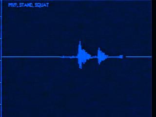
71 71 Still s murmur versus subaortic stenosis Subaortic stenosis can mimic Still s: both can be musical

72 72 Still s murmur versus Subaortic Stenosis Use positional changes to help distinguish subas from Still s Dynamic obstruction in HCM accentuated with decrease in filling: murmur gets louder with stand Discrete sub AS will not usually get louder, but will also not diminish with stand
73 73 Innocent Murmur: Venous Hum Venous Hum: innocent continuous murmur Turbulent flow merging from internal jugular and subclavian veins into SVC Louder in diastole Disappear when patient lies supine or turns head Audible along infraclavicular area, and low anterior neck (not the head) I- III/VI


74 74 Innocent Murmur: Venous Hum Continuous murmur: whining, roaring, whirring, waterfall
75 75 Venous Hum versus Pathologic Continuous Murmurs PDA Coronary fistula Cranial AVM AO- PA shunt
76 76 Innocent Murmur: Peripheral Pulmonary Stenosis PPS in infant under 6 mos: same pitch as respirations As loud or loudest in back or axilla Systolic, high pitched, blowing Relatively small branch Pas arising at acute angle from large MPA
77 77 PPS versus Pathologic Murmurs PPS- distinguish from Tiny VSD: better heard at mid to LLSB, not back/axilla PS: has a click ASD: Has abnl S2
78 78 Position Changes Distinguish innocent Still s murmurs from LVOTO Detect gallops: apex, left lateral decubitus Distinguish venous hums from non- innocent continuous murmurs Mitral valve prolapse


79 Mitral Valve Prolapse 79
80 80 Auscultation Artificial valves: should be audible without a stethoscope Artificial aortic valves should have a mechanical S2 Artificial mitral valves should have a mechanical S1 Worry if it goes away- valve thrombosis
81 81 Tips for better exams Quiet room Recognize that naptime, stranger anxiety, hunger can adversely affect the situation Make the child as comfortable as possible: Silent distracters to entertain the child- flashlight, ID badge, toys, siblings
82 82 Tips for Better Exams Tiny bodies: Use the right size stethoscope to minimize ambient noise and to accurately determine the presence and location of a murmur Change the order of the exam to fit the child Warm hands and scopes
83 83 Remember- Always not normal: RV impulse, thrills, apical murmurs, murmurs that increase with sitting or standing, murmurs with extra heart sounds, diastolic murmurs Need to have a normally split S2 to be normal If it does not sound innocent- needs further evaluation Thank you.
Heart sounds and murmurs. Dr. Szathmári Miklós Semmelweis University First Department of Medicine 15. Oct
 Heart sounds and murmurs Dr. Szathmári Miklós Semmelweis University First Department of Medicine 15. Oct. 2013. Conditions for auscultation of the heart Quiet room Patient comfortable Chest fully exposed
What s That Sound? Pediatric Murmur Evaluation
 What s That Sound? Pediatric Murmur Evaluation Jamie S. Sutherell, M.D, M.Ed. Associate Professor, Pediatrics Division of Cardiology Director, Medical Student Education in Pediatrics Director, Pediatric
Murmur Sounds made by turbulence in the heart or blood stream. 1. Timing. 5. Intensity 2. Shape. 6. Pitch 3. Location of maximum intensity
 Definition Items in description of Timing Shape Location of maximum intensity Murmur Sounds made by turbulence in the heart or blood stream. 1. Timing 5. Intensity 2. Shape 6. Pitch 3. Location of maximum
Cardiac Ausculation in the Elderly
 Cardiac Ausculation in the Elderly 박성하 신촌세브란스병원심장혈관병원심장내과 Anatomy Surface projection of the Heart and Great Vessels Evaluating pulsation Superior vena cava Rt. pulmonary artery Right atrium Right ventricle
CARDIAC EXAMINATION MINI-QUIZ
 CARDIAC EXAMINATION MINI-QUIZ 1. Sitting bolt upright, your dyspneic (short of breath) patient has visible jugular venous pulsations to the angle of his jaw, which is 12 cm above his sternal angle. What
Cardiac Examination. Pediatrics Clinical Examination
 Pediatrics Clinical Examination Symptoms of Cardiovascular Affection: Cardiac Examination 1. Perinatal history: Maternal DM, cyanosis, respiratory distress 2. Symptoms of lung congestion: Poor interrupted
Congenital heart disease. By Dr Saima Ali Professor of pediatrics
 Congenital heart disease By Dr Saima Ali Professor of pediatrics What is the most striking clinical finding in this child? Learning objectives By the end of this lecture, final year student should be able
PEDIATRIC HEART MURMURS. Manish Bansal, MD Clinical Assistant Professor Division of Pediatric Cardiology University of Iowa
 PEDIATRIC HEART MURMURS Manish Bansal, MD Clinical Assistant Professor Division of Pediatric Cardiology University of Iowa Murmur murmur. (n.d.). Dictionary.com Unabridged. Retrieved March 9, 2018 from
2. The heart sounds are produced by a summed series of mechanical events, as follows:
 Heart Sounds. Phonocardiography 1 Objectives 1. Phonocardiography - Definition 2. What produces the heart sounds 3. Where to listen for the heart sounds 4. How to record a phonocardiogram 5. Normal heart
Congenital Heart Disease: Physiology and Common Defects
 Congenital Heart Disease: Physiology and Common Defects Jamie S. Sutherell, M.D, M.Ed. Associate Professor, Pediatrics Division of Cardiology Director, Medical Student Education in Pediatrics Director,
HISTORY. Question: What category of heart disease is suggested by this history? CHIEF COMPLAINT: Heart murmur present since early infancy.
 HISTORY 18-year-old man. CHIEF COMPLAINT: Heart murmur present since early infancy. PRESENT ILLNESS: Although normal at birth, a heart murmur was heard at the six week check-up and has persisted since
HISTORY. Question: What type of heart disease is suggested by this history? CHIEF COMPLAINT: Decreasing exercise tolerance.
 HISTORY 15-year-old male. CHIEF COMPLAINT: Decreasing exercise tolerance. PRESENT ILLNESS: A heart murmur was noted in childhood, but subsequent medical care was sporadic. Easy fatigability and slight
Physical Exam Part II
 Physical Exam Part II University of Michigan Cardiovascular Center Kim A. Eagle, MD Albion Walter Hewlett Professor Director Physical Exam: Part II Heart Sounds Heart Murmurs HEART SOUNDS S1 MITRAL + TRICUSPID
ECHOCARDIOGRAPHIC APPROACH TO CONGENITAL HEART DISEASE: THE UNOPERATED ADULT
 ECHOCARDIOGRAPHIC APPROACH TO CONGENITAL HEART DISEASE: THE UNOPERATED ADULT Karen Stout, MD, FACC Divisions of Cardiology University of Washington Medical Center Seattle Children s Hospital NO DISCLOSURES
2) VSD & PDA - Dr. Aso
 VSD & PDA - Dr. Aso") 2) VSD & PDA - Dr. Aso Ventricular Septal Defect (VSD) Most common cardiac malformation 25-30 % Types of VSD: According to position perimembranous, inlet, muscular. According to size small, medium, large.
2) VSD & PDA - Dr. Aso Ventricular Septal Defect (VSD) Most common cardiac malformation 25-30 % Types of VSD: According to position perimembranous, inlet, muscular. According to size small, medium, large.
Uptofate Study Summary
 CONGENITAL HEART DISEASE Uptofate Study Summary Acyanotic Atrial septal defect Ventricular septal defect Patent foramen ovale Patent ductus arteriosus Aortic coartation Pulmonary stenosis Cyanotic Tetralogy
Clinical significance of cardiac murmurs: Get the sound and rhythm!
 Clinical significance of cardiac murmurs: Get the sound and rhythm! Prof. dr. Gunther van Loon, DVM, PhD, Ass Member ECVDI, Dip ECEIM Dept. of Large Animal Internal Medicine Ghent University, Belgium Murmurs
Paediatrics Revision Session Cardiology. Emma Walker 7 th May 2016
 Paediatrics Revision Session Cardiology Emma Walker 7 th May 2016 Cardiovascular Examination! General:! Make it fun!! Change how you act depending on their age! Introduction! Introduce yourself & check
Paediatric Cardiology. Acyanotic CHD. Prof F F Takawira
 Paediatric Cardiology Acyanotic CHD Prof F F Takawira Aetiology Chromosomal Down syndrome, T13, T18 Genetic syndromes (gene defects) Velo-Cardio-facial (22 del) Genetic syndromes (undefined aetiology)
COLIC AND MURMURS: AN OVERVIEW
 COLIC AND MURMURS: AN OVERVIEW Gunther van Loon, DVM, PhD, Department of Large Animal Internal Medicine, Ghent University, Merelbeke, Belgium Introduction Many horses with colic present with a cardiac
1. how a careful cardiovascular evaluation can accurately assess pathology and physiology at the bedside, and
 This program will demonstrate: 1. how a careful cardiovascular evaluation can accurately assess pathology and physiology at the bedside, and 2. the importance of integrating this information with selected
Notes by Sandra Dankwa 2009 HF- Heart Failure DS- Down Syndrome IE- Infective Endocarditis ET- Exercise Tolerance. Small VSD Symptoms -asymptomatic
 Congenital Heart Disease: Notes. Condition Pathology PC Ix Rx Ventricular septal defect (VSD) L R shuntsdefect anywhere in the ventricle, usually perimembranous (next to the tricuspid valve) 30% 1)small
SMALL GROUP SESSION 19 January 30 th or February 1st. Groups 1-12: Cardiac Case and Cardiac Exam Workshop
 SMALL GROUP SESSION 19 January 30 th or February 1st Groups 1-12: Cardiac Case and Cardiac Exam Workshop Readings: Complete the cardiac examination tutorial on the POM1 web site. Optional: http://medicine.ucsd.edu/clinicalmed/heart.htm
Pathophysiology: Left To Right Shunts
 Pathophysiology: Left To Right Shunts Daphne T. Hsu, MD dh17@columbia.edu Learning Objectives Learn the relationships between pressure, blood flow, and resistance Review the transition from fetal to mature
Absent Pulmonary Valve Syndrome
 Absent Pulmonary Valve Syndrome Fact sheet on Absent Pulmonary Valve Syndrome In this condition, which has some similarities to Fallot's Tetralogy, there is a VSD with narrowing at the pulmonary valve.
Pathophysiology: Left To Right Shunts
 Pathophysiology: Left To Right Shunts Daphne T. Hsu, MD dh17@columbia.edu Learning Objectives Learn the relationships between pressure, blood flow, and resistance Review the transition from fetal to mature
Congenital Heart Disease. Mohamed Waheed Elsharief.
 Congenital Heart Disease Mohamed Waheed Elsharief. Objectives l By the end of this lecture you should be able to Fetal Circulation l For the fetus the placenta is the oxygenator so the lungs do little
Congenital Heart Disease
 Congenital Heart Disease Mohammed Alghamdi, MD, FRCPC, FAAP, FACC Associate Professor and Consultant Pediatric Cardiology, Cardiac Science King Fahad Cardiac Centre King Saud University INTRODUCTION CHD
Pediatric Board Review Congenital Heart Disease. Steven H. Todman, M.D. Pediatric Cardiologist Louisiana State University
 Pediatric Board Review Congenital Heart Disease Steven H. Todman, M.D. Pediatric Cardiologist Louisiana State University Our Mission To discuss various types of congenital heart disease that are commonly
Echocardiographic assessment in Adult Patients with Congenital Heart Diseases
 Echocardiographic assessment in Adult Patients with Congenital Heart Diseases Athanasios Koutsakis Cardiologist, Cl. Research Fellow George Giannakoulas Ass. Professor in Cardiology 1st Cardiology Department,
Are you too quick to refer?
 HEART MURMURS IN CHILDREN Are you too quick to refer? Many children with murmurs are unnecessarily referred, say the authors, who describe a thorough cardiovascular exam you can perform in three to five
HEART MURMURS: DECIPHERING THEIR CAUSE AND SIGNIFICANCE
 Vet Times The website for the veterinary profession https://www.vettimes.co.uk HEART MURMURS: DECIPHERING THEIR CAUSE AND SIGNIFICANCE Author : Pedro Oliveira Categories : Vets Date : May 27, 2013 PEDRO
Heart and Neck Vessels
 CHAPTER 12 Heart and Neck Vessels ANATOMY The precordium is the area on the anterior chest overlying the heart and great vessels. The heart extends from the second to the fifth intercostal space, and from
Introduction. Pediatric Cardiology. General Appearance. Tools of Assessment. Auscultation. Vital Signs
 Introduction Pediatric Cardiology An introduction to the pediatric patient with heart disease: M-III Lecture Douglas R. Allen, M.D. Assistant Professor and Director of Community Pediatric Cardiology at
Most common fetal cardiac anomalies
 Most common fetal cardiac anomalies Common congenital heart defects CHD % of cardiac defects Chromosomal Infants Fetuses anomaly (%) 22q11 deletion (%) VSD 30 5~10 20~40 10 PS 9 5 (PA w/ VSD) HLHS 7~9
Congenital Heart Disease An Approach for Simple and Complex Anomalies
 Congenital Heart Disease An Approach for Simple and Complex Anomalies Michael D. Pettersen, MD Director, Echocardiography Rocky Mountain Hospital for Children Denver, CO None Disclosures 1 ASCeXAM Contains
CARDIOVASCULAR PHYSIOLOGY
 CARDIOVASCULAR PHYSIOLOGY LECTURE 4 Cardiac cycle Polygram - analysis of cardiac activity Ana-Maria Zagrean MD, PhD The Cardiac Cycle - definitions: the sequence of electrical and mechanical events that
Patent ductus arteriosus PDA
 Patent ductus arteriosus PDA Is connecting between the aortic end just distal to the origin of the LT sub clavian artery& the pulmonary artery at its bifurcation. Female/male ratio is 2:1 and it is more
Congenital heart disease: When to act and what to do?
 Leading Article Congenital heart disease: When to act and what to do? Duminda Samarasinghe 1 Sri Lanka Journal of Child Health, 2010; 39: 39-43 (Key words: Congenital heart disease) Congenital heart disease
CONGENITAL HEART DISEASE (CHD)
") CONGENITAL HEART DISEASE (CHD) DEFINITION It is the result of a structural or functional abnormality of the cardiovascular system at birth GENERAL FEATURES OF CHD Structural defects due to specific disturbance
CONGENITAL HEART DISEASE (CHD) DEFINITION It is the result of a structural or functional abnormality of the cardiovascular system at birth GENERAL FEATURES OF CHD Structural defects due to specific disturbance
The production of murmurs is due to 3 main factors:
 Heart murmurs The production of murmurs is due to 3 main factors: high blood flow rate through normal or abnormal orifices forward flow through a narrowed or irregular orifice into a dilated vessel or
The Cardiac Cycle Clive M. Baumgarten, Ph.D.
 The Cardiac Cycle Clive M. Baumgarten, Ph.D. OBJECTIVES: 1. Describe periods comprising cardiac cycle and events within each period 2. Describe the temporal relationships between pressure, blood flow,
By Dickens ATURWANAHO & ORIBA DAN LANGOYA MAKchs, MBchB CONGENTAL HEART DISEASE
 By Dickens ATURWANAHO & ORIBA DAN LANGOYA MAKchs, MBchB CONGENTAL HEART DISEASE Introduction CHDs are abnormalities of the heart or great vessels that are present at birth. Common type of heart disease
HISTORY. Question: What category of heart disease is suggested by the fact that a murmur was heard at birth?
 HISTORY 23-year-old man. CHIEF COMPLAINT: Decreasing exercise tolerance of several years duration. PRESENT ILLNESS: The patient is the product of an uncomplicated term pregnancy. A heart murmur was discovered
بسم هللا الرحمن الرحيم. The cardio vascular system By Dr.Rawa Younis Mahmood
 بسم هللا الرحمن الرحيم The cardio vascular system By Dr.Rawa Younis Mahmood Introduction Evaluation of the cardio vascular system depend on history and physical examination by : Asking about cyanosis,blueness
DORV: The Great Chameleon. Heart Conference October 15, 2016 Tina Kwan, MD
 DORV: The Great Chameleon Heart Conference October 15, 2016 Tina Kwan, MD Kenneth Maehara, Ph.D. May 7, 1942 - August 26, 2013 A.R. A classic case of broken heart 38 week AGA F born at an OSH to
5.8 Congenital Heart Disease
 5.8 Congenital Heart Disease Congenital heart diseases (CHD) refer to structural or functional heart diseases, which are present at birth. Some of these lesions may be discovered later. prevalence of Chd
CMR for Congenital Heart Disease
 CMR for Congenital Heart Disease * Second-line tool after TTE * Strengths of CMR : tissue characterisation, comprehensive access and coverage, relatively accurate measurements of biventricular function/
Leicester Medical School
 Leicester Medical School THE CARDIOVASCULAR SYSTEM PHYSICAL EXAMINATION Overview The cardiovascular examination should include the following: - General inspection from the end of the bed. - General examination
Congenital Heart Disease: Cyanotic Lesions. Amitesh Aggarwal
 Congenital Heart Disease: Cyanotic Lesions Amitesh Aggarwal 12 y/o male admitted because of dyspnea and cyanosis Patient has been cyanotic since few months after birth Has episodes of tachypnea and worsening
Adult Congenital Heart Disease: What All Echocardiographers Should Know Sharon L. Roble, MD, FACC Echo Hawaii 2016
 1 Adult Congenital Heart Disease: What All Echocardiographers Should Know Sharon L. Roble, MD, FACC Echo Hawaii 2016 DISCLOSURES I have no disclosures relevant to today s talk 2 Why should all echocardiographers
Cardiac Evaluation of the Adolescent: The Examination
 Cardiac Evaluation of the Adolescent: The Examination murmurlab.org The Helen B. Taussig Children s Heart Center W. Reid Thompson, M.D. Department of Pediatrics The Johns Hopkins University School of Medicine
ADULT CONGENITAL HEART DISEASE. Stuart Lilley
 ADULT CONGENITAL HEART DISEASE Stuart Lilley More adults than children have congenital heart disease Huge variety of congenital lesions from minor to major Heart failure, re-operation and arrhythmia are
Anatomy & Physiology
 1 Anatomy & Physiology Heart is divided into four chambers, two atrias & two ventricles. Atrioventricular valves (tricuspid & mitral) separate the atria from ventricles. they open & close to control flow
Cardiac Catheterization Cases Primary Cardiac Diagnoses Facility 12 month period from to PRIMARY DIAGNOSES (one per patient)
") PRIMARY DIAGNOSES (one per patient) Septal Defects ASD (Atrial Septal Defect) PFO (Patent Foramen Ovale) ASD, Secundum ASD, Sinus venosus ASD, Coronary sinus ASD, Common atrium (single atrium) VSD (Ventricular
PRIMARY DIAGNOSES (one per patient) Septal Defects ASD (Atrial Septal Defect) PFO (Patent Foramen Ovale) ASD, Secundum ASD, Sinus venosus ASD, Coronary sinus ASD, Common atrium (single atrium) VSD (Ventricular
The production of murmurs is due to 3 main factors:
 Heart murmurs The production of murmurs is due to 3 main factors: high blood flow rate through normal or abnormal orifices forward flow through a narrowed or irregular orifice into a dilated vessel or
Cases in Adult Congenital Heart Disease
 Cases in Adult Congenital Heart Disease Sabrina Phillips, MD FACC FASE Associate Professor of Medicine The University of Oklahoma Health Sciences Center No Disclosures I Have Palpitations 18 Year old Man
CongHeartDis.doc. Андрій Миколайович Лобода
 CongHeartDis.doc Андрій Миколайович Лобода 2015 Зміст 3 Зміст Зміст 4 A child with tetralogy of Fallot is most likely to exhibit: -Increased pulmonary blood flow -Increased pressure in the right ventricle
PHONOCARDIOGRAPHY (PCG)
") PHONOCARDIOGRAPHY (PCG) The technique of listening to sounds produced by the organs and vessels of the body is called auscultation. The areas at which the heart sounds are heard better are called auscultation
PHONOCARDIOGRAPHY (PCG) The technique of listening to sounds produced by the organs and vessels of the body is called auscultation. The areas at which the heart sounds are heard better are called auscultation
PedsCases Podcast Scripts
 PedsCases Podcast Scripts This is a text version of a podcast from Pedscases.com on Acyanotic Congenital Heart Lesions. These podcasts are designed to give medical students an overview of key topics in
Adults with Congenital Heart Disease
 Adults with Congenital Heart Disease Edward K. Rhee, MD, FACC Director, Pediatric-Adult Congenital Arrhythmia Service SJHMC Disclosures & Disclaimer I have no lucrative financial relationships with industry
PATENT DUCTUS ARTERIOSUS (PDA)
") PATENT DUCTUS ARTERIOSUS (PDA) It is a channel that connect the pulmonary artery with the descending aorta (isthumus part). It results from the persistence of patency of the fetal ductus arteriosus after
PATENT DUCTUS ARTERIOSUS (PDA) It is a channel that connect the pulmonary artery with the descending aorta (isthumus part). It results from the persistence of patency of the fetal ductus arteriosus after
3/14/2011 MANAGEMENT OF NEWBORNS CARDIAC INTENSIVE CARE CONFERENCE FOR HEALTH PROFESSIONALS IRVINE, CA. MARCH 7, 2011 WITH HEART DEFECTS
 CONFERENCE FOR HEALTH PROFESSIONALS IRVINE, CA. MARCH 7, 2011 MANAGEMENT OF NEWBORNS WITH HEART DEFECTS A NTHONY C. CHANG, MD, MBA, MPH M E D I C AL D I RE C T OR, HEART I N S T I T U T E C H I LDRE N
Congenital Heart Defects
 Normal Heart Congenital Heart Defects 1. Patent Ductus Arteriosus The ductus arteriosus connects the main pulmonary artery to the aorta. In utero, it allows the blood leaving the right ventricle to bypass
Echocardiography in Adult Congenital Heart Disease
 Echocardiography in Adult Congenital Heart Disease Michael Vogel Kinderherz-Praxis München CHD missed in childhood Subsequent lesions after repaired CHD Follow-up of cyanotic heart disease CHD missed in
Tracheal normal sound heard over trachea loud tubular quality high-pitched expiration equal to or slightly longer than inspiration
 = listening for sounds produced in the body over chest to ID normal & abnormal lung sounds all BS made by turbulent flow in the airways useful in making initial D & evaluating effects of R 4 characteristics
CARDIOVASCULAR PHYSICAL EXAMINATION
 CARDIOVASCULAR PHYSICAL EXAMINATION Clarke Atkins, DVM, Diplomate, ACVIM (Internal Medicine and Cardiology) Jane Lewis Seaks Distinguished Professor Emeritus North Carolina State University, College of
Slide 1. Slide 2. Slide 3 CONGENITAL HEART DISEASE. Papworth Hospital NHS Trust INTRODUCTION. Jakub Kadlec/Catherine Sudarshan INTRODUCTION
 Slide 1 CONGENITAL HEART DISEASE Jakub Kadlec/Catherine Sudarshan NHS Trust Slide 2 INTRODUCTION Most common congenital illness in the newborn Affects about 4 9 / 1000 full-term live births in the UK 1.5
SAMPLE HLTEN610A. TAFE NSW Training and Education Support Industry Skills Unit, Meadowbank. Practise in the cardiovascular nursing environment
 TAFE NSW Training and Education Support Industry Skills Unit, Meadowbank HLTEN610A Practise in the cardiovascular nursing environment Version 1.0 Flexible Learner Resource Product Code: ISO 9001 HLTEN610A
September 26, 2012 Philip Stockwell, MD Lifespan CVI Assistant Professor of Medicine (Clinical)
") September 26, 2012 Philip Stockwell, MD Lifespan CVI Assistant Professor of Medicine (Clinical) Advances in cardiac surgery have created a new population of adult patients with repaired congenital heart
September 26, 2012 Philip Stockwell, MD Lifespan CVI Assistant Professor of Medicine (Clinical) Advances in cardiac surgery have created a new population of adult patients with repaired congenital heart
Cardiovascular System
 Cardiovascular System Chapter 8 1 Cardiovascular System Functions: pump saturated oxygenated blood into arterial system cells pump desaturated deoxygenated blood to lungs via veins for reoxygenation Heart
Cardiovascular Pathophysiology: Right to Left Shunts aka Cyanotic Lesions
 Cardiovascular Pathophysiology: Right to Left Shunts aka Cyanotic Lesions Ismee A. Williams, MD, MS iib6@columbia.edu Pediatric Cardiology Learning Objectives To discuss the hemodynamic significance of
Cardiovascular Pathophysiology: Right to Left Shunts aka Cyanotic Lesions Ismee A. Williams, MD, MS Pediatric Cardiology
 Cardiovascular Pathophysiology: Right to Left Shunts aka Cyanotic Lesions Ismee A. Williams, MD, MS iib6@columbia.edu Pediatric Cardiology Learning Objectives To discuss the hemodynamic significance of
NASCO LF01199 AUSCULTATION POSTERIOR BOARD INSTRUCTION SHEET
 NASCO LF01199 AUSCULTATION POSTERIOR BOARD INSTRUCTION SHEET Attaching to a Case Without Side Rails If you have a case without rails on the inside of the cover, with the metal Serial Tag riveted in the
Cardiac Cycle MCQ. Professor of Cardiovascular Physiology. Cairo University 2007
 Cardiac Cycle MCQ Abdel Moniem Ibrahim Ahmed, MD Professor of Cardiovascular Physiology Cairo University 2007 1- Regarding the length of systole and diastole: a- At heart rate 75 b/min, the duration of
Case 47 Clinical Presentation
 93 Case 47 C Clinical Presentation 45-year-old man presents with chest pain and new onset of a murmur. Echocardiography shows severe aortic insufficiency. 94 RadCases Cardiac Imaging Imaging Findings C
Large Arteries of Heart
 Cardiovascular System (Part A-2) Module 5 -Chapter 8 Overview Arteries Capillaries Veins Heart Anatomy Conduction System Blood pressure Fetal circulation Susie Turner, M.D. 1/5/13 Large Arteries of Heart
THE SOUNDS AND MURMURS IN TRANSPOSITION OF THE
 Brit. Heart J., 25, 1963, 748. THE SOUNDS AND MURMURS IN TRANSPOSITION OF THE GREAT VESSELS BY BERTRAND WELLS From The Hospital for Sick Children, Great Ormond Street, London W. C.J Received April 18,
Physical Exam Of The Cardio- Vascular System. James G. Laws D.O. MACOI
 Physical Exam Of The Cardio- Vascular System James G. Laws D.O. MACOI An Important Part Of The Physical Exam Of The Cardio-Vascular System That Is Commonly Overlooked Is? 20% 20% 20% 20% 20% 1. Palpation
Hypoplastic Left Heart Syndrome: Echocardiographic Assessment
 Hypoplastic Left Heart Syndrome: Echocardiographic Assessment Craig E Fleishman, MD, FACC, FASE Director, Non-invasive Cardiac Imaging The Hear Center at Arnold Palmer Hospital for Children, Orlando SCAI
Giovanni Di Salvo MD, PhD, FESC Second University of Naples Monaldi Hospital
 Giovanni Di Salvo MD, PhD, FESC Second University of Naples Monaldi Hospital VSD is one of the most common congenital cardiac abnormalities in the newborn. It can occur as an isolated finding or in combination
Atrial Septal Defects
 Supplementary ACHD Echo Acquisition Protocol for Atrial Septal Defects The following protocol for echo in adult patients with atrial septal defects (ASDs) is a guide for performing a comprehensive assessment
The background of the Cardiac Sonographer Network News masthead is a diagnostic image:
 Number 5 Welcome Number 5 Welcome to the newsletter created just for you: sonographers who perform pediatric echocardiograms in primarily adult echo labs. Each issue features tips on echocardiography of
Heart and Soul Evaluation of the Fetal Heart
 Heart and Soul Evaluation of the Fetal Heart Ivana M. Vettraino, M.D., M.B.A. Clinical Associate Professor, Michigan State University College of Human Medicine Objectives Review the embryology of the formation
Common Defects With Expected Adult Survival:
 Common Defects With Expected Adult Survival: Bicuspid aortic valve :Acyanotic Mitral valve prolapse Coarctation of aorta Pulmonary valve stenosis Atrial septal defect Patent ductus arteriosus (V.S.D.)
M-Mode Echocardiography Is it still Alive? Itzhak Kronzon, MD,FASE. Sampling Rate M-Mode: 1800 / sec 2D: 30 / sec
 M-Mode Echocardiography Is it still Alive? Itzhak Kronzon, MD,FASE Honoraria: Philips Classical M-mode Echocardiography M-Mode offers better time and image resolution. Sampling Rate M-Mode: 1800 / sec
HISTORY. Question: How do you interpret the patient s history? CHIEF COMPLAINT: Dyspnea of two days duration. PRESENT ILLNESS: 45-year-old man.
 HISTORY 45-year-old man. CHIEF COMPLAINT: Dyspnea of two days duration. PRESENT ILLNESS: His dyspnea began suddenly and has been associated with orthopnea, but no chest pain. For two months he has felt
Adult Congenital Heart Disease for the Internist
 Adult Congenital Heart Disease for the Internist Saurabh Rajpal, MBBS, MD Assistant Professor Department of Internal Medicine Division of Cardiovascular Medicine The Ohio State University Wexner Medical
Adult Congenital Heart Disease for the Internist
 Adult Congenital Heart Disease for the Internist Saurabh Rajpal, MBBS, MD Assistant Professor Department of Internal Medicine Division of Cardiovascular Medicine The Ohio State University Wexner Medical
See below for descriptions of the waveform
 The internal jugular vein (IJV) connects to the right atrium without any intervening valves. The pulsation of the right atrium therefore causes the column of blood in the IJV to rise and fall this is called
DEVELOPMENT OF THE CIRCULATORY SYSTEM L E C T U R E 5
 DEVELOPMENT OF THE CIRCULATORY SYSTEM L E C T U R E 5 REVIEW OF CARDIAC ANATOMY Heart 4 chambers Base and apex Valves Pericardial sac 3 layers: epi, myo, endo cardium Major blood vessels Aorta and its
CARDIAC CYCLE CONTENTS. Divisions of cardiac cycle 11/13/13. Definition. Badri Paudel GMC
 CARDIAC CYCLE Badri Paudel GMC CONTENTS Ø DEFINATION Ø DIVISION OF CARDIAC CYCLE Ø SUB DIVISION AND DURATION OF CARDIAC CYCLE Ø SYSTOLE Ø DIASTOLE Ø DESCRIPTION OF EVENTS OF CARDIAC CYCLE Ø SUMMARY Ø ELECTROCARDIOGRAPHY
Ummeenatrbilaoiasetptiwmsaiiri
 atrial This This atrial CIRCULATORY CHANGES My My pressure In the foetus the left atrial is low as relatively Ummeenatrbilaoiasetptiwmsaiiri ze@fgffmftheyubsidtritupyeiirieminfyifjjtajefjjieiminylntentiiiarmmnitnteimiiiinc1udingfromthepl9centaj
Proceeding of the LAVC Latin American Veterinary Conference Apr , 2016 Lima, Perú
 Close this window to return to IVIS www.ivis.org Proceeding of the LAVC Latin American Veterinary Conference Apr. 20-22, 2016 Lima, Perú Next LAVC Conference: Apr. 4-7, 2017 Lima, Perú Reprinted in the
ASCeXAM / ReASCE. Practice Board Exam Questions. Tuesday Morning
 ASCeXAM / ReASCE Practice Board Exam Questions Tuesday Morning Congenital Heart Disease in Adults Congenital Heart Disease Cases Diastolic Function Pericardial Disease Congenital Heart Disease Cases Michael
Adult Echocardiography Examination Content Outline
 Adult Echocardiography Examination Content Outline (Outline Summary) # Domain Subdomain Percentage 1 2 3 4 5 Anatomy and Physiology Pathology Clinical Care and Safety Measurement Techniques, Maneuvers,
Congenital Heart Disease. Children s Hospital Zhejiang University School of Medicine
 Congenital Heart Disease Children s Hospital Zhejiang University School of Medicine Cardiovascular Development Key periods:the 2 nd ~8 th weeks Fetal Circulation Physiological Changes in the Circulation
CARDIOLOGY FOR DUMMIES
 CARDIOLOGY FOR DUMMIES Prof. dr. Gunther van Loon, DVM, PhD, Dipl ECEIM Dept. of Large Animal Internal Medicine, Fac. of Veterinary Medicine, Ghent University, Salisburylaan 133, B-9820 Merelbeke, Belgium
Miscellaneous Cardiology Topics pregnancy - congenital - myocarditis - pericardial disease. Pregnancy and Cardiovascular Disease MCQ
 Miscellaneous Cardiology Topics pregnancy - congenital - myocarditis - pericardial disease Maan Jokhadar, MD, FACC Emory Center for Advanced Heart Failure Therapy Emory Adult Congenital Heart Center Pregnancy
Neonatal Cardiac Assessment and Congenial Heart Disease compiled by Violet Stephens based on the works of Patricia Hartley
 Neonatal Cardiac Assessment and Congenial Heart Disease compiled by Violet Stephens based on the works of Patricia Hartley Course Contents Purpose Objectives Introduction Fetal Circulation Neonatal Circulation