Chapter 11. The Cardiovascular System
|
|
|
- Basil Powell
- 5 years ago
- Views:
Transcription

1 Chapter 11 The Cardiovascular System
2 The Cardiovascular System
3 The Cardiovascular System A closed system of the heart and blood vessels Heart pumps blood Blood vessels circulate blood to all parts of the body Functions of the cardiovascular system: Deliver oxygen and nutrients to cells and tissues Remove carbon dioxide and other waste products from cells and tissues
4 Anatomy of the Heart External Structures Blood Circulation pulmonary and systemic Text p WB Quiz: Identify External Features; Blood pathway/circulation


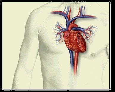
5 The Heart Location Thorax, between the lungs in the inferior mediastinum Orientation Pointed apex directed toward left hip Base points toward right shoulder About the size of a human fist
 Apex of heart (a)")
6 Heart in the Thoracic Cavity Superior vena cava Pulmonary trunk Diaphragm Aorta Pericardium (cut) Apex of heart (a)

7 Heart Location in Thoracic Cavity Midsternal line 2nd rib Diaphragm Sternum

8 Heart Location in the Thoracic Cavity Mediastinum Heart Left lung Posterior

9 Chambers and Associated Great Vessels Right and left side act as separate pumps Four chambers: Atria - Receiving chambers Ventricles - Discharging chambers

10 2015 Pearson Education, Inc.

11 2015 Pearson Education, Inc.
12 Chambers and Associated Great Vessels Pulmonary circulation Blood flows from the right side of the heart to the lungs and back to the left side of the heart Blood is pumped out of right side through the pulmonary trunk, which splits into pulmonary arteries and takes oxygen-poor blood to lungs Oxygen-rich blood returns to the heart from the lungs via pulmonary veins
13 Chambers and Associated Great Vessels Systemic circulation Blood flows from the left side of the heart through body tissues, and back to the right side of the heart Blood returned to the left side of the heart is pumped out into the aorta Oxygen-poor blood circulates to systemic tissues, and returns to the right atrium via systemic veins, which empty blood into the superior and inferior venae cavae
14 Figure 11.4 The systemic and pulmonary circulations. Capillary beds of lungs where gas exchange occurs Pulmonary Circuit Pulmonary arteries Pulmonary veins Venae cavae Right atrium Right ventricle Heart Left atrium Systemic Circuit Left ventricle Aorta and branches KEY: Oxygen-rich, CO 2 -poor blood Oxygen-poor, CO 2 -rich blood Capillary beds of all body tissues where gas exchange occurs 2015 Pearson Education, Inc.

15 Blood Flow Right side pulmonary region left side Left side body region right side Vein heart atrium ventricle artery Artery capillary bed vein 2015 Pearson Education, Inc.

16 2015 Pearson Education, Inc.
17 Heart Valves Allow blood to flow in only one direction, to prevent backflow Four valves Atrioventricular (AV) valves between atria and ventricles Bicuspid (mitral) valve (left side of heart) Tricuspid valve (right side of heart) Semilunar valves between ventricle and artery Pulmonary semilunar valve Aortic semilunar valve
18 Heart Valves AV valves Anchored in place by chordae tendineae ( heart strings ) Open during heart relaxation and closed during ventricular contraction Semilunar valves Closed during heart relaxation but open during ventricular contraction These valves open and close in response to pressure changes in the heart
19 Figure 11.6a Operation of the heart valves. Slide 2 (a) Operation of the AV valves 1 Blood returning to the atria puts pressure against AV valves; the AV valves are forced open. Ventricles 2015 Pearson Education, Inc.
20 Figure 11.6a Operation of the heart valves. Slide 3 (a) Operation of the AV valves 1 Blood returning to the atria puts pressure against AV valves; the AV valves are forced open. 2 As the ventricles fill, AV valve flaps hang limply into ventricles. Ventricles 2015 Pearson Education, Inc.
21 Figure 11.6a Operation of the heart valves. Slide 4 (a) Operation of the AV valves 1 Blood returning to the atria puts pressure against AV valves; the AV valves are forced open. 2 As the ventricles fill, AV valve flaps hang limply into ventricles. 3 Atria contract, forcing additional blood into ventricles. Ventricles AV valves open; atrial pressure greater than ventricular pressure 2015 Pearson Education, Inc.
22 Figure 11.6a Operation of the heart valves. Slide 5 (a) Operation of the AV valves 1 Blood returning to the atria puts pressure against AV valves; the AV valves are forced open. 4 Ventricles contract, forcing blood against AV valve flaps. 2 As the ventricles fill, AV valve flaps hang limply into ventricles. 3 Atria contract, forcing additional blood into ventricles. Ventricles AV valves open; atrial pressure greater than ventricular pressure 2015 Pearson Education, Inc.
23 Figure 11.6a Operation of the heart valves. Slide 6 (a) Operation of the AV valves 1 Blood returning to the atria puts pressure against AV valves; the AV valves are forced open. 4 Ventricles contract, forcing blood against AV valve flaps. 5 AV valves close. 2 As the ventricles fill, AV valve flaps hang limply into ventricles. 3 Atria contract, forcing additional blood into ventricles. Ventricles AV valves open; atrial pressure greater than ventricular pressure 2015 Pearson Education, Inc.
24 Figure 11.6a Operation of the heart valves. Slide 7 (a) Operation of the AV valves 1 Blood returning to the atria puts pressure against AV valves; the AV valves are forced open. 2 As the ventricles fill, AV valve flaps hang limply into ventricles. 3 Atria contract, forcing additional blood into ventricles. Ventricles 4 Ventricles contract, forcing blood against AV valve flaps. 5 AV valves close. 6 Chordae tendineae tighten, preventing valve flaps from everting into atria. AV valves open; atrial pressure greater than ventricular pressure AV valves closed; atrial pressure less than ventricular pressure 2015 Pearson Education, Inc.
25 Figure 11.6b Operation of the heart valves. Slide 2 (b) Operation of the semilunar valves Pulmonary trunk 1 As ventricles contract and intraventricular pressure rises, blood is pushed up against semilunar valves, forcing them open. Aorta Semilunar valves open 2015 Pearson Education, Inc.
 Operation of the semilunar valves Pulmonary trunk Aorta 1 As ventricles contract 2 As ventricles relax and intraventricular pressure rises, blood is pushed up against semilunar valves,")
26 Figure 11.6b Operation of the heart valves. Slide 3 (b) Operation of the semilunar valves Pulmonary trunk Aorta 1 As ventricles contract 2 As ventricles relax and intraventricular pressure rises, blood is pushed up against semilunar valves, forcing them open. and intraventricular pressure falls, blood flows back from arteries, filling the leaflets of semilunar valves and forcing them to close. Semilunar valves open Semilunar valves closed 2015 Pearson Education, Inc.
27 Cardiac Circulation Blood in the heart chambers does not nourish the myocardium The heart has its own nourishing circulatory system consisting of: Coronary arteries branch from the aorta to supply the heart muscle with oxygenated blood Cardiac veins drain the myocardium of blood Coronary sinus a large vein on the posterior of the heart, receives blood from cardiac veins Blood empties into the right atrium via the coronary sinus
28 Blood Flow Through the Heart Superior and inferior venae cavae dump blood into the right atrium From right atrium, through the tricuspid valve, blood travels to the right ventricle From the right ventricle, blood leaves the heart as it passes through the pulmonary semilunar valve into the pulmonary trunk Pulmonary trunk splits into right and left pulmonary arteries, which carry blood to the lungs
29 Blood Flow Through the Heart In the lungs, blood picks up oxygen and drops off carbon dioxide Oxygen-rich blood returns to the heart through the four pulmonary veins Blood enters the left atrium and travels through the bicuspid valve into the left ventricle From the left ventricle, blood leaves the heart via the aortic semilunar valve and aorta
30 Figure 11.4 The systemic and pulmonary circulations. Capillary beds of lungs where gas exchange occurs Pulmonary Circuit Pulmonary arteries Pulmonary veins Venae cavae Right atrium Right ventricle Heart Left atrium Systemic Circuit Left ventricle Aorta and branches KEY: Oxygen-rich, CO 2 -poor blood Oxygen-poor, CO 2 -rich blood Capillary beds of all body tissues where gas exchange occurs 2015 Pearson Education, Inc.
31 Physiology of the Heart Text p WB
32 Intrinsic Conduction System of the Heart: Setting the Basic Rhythm Cardiac muscle is able to initiate its own contraction in a regular way, but its rate is influenced by both intrinsic and extrinsic factors The intrinsic conduction (nodal) system increases the rate of heart contraction and ensures that the heart beats as a unit
33 Intrinsic Conduction System of the Heart: Setting the Basic Rhythm Sinoatrial (SA) node is the heart s pacemaker Atrioventricular (AV) node is at the junction of the atria and ventricles Atrioventricular (AV) bundle (bundle of His) is in the interventricular septum Bundle branches are in the interventricular septum Purkinje fibers spread within the ventricle wall muscles
 node (pacemaker) Left atrium Atrioventricular (AV) node Right atrium Bundle branches Purkinje fibers Atrioventricular")
34 Figure 11.7 The intrinsic conduction system of the heart. Superior vena cava Sinoatrial (SA) node (pacemaker) Left atrium Atrioventricular (AV) node Right atrium Bundle branches Purkinje fibers Atrioventricular (AV) bundle (bundle of His) Purkinje fibers Interventricular septum 2015 Pearson Education, Inc.
35 Heart Contractions Intrinsic conduction system enforces 75 beats per minute Contraction is initiated by the sinoatrial node (SA node) Sequential stimulation occurs at other autorhythmic cells Force cardiac muscle depolarization in one direction from atria to ventricles
36 Heart Contractions Once SA node starts the heartbeat Impulse spreads to the AV node Then the atria contract At the AV node, the impulse passes through the AV bundle, bundle branches, and Purkinje fibers Blood is ejected from the ventricles to the aorta and pulmonary trunk as the ventricles contract
37 Heart Contractions Homeostatic imbalance Heart block damaged AV node releases the ventricles from control of the SA node; result is in a slower heart rate as ventricles contract at their own rate Ischemia lack of adequate oxygen supply to heart muscle Fibrillation a rapid, uncoordinated shuddering of the heart muscle
38 Heart Contractions Homeostatic imbalance (continued) Tachycardia rapid heart rate over 100 beats per minute Bradycardia slow heart rate less than 60 beats per minutes
39 Cardiac Cycle & Heart Sounds Cardiac cycle refers to one complete heartbeat Systole contraction Diastole relaxation Heart beats approximately 75 times per minute Cardiac cycle length is normally 0.8 second
40 Cardiac Cycle & Heart Sounds Mid-to-late diastole Pressure in heart is low Blood flows passively into the atria and into ventricles Semilunar valves are closed Atrioventricular valves are open Atria contract and force blood into ventricles
41 Cardiac Cycle & Heart Sounds Ventricular systole Blood pressure rises as ventricles prepare to contract Atrioventricular valves close causing first heart sound, lub Semilunar valves open as blood pushes against them Blood travels out of the ventricles through pulmonary trunk and aorta Atria are relaxed and filling with blood
42 Cardiac Cycle & Heart Sounds Early diastole At the end of systole, all four valves are briefly closed at the same time Second heart sound is heard as semilunar valves close, causing dup sound Closure prevents blood backflow into ventricles Atria finish refilling as pressure in heart drops Ventricular pressure is low Atrioventricular valves open

43 Figure 11.8 Summary of events occurring during the cardiac cycle. Left atrium Right atrium Left ventricle Right ventricle Ventricular filling Atrial contraction Isovolumetric contraction phase Ventricular ejection phase Isovolumetric relaxation 1 Mid-to-late diastole (ventricular filling) 2 Ventricular systole (atria in diastole) 3 Early diastole 2015 Pearson Education, Inc.
44 Homeostatic Imbalance Faulty valves reduce the efficiency of the heart as a pump and result in abnormal heart sounds (murmurs)
45 Cardiac Output Text p WB
46 Cardiac Output Cardiac output (CO) Amount of blood pumped by each side (ventricle) of the heart in one minute Stroke volume (SV) Volume of blood pumped by each ventricle in one contraction (each heartbeat) About 70 ml of blood is pumped out of the left ventricle with each heartbeat Heart rate (HR) Typically 75 beats per minute CO = HR SV CO = HR (75 beats/min) SV (70 ml/beat) CO = 5250 ml/min = 5.25 L/min
47 Regulation of Stroke Volume 60 percent of blood in ventricles (about 70 ml) is pumped with each heartbeat Starling s law of the heart: Critical factor controlling SV The more the cardiac muscle is stretched, the stronger the contraction SV rises or falls with the volume of venous return
48 Regulation of Heart Rate Heart rate is modified by: 1. Neural (ANS) controls Sympathetic nervous system Parasympathetic nervous system 2. Hormones and ions 3. Physical factors
 Low blood pressure High blood pressure or blood volume Exercise Decreased blood volume (hemorrhage) Sympathetic")
49 Figure 11.9 Influence of selected factors on cardiac output. Crisis stressors (physical or emotional trauma; increased body temperature; exercise) Low blood pressure High blood pressure or blood volume Exercise Decreased blood volume (hemorrhage) Sympathetic nervous system activity Activation of skeletal muscle and respiratory pumps Crisis has passed Hormones: epinephrine, thyroxine Increased venous return Decreased venous return Parasympathetic nervous system controls (via vagus nerves) Increased contractile force of cardiac muscle Heart rate (beats/min) Stroke volume (ml/beat) KEY: Increases, stimulates Reduces, inhibits Initial stimulus Physiological response End result Cardiac output (ml/min) 2015 Pearson Education, Inc.
50 Blood Vessels Text p WB

51 Blood Vessels
52 Blood Vessels: The Vascular System Vascular system transports blood to the tissues and back to the heart Vessels that carry blood away from the heart: Arteries and arterioles Vessels that play a role in exchanges between tissues and blood: Capillary beds Vessels that return blood toward the heart: Venules and veins
53 Microscopic Anatomy of Blood Vessels Three layers (tunics) in blood vessels (except the capillaries): Tunica intima forms a friction-reducing lining Endothelium Tunica media Smooth muscle and elastic tissue Controlled by sympathetic nervous system Tunica externa forms protective outermost covering Mostly fibrous connective tissue
 Artery Vein 2015 Pearson Education,")
54 Figure 11.10a Structure of blood vessels. (a) Artery Vein 2015 Pearson Education, Inc.

55 Figure 11.10b Structure of blood vessels. Artery Tunica intima Endothelium Loose connective tissue Internal elastic lamina Tunica media Smooth muscle Elastic fibers External elastic lamina Tunica externa Collagen fibers Vein Valve Lumen Arteriole Venule Capillary network Lumen Basement membrane Endothelial cells (b) Capillary 2015 Pearson Education, Inc.
56 Structural Differences in Arteries, Veins, Capillaries Arteries have a thicker tunica media than veins to withstand changes in pressure Veins have a thinner tunica media than arteries and operate under low pressure Veins also have valves to prevent backflow of blood Lumen of veins is larger than that of arteries Skeletal muscle milks blood in veins toward the heart
 Contracted skeletal muscle Valve (closed) Vein Direction of blood")
57 Figure Operation of the muscular pump. Valve (open) Contracted skeletal muscle Valve (closed) Vein Direction of blood flow 2015 Pearson Education, Inc.
58 Structural Differences in Arteries, Veins, Capillaries Capillaries are only one cell layer thick (tunica intima), to allow for exchanges between blood and tissue Capillaries form networks called capillary beds Blood flow through a capillary bed is known as microcirculation
59 Structural Differences in Arteries, Veins, Capillaries Capillary beds consist of two types of vessels: 1. Vascular shunt 2. True capillaries Entrances to capillary beds are guarded by precapillary sphincters Exchanges with tissue cells occur across walls of true capillaries When precapillary sphincters are closed, blood bypasses the local area via the vascular shunt
 Sphincters")
60 Figure 11.12a Anatomy of a capillary bed. Precapillary sphincters Vascular shunt True capillaries Terminal arteriole Postcapillary venule (a) Sphincters open; blood flows through true capillaries Pearson Education, Inc.
 Sphincters closed; blood flows through")
61 Figure 11.12b Anatomy of a capillary bed. Terminal arteriole Postcapillary venule (b) Sphincters closed; blood flows through vascular shunt Pearson Education, Inc.
62 Homeostatic Imbalance Varicose veins Structural defect due to incompetent valves Common vascular problem, especially in people who are obese and people who stand for long periods of time Predisposing factor for thrombophlebitis
63 Major Arteries of the Systemic Circulation Aorta Largest artery in the body Leaves from the left ventricle of the heart Regions Ascending aorta leaves the left ventricle Aortic arch arches to the left Thoracic aorta travels downward through the thorax Abdominal aorta passes through the diaphragm into the abdominopelvic cavity
64 Blood Vessels: Major Arteries and Veins Hepatic Portal Circulation Fetal Circulation Text p WB
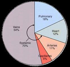
65 Distribution of Blood

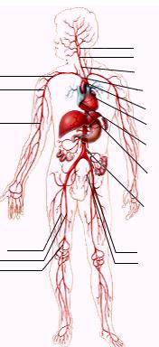
66 Figure Major arteries of the systemic circulation, anterior view. Arteries of the head and trunk Common carotid arteries Subclavian artery Aortic arch Arteries that supply the upper limb Subclavian artery Axillary artery Coronary artery Brachial artery Abdominal aorta Radial artery Ulnar artery Renal artery Arteries that supply the lower limb Common iliac artery Femoral artery 2015 Pearson Education, Inc.
67 Major Veins of Systemic Circulation Superior and inferior venae cavae enter the right atrium of the heart Superior vena cava drains the head and arms Inferior vena cava drains the lower body Superior vena cava Inferior vena cava
68 Figure Major veins of the systemic circulation, anterior view. Veins of the head and trunk Internal jugular vein Veins that drain the upper limb Subclavian vein Superior vena cava Brachial vein Ulnar vein Radial vein Inferior mesenteric vein Digital veins Veins that drain the lower limb Inferior vena cava Femoral vein 2015 Pearson Education, Inc.
69 Fetal Circulation Fetal circulation is a temporary circulation seen only in the fetus Fetus receives exchanges of gases, nutrients, and wastes through the placenta Umbilical cord contains three vessels: 1. Umbilical vein carries blood rich in nutrients and oxygen to the fetus 2 3. Umbilical arteries (2) carry carbon dioxide and debris-laden blood from fetus to placenta

70 Figure Schematic of the fetal circulation. Superior vena cava Foramen ovale Ductus arteriosus Pulmonary artery Pulmonary veins Inferior vena cava Hepatic vein Ductus venosus Hepatic portal vein Inferior vena cava Umbilical vein Fetal umbilicus Umbilical cord Umbilical arteries Aorta Common iliac artery External iliac artery Internal iliac artery Urinary bladder KEY: High oxygenation Moderate oxygenation Low oxygenation Very low oxygenation Placenta 2015 Pearson Education, Inc.
71 Fetal Circulation Shunts bypassing the lungs and liver are also present. Blood flow bypasses the liver through the ductus venosus and enters the inferior vena cava right atrium of heart Blood flow bypasses the lungs Blood entering right atrium is shunted directly into left atrium through foramen ovale (becomes fossa ovalis at or after birth) Ductus arteriosus connects aorta and pulmonary trunk (becomes ligamentum arteriosum at birth)
72 Physiology of Circulation: Pulse Blood Pressure Atherosclerosis Text p WB
73 Arterial Pulse Pulse Alternate expansion and recoil of a blood vessel wall (the pressure wave) that occurs as the heart beats Monitored at pressure points in superficial arteries where pulse is easily palpated Pulse averages 70 to 76 beats per minute at rest, in a healthy person
74 Blood Pressure The pressure blood exerts against the inner walls of the blood vessels The force that causes blood to continue to flow in the blood vessels
75 Blood Pressure Gradient Blood is forced along a descending pressure gradient Pressure in blood vessels decreases as distance from the heart increases Pressure is high in the arteries, lower in the capillaries, and lowest in the veins

76 Aorta Arteries Arterioles Capillaries Venules Veins Venae cavae Pressure (mm Hg) Figure Blood pressure in various areas of the cardiovascular system. 120 Systolic pressure Diastolic pressure Pearson Education, Inc.
77 Measuring Blood Pressure Health professionals measure the pressure in large arteries Systolic pressure at the peak of ventricular contraction Diastolic pressure when ventricles relax Expressed as systolic pressure over diastolic pressure: for example, 120/80 mm Hg
 Brachial artery 2015 Pearson Education, Inc.")
78 Figure 11.21a Measuring blood pressure. Blood pressure 120 systolic 70 diastolic (to be measured) Brachial artery 2015 Pearson Education, Inc. (a) The course of the brachial artery of the arm. Assume a blood pressure of 120/70 in a young, healthy person.
79 Blood Pressure Systolic pressure 120 contraction Diastolic pressure 80 relaxation Healthy range:
 The blood pressure cuff is wrapped snugly around the arm just above the elbow and inflated until the cuff pressure exceeds the systolic blood pressure.")
80 Figure 11.21b Measuring blood pressure. Pressure in cuff above 120; no sounds audible Rubber cuff inflated with air 120 mm Hg Brachial artery closed 2015 Pearson Education, Inc. (b) The blood pressure cuff is wrapped snugly around the arm just above the elbow and inflated until the cuff pressure exceeds the systolic blood pressure. At this point, blood flow into the arm is stopped, and a brachial pulse cannot be felt or heard.
 The pressure in the cuff is gradually reduced while the examiner listens (auscultates) for sounds in the")
81 Figure 11.21c Measuring blood pressure. Pressure in cuff below 120, but above mm Hg 70 mm Hg Sounds audible in stethoscope (c) The pressure in the cuff is gradually reduced while the examiner listens (auscultates) for sounds in the brachial artery with a stethoscope. The pressure read as the first soft tapping sounds are heard (the first point at which a small amount of blood is spurting through the constricted artery) is recorded as the systolic pressure Pearson Education, Inc.
 As the pressure is reduced still further, the sounds become louder and more distinct; when the artery is no longer constricted and blood flows freely, the sounds can no longer be heard.")
82 Figure 11.21d Measuring blood pressure. Pressure in cuff below 70; no sounds audible 70 mm Hg 2015 Pearson Education, Inc. (d) As the pressure is reduced still further, the sounds become louder and more distinct; when the artery is no longer constricted and blood flows freely, the sounds can no longer be heard. The pressure at which the sounds disappear is recorded as the diastolic pressure.
83 Effects of Various Factors on Blood Pressure BP CO PR BP blood pressure CO the amount of blood pumped out of the left ventricle per minute PR peripheral resistance, or the amount of friction blood encounters as it flows through vessels
84 Effects of Various Factors on Blood Pressure 1. Neural factors: the autonomic nervous system Sympathetic nervous system promotes narrowing of vessels (vasoconstriction) Vasoconstriction increases blood pressure
85 Effects of Various Factors on Blood Pressure 2. Renal factors: the kidneys Regulation by altering blood volume Renin, an enzyme, is released when arterial pressure is low Renin triggers formation of angiotensin II, a vasoconstrictor Angiotensin II stimulates release of aldosterone Aldosterone enhances sodium reabsorption (and water) by kidneys
86 Effects of Various Factors on Blood Pressure 3. Temperature Heat has a vasodilating effect Cold has a vasoconstricting effect 4. Chemicals Various substances can cause increases or decreases in blood pressure Epinephrine increases heart rate and blood pressure

87 2015 Pearson Education, Inc. Control of Blood
88 Effects of Various Factors on Blood Pressure 5. Diet Commonly believed that a diet low in salt, saturated fats, and cholesterol prevents hypertension (high blood pressure)
 Increased blood viscosity Sympathetic nervous system centers Kidney conserves water and salt Increased stroke")
89 Figure Summary of factors that increase arterial blood pressure. Decreased blood volume Exercise Postural changes Chemicals (renin, nicotine and others) Increased blood viscosity Sympathetic nervous system centers Kidney conserves water and salt Increased stroke volume Increased heart rate Vasoconstriction Increased cardiac output Increased peripheral resistance KEY: Increases, stimulates Initial stimulus Physiological response End result Increased arterial blood pressure 2015 Pearson Education, Inc.
90 Variations in Blood Pressure Normal human range is variable Normal 140 to 110 mm Hg systolic 80 to 70 mm Hg diastolic Hypotension (low blood pressure) Low systolic (below 100 mm Hg) Often associated with illness Acute hypotension is a warning sign for circulatory shock Hypertension (high blood pressure) Sustained elevated arterial pressure of 140/90 mm Hg Warns of increased peripheral resistance
91 Capillaries and Gas Exchange Text p WB

92 Capillary System

93 Capillary exchange
94 Capillary Exchange of Gases & Nutrients Substances move to and from the blood and tissue cells through capillary walls Exchange is due to concentration gradients Oxygen and nutrients leave the blood and move into tissue cells Carbon dioxide and other wastes exit tissue cells and enter the blood Recall that interstitial fluid (tissue fluid) is found between cells
95 Capillary Exchange of Gases & Nutrients Substances enter or leave the blood via: 1. Direct diffusion through membranes 2. Diffusion through intercellular clefts 3. Diffusion through pores of fenestrated capillaries 4. Transport via vesicles
96 Figure Capillary transport mechanisms. Lumen of capillary Vesicles Intercellular cleft Fenestra (pore) 4 Transport via vesicles 3 Diffusion through pore 1 Direct diffusion through membrane 2 Diffusion through intercellular cleft Interstitial fluid 2015 Pearson Education, Inc.
97 Fluid Movements at Capillary Beds Whether fluid moves out of or into a capillary depends on the difference between the two pressures: 1. Blood pressure forces fluid and solutes out of capillaries 2. Osmotic pressure draws fluid into capillaries Blood pressure is higher than osmotic pressure at the arterial end of the capillary bed Blood pressure is lower than osmotic pressure at the venous end of the capillary bed


98 Figure Bulk fluid flows across capillary walls depend largely on the difference between the blood pressure and the osmotic pressure at different regions of the capillary bed. Tissue cell Interstitial fluid Net fluid movement out Net fluid movement in Arterial end of capillary Venule end of capillary At the arterial end of a capillary, blood pressure is more than osmotic pressure, and fluid flows out of the capillary and into the interstitial fluid. At the venule end of the capillary, blood pressure is less than osmotic pressure, and fluid flows from the interstitial fluid into the capillary. Blood pressure is higher than osmotic pressure Osmotic pressure remains steady in capillary bed 2015 Pearson Education, Inc. Blood pressure is lower than osmotic pressure
99 Developmental Aspects and Review Text p WB
100 Developmental Aspects of the Cardiovascular System A simple tube heart develops in the embryo and pumps by week 4 The heart becomes a four-chambered organ by the end of 7 weeks Few structural changes occur after week 7 Congenital heart defects account for half of all infant deaths resulting from congenital problems
101 Developmental Aspects of the Cardiovascular System Age-related problems associated with the cardiovascular system include: Weakening of venous valves Varicose veins Progressive arteriosclerosis Hypertension resulting from loss of elasticity of vessels Coronary artery disease resulting from fatty, calcified deposits in the vessels
102 Developmental Aspects of the Cardiovascular System Modifications in diet (decreased consumption of fats, cholesterol, and salt), stopping smoking, and regular aerobic exercise may help to reverse the atherosclerotic process and prolong life
103 Heart Dissection D block long block Complete Slideshow by
Figure ) The specific chamber of the heart that is indicated by letter A is called the. Diff: 1 Page Ref: 364
 The specific chamber of the heart that is indicated by letter A is called the. Diff: 1 Page Ref: 364") Essentials of Anatomy and Physiology, 9e (Marieb) Chapter 11 The Cardiovascular System Short Answer Figure 11.1 Using Figure 11.1, identify the following: 1) The Purkinje fibers are indicated by label.
Essentials of Anatomy and Physiology, 9e (Marieb) Chapter 11 The Cardiovascular System Short Answer Figure 11.1 Using Figure 11.1, identify the following: 1) The Purkinje fibers are indicated by label.
The Cardiovascular System
 The Cardiovascular System The Cardiovascular System A closed system of the heart and blood vessels The heart pumps blood Blood vessels allow blood to circulate to all parts of the body The function of
Function: Transportation of. Oxygen Nutrients Waste Hormones gases
 Function: Transportation of Oxygen Nutrients Waste Hormones gases Pericardium: double sac of serous membrane filled with fluid (pericardial fluid to be exact) that surrounds the heart. Parietal pericardium:
The Cardiovascular System
 PowerPoint Lecture Slide Presentation by Patty Bostwick-Taylor, Florence-Darlington Technical College The Cardiovascular System 11PART B The Heart: Cardiac Output Cardiac output (CO) Amount of blood pumped
The Cardiovascular System
 PowerPoint Lecture Slide Presentation by Patty Bostwick-Taylor, Florence-Darlington Technical College The Cardiovascular System 11PART A The Cardiovascular System A closed system of the heart and blood
Unit 11 - The Cardiovascular System 1
 Unit 11 - The Cardiovascular System 1 I. Unit 11: The Cardiovascular System A. The Cardiovascular System 1. A closed system of the heart and blood vessels a) The heart pumps blood b) Blood vessels allow
Major Function of the Cardiovascular System. Transportation. Structures of the Cardiovascular System. Heart - muscular pump
 Structures of the Cardiovascular System Heart - muscular pump Blood vessels - network of tubes Blood - liquid transport vehicle brachiocephalic trunk superior vena cava right pulmonary arteries right pulmonary
Unit 11: The Cardiovascular System
 Unit 11: The Cardiovascular System I. The Cardiovascular System A. A closed system of the heart and blood vessels 1. The heart pumps blood 2. Blood vessels allow blood to circulate to all parts of the
10. Thick deposits of lipids on the walls of blood vessels, called, can lead to serious circulatory issues. A. aneurysm B. atherosclerosis C.
 Heart Student: 1. carry blood away from the heart. A. Arteries B. Veins C. Capillaries 2. What is the leading cause of heart attack and stroke in North America? A. alcohol B. smoking C. arteriosclerosis
The Cardiovascular System
 Essentials of Human Anatomy & Physiology Elaine N. Marieb Seventh Edition Chapter 11 The Cardiovascular System Slides 11.1 11.19 Lecture Slides in PowerPoint by Jerry L. Cook The Cardiovascular System
The Cardiovascular System
 PowerPoint Lecture Slide Presentation by Patty Bostwick-Taylor, Florence-Darlington Technical College The Cardiovascular System 11 PART A The Cardiovascular System A closed system of the heart and blood
The Cardiovascular System
 11 The Cardiovascular System Yong Jeong, MD, PhD Department of Bio and Brain Engineering The Cardiovascular System A closed system of the heart and blood vessels The heart pumps blood Blood vessels allow
Chapter 14. The Cardiovascular System
 Chapter 14 The Cardiovascular System Introduction Cardiovascular system - heart, blood and blood vessels Cardiac muscle makes up bulk of heart provides force to pump blood Function - transports blood 2
IB TOPIC 6.2 THE BLOOD SYSTEM
 IB TOPIC 6.2 THE BLOOD SYSTEM TERMS TO KNOW circulation ventricle artery vein THE BLOOD SYSTEM 6.2.U1 - Arteries convey blood at high pressure from the ventricles to the tissues of the body Circulation
1. Which of the following blood vessels has a thin elastic layer? A. Aorta. B. Pulmonary artery. C. Posterior vena cava. D. Mesenteric capillary.
 CIRCULATORY SYSTEM 1. Which of the following blood vessels has a thin elastic layer? A. Aorta. B. Pulmonary artery. C. Posterior vena cava. D. Mesenteric capillary. 2. Capillary beds are equipped with
Blood Vessels. Types of Blood Vessels Arteries carry blood away from the heart Capillaries smallest blood vessels. Veins carry blood toward the heart
 C H A P T E R Blood Vessels 20 Types of Blood Vessels Arteries carry blood away from the heart Capillaries smallest blood vessels The site of exchange of molecules between blood and tissue fluid Veins
The Cardiovascular System
 11 PART A The Cardiovascular System PowerPoint Lecture Slide Presentation by Jerry L. Cook, Sam Houston University ESSENTIALS OF HUMAN ANATOMY & PHYSIOLOGY EIGHTH EDITION ELAINE N. MARIEB The Cardiovascular
Pearson's Comprehensive Medical Assisting Administrative and Clinical Competencies
 Pearson's Comprehensive Medical Assisting Administrative and Clinical Competencies THIRD EDITION CHAPTER 27 The Cardiovascular System Lesson 1: Overview of the Cardiovascular System Lesson Objectives Upon
IB TOPIC 6.2 THE BLOOD SYSTEM
 IB TOPIC 6.2 THE BLOOD SYSTEM THE BLOOD SYSTEM TERMS TO KNOW circulation ventricle artery vein 6.2.U1 - Arteries convey blood at high pressure from the ventricles to the tissues of the body Circulation
The Cardiovascular System. The Structure of Blood Vessels. The Structure of Blood Vessels. The Blood Vessels. Blood Vessel Review
 The Cardiovascular System The Blood Vessels The Structure of Blood Vessels Blood Vessel Review Arteries carry blood away from the heart Pulmonary trunk to lungs Aorta to everything else Microcirculation
Chapter 12 Lecture Outline
 Chapter 12 Lecture Outline See separate PowerPoint slides for all figures and tables preinserted into PowerPoint without notes. Copyright The McGraw-Hill Companies, Inc. Permission required for reproduction
THE CARDIOVASCULAR SYSTEM. Part 1
 THE CARDIOVASCULAR SYSTEM Part 1 CARDIOVASCULAR SYSTEM Blood Heart Blood vessels What is the function of this system? What other systems does it affect? CARDIOVASCULAR SYSTEM Functions Transport gases,
The Heart. The Heart A muscular double pump. The Pulmonary and Systemic Circuits
 C H A P T E R 19 The Heart The Heart A muscular double pump circuit takes blood to and from the lungs Systemic circuit vessels transport blood to and from body tissues Atria receive blood from the pulmonary
The Cardiovascular System. Preview of Heart Action. The CV system provides oxygen & nutrients to tissues-removes wastes.
 The Cardiovascular System BIO 250 Human Anatomy & Physiology Preview of Heart Action http://www.youtube.com/watch?v=d3zdj gfddk0&nr=1 The CV system provides oxygen & nutrients to tissues-removes wastes.
Cardiovascular System
 Cardiovascular System Purpose Transport oxygen and nutrients Take waste products away from tissues & organs Things we learned Blood pressure: the force of blood pushing against the walls of blood vessels
Circulation. Sinoatrial (SA) Node. Atrioventricular (AV) Node. Cardiac Conduction System. Cardiac Conduction System. Linked to the nervous system
 Node. Atrioventricular (AV) Node. Cardiac Conduction System. Cardiac Conduction System. Linked to the nervous system") Circulation Cardiac Conduction System AHS A H S Your body resembles a large roadmap. There are routes or arteries that take you downtown to the heart of the city and veins that take you to the outskirts
Circulation Cardiac Conduction System AHS A H S Your body resembles a large roadmap. There are routes or arteries that take you downtown to the heart of the city and veins that take you to the outskirts
CARDIOVASCULAR SYSTEM
 CARDIOVASCULAR SYSTEM CARDIAC SYSTEM TWO TYPES OF CIRCULATION Systemic system delivers blood to ALL body cells and carries away waste. The red blood cells use hemoglobin to carry oxygen to the cells Pulmonary
Cardiovascular Anatomy Dr. Gary Mumaugh
 Cardiovascular Anatomy Dr. Gary Mumaugh Location of Heart Approximately the size of your fist Location o Superior surface of diaphragm o Left of the midline in mediastinum o Anterior to the vertebral column,
The Cardiovascular System (Heart)
") The Cardiovascular System The Cardiovascular System (Heart) A closed system of the heart and blood vessels The heart pumps blood Blood vessels allow blood to circulate to all parts of the body The function
The Cardiovascular System The Cardiovascular System (Heart) A closed system of the heart and blood vessels The heart pumps blood Blood vessels allow blood to circulate to all parts of the body The function
The Cardiovascular System
 Essentials of Human Anatomy & Physiology Elaine N. Marieb Slides 11.1 11.19 Seventh Edition Chapter 11 The Cardiovascular System Functions of the Cardiovascular system Function of the heart: to pump blood
The Circulatory System (p )
") The Circulatory System (p. 268-281) How Does Gravity Affect Blood Circulation? As with all land animals, the giraffe and the corn snake are constantly subject to the force of gravity The circulatory system
The Circulatory System (p. 268-281) How Does Gravity Affect Blood Circulation? As with all land animals, the giraffe and the corn snake are constantly subject to the force of gravity The circulatory system
37 1 The Circulatory System
 H T H E E A R T 37 1 The Circulatory System The circulatory system and respiratory system work together to supply cells with the nutrients and oxygen they need to stay alive. a) The respiratory system:
The Cardiovascular and Lymphatic Systems Cardiovascular System Blood Vessels Blood Vessels Arteries Arteries Arteries
 CH 12 The Cardiovascular and s The Cardiovascular and s OUTLINE: Cardiovascular System Blood Vessels Blood Pressure Cardiovascular System The cardiovascular system is composed of Blood vessels This system
Circulatory System Review
 Circulatory System Review 1. Know the diagrams of the heart, internal and external. a) What is the pericardium? What is myocardium? What is the septum? b) Explain the 4 valves of the heart. What is their
Cardiovascular System. Biology 105 Lecture 15 Chapter 12
 Cardiovascular System Biology 105 Lecture 15 Chapter 12 Outline I. Functions of cardiovascular system II. Components of the cardiovascular system: I. Blood vessels II. Heart III. Regulation of the heartbeat
Anatomy & Physiology of Cardiovascular System. Chapter 18 & 19
 Anatomy & Physiology of Cardiovascular System Chapter 18 & 19 Objectives..cont 1. Discuss the physiological stages of cardiac muscle contraction. 2. Trace a typical ECG and label each wave or complex 3.
CIRCULATORY SYSTEM BLOOD VESSELS
 Name: Block: CIRCULATORY SYSTEM Multicellular organisms (above the level of roundworms) rely on a circulatory system to bring nutrients to, and take wastes away from, cells. In higher organisms such as
A closed system of the heart/blood. Function: The heart pumps blood. Blood vessels allow blood to circulate throughout the body
 A closed system of the heart/blood The heart pumps blood It is no more than a transportation pump Blood vessels allow blood to circulate throughout the body MILES of blood vessels intricate network At
Cardiovascular system
 Cardiovascular system 1 Essential Question: How does the structure of the heart allow it to function in pumping blood? 2 Function? 3 Location of Heart 4 Heart coverings double sac of serous membrane pericardium
Large Arteries of Heart
 Cardiovascular System (Part A-2) Module 5 -Chapter 8 Overview Arteries Capillaries Veins Heart Anatomy Conduction System Blood pressure Fetal circulation Susie Turner, M.D. 1/5/13 Large Arteries of Heart
The Cardiovascular System. Chapter 15. Cardiovascular System FYI. Cardiology Closed systemof the heart & blood vessels. Functions
 Chapter 15 Cardiovascular System FYI The heart pumps 7,000 liters (4000 gallons) of blood through the body each day The heart contracts 2.5 billion times in an avg. lifetime The heart & all blood vessels
Cardiovascular System
 Cardiovascular System I. Structure of the Heart A. Average adult heart is 14 cm long and 9 cm wide. B. Lies in the mediastinum. C. Enclosed in the pericardium. 1. Fibrous pericardium- Outer, tough connective
Cardiovascular. Function of the cardiovascular system is to transport blood containing: Nutrients Waste Hormones Immune cells Oxygen
 Cardiovascular The Cardiovascular System - Arteries Arteries Cardiovascular System Function of the cardiovascular system is to transport blood containing: Carry blood away from heart Carotid arteries Deliver
Approximately the size of your fist Location Superior surface of diaphragm Left of the midline in mediastinum Anterior to the vertebral column,
 Dr. Gary Mumaugh Approximately the size of your fist Location Superior surface of diaphragm Left of the midline in mediastinum Anterior to the vertebral column, posterior to the sternum Posteriorly the
- what other structures, besides the heart, does the mediastinum contain?
 Basic A & P II Dr. L. Bacha Chapter Outline (Martini & Nath 2010) An Introduction to the Cardiovascular System - read the paragraphs under this heading on page 580 The Heart is a Four Chambered Organ describe
4. The two inferior chambers of the heart are known as the atria. the superior and inferior vena cava, which empty into the left atrium.
 Answer each statement true or false. If the statement is false, change the underlined word to make it true. 1. The heart is located approximately between the second and fifth ribs and posterior to the
The Cardiovascular and Lymphatic Systems
 BIOLOGY OF HUMANS Concepts, Applications, and Issues Fifth Edition Judith Goodenough Betty McGuire 12 The Cardiovascular and Lymphatic Systems Lecture Presentation Anne Gasc Hawaii Pacific University and
1. Distinguish among the types of blood vessels on the basis of their structure and function.
 Blood Vessels and Circulation Objectives This chapter describes the structure and functions of the blood vessels Additional subjects contained in Chapter 13 include cardiovascular physiology, regulation,
UNIT 11: THE CARDIOVASCULAR SYSTEM
 UNIT 11: THE CARDIOVASCULAR SYSTEM Functions of the Heart PUMPS Blood Transports Oxygen and Nutrients Removes Carbon Dioxide and Metabolic Wastes Thermoregulation Immunological Function Clotting Mechanisms
Approximately the size of your fist Location. Pericardial physiology
 Heart Anatomy Approximately the size of your fist Location Superior surface of diaphragm Left of the midline Anterior to the vertebral column, posterior to the sternum Wednesday, March 28, 2012 Muscle
The HEART. What is it???? Pericardium. Heart Facts. This muscle never stops working It works when you are asleep
 This muscle never stops working It works when you are asleep The HEART It works when you eat It really works when you exercise. What is it???? Located between the lungs in the mid thoracic region Apex
d) Cardiovascular System Higher Human Biology
 Cardiovascular System Higher Human Biology") d) Cardiovascular System Higher Human Biology What can your remember about the heart and blood vessels? What is the Cardiovascular System? The cardiovascular system, also known as the circulatory system,
d) Cardiovascular System Higher Human Biology What can your remember about the heart and blood vessels? What is the Cardiovascular System? The cardiovascular system, also known as the circulatory system,
Human Anatomy, First Edition
 Human Anatomy, First Edition McKinley & O'Loughlin Chapter 22 : Heart 1 Functions of the Heart Center of the cardiovascular system, the heart. Connects to blood vessels that transport blood between the
Circulatory System Notes
 Circulatory System Notes Functions of Circulatory System A. Transports B. Transports C. Transports D. Transports E. of fluids F. G. Regulate temperature H. Blood clotting Characteristics of various blood
11/10/2014. Muscular pump Two atria Two ventricles. In mediastinum of thoracic cavity 2/3 of heart's mass lies left of midline of sternum
 It beats over 100,000 times a day to pump over 1,800 gallons of blood per day through over 60,000 miles of blood vessels. During the average lifetime, the heart pumps nearly 3 billion times, delivering
Circulation. Circulation = is a process used for the transport of oxygen, carbon! dioxide, nutrients and wastes through-out the body
 Circulation Circulation = is a process used for the transport of oxygen, carbon! dioxide, nutrients and wastes through-out the body Heart = muscular organ about the size of your fist which pumps blood.
Ch. 12 The Circulatory System. The heart. The heart is a double pump. A quick note on arteries vs. veins. = the muscular pump of the CV system
 Ch. 12 The Circulatory System The heart A.k.a. the cardiovascular system Blood was discussed in Ch. 11 Focus of Ch. 12: heart and blood vessels = the muscular pump of the CV system ~ 100,000 heartbeats/day!
Chapter 18 - Heart. I. Heart Anatomy: size of your fist; located in mediastinum (medial cavity)
") Chapter 18 - Heart I. Heart Anatomy: size of your fist; located in mediastinum (medial cavity) A. Coverings: heart enclosed in double walled sac called the pericardium 1. Fibrous pericardium: dense connective
Chapter 18 - Heart I. Heart Anatomy: size of your fist; located in mediastinum (medial cavity) A. Coverings: heart enclosed in double walled sac called the pericardium 1. Fibrous pericardium: dense connective
1. What kind of blood is found in the rt. atrium? (oxygenated or deoxygenated)
") Carl Christennsen, PhD Chap. 19, 20, & 21 - Circulatory System Bio. 2304 Human Anatomy HEART 1. What kind of blood is found in the rt. atrium? (oxygenated or deoxygenated) Where does this blood come from?
Carl Christennsen, PhD Chap. 19, 20, & 21 - Circulatory System Bio. 2304 Human Anatomy HEART 1. What kind of blood is found in the rt. atrium? (oxygenated or deoxygenated) Where does this blood come from?
Cardiovascular System Notes: Physiology of the Heart
 Cardiovascular System Notes: Physiology of the Heart Interesting Heart Fact Capillaries are so small it takes ten of them to equal the thickness of a human hair. Review What are the 3 parts of the cardiovascular
The ancient Babylonians, Egyptians, Indians and Chinese believed the heart was the centre of thinking and emotions
 The Concept of Mind The ancient Babylonians, Egyptians, Indians and Chinese believed the heart was the centre of thinking and emotions Hippocrates 460 BC 370 BC - Thoughts, ideas, and feelings come from
Vascular System Part One
 Vascular System Part One Objectives Trace the route taken by blood as it leaves, and then returns to the heart. Describe the structure of the walls of arteries and veins. Discuss the structure and function
Cardiovascular system
 BIO 301 Human Physiology Cardiovascular system The Cardiovascular System: consists of the heart plus all the blood vessels transports blood to all parts of the body in two 'circulations': pulmonary (lungs)
The Heart and Cardiovascular System
 The Heart and Cardiovascular System What you will learn The location of the heart 3 layers and covering of the heart Explain the function of the heart as 2 separate pumps Identify the 4 chambers of the
THE HEART. A. The Pericardium - a double sac of serous membrane surrounding the heart
 THE HEART I. Size and Location: A. Fist-size weighing less than a pound (250 to 350 grams). B. Located in the mediastinum between the 2 nd rib and the 5 th intercostal space. 1. Tipped to the left, resting
THE CIRCULATORY SYSTEM
 Biology 30S THE CIRCULATORY SYSTEM Name: This module adapted from bblearn.merlin.mb.ca 1 Introduction to Circulation The first organ to form, and the last organ to die. The heart is the pump of life. The
the Cardiovascular System I
 the Cardiovascular System I By: Dr. Nabil A Khouri MD, MsC, Ph.D MEDIASTINUM 1. Superior Mediastinum 2. inferior Mediastinum Anterior mediastinum. Middle mediastinum. Posterior mediastinum Anatomy of
Chapter 05 Cardiovascular System
 Chapter 05 Cardiovascular System 1 Cardiovascular System: Heart and Blood Vessels 2 Points to ponder What are the functions of the cardiovascular system? What is the anatomy of the heart? Of blood vessels,
HUMAN HEART. Learn the following structures on the heart models.
 HUMAN HEART Learn the following structures on the heart models. The human heart has four chambers that consist of the right atrium, left atrium, right ventricle, and left ventricle. The atria are smaller
Chapter 20 (1) The Heart
 The Heart") Chapter 20 (1) The Heart Learning Objectives Describe the location and structure of the heart Describe the path of a drop of blood from the superior vena cava or inferior vena cava through the heart out
Chapter 20 (1) The Heart Learning Objectives Describe the location and structure of the heart Describe the path of a drop of blood from the superior vena cava or inferior vena cava through the heart out
Heart. Large lymphatic vessels Lymph node. Lymphatic. system Arteriovenous anastomosis. (exchange vessels)
") Venous system Large veins (capacitance vessels) Small veins (capacitance vessels) Postcapillary venule Thoroughfare channel Heart Large lymphatic vessels Lymph node Lymphatic system Arteriovenous anastomosis
Venous system Large veins (capacitance vessels) Small veins (capacitance vessels) Postcapillary venule Thoroughfare channel Heart Large lymphatic vessels Lymph node Lymphatic system Arteriovenous anastomosis
2. capillaries - allow exchange of materials between blood and tissue fluid
 Chapter 19 - Vascular System A. categories and general functions: 1. arteries - carry blood away from heart 2. capillaries - allow exchange of materials between blood and tissue fluid 3. veins - return
The Heart. Happy Friday! #takeoutyournotes #testnotgradedyet
 The Heart Happy Friday! #takeoutyournotes #testnotgradedyet Introduction Cardiovascular system distributes blood Pump (heart) Distribution areas (capillaries) Heart has 4 compartments 2 receive blood (atria)
Lab Activity 23. Cardiac Anatomy. Portland Community College BI 232
 Lab Activity 23 Cardiac Anatomy Portland Community College BI 232 Cardiac Muscle Histology Branching cells Intercalated disc: contains many gap junctions connecting the adjacent cell cytoplasm, creates
Functions of Blood. Blood Vessels. Lymphatic System. Components of the Cardiovascular System. Unit 5 Cardiovascular System: Heart and Blood Vessels
 Unit 5 Cardiovascular System: Heart and Blood Vessels Components of the Cardiovascular System Heart pumps blood Blood vessels the tubes through which the blood flows Functions of Blood Blood removes wastes
Chapter 23. Circulation
 Chapter 23 Circulation Standards CORE: I can describe the components and function of blood. I can describe structure and function of blood vessels. I can compare and contrast systemic and pulmonary systems.
THE HEART OBJECTIVES: LOCATION OF THE HEART IN THE THORACIC CAVITY CARDIOVASCULAR SYSTEM
 BIOLOGY II CARDIOVASCULAR SYSTEM ACTIVITY #3 NAME DATE HOUR THE HEART OBJECTIVES: Describe the anatomy of the heart and identify and give the functions of all parts. (pp. 356 363) Trace the flow of blood
The Circulatory System. Lesson Overview. Lesson Overview The Circulatory System
 33.1 THINK ABOUT IT More than one-third of the 1.2 million Americans who suffer a heart attack each year die. This grim evidence shows that the heart and the circulatory system it powers are vital to life.
Biology 12 - Circulation - Chapter Notes
 Biology 12 - Circulation - Chapter Notes Multicellular organisms (above the level of roundworms) rely on a circulatory system to bring nutrients to, and take wastes away from, cells. In higher organisms
The Heart. Size, Form, and Location of the Heart. 1. Blunt, rounded point; most inferior part of the heart.
 12 The Heart FOCUS: The heart is composed of cardiac muscle cells, which are elongated, branching cells that appear striated. Cardiac muscle cells behave as a single electrical unit, and the highly coordinated
The Mammalian Circulatory System
 The Mammalian Heart The Mammalian Circulatory System Recall: What are the 3 cycles of the mammalian circulatory system? What are their functions? What are the three main vessel types in the mammalian circulatory
Cardiovascular System
 Cardiovascular System The Heart Cardiovascular System The Heart Overview What does the heart do? By timed muscular contractions creates pressure gradients blood moves then from high pressure to low pressure
INTRODUCTORY REMARKS:
 INTRODUCTORY REMARKS: The circulatory system provides a way for the blood to be transported throughout the body. This provides nutrients to the cells and allows wastes to be removed. Open vs. Closed Circulatory
Do Now. Get out work from last class to be checked
 Do Now Get out work from last class to be checked Heart Actions Cardiac Cycle: One complete heartbeat. The contraction of a heart chamber is called systole and the relaxation of a chamber is called diastole.
Chapter 13. Cardiovascular System
 Chapter 13 Cardiovascular System 1 Introduction A. The cardiovascular system consists of the heart and vessels (arteries, capillaries and veins.) B. A functional cardiovascular system is vital for supplying
10/23/2017. Muscular pump Two atria Two ventricles. In mediastinum of thoracic cavity 2/3 of heart's mass lies left of midline of sternum
 It beats over 100,000 times a day to pump over 1,800 gallons of blood per day through over 60,000 miles of blood vessels. During the average lifetime, the heart pumps nearly 3 billion times, delivering
Unit 1: Human Systems. The Circulatory System
 Unit 1: Human Systems The Circulatory System nourish all cells with oxygen, glucose, amino acids and other nutrients and carry away carbon dioxide, urea and other wastes Purposes Transport chemical messengers
Health Science 20 Circulatory System Notes
 Health Science 20 Circulatory System Notes Functions of the Circulatory System The circulatory system functions mainly as the body s transport system. It transports: o Oxygen o Nutrients o Cell waste o
Chp. 5 The cardiovascular system. What are the function of the cardiovascular system? Arteries and arterioles:
 5.1 Overview of the cardiovascular system Chp. 5 The cardiovascular system Includes the heart and blood vessels Brings nutrients to cells and helps get rid of wastes Blood is refreshed in the lung, kidneys,
BIOL 219 Spring Chapters 14&15 Cardiovascular System
 1 BIOL 219 Spring 2013 Chapters 14&15 Cardiovascular System Outline: Components of the CV system Heart anatomy Layers of the heart wall Pericardium Heart chambers, valves, blood vessels, septum Atrioventricular
BIOL 4350 Cardiovascular Physiology Dr. Hamilton. Using the figure above, match the following: 1. Purkinje fibers. 2. SA node. 3. AV node.
 BIOL 4350 Cardiovascular Physiology Dr. Hamilton Using the figure above, match the following: 1. Purkinje fibers. 2. SA node. 3. AV node. 1 Using the figure above, match the following: 4. Atrial depolarization.
Circulatory system. Terminology. Ventricles and resistance. Pressure gradients move blood through the heart and vessels.
 Circulatory system Pressure gradients move blood through the heart and vessels. Pulmonary circulation vs. systemic circulation (to pulmonary circuit) liver head and arms heart aorta diaphragm (from pulmonary
Principles of Anatomy and Physiology
 Principles of Anatomy and Physiology 14 th Edition CHAPTER 20 The Cardiovascular System: The Heart Introduction The purpose of the chapter is to: 1. Learn about the components of the cardiovascular system
CV Anatomy Quiz. Dr Ella Kim Dr Pip Green
 CV Anatomy Quiz Dr Ella Kim Dr Pip Green Q1 The location of the heart is correctly described as A) lateral to the lungs. B) medial to the sternum. C) superior to the diaphragm. D) posterior to the spinal
AN ATOMY OF THE CARDIOVASCULAR SYSTEM
 Student Name CHAPTER 18 AN ATOMY OF THE CARDIOVASCULAR SYSTEM T he heart is actually two pumps one moves blood to the lungs, the other pushes it out into the body. These two functions seem rather elementary
Chapter 27 The Heart and Blood Vessels
 Chapter 27 The Heart and Blood Vessels Most animals have a closed blood system. The blood flows continuously in vessels back to the heart. In an open system the blood is pumped into open ended tubes and
Chapter 20: Cardiovascular System: The Heart
 Chapter 20: Cardiovascular System: The Heart I. Functions of the Heart A. List and describe the four functions of the heart: 1. 2. 3. 4. II. Size, Shape, and Location of the Heart A. Size and Shape 1.
Structure and organization of blood vessels
 The cardiovascular system Structure of the heart The cardiac cycle Structure and organization of blood vessels What is the cardiovascular system? The heart is a double pump heart arteries arterioles veins
Chapter 12. Capillaries. Circulation. The circulatory system connects with all body tissues
 Chapter 12 Circulation The circulatory system connects with all body s In many animals, microscopic blood vessels called capillaries Form an intricate network among the Red blood cell song Figure 23.1A