MRI Sequences: What to use for what
|
|
|
- Opal Collins
- 5 years ago
- Views:
Transcription
 T1 Special protocols MRA / Flow Summary Outline MRI Basics Measures signal from")

1 MRI Sequences: What to use for what MRI basics T 1 and T 2 relaxation Common Imaging Protocols Mechanical function (cine) Tissue characterization LGE Edema imaging (T 2 weighted) T1 Special protocols MRA / Flow Summary Outline MRI Basics Measures signal from water (protons) Body is 60% water Each proton is like a small magnet Signal (contrast) depends on: Concentration Flow Local environment Blood vs. fat 3D vdw.png 1
 Acquire signal Radio waves emitted by excited H + Turn signal into pictures Net Magnetization T 1 Relaxation Spin lattice or")
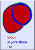
: Blood: 1650 ms Myocardium: 1175 ms Fat: 250 ms T 2 (and T 2* ) Relaxation Spin spin, or transverse relaxation Decay of excited transverse")
2 MRI Basics 3 Easy Steps 1) Prepare sample Line up H + in strong magnetic field 2) Excite sample Excite H + using radio waves Excited H + precess in unison 3) Acquire signal Radio waves emitted by excited H + Turn signal into pictures Net Magnetization T 1 Relaxation Spin lattice or longitudinal relaxation Recovery of longitudinal magnetization Typical values (1.5T): Blood: 1650 ms Myocardium: 1175 ms Fat: 250 ms T 2 (and T 2* ) Relaxation Spin spin, or transverse relaxation Decay of excited transverse magnetization Typical values (1.5T): Blood: 250 ms Myocardium: 50 ms Fat: 60 ms 2


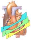
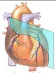
3 Mechanical Function Injury/Viability Anatomy Blood Velocity Perfusion Cine Imaging: LV RV Function Mechanical Function Short Axis Long Axis 3
 Qualitative Morphology wall motion abnormalities Visual assessment of Mitral and")

4 LV Function Protocol: (15 min) Quantitative: Volumetrics: (ml/m2) Indexed to BSA LVEDVI LVESVI RVEDVI RVESVI LV mass / wall thickness LA volume LV Function Protocol: (15 min) Qualitative Morphology wall motion abnormalities Visual assessment of Mitral and Tricuspid valve function/morphology Indications: Monitor medical treatment Assess for ICD Cardio oncology Myocardial Infarction Healthy Cine RV stack + LV RV Short axis function: ARVC protocol RV volume RV Wall motion abnormalities Marcus FI, et al. Circulation 2010; 121 4


5 Cine Imaging Let s make a movie! Image Acquisition Single shot Segmented Gated segmented Imaging Image 1 Image 2 Image 3 A portion of several images is acquired every heartbeat Each image has a different cardiac phase Requires no motion between heartbeats (good breath holding!) 5
")


6 LGE + LV function: Chronic Cardiomyopathy Protocol Ischemic vs non ischemic vs infiltrative cardiomyopathies LGE: Myocardial Fibrosis Contrast agent (Gd) reduces T 1 values Contrast cannot enter intact cells Greater accumulation of contrast in scarred tissue Weinsaft JW et al. Cardiol Clin 2007;25:35 56 Inversion Recovery 250 ms 425 ms 1000 ms Image contrast varies with inversion time Healthy myocardium can be made dark by choosing a specific inversion time 6
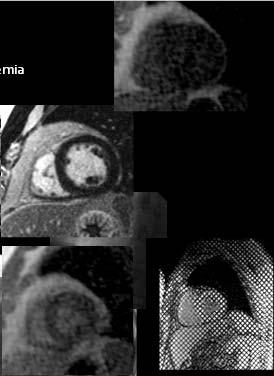
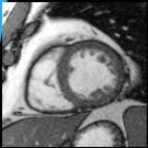
 Healthy? Scar Healthy?")
7 LGE Imaging: Identifying Etiology Ischemic vs non ischemic vs infiltrative cardiomyopathy White, JA, Patel, MR. CMR in Heart Failure and the Cardiomyopathies Cardiology Clinics, 2007 Limitations of Weighted Imaging (More) Healthy? Scar Healthy? LGE imaging identifies scar relative to healthy myocardium Anatomic Late Enhancement Global or diffuse disease may not be readily identifiable T2 + (LV function + LGE) : Acute Cardiomyopathy Protocol T2 imaging: Highlights edema like/water signal T2 STIR vs T2 Mapping vs SPAIR 7


 STIR When to add.")

8 T 2 Imaging T 2 imaging is sensitive to pathological tissue changes Turbo spin echo imaging has good T 2 contrast, but is sensitive to flow artifacts?? Simonetti O et al. Radiology 1996;199:49 57 Triple Inversion Recovery Global INV Slice INV Global INV IMG Additional inversion can null both blood and fat Signal to noise penalty Termed Short Tau Inversion Recovery (STIR) STIR When to add... T2 Weighted Imaging Ischemic Non Ischemic Disease Acuity? Disease reversibility? LGE LGE T2 T2 8

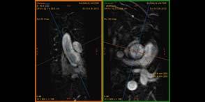
9 Perfusion Imaging Short Axis Special Protocols: T2* Free breathing + Tagging T1 mapping MRA Flow T2* + LV Function: Iron Overload Protocol 9


10 T2* imaging of iron overload T2* decay (calculate slope of line) T2* imaging of iron overload > 20ms is normal at 1.5T 10 20ms is possible >10ms is positive Tagging + Free breathing cine: Pericardial Disease LV Function/cine images: septal bounce Pericardial thickening (enhancement) 10
 Looking for D shaped septum")

 LV RV function + LGE Sarcoidosis myocarditis T1 mapping:")
11 Tagging + Free breathing cine: Pericardial Disease Real time Cine (free breathing, with deep inspiration) Looking for D shaped septum constrictive physiology Tagging + Free breathing cine: Pericardial Disease Tagged imaging Direct visualization of pericardial adhesions T1 Mapping + LGE + LV Function: Chronic Cardiomyopathy ( esp Amyloidosis or Fabry s) LV RV function + LGE Sarcoidosis myocarditis T1 mapping: Amyloidosis Fabry s disease 11
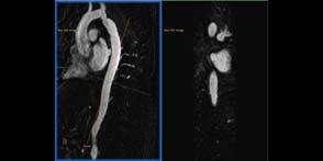
12 T1 Mapping Pulse Iron Fat Normal Fibrous (scar) Edema (water) Bulluck H, et al Circulation Journal (2015) 3D Gadolinium enhanced MRA / MRV 3D volumetric acquisition allows multiplanar post processing Imaging of the Aorta Aortopathy/aneurysm/dissection Coarctation/Bicuspid aortic valve 3D Gadolinium enhanced MRA / MRV Imaging of the Aorta Aortopathy/aneurysm/dissection Coarctation/Bicuspid aortic valve Pulmonary vessels Pulmonary vein mapping AFib Pulmonary arteries congenital 12
")


13 Bright blood (cine/trufi) Dark blood double inversion pulse Null signal of blood Reduces susceptibility artefact from clips Vessel wall evaluation 13
14 14
15 15


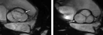
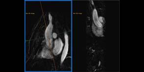
16 Cine Valve: (Bicuspid Aortic Valve) 16
 +218 mls - 52 mls (24% regurgitant")

 Qp:Qs quantify flow through MPA")
17 Flow Assessment Flow Assessment: (Aortic regurgitation) +218 mls - 52 mls (24% regurgitant fraction) Flow Assessment: Valvular stenosis regurgitation in plane or through plane Quantify pressure gradients at areas of stenosis (modified Bernoulli Equation) Qp:Qs quantify flow through MPA vs Ascending aorta 17


18 T2/STIR Cine SUMMARY LGE Tagging T1 Map T2* Black blood Flow Perfusion MRA Summary Many different MRI sequences Ways to generate image contrast permit tissue characterization ** a unique feature of MRI! Tailor the protocol to answer the clinical question. Each sequence takes time patients can only tolerate so long in the scanner Important to use an MRI scanner to capacity. Having a preset group of protocols may benefit workflow New sequences and means to generate image contrast are still being discovered! Acknowledgements: Stephenson Cardiovascular MRI Centre, Director Dr. James White Kelvin Chow, Senior Scientist, MR R&D Collaborations, Siemens Medical Solutions 18
 Bulluck H, et al Circulation Journal (2015) Calculating ECV Pulse Post contrast T1 ECV Native T1 (pre contrast) myocardial ECV")
19 SUMMARY: Cine LV RV function LGE (+ LV RV function) Chronic CM T2 (+ LGE + LV RV function) acute CM 3D MRA + Flow Special protocols (T2*, T1, Tagging, Free breathing, perfusion). Post contrast T1 Mapping Pulse Post contrast T1 Native T1 (pre contrast) Bulluck H, et al Circulation Journal (2015) Calculating ECV Pulse Post contrast T1 ECV Native T1 (pre contrast) myocardial ECV = (1 hematocrit) (ΔR1myocardium/ΔR1blood), where R1 = 1/T1 19


20 CMR = Versatility Ischemia Inflammation Infiltration Cardiac Ischemia Masses CMR s Incremental Role LGE Ischemic Non Ischemic Disease Etiology Disease activity Disease reversibility Response to Rx Risk stratification Impact Therapy White, JA, Patel, MR. Cardiology Clinics,


21 When to add... T1 Weighted Imaging ARVC: The 3 F s Disease Etiology Task Force Disease activity Fat Disease reversibility Response to Rx Function Risk stratification Marcus FI, et al. Circulation 2010; 121 Impact Therapy Fibrosis Beyond Diagnosis LGE Does CMR Alter Therapeutic Decisions? Disease Etiology Patients yo male Presenting with SCD, normal with Resuscitated echo and normal SCD or cath Unstable VT Disease activity Disease reversibility Response to Rx Risk stratification Impact Therapy White, JA, Fine N, Warren H, et al: Circulation: Card Imaging 2012 T 2 Imaging and Bright Blood T 2 imaging is sensitive to pathological tissue changes Turbo spin echo imaging has good T 2 contrast, but is sensitive to flow artifacts?? Simonetti O et al. Radiology 1996;199:
 Quantification of reguritant fractions Quantify pressure")
22 Cardiac MRI Sequences: 3D Balanced steady state free precession (3D b ssfe) ECG triggered, navigator gated free breathing For assessment of morphology 3D volumetric acquisition allows multiplanar postprocessing Cardiac MRI Sequences: Phase Contrast Allows measurement of intracardiac shunts Pulmonary : Systemic blood flow ratios (Qp:Qs) Quantification of reguritant fractions Quantify pressure gradients at areas of stenosis (modified Bernoulli Equation) MRI Contrast Agents Paramagnetic materials have unpaired electrons that catalyze proton interactions Result in shorter T 1 and T 2 times Gadolinium (Gd) has 7 unpaired electrons! Contrast agents are chelated Gd so that they can be biologically safe 22


 Need two")
23 Myocardial Perfusion Heart requires constant blood flow to function Bolus IV contrast flows along with the blood Contrast causes increase in brightness Repeated imaging can image contrast dynamics Kellman P et al. J Magn Reson Imag 2012;36: Quantitative T 2 Imaging Used to assess severity of edema Can detect global edema Standard imaging for all studies Long axis planes HLA, VLA, LVOT, LVOT coronal, (RVOT +/ RV inflow if right sided lesions) Need two perpendicular views of the valve(s) in question LV & RV function 23
 Mitral regurgitation")

24 Don t forget the aorta in your valve assessment! Aortic disease Residual root dissection in a patient with a previous type A dissection repair (interpositional graft) Mitral regurgitation Standard methods of quantification are indirect: 1) Regurgitant flow = LVSV - Ao systolic flow (independent of other valve lesions) 2) Regurgitant flow = LVSV - RVSV Mitral stenosis Can assess mitral valve area by direct planimetry Important to ensure correct slice positioning at MV tips (as for echo) Diastolic flow (volume and velocity) is feasible though temporal resolution is lower than echo 24


25 Mitral stenosis (2) Planimetry of the MV tips: Pulmonary stenosis Horizontal RVOT planned RVOT planned from transverse slices from previous RVOT Good visualisation of pulmonary valve motion Accurate velocity assessment RVOT sizing for potential surgery / balloon valvuloplasty / percutaneous valve replacement Pulmonary regurgitation Quantification of PR Size & shape of RVOT?percutaneous stentvalve replacement Size & function of RV Forward flow: 72mls Regurgitant flow: 27mls (38% regurgitant fraction) 25


.")
 Stenosis can be assessed with direct")
26 Pulmonary valve disease (3) CMR is also important for: determining RV mass & volumes assessing RVOT morphology Dilated RV secondary to chronic PR Supravalvular Complex stenosis with pulmonary previous surgical disease widening Now recurrent supravalvular stenosis & valvular regurgitation Dilated post stenotic pulmonary artery Tricuspid disease Severe TR (note low velocity causes minimal turbulence from dephased spins). Also has pericardial effusion Regurgitation can be quantified similarly to mitral regurgitation (RVSV pulmonary flow) Stenosis can be assessed with direct planimetry of the tips RV volumes & function for all 26


27 Multiple valve disease Detailed assessment of severity of each lesion & LV function Mixed aortic and mixed mitral valve disease Proceed sequentially through assessment of each lesion, including LV/RV funciton assessment Global INV Slice INV IMG Double Inversion Recovery Double inversion has no effect on imaging slice Inverted blood flows into slice Imaging occurs when inverted blood has no signal (due to T 1 recovery) Aortic Valve 27

 Choose the best LVOT view for in plane flow assessment (the one with the best view of the core jet) Aortic stenosis (3) Measure the peak velocity, either from the in plane flow itself,")
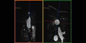
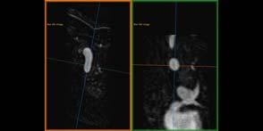
28 Aortic stenosis (1) SA pilot LVOT view Coronal LVOT view Plan initial LVOT view from short axis pilot scan, with the plane through the aortic root/valve The second LVOT (coronal) plane is planned through this, aligned with the stenotic jet. There is often a central core in the jet comprising laminar flow, with turbulent flow (black/low intensity on gradient echo) surrounding this Align planes with AS jet rather than Ao root Aortic stenosis (2) Choose the best LVOT view for in plane flow assessment (the one with the best view of the core jet) Aortic stenosis (3) Measure the peak velocity, either from the in plane flow itself, or using the in plane flow to identify the point of peak velocity and acquire a through plane flow sequence at this point: Position for through plane flow acquisition 28


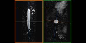
29 Aortic stenosis (4) Measure the valve area by direct planimetry, by acquiring a thin (5 6mm) slice through the tips of the aortic valve in systole, piloted from the 2 LVOT views. It is important to ensure you are at the tips, as you may overestimate the valve area otherwise Need still image of valve in systole here, including planimetry Valve tips in systole area = 1.0cm 2 Aortic stenosis (5) Correct alignment with AS jet Accurate trans valvular velocity (in plane / through plane) avoids underestimation with angulated roots Valve orifice area (direct planimetry) LV mass & volumes to assess impact on LV Aortic regurgitation Through plane flow measurement Allows quantification of regurgitation 29


30 Saturation recovery imaging Short T 1 tissue recover magnetization faster and are therefore brighter Longer T 1 means slower recovery and are darker Image brightness can be used to calculate T 1 and also contrast concentration Quantitative T 1 Imaging T 1 imaging is used to assess fibrosis Extracellular volume can also be calculated 30
Can SCMR CMR protocol recommendations
 Can SCMR CMR protocol recommendations V1.3 - April 2009 CanSCMR CMR Protocol and SOP Recommendation 2009 (15 minutes) 2 Planning of LV fct. real time multiple axes Realtime 3 cine long axis 6 long axes
Cardiac MRI: Clinical Application to Disease
 Cardiac MRI: Clinical Application to Disease Jessi Smith, MD Cardiothoracic imaging, Indiana University Slides courtesy of Stacy Rissing, MD Outline Imaging planes Disease findings Pulse sequences used
Cardiac MRI in ACHD What We. ACHD Patients
 Cardiac MRI in ACHD What We Have Learned to Apply to ACHD Patients Faris Al Mousily, MBChB, FAAC, FACC Consultant, Pediatric Cardiology, KFSH&RC/Jeddah Adjunct Faculty, Division of Pediatric Cardiology
Why Cardiac MRI? Presented by:
 Why Cardiac MRI? Presented by: Lisa G. Carkner, MD, FACC 1 Disclosures I have no financial disclosures Objectives Review basic principles of Cardiac MRI. What patient characteristics do I need to consider
cardiac imaging planes planning basic cardiac & aortic views for MR
 cardiac imaging planes planning basic cardiac & aortic views for MR Dianna M. E. Bardo, M. D. Assistant Professor of Radiology & Cardiovascular Medicine Director of Cardiac Imaging cardiac imaging planes
MR Advance Techniques. Vascular Imaging. Class II
 MR Advance Techniques Vascular Imaging Class II 1 Vascular Imaging There are several methods that can be used to evaluate the cardiovascular systems with the use of MRI. MRI will aloud to evaluate morphology
Cardiac MRI: Clinical Application to Disease
 Cardiac MRI: Clinical Application to Disease Stacy Rissing, MD! Cardiothoracic imaging, Indiana University! Outline Imaging planes Disease findings Pulse sequences used for each indication Pathophysiology
Objectives 8/17/2011. Challenges in Cardiac Imaging. Challenges in Cardiac Imaging. Basic Cardiac MRI Sequences
 8/17/2011 Traditional Protocol Model for Tomographic Imaging Cardiac MRI Sequences and Protocols Frandics Chan, M.D., Ph.D. Stanford University Medical Center Interpretation Lucile Packard Children s Hospital
MR Advance Techniques. Cardiac Imaging. Class IV
 MR Advance Techniques Cardiac Imaging Class IV Heart The heart is a muscular organ responsible for pumping blood through the blood vessels by repeated, rhythmic contractions. Layers of the heart Endocardium
MRI protocol for post-repaired TOF
 2012 NASCI MRI protocol for post-repaired TOF Taylor Chung, M.D. Associate Director, Body and Cardiovascular Imaging Department of Diagnostic Imaging Children s Hospital & Research Center Oakland Oakland,
Imaging in Heart Failure: A Multimodality Approach. Thomas Ryan, MD
 Imaging in Heart Failure: A Multimodality Approach Thomas Ryan, MD Heart Failure HFrEF HFpEF EF50% Lifetime risk 20% Prevalence 6M Americans Societal costs - $30B 50% 5-year survival 1 Systolic
Cardiac Imaging. Kimberly Delcour, DO, FACC. Mahi Ashwath, MD, FACC, FASE. Director, Cardiac CT. Director, Cardiac MRI
 Cardiac Imaging Kimberly Delcour, DO, FACC Director, Cardiac CT Mahi Ashwath, MD, FACC, FASE Director, Cardiac MRI Cardiac Imaging Discuss the clinical applications of and indications for: Cardiac CT Nuclear
Magnetic Resonance Angiography
 Magnetic Resonance Angiography 1 Magnetic Resonance Angiography exploits flow enhancement of GR sequences saturation of venous flow allows arterial visualization saturation of arterial flow allows venous
Clinical Applications
 C H A P T E R 16 Clinical Applications In selecting pulse sequences and measurement parameters for a specific application, MRI allows the user tremendous flexibility to produce variations in contrast between
Cardiac MRI: Cardiomyopathy
 Cardiac MRI: Cardiomyopathy Laura E. Heyneman, MD I do not have any relevant financial relationships with any commercial interests Cardiac MRI: Cardiomyopathy Laura E. Heyneman, MD Duke University Medical
What s New in Cardiac MRI
 What s New in Cardiac MRI Katie M. Hawthorne, MD Director, Cardiac MRI Main Line Health Philadelphia Cardiovascular Summit November 18, 2017 Cardiac MRI: Disclosure 2 Disclosures No financial disclosures
CARDIAC MRI. Cardiovascular Disease. Cardiovascular Disease. Cardiovascular Disease. Overview
 CARDIAC MRI Dr Yang Faridah A. Aziz Department of Biomedical Imaging University of Malaya Medical Centre Cardiovascular Disease Diseases of the circulatory system, also called cardiovascular disease (CVD),
Constrictive Pericarditis Pitfalls in MR Diagnosis Cylen Javidan-Nejad Associate Professor Mallinckrodt Institute of Radiology Washington University
 Constrictive Pericarditis Pitfalls in MR Diagnosis Cylen Javidan-Nejad Associate Professor Mallinckrodt Institute of Radiology Washington University in St. Louis Goal o To review the imaging criteria of
Rotation: Imaging 2. Nuclear Cardiology (in Imaging 1 and 2)
") Rotation: Imaging 2 Imaging 2 provides addition nuclear cardiology experience and COCATS Level 1 cardiac MRI experience. Fellows administer, process, and read VHVI cardiac nuclear studies with cardiology
Rotation: Imaging 2 Imaging 2 provides addition nuclear cardiology experience and COCATS Level 1 cardiac MRI experience. Fellows administer, process, and read VHVI cardiac nuclear studies with cardiology
ρ = 4(νp)2 Scale -200 to 200 V = m/s Grad = 34 mmhg V = 1.9 m/s Grad = 14 mmhg Types
2 Scale -200 to 200 V = m/s Grad = 34 mmhg V = 1.9 m/s Grad = 14 mmhg Types") Pre and Post Operative Evaluation of the Aorta and Aortic Valve Andrew J. Bierhals, MD The Pre and Post-Operative Evaluation of the Aorta and Aortic Valve Andrew Bierhals, MD, MPH Mallinckrodt Institute
Pre and Post Operative Evaluation of the Aorta and Aortic Valve Andrew J. Bierhals, MD The Pre and Post-Operative Evaluation of the Aorta and Aortic Valve Andrew Bierhals, MD, MPH Mallinckrodt Institute
1Pulse sequences for non CE MRA
 MRI: Principles and Applications, Friday, 8.30 9.20 am Pulse sequences for non CE MRA S. I. Gonçalves, PhD Radiology Department University Hospital Coimbra Autumn Semester, 2011 1 Magnetic resonance angiography
ADVANCED CARDIOVASCULAR IMAGING. Medical Knowledge. Goals and Objectives PF EF MF LF Aspirational
 Medical Knowledge Goals and Objectives PF EF MF LF Aspirational Know the basic principles of magnetic resonance imaging (MRI) including the role of the magnetic fields and gradient coil systems, generation
Non Contrast MRA. Mayil Krishnam. Director, Cardiovascular and Thoracic Imaging University of California, Irvine
 Non Contrast MRA Mayil Krishnam Director, Cardiovascular and Thoracic Imaging University of California, Irvine No disclosures Non contrast MRA-Why? Limitations of CTA Radiation exposure Iodinated contrast
Case based learning: CMR in Heart Failure
 Case based learning: CMR in Heart Failure Milind Y Desai, MD FACC FAHA FESC Associate Professor of Medicine Heart and Vascular Institute, Cleveland Clinic Cleveland, OH Disclosures: none Use of Gadolinium
CT for Myocardial Characterization of Cardiomyopathy. Byoung Wook Choi, Yonsei University Severance Hospital, Seoul, Korea
 CT for Myocardial Characterization of Cardiomyopathy Byoung Wook Choi, Yonsei University Severance Hospital, Seoul, Korea Cardiomyopathy Elliott P et al. Eur Heart J 2008;29:270-276 The European Society
MITRAL STENOSIS. Joanne Cusack
 MITRAL STENOSIS Joanne Cusack BSE Breakdown Recognition of rheumatic mitral stenosis Qualitative description of valve and sub-valve calcification and fibrosis Measurement of orifice area by planimetry
Current Indications for Cardiac MRI: What You See is What You Get?
 Current Indications for Cardiac MRI: What You See is What You Get? Javier Ganame, MD, PhD, FASE No disclosures Cardiology Update, Niagara, Sept 24th, 2016 The Ideal Diagnostic Technique Easy to apply Accurate
Cardiac Stress MRI: Detection of Ischemia. Disclosures: Dobutamine Stress MR. April 28, 2018
 Cardiac MRI: Detection of Ischemia Cardiac MRI in Today s Clinical Practice Foundations of Cardiovascular Magnetic Resonance Daniel C. Lee, MD, MSc Assistant Professor of Medicine and Radiology Co-Director,
Normal TTE/TEE Examinations
 Normal TTE/TEE Examinations Geoffrey A. Rose, MD FACC FASE Sanger Heart & Vascular Institute Before you begin imaging... Obtain the patient s Height Weight BP PLAX View PLAX View Is apex @ 9-10 o clock?
Cardiac Imaging Tests
 Cardiac Imaging Tests http://www.medpagetoday.com/upload/2010/11/15/23347.jpg Standard imaging tests include echocardiography, chest x-ray, CT, MRI, and various radionuclide techniques. Standard CT and
Multiparametric T1, T2 and T2* MR Imaging
 Multiparametric T1, T2 and T2* MR Imaging Kate Hanneman MD Assistant Professor of Radiology Joint Department of Medical Imaging, University of Toronto Disclosure Neither I nor my immediate family members
UK Biobank. Imaging modality Cardiovascular Magnetic Resonance (CMR) Version th Oct 2015
 Version th Oct 2015") Imaging modality Cardiovascular Magnetic Resonance (CMR) Version 1.0 http://www.ukbiobank.ac.uk/ 30 th Oct 2015 This document details the procedure for the CMR scan performed at an Imaging assessment centre
Imaging modality Cardiovascular Magnetic Resonance (CMR) Version 1.0 http://www.ukbiobank.ac.uk/ 30 th Oct 2015 This document details the procedure for the CMR scan performed at an Imaging assessment centre
New Cardiovascular Devices and Interventions: Non-Contrast MRI for TAVR Abhishek Chaturvedi Assistant Professor. Cardiothoracic Radiology
 New Cardiovascular Devices and Interventions: Non-Contrast MRI for TAVR Abhishek Chaturvedi Assistant Professor Cardiothoracic Radiology Disclosure I have no disclosure pertinent to this presentation.
MRI (AND CT) FOR REPAIRED TETRALOGY OF FALLOT
 FOR REPAIRED TETRALOGY OF FALLOT") MRI (AND CT) FOR REPAIRED TETRALOGY OF FALLOT Linda B Haramati MD, MS Departments of Radiology and Medicine Bronx, New York OUTLINE Pathogenesis Variants Initial surgical treatments Basic MR protocols
MRI (AND CT) FOR REPAIRED TETRALOGY OF FALLOT Linda B Haramati MD, MS Departments of Radiology and Medicine Bronx, New York OUTLINE Pathogenesis Variants Initial surgical treatments Basic MR protocols
Doppler Basic & Hemodynamic Calculations
 Doppler Basic & Hemodynamic Calculations August 19, 2017 Smonporn Boonyaratavej MD Division of Cardiology, Department of Medicine Chulalongkorn University Cardiac Center, King Chulalongkorn Memorial Hospital
Cardiac MRI: Appropriateness. Scott Mattson, DO, FACC Lutheran Medical Group Fort Wayne, IN
 Cardiac MRI: Appropriateness Scott Mattson, DO, FACC Lutheran Medical Group Fort Wayne, IN Approaches to Appropriateness The indication The patient The scanner and technologists The interpreting physician
Objectives. CMR Volumetric Analysis 8/25/11. CMR Volumetric Analysis Technique. Cardiac imaging plane acquisition. CMR Volumetric Analysis
 Objectives Cynthia K. Rigsby Children s Memorial Hospital Chicago, IL CMR volumetric analysis Techniques Normalized data Sources of error CMR phase contrast flow analysis Techniques What we can do with
What are the best diagnostic tools to quantify aortic regurgitation?
 What are the best diagnostic tools to quantify aortic regurgitation? Agnès Pasquet, MD, PhD Pôle de Recherche Cardiovasculaire Institut de Recherche Expérimentale et Clinique Université catholique de Louvain
Advanced MR Imaging in Myocarditis
 Naeem Merchant MD FRCP Professor of Medicine Department of Radiology Department of Cardiac Sciences Cumming School of Medicine University of Calgary Advanced MR Imaging in Myocarditis The Lake Louise Criteria
Cardiac Computed Tomography
 Cardiac Computed Tomography Authored and approved by Koen Nieman Stephan Achenbach Francesca Pugliese Bernard Cosyns Patrizio Lancellotti Anastasia Kitsiou Contents CARDIAC COMPUTED TOMOGRAPHY Page 1.
Outils d évaluation myocardique par relaxométrie (Myomaps)
") Cardiovascular sciences Outils d évaluation myocardique par relaxométrie (Myomaps) Alain Nchimi Geva T. Magnetic resonance imaging: historical perspective. J Cardiovasc Magn Reson. 2006;8(4):573-80. CMR
Cardiovascular sciences Outils d évaluation myocardique par relaxométrie (Myomaps) Alain Nchimi Geva T. Magnetic resonance imaging: historical perspective. J Cardiovasc Magn Reson. 2006;8(4):573-80. CMR
Introduction. Cardiac Imaging Modalities MRI. Overview. MRI (Continued) MRI (Continued) Arnaud Bistoquet 12/19/03
 MRI (Continued) Arnaud Bistoquet 12/19/03") Introduction Cardiac Imaging Modalities Arnaud Bistoquet 12/19/03 Coronary heart disease: the vessels that supply oxygen-carrying blood to the heart, become narrowed and unable to carry a normal amount
Introduction Cardiac Imaging Modalities Arnaud Bistoquet 12/19/03 Coronary heart disease: the vessels that supply oxygen-carrying blood to the heart, become narrowed and unable to carry a normal amount
Echocardiography as a diagnostic and management tool in medical emergencies
 Echocardiography as a diagnostic and management tool in medical emergencies Frank van der Heusen MD Department of Anesthesia and perioperative Care UCSF Medical Center Objective of this presentation Indications
Policy #: 222 Latest Review Date: March 2009
 Name of Policy: MRI Phase-Contrast Flow Measurement Policy #: 222 Latest Review Date: March 2009 Category: Radiology Policy Grade: Active Policy but no longer scheduled for regular literature reviews and
Conflict Disclosures. Vermont Cardiac Network. Outline. Series Learning Objectives 4/27/2016. Scott E. Friedman April 28, 2016
 Conflict Disclosures Vermont Cardiac Network The Speaker has reported no significant financial relationship with any companies whose product may be germane to the content of their presentations or who
Tricuspid and Pulmonary Valve Disease
 Tricuspid and Pulmonary Valve Disease Lawrence Rudski MD FRCPC FACC FASE Professor of Medicine Director, Division of Cardiology Jewish General Hospital McGill University Question 1 All of the following
Go With The Flow: Role of 4D Flow Imaging
 4D Flow Go With The Flow: Role of 4D Flow Imaging Niti R. Aggarwal, MD Associate Director of Cardiac MRI Assistant Professor of Medicine & Radiology University of Wisconsin Madison Disclosures GE Healthcare
Adult Echocardiography Examination Content Outline
 Adult Echocardiography Examination Content Outline (Outline Summary) # Domain Subdomain Percentage 1 2 3 4 5 Anatomy and Physiology Pathology Clinical Care and Safety Measurement Techniques, Maneuvers,
Cardiovascular MRI of Adult Congenital Heart Disease
 Cardiovascular MRI of Adult Congenital Heart Disease Anil K. Attili, MD Cardiovascular Magnetic Resonance imaging of Adult Congenital Heart Disease Anil Attili, M.D. Assistant Professor of Radiology /Cardiology
Ve V rmont rmon Card Car iac d Netw Ne ork tw Scott E. Friedman April 28, 2016
 Vermont Cardiac Network Scott E. Friedman April 28, 2016 Conflict Disclosures Th S k h d i ifi fi i l l i hi ih The Speaker has reported no significant financial relationship with any companies whose product
Adel Hasanin Ahmed 1
 Adel Hasanin Ahmed 1 PERICARDIAL DISEASE The pericardial effusion ends anteriorly to the descending aorta and is best visualised in the PLAX. PSAX is actually very useful sometimes for looking at posterior
A Light in the Dark: Cardiac MRI and Risk Mitigation. J. Ronald Mikolich MD Professor of Internal Medicine Northeast Ohio Medical University (NEOMED)
") A Light in the Dark: Cardiac MRI and Risk Mitigation J. Ronald Mikolich MD Professor of Internal Medicine Northeast Ohio Medical University (NEOMED) Dr. Mikolich has NO financial disclosures relative to
A Light in the Dark: Cardiac MRI and Risk Mitigation J. Ronald Mikolich MD Professor of Internal Medicine Northeast Ohio Medical University (NEOMED) Dr. Mikolich has NO financial disclosures relative to
Functional Chest MRI in Children Hyun Woo Goo
 Functional Chest MRI in Children Hyun Woo Goo Department of Radiology and Research Institute of Radiology Asan Medical Center, University of Ulsan College of Medicine, Seoul, Korea No ionizing radiation
Φαινόμενο No-Reflow. Απεικόνιση με CMR, κλινική συσχέτιση και προγνωστική σημασία
 Φαινόμενο No-Reflow. Απεικόνιση με CMR, κλινική συσχέτιση και προγνωστική σημασία Θεόδωρος. Καραμήτσος MD PhD Honorary Consultant in Cardiology University of Oxford Centre for Clinical Magnetic Resonance
Comprehensive Echo Assessment of Aortic Stenosis
 Comprehensive Echo Assessment of Aortic Stenosis Smonporn Boonyaratavej, MD, MSc King Chulalongkorn Memorial Hospital Bangkok, Thailand Management of Valvular AS Medical and interventional approaches to
The Doppler Examination. Katie Twomley, MD Wake Forest Baptist Health - Lexington
 The Doppler Examination Katie Twomley, MD Wake Forest Baptist Health - Lexington OUTLINE Principles/Physics Use in valvular assessment Aortic stenosis (continuity equation) Aortic regurgitation (pressure
Imaging and heart failure
 Imaging and heart failure Jeroen J Bax Dept of Cardiology Leiden Univ Medical Center The Netherlands Davos, feb 2013 Research grants: Medtronic, Biotronik, Boston, St Jude, BMS imaging, GE Healthcare,
ViosWorks: A Paradigm Shift in Cardiac MR Imaging
 Figure 1. ViosWorks image of a patient with shunted pulmonary venous return. Image courtesy of Dr. Shreyas Vasanawala, Stanford University. ViosWorks: A Paradigm Shift in Cardiac MR Imaging The value of
Essentials of Clinical MR, 2 nd edition. 99. MRA Principles and Carotid MRA
 99. MRA Principles and Carotid MRA As described in Chapter 12, time of flight (TOF) magnetic resonance angiography (MRA) is commonly utilized in the evaluation of the circle of Willis. TOF MRA allows depiction
Comprehensive Hemodynamics By Doppler Echocardiography. The Echocardiographic Swan-Ganz Catheter.
 Comprehensive Hemodynamics By Doppler Echocardiography. The Echocardiographic Swan-Ganz Catheter. Itzhak Kronzon, MD, FASE, FACC, FESC, FAHA, FACP, FCCP North Shore HS, LIJ/Lenox Hill Hospital, New York
DECLARATION OF CONFLICT OF INTEREST
 DECLARATION OF CONFLICT OF INTEREST Cardiovascular magnetic resonance for timing pulmonary valve replacement E.Valsangiacomo Buechel University Children s Hospital Zurich Outline Introduction Pulmonary
Sung A Chang Department of Internal Medicine, Division of Cardiology, Sungkyunkwan University School of Medicine, Samsung Medical Center
 CMR Perfusion and Viability A STICH Out of Time? Sung A Chang Department of Internal Medicine, Division of Cardiology, Sungkyunkwan University School of Medicine, Samsung Medical Center Can Imaging Improve
LV FUNCTION ASSESSMENT: WHAT IS BEYOND EJECTION FRACTION
 LV FUNCTION ASSESSMENT: WHAT IS BEYOND EJECTION FRACTION Jamilah S AlRahimi Assistant Professor, KSU-HS Consultant Noninvasive Cardiology KFCC, MNGHA-WR Introduction LV function assessment in Heart Failure:
PROSTHETIC VALVE BOARD REVIEW
 PROSTHETIC VALVE BOARD REVIEW The correct answer D This two chamber view shows a porcine mitral prosthesis with the typical appearance of the struts although the leaflets are not well seen. The valve
How to Assess and Treat Obstructive Lesions
 How to Assess and Treat Obstructive Lesions Erwin Oechslin, MD, FESC, FRCPC, Director, Congenital Cardiac Centre for Adults Peter Munk Cardiac Centre University Health Network/Toronto General Hospital
Normal values for cardiovascular magnetic resonance in adults and children
 Kawel-Boehm et al. Journal of Cardiovascular Magnetic Resonance (2015) 17:29 DOI 10.1186/s12968-015-0111-7 REVIEW Normal values for cardiovascular magnetic resonance in adults and children Nadine Kawel-Boehm
General Cardiovascular Magnetic Resonance Imaging
 2 General Cardiovascular Magnetic Resonance Imaging 19 Peter G. Danias, Cardiovascular MRI: 150 Multiple-Choice Questions and Answers Humana Press 2008 20 Cardiovascular MRI: 150 Multiple-Choice Questions
CMR for Congenital Heart Disease
 CMR for Congenital Heart Disease * Second-line tool after TTE * Strengths of CMR : tissue characterisation, comprehensive access and coverage, relatively accurate measurements of biventricular function/
Review of Cardiac Imaging Modalities in the Renal Patient. George Youssef
 Review of Cardiac Imaging Modalities in the Renal Patient George Youssef ECHO Left ventricular hypertrophy (LVH) assessment Diastolic dysfunction Stress ECHO Cardiac CT angiography Echocardiography - positives
Role of CMR in heart failure and cardiomyopathy
 Role of CMR in heart failure and cardiomyopathy Hajime Sakuma Department of Radiology, Mie University Late gadolinium enhancement (LGE) LGE MRI can demonstrate site of necrosis, fibrosis or deposition
CMS Limitations Guide - Radiology Services
 CMS Limitations Guide - Radiology Services Starting October 1, 2015, CMS will update their existing medical necessity limitations on tests and procedures to correspond to ICD-10 codes. This limitations
Atlas of Practical Cardiac Applications of MRI
 Atlas of Practical Cardiac Applications of MRI Atlas of Practical Cardiac Applications of MRI Guillcm Pons-LIado, MD. Director, Cardiac Imaging Unit, Cardiology Department, Hospital de la Santa Creu i
Fulfilling the Promise
 Fulfilling the Promise of Cardiac MR Non-contrast, free-breathing technique generates comprehensive evaluation of the coronary arteries By Maggie Fung, MR Cardiovascular Clinical Development Manager; Wei
Epicardial VT Ablation The Cleveland Clinic Experience
 Epicardial VT Ablation The Cleveland Clinic Experience Walid Saliba, MD, FHRS Director, EP Lab Cardiac Electrophysiology Heart and Vascular Institute Epicardial Access in the EP Lab Why Epicardial Special
I have no financial disclosures
 Manpreet Singh MD I have no financial disclosures Exercise Treadmill Bicycle Functional capacity assessment Well validated prognostic value Ischemic assessment ECG changes ST segments Arrhythmias Hemodynamic
Cardiac magnetic resonance imaging in rheumatoid arthritis: promising or misleading? Sophie Mavrogeni MD FESC
 Cardiac magnetic resonance imaging in rheumatoid arthritis: promising or misleading? Sophie Mavrogeni MD FESC Onassis Cardiac Surgery Center Athens Greece Nothing to disclose Financial disclosure Cardiac
Perfusion, Viability, Edema and Hemorrhage: How it Can (and Should) Change Clinical Practice. Rohan Dharmakumar, Ph.D.
 Change Clinical Practice. Rohan Dharmakumar, Ph.D.") Perfusion, Viability, Edema and Hemorrhage: How it Can (and Should) Change Clinical Practice Rohan Dharmakumar, Ph.D. Director, Translational Cardiac Imaging Research Associate Director, Biomedical Imaging
Perfusion, Viability, Edema and Hemorrhage: How it Can (and Should) Change Clinical Practice Rohan Dharmakumar, Ph.D. Director, Translational Cardiac Imaging Research Associate Director, Biomedical Imaging
Imaging Assessment of the Pulmonary Valve in Stenosis/Atresia and Regurgitation
 Imaging Assessment of the Pulmonary Valve in Stenosis/Atresia and Regurgitation Craig E Fleishman, MD FACC FASE The Heart Center at Arnold Palmer Hospital for Children SCAI Fall Fellows Course 2014 Las
HEMODYNAMIC ASSESSMENT
 HEMODYNAMIC ASSESSMENT INTRODUCTION Conventionally hemodynamics were obtained by cardiac catheterization. It is possible to determine the same by echocardiography. Methods M-mode & 2D echo alone can provide
Valvular disease : Ο ρόλος του CMR. Sophie Mavrogeni MD FESC. Onassis Cardiac Surgery Center Athens Greece
 Valvular disease : Ο ρόλος του CMR Sophie Mavrogeni MD FESC Onassis Cardiac Surgery Center Athens Greece Aortic Valve CMR EVALUATION OF AORTIC VALVE Phase-contrast CMR of the aorta to determine aortic
Case based learning: CMR in Heart Failure
 Case based learning: CMR in Heart Failure Milind Y Desai, MD FACC FAHA FESC Associate Professor of Medicine Heart and Vascular Institute, Cleveland Clinic Cleveland, OH Disclosures: none Use of Gadolinium
Certificate in Clinician Performed Ultrasound (CCPU) Syllabus. Rapid Cardiac Echo (RCE)
 Syllabus. Rapid Cardiac Echo (RCE)") Certificate in Clinician Performed Ultrasound (CCPU) Syllabus Rapid Cardiac Echo (RCE) Purpose: Rapid Cardiac Echocardiography (RCE) This unit is designed to cover the theoretical and practical curriculum
Certificate in Clinician Performed Ultrasound (CCPU) Syllabus Rapid Cardiac Echo (RCE) Purpose: Rapid Cardiac Echocardiography (RCE) This unit is designed to cover the theoretical and practical curriculum
Multimodality Imaging of Septal Defects
 Multimodality Imaging of Septal Defects Ohio-ACC 2018 Annual Meeting October 27, 2018 Kan N. Hor, MD Director, Cardiac Magnetic Resonance Imaging Associate Professor of Pediatrics The Heart Center, Nationwide
HISTORY. Question: What category of heart disease is suggested by the fact that a murmur was heard at birth?
 HISTORY 23-year-old man. CHIEF COMPLAINT: Decreasing exercise tolerance of several years duration. PRESENT ILLNESS: The patient is the product of an uncomplicated term pregnancy. A heart murmur was discovered
Giovanni Di Salvo MD, PhD, FESC Second University of Naples Monaldi Hospital
 Giovanni Di Salvo MD, PhD, FESC Second University of Naples Monaldi Hospital VSD is one of the most common congenital cardiac abnormalities in the newborn. It can occur as an isolated finding or in combination
Appendix II: ECHOCARDIOGRAPHY ANALYSIS
 Appendix II: ECHOCARDIOGRAPHY ANALYSIS Two-Dimensional (2D) imaging was performed using the Vivid 7 Advantage cardiovascular ultrasound system (GE Medical Systems, Milwaukee) with a frame rate of 400 frames
Cardiovascular manifestations of HIV
 Cardiovascular manifestations of HIV Prabhakar Rajiah, MBBS, MD, FRCR Associate Professor of Radiology Associate Director, Cardiac CT and MRI University of Texas Southwestern Medical Center, Dallas, USA
When Does 3D Echo Make A Difference?
 When Does 3D Echo Make A Difference? Wendy Tsang, MD, SM Assistant Professor, University of Toronto Toronto General Hospital, University Health Network 1 Practical Applications of 3D Echocardiography Recommended
Advanced Imaging MRI and CTA
 Advanced Imaging MRI and CTA Who and why may benefit. Matthew W. Martinez, M.D. FACC Lehigh Valley Health Network Director, Cardiovascular Imaging Learning Objectives Review basics of CMR and CTA Review
A Magnetic Resonance Imaging Method for
 Journal of Cardiovascular Magnetic Resonance, 1(1), 59-64 (1999) INVITED PAPER Use of MRI in ASD Asessment A Magnetic Resonance Imaging Method for Evaluating Atrial Septa1 Defects Godtfred Holmvang Cardiac
Usefulness of Delayed Enhancement by Magnetic Resonance Imaging in Hypertrophic Cardiomyopathy as a Marker of Disease and Its Severity
 Usefulness of Delayed Enhancement by Magnetic Resonance Imaging in Hypertrophic Cardiomyopathy as a Marker of Disease and Its Severity G.D.Aquaro, MD Fondazione G.Monasterio Regione Toscana/CNR Pisa, Italy
Titel Kardiologie-SG.ch hot topics in heart failure and mitral regurgitation
 Titel Kardiologie-SG.ch hot topics in heart failure and mitral regurgitation where and how to quantify mitral regurgitation: Echolab, Cathlab or MRI? Philipp K. Haager, St. Gallen Measuring mitral regurgitation?
Tricuspid and Pulmonary Valve Disease
 Tricuspid and Pulmonary Valve Disease Lawrence Rudski MD FRCPC FACC FASE Professor of Medicine Director, Division of Cardiology Jewish General Hospital McGill University Right Sided Failure Edema Gut congestion
Managing Hypertrophic Cardiomyopathy with Imaging. Gisela C. Mueller University of Michigan Department of Radiology
 Managing Hypertrophic Cardiomyopathy with Imaging Gisela C. Mueller University of Michigan Department of Radiology Disclosures Gadolinium contrast material for cardiac MRI Acronyms Afib CAD Atrial fibrillation
Hemodynamic Assessment. Assessment of Systolic Function Doppler Hemodynamics
 Hemodynamic Assessment Matt M. Umland, RDCS, FASE Aurora Medical Group Milwaukee, WI Assessment of Systolic Function Doppler Hemodynamics Stroke Volume Cardiac Output Cardiac Index Tei Index/Index of myocardial
Acute Myocarditis Mimicking ST-segment Elevation Myocardial Infarction: Relation Between ECG Changes And Myocardial Damage As Assessed By CMR
 Acute Myocarditis Mimicking ST-segment Elevation Myocardial Infarction: Relation Between ECG Changes And Myocardial Damage As Assessed By CMR G. Nucifora 1, A. Di Chiara 2, D. Miani 1, G. Piccoli 3, M.
RVOTO adult and post-op
 Right ventricular outflow tract obstruction in the adult: native and post-op Helmut Baumgartner Westfälische Wilhelms-Universität Münster Adult Congenital and Valvular Heart Disease Center University of
Tools for cardiovascular magnetic resonance imaging
 Review Article Tools for cardiovascular magnetic resonance imaging Ramkumar Krishnamurthy, Benjamin Cheong, Raja Muthupillai Department of Diagnostic and Interventional Radiology, CHI St. Luke s Health,
Essential tools for Clinical Cardiovascular MRI Raja Muthupillai, PhD,DABMP, DABR
 Essential tools for Clinical Cardiovascular MRI Raja Muthupillai, PhD,DABMP, DABR Director of Imaging Research Department of Diagnostic and Interventional Radiology Baylor St Luke s Medical Center, Houston,
J. Schwitter, MD, FESC Section of Cardiology
 J. Schwitter, MD, FESC Section of Cardiology CMR Center of the CHUV University Hospital Lausanne - CHUV Switzerland Centre de RM Cardiaque J. Schwitter, MD, FESC Section of Cardiology CMR Center of the