URINARY TRACT IMAGING - BASIC PRINCIPLES
|
|
|
- Joleen Atkins
- 5 years ago
- Views:
Transcription

1 URINARY TRACT IMAGING - BASIC PRINCIPLES Clinical Radiology Every physician needs a basic understanding of diagnostic imaging to understand how to order the appropriate studies and to understand the resulting radiologist's report.
2 What is the difference between a urologist and a nephrologist? Urologists and nephrologists both treat kidney problems. Urologists are surgical specialists who focus on anatomical or structural disorders of the kidneys and urinary tract. They treat problems such as kidney stones, kidney blockages, and kidney cancer. Urologists are qualified to perform surgery and outpatient medical procedures to correct such conditions. Nephrologists are medical specialists who focus on disorders that affect the way the kidneys work such as diabetes and chronic kidney disease. Nephrologists prescribe nonsurgical medical treatments for these disorders.
3 Urologic Radiology Science has given urologists a bevy of tools to probe the most private parts of the body in diagnosing urinary and renal disease. Every modern imaging technology, from conventional X-rays to radionuclide imaging, has found its way into urologic radiology's arsenal. The good news for physicians is that they have many options to explore the kidneys, ureters, bladder and surrounding structures. The better news for patients is that today's tests are thorough, relatively pain-free and often quick.
4 What problems could require imaging of the urinary tract? Imaging can help the health care provider find the cause of 1.urinary retention the inability to empty the bladder completely 2.urinary frequency urination eight or more times a day 3.urinary urgency the inability to delay urination 4.urinary incontinence the accidental loss of urine 5.blockage of urine 6.abdominal mass 7.pain in the groin or lower back 8.blood in the urine 9.high blood pressure 10.kidney failure


 MRI")
5 METHODS OF INVESTIGATION ULTRASONOGRAPHY RADIOLOGY Simple abdominal X-ray Intravenous urography Retrograde pieloureterography Cystography Uretrography NUCLEAR MEDICINE Static studies: static renal scintigraphy Dynamic studies: renogram CT (without, with contrast, Angio) MRI (without, with contrast, Angio)
6 Imaging the urinary tract which modality to use for first-line examination? In imaging the urinary tract, the modality of choice for the initial examination will almost universally be ultrasound (US). US is inexpensive, immediate, painless, requires no sedation or anesthetic, is widely available, and is radiation free. US can be used to scan in any plane at the discretion of the operator, and whereas the technique is entirely operator dependent, most centers have staff with a high level of skill.

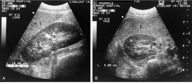
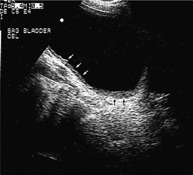
7 Doppler USG ULTRASOUND High frequency- high resolution but low penetration depth Renal- parenchyma evaluate hematuria, solid mass, cysts congenital abnormalities, stones Adrenal- CT/MRI better Nodules, cysts, hemorrhage, location, tumors Bladder- examine wall, lesions Transvaginal, transabdominal, transrectal Normal wall >= 6 mm bladder volume Prostate- transrectal, access for biopsy
8 ULTRASOUND Scrotal- Evaluate- mass, pain, torsion, orchitis, epididymitis, hydrocele, hernia, varicoceles Testicle- 4 x 3 cm Veins- >2mm= varicocele- evaluate in erect position
9

10

11 Doppler Ultrasound
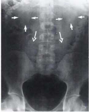
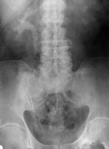
12 PLAIN FILM OF THE ABDOMEN The kidneys-ureters-bladder is often the first imaging study performed to visualize the abdomen and urinary tract The film is taken with the patient supine and should include the entire abdomen from the base of the sternum to the pubic symphisis Can show bony abnormalities, calcification and large soft tissue masses

13

14

15
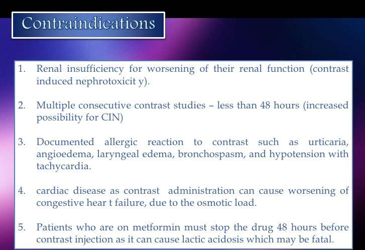
16 CONTRAST FILMS Rapidly concentrated by kidneys and opacifies urinary tract Low osmolar iodine nonionic contrast material less osmolar load- fewer complications than high osmolar Reactions: Allergic, renal toxicity, shock
17 UROGRAPHY Involves instillation of contrast material to better visualize the collecting or lumenal structures of the kidneys, ureters, bladder, and urethra This can be done after i/v injection or direct instillation into the urinary tract 1) Intravenous urography 2) Cystourethroography 3) Retrograde urethrography
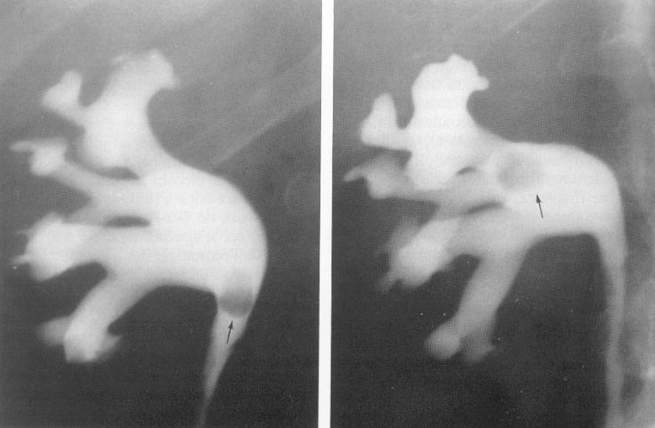
18 INTRAVENOUS UROGRAPHY IVU/ intravenous pyelogram is the classic modality of imaging the entire urethelial tract from pyelocalyceal system trhough the ureters and bladder Excellent for indentifying small urethelial lesions as well as the severity of obstruction from calculi Provides anatomical and qualitative functional information about the kidneys

19
20

21

22
23 CYSTOGRAPHY Permits imaging of an opacified urinary bladder after retrograde instillation of contrast media through a urethral or suprapubic catheter Imaging is performed to demonstrate a suspected urine leak, either from traumatic bladder rupture or after bladder surgery Can also demonstrate a presence of a fistula between the bladder and vagina or to characterize bladder diverticuli

24 NORMAL MALE CYSTOGRAM
25 RETROGRADE URETHROGRAPHY (RUG) Complete evaluation of the urethra includes both antegrade and retrograde urethrography Allows visualization of the anterior male urethra Used for evaluating a suspected traumatic urethral injury or urethral stricture Can also be useful for diagnosis of a urethral diverticulum in females Evaluate anterior and posterior urethra- strictures, trauma 8-16 F foley in fossa navicularis, fill balloon with 1-2 ml and inject 30-50% contrast while filming obliquely

26 NORMAL RUG


27 Multi-Detector Computed Tomography (MDCT)
, kidney stones, renal cysts and masses.")
28 CT + ANGIO CT SCAN often used examine structures in the abdomen and pelvis (liver, pancreas, gallbladder, spleen and intestines). CT Scans are a diagnostic tool that urologists use to detect and diagnose: recurrent urinary tract infections, sources of blood in the urine (hematuria), kidney stones, renal cysts and masses. It can help urologists rule out prostate, bladder and renal cancers
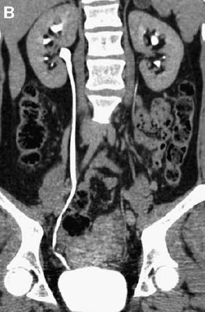
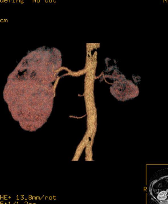
29 CT + ANGIO CT Contrast- parenchyma, adrenals 3-D to evaluate vascular abnormality ml i/v bolus injection Renal- stages: Precontrast- stones, parenchyma, vascular calcifications, renal contour 30 sec- cortex vs medulla Nephrographic- 100 sec- uniform enhancement of parencyma (masses) Pyelographic- excretory- collecting system

30 NO CONTRAST CT

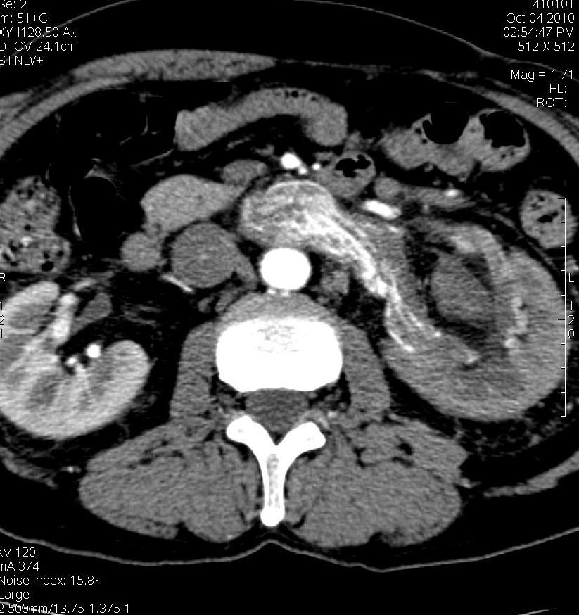
31 CONTRAST CT
32 MRI No iodinated contrast Soft tissue resolution better than CT Contraindications- pacemaker, aneurysm clips T1- fluid dark, fat bright T2- fluid bright, fat dark

33 MRI
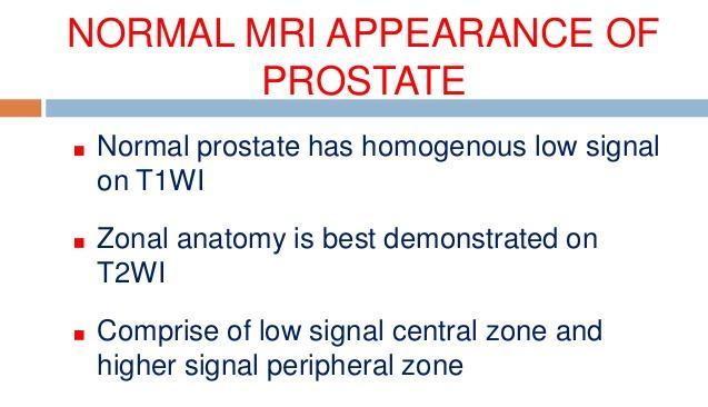
34 MRI Renal- will not evaluate stones, determine tumor Adrenal- contain more fat than cancers, bright on T2, isodense with liver Bladder- to determin invasion of wall by cell cancer or other pelvic neoplasms (on T2) Prostate- evaluate prostate cancer for capsular invasion. T1- distinct from surrounding fat/seminal vesicles (intermediate intensity), T2- peripheral zone (high intensity), central (intermediate), neurovascular bundles bright, seminal vesicles (high)
35 Table 1 Comparison of advantages and disadvantages between computed tomography (CT) and magnetic resonance (MR) imaging modalities CT Uses ionizing radiation, high-dose procedure Excellent spatial resolution Actual scanning time measured in seconds (typically <10 s) Rarely requires general anesthetic in children MRI Uses magnetic resonance, no ionizing radiation Excellent contrast resolution Actual scanning time measured in minutes (typically 45 min) Frequently requires general anesthetic in children, depending on age
36 Excellent at showing calcification Poor at showing edema or pathological changes in specific tissue types Usually requires intravenous contrast (unless looking for calcification when not required) No known risk of nephrogenic systemic fibrosis (NSF) Less expensive Usually available as an emergency imaging technique No significant contraindications Open-style scanners Poor at showing calcification (signal void) Excellent at showing edema and pathological changes in specific tissue types Usually requires intravenous administration of contrast (but certain sequences can be tailored if this is contraindicated) Risk of NSF (rare, but renal patients believed to be at increased risk) Expensive Not routinely available as an emergency technique Contraindicated in patients with any internal ferrous objects (pacemakers, defibrillators, recent orthopedic metalware, other implanted metallic devices, metallic foreign bodies) Generally quite enclosed scanners risk of claustrophobia
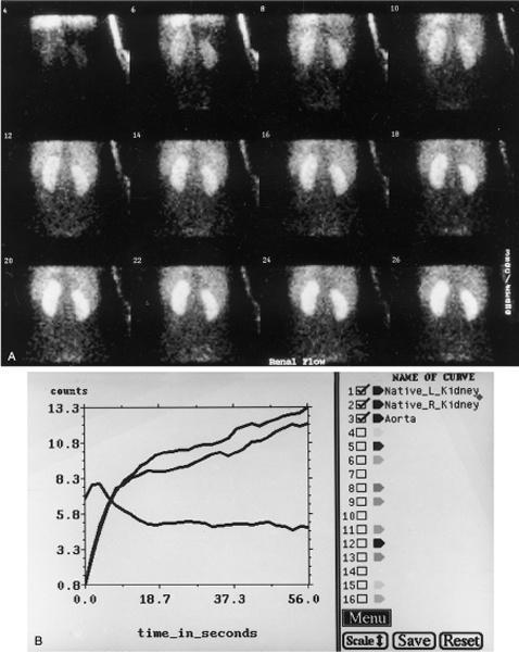
37 NUCLEAR MEDICINE uses the radiation released by radionuclides (called nuclear decay) to produce images A radionuclide, usually technetium-99m, is combined with different stable, metabolically active compounds to form a radiopharmaceutical that localizes to a particular anatomic or diseased structure (target tissue). tracer goes to the target organ and can then be imaged with a gamma camera, which takes pictures of the radiation photons emitted by the radioactive tracer Physiologic and anatomic info
38

39 ANGIOGRAPHY

40 AORTOGRAPHY: LEFT RENAL ARTERY THROMBOSIS

41 Left renal artery stenosis MR ANGIOGRAPHY
42 RADIOLOGIC EXAMINATION PROCEDURE Imagistic Criteria for Hematuria APPROPRIATENESS RATING COMMENTS Multidetector CT urography 8 This is becoming the method of choice for hematuria, supplanting intravenous pyelography Radiography, intravenous urography (intravenous pyelogram, excretory urography) Ultrasonography, kidney and bladder, transabdominal 8 6 May miss ureteral and urothelial lesions; abdominal radiography, retrograde pyelography, and cystoscopy are useful adjuncts Radiography, retrograde urography 5 MRI urography 4 CT, abdomen and pelvis 4 CT may follow intravenous pyelogram or ultrasonography if initial findings are ambiguous Kidney, angiography 4 Rarely, vascular malformations may cause hematuria and require angiography for diagnosis Radiography, abdomen, KUB 2 It is assumed that a plain film of the abdomen will be part of the indicated intravenous pyelogram; if an intravenous pyelogram is not performed, KUB may be performed with ultrasonography MRI, abdomen, and pelvis 2 Urinary tract scintigraphy 2 Virtual cystoscopy 2


43 Testicular torsion US testes was performed which demonstrate the left testicle assuming an abnormal orientation and lack normal color and power Doppler flow with maintained testicular normal echogenicity, consistent with acute testicular torsion. The right testicle is within normal.
44 Ultrasound - stones

45 Nephrocalcinosis Calcification which appears medullary over the left renal shadow.

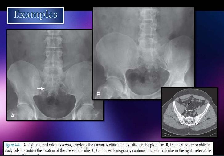
46 PLAIN FILM- LEFT DISTAL URETERAL CALCULUS

47 Intravenous urography
48

49 Retrograde urethrogram 20mm stricture in the bulbous urethra.


50 Anatomy of the normal ureter on ascending urethrogram. Urethral stricture

51
52


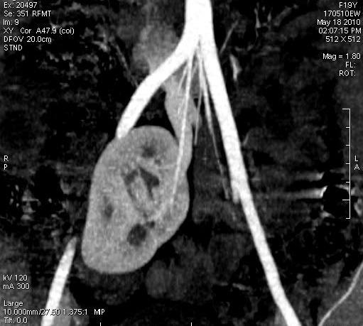
53 Renal hypoplasia


54 Cazul 2 Renal hypoplasia

55 Ectopic kidney


56 Ectopic kidney


57 Ectopic kidney

58 Ectopic kidney


59 Ectopic kidney

60 Tumors

61 Tumors

62 Tumor

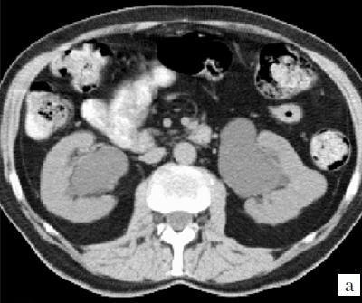
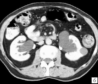
63 TUMOR CT

64 TUMOR CT


65 Hypervascular process left kidney
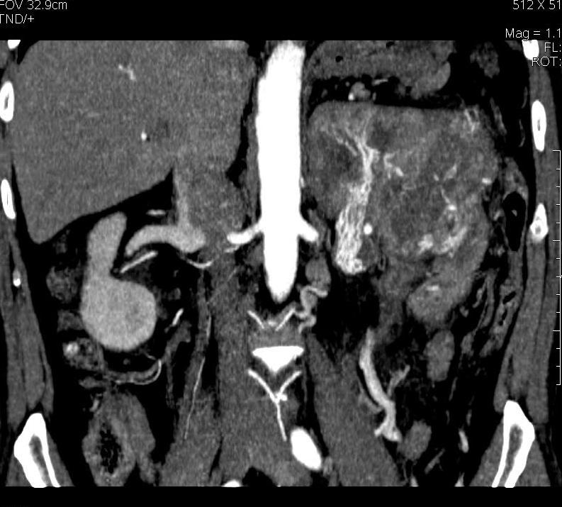
66 Hypervascular process left kidney

67 Hypervascular process left kidney
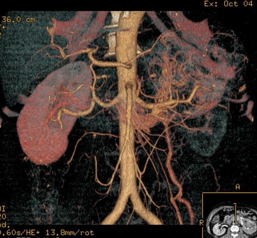

68 Hypervascular process left kidney

69 renal carcinoma MRI


70 PARAPELVICAL CYST
71 Bosniak renal cyst classification The Bosniak classification system for CT evaluation of renal cysts is helpful in determining both malignant risk and required follow-up and/or treatment. Bosniak 1 simple cyst, imperceptible wall, rounded work up : nil % malignant : ~ 0% Bosniak 2 minimally complex, a few thin (< 1mm) septa, thin Ca++; non-enhancing high-attenuation (due to to proteinaceous or haemorrhagic fluid) renal lesions of less than 3 cm are also included in this category; these lesions are generally well marginated. work up : nil % malignant : ~ 0% Bosniak 2F minimally complex but requiring follow up. increased number of septa, minimally thickened or enhancing septa or wall thick Ca++, hyperdense cyst that is: > 3 cm diameter, mostly intrarenal (less than 25% of wall visible); no enhancement work up : needs ultrasound / CT follow up % malignant : ~ 25 % 6
72 Bosniak 3 indeterminate, thick or multiple septations, mural nodule, hyperdense on CT (see 2F) treatment / work up : partial nephrectomy or RF ablation in elderly / poor surgical risk % malignant : ~ 54% 6 Bosniak 4 clearly malignant, solid mass with large cystic or necrotic component treatment: partial / total nephrectomy % malignant : ~100%


73 Extrarenal renal cyst expansion


74 Extrarenal renal cyst expansion
75 Extrarenal renal cyst expansion
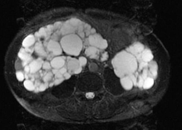
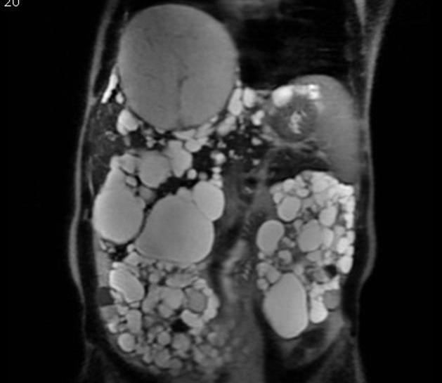


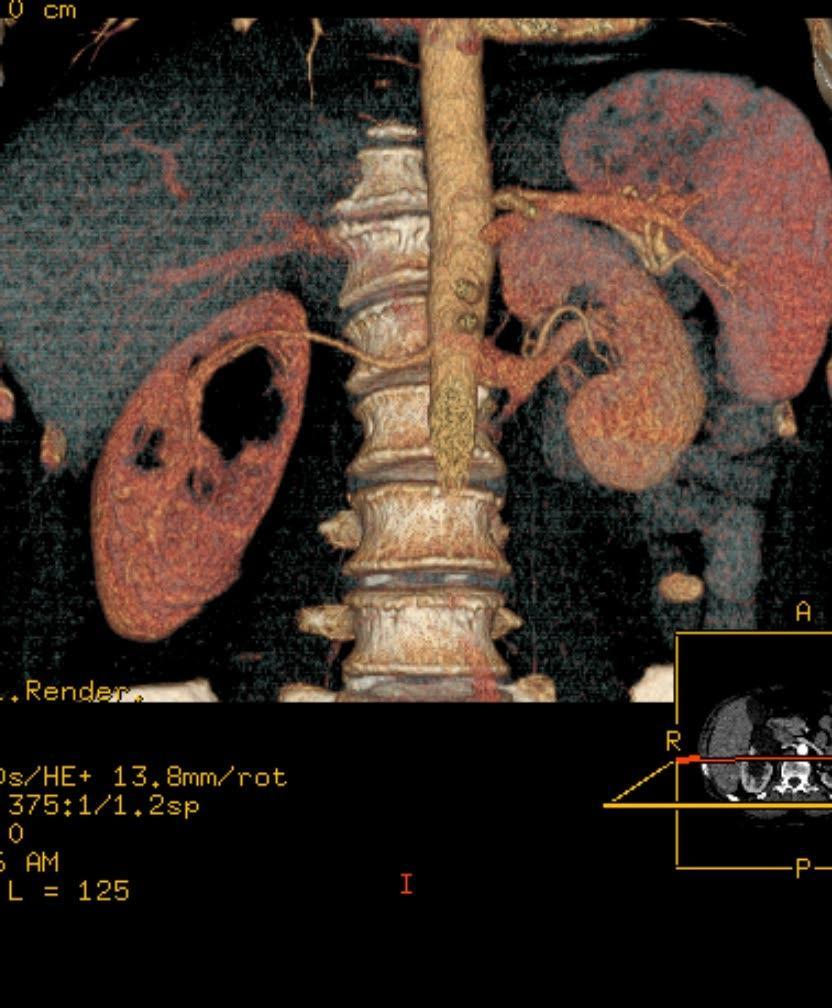
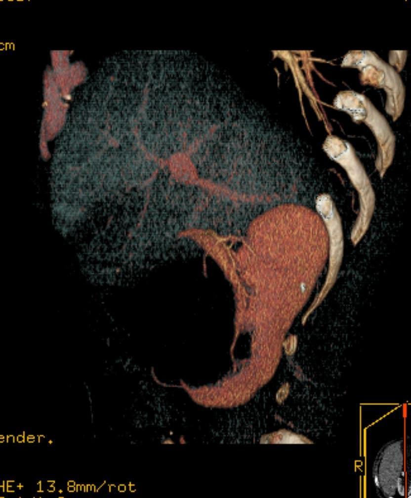
76 Polycystic kidney disease: CT vs MRI

77 Polytrauma extensive skin emphysema kidney contusion.

78 TRAUMA CT


79 Normal prostate gland Axial T2


80
81


82 Prostate carcinoma Axial T2 Ill-defined hypointense lesions in the peripheral zone of the prostate gland - this appearance is highly likely of prostatic carcinoma.
PROFESSIONAL SKILLS 1 3RD YEAR SEMESTER 6 RADIOGRAPHY. THE URINARY SYSTEM Uz. Fatema shmus aldeen Tel
 PROFESSIONAL SKILLS 1 3RD YEAR SEMESTER 6 RADIOGRAPHY THE URINARY SYSTEM Uz. Fatema shmus aldeen Tel. 0925111552 Professional skills-2 THE URINARY SYSTEM The urinary system (review anatomy and physiology)
Excretory urography (EU) or IVP US CT & radionuclide imaging
 or IVP US CT & radionuclide imaging") Excretory urography (EU) or IVP US CT & radionuclide imaging MRI arteriography studies requiring catherization or direct puncture of collecting system EU & to a lesser extent CT provide both functional
Excretory urography (EU) or IVP US CT & radionuclide imaging MRI arteriography studies requiring catherization or direct puncture of collecting system EU & to a lesser extent CT provide both functional
Genitourinary. Common Clinical Scenarios Protocoling Module. Patty Ojeda & Mariam Shehata
 The following training module was developed as a quality improvement project to serve as an educational tool for junior radiology residents. The following diagnostic radiology protocoling modules were
Contents. Review anatomy of the urinary tract Imaging modalities
 Contents Review anatomy of the urinary tract Imaging modalities The Urinary Tract Kidneys ตาแหน งไต (position) อย ใน retroperitoneum ระด บ T12-L3 โดยไต ขวาจะม ระด บตากว าไตซ ายเล กน อย ร ปร าง (shape)
Lec-8 جراحة بولية د.نعمان
 4th stage Lec-8 جراحة بولية د.نعمان 11/10/2015 بسم هللا الرحمن الرحيم Ureteric, Vesical, & urethral stones Ureteric Calculus Epidemiology like renal stones Etiology like renal stones Risk factors like
R adio logical investigations of urinary system
 R adio logical investigations of urinary system There are 4 main radiological Ix: 1 IVU: Intravenous urography. 2- U/S 3-CT scan 4-Radioisotope scan. Others (not frequently used): MRI, arteriography, antegrade
Outline. Introduction to imaging modalities of the urinary system. Case base learning of common diseases in urinary tract
 Outline Introduction to imaging modalities of the urinary system Case base learning of common diseases in urinary tract Outline Introduction to imaging modalities of the urinary system Case base learning
Outline. Introduction to imaging modalities of the urinary system. Case base learning of common diseases in urinary tract
 Outline Introduction to imaging modalities of the urinary system Case base learning of common diseases in urinary tract Diagnostic Investigations in Urinary System PLAIN KUB EXCRETORY UROGRAPHY RETROGRADE
Uroradiology For Medical Students
 Uroradiology For Medical Students Lesson 4: Cystography & Urethrography - Part 2 American Urological Association Review Cystography is useful in evaluating the bladder, the urethra and the competence of
Imaging of the Urinary Tract
 Imaging of the Urinary Tract National Kidney and Urologic Diseases Information Clearinghouse U.S. Department of Health and Human Services NATIONAL INSTITUTES OF HEALTH What is the urinary tract? The urinary
UBC Department of Urologic Sciences Lecture Series. Urological Trauma
 UBC Department of Urologic Sciences Lecture Series Urological Trauma Disclaimer: This is a lot of information to cover and we are unlikely to cover it all today These slides are to be utilized for your
Autosomal Dominant Polycystic Kidney Disease
 Case Studies [1] July 01, 2014 By Amar Udare, MBBS [2] Case History: 45-year-old female with vague pain in the abdomen. Case History: A 45-year-old female presented with vague pain in the abdomen. A USG
Imaging Ejaculatory Disorders and Hematospermia
 ATHENS 4-6 October 2018 European Society of Urogenital Radiology Imaging Ejaculatory Disorders and Hematospermia Parvati Ramchandani, MD Professor, Radiology and Surgery University of Pennsylvania Medical
Bladder Trauma Data Collection Sheet
 Bladder Trauma Data Collection Sheet If there was no traumatic injury with PENETRATION of the bladder DO NOT proceed Date of injury: / / Time of injury: Date of hospital arrival: / / Time of hospital arrival:
January Dear Medical Director:
 January 2010 Dear Medical Director: It is the position of the American Urological Association (AUA) that urologists are appropriately trained in the performance of sonographic procedures. In spite of this,
4 th Year Urology Core Objectives Keith Rourke (Revised June 1, 2007)
") 4 th Year Urology Core Objectives Keith Rourke (Revised June 1, 2007) I. Genitourinary Trauma: 1. Goal: The student will be able to demonstrate a basic clinical approach to the management & diagnosis of
4 th Year Urology Core Objectives Keith Rourke (Revised June 1, 2007) I. Genitourinary Trauma: 1. Goal: The student will be able to demonstrate a basic clinical approach to the management & diagnosis of
ASSESSING THE PLAIN ABDOMINAL RADIOGRAPH M A A M E F O S U A A M P O F O
 ASSESSING THE PLAIN ABDOMINAL RADIOGRAPH M A A M E F O S U A A M P O F O Introduction The abdomen (less formally called the belly, stomach, is that part of the body between the thorax (chest) and pelvis,
US in non-traumatic acute abdomen. Lalita, M.D. Radiologist Department of radiology Faculty of Medicine ChiangMai university
 US in non-traumatic acute abdomen Lalita, M.D. Radiologist Department of radiology Faculty of Medicine ChiangMai university Sagittal Orientation Transverse (Axial) Orientation Coronal Orientation Intercostal
Renal masses - the role of diagnostic imaging
 Renal masses - the role of diagnostic imaging Poster No.: C-2471 Congress: ECR 2015 Type: Educational Exhibit Authors: V. Rai#; Bjelovar/HR Keywords: Cysts, Cancer, Structured reporting, Ultrasound, MR,
Coding Companion for Urology/Nephrology. A comprehensive illustrated guide to coding and reimbursement
 Coding Companion for Urology/Nephrology A comprehensive illustrated guide to coding and reimbursement 2014 Contents Getting Started with Coding Companion...i Integumentary...1 Arteries and Veins...15 Lymph
FOR CMS (MEDICARE) MEMBERS ONLY NATIONAL COVERAGE DETERMINATION (NCD) FOR MAGNETIC RESONANCE IMAGING:
 MEMBERS ONLY NATIONAL COVERAGE DETERMINATION (NCD) FOR MAGNETIC RESONANCE IMAGING:") National Imaging Associates, Inc. Clinical guidelines SINUS MRI Original Date: November 2007 Page 1 of 5 CPT Codes: 70540, 70542, 70543 Last Review Date: July 2014 NCD 220.2 MRI Last Effective Date: July
National Imaging Associates, Inc. Clinical guidelines SINUS MRI Original Date: November 2007 Page 1 of 5 CPT Codes: 70540, 70542, 70543 Last Review Date: July 2014 NCD 220.2 MRI Last Effective Date: July
Abdomen Sonography Examination Content Outline
 Abdomen Sonography Examination Content Outline (Outline Summary) # Domain Subdomain Percentage 1 2 3 Anatomy, Perfusion, and Function Pathology, Vascular Abnormalities, Trauma, and Postoperative Anatomy
Index. Note: Page numbers of article titles are in boldface type.
 Magn Reson Imaging Clin N Am 12 (2004) 587 591 Index Note: Page numbers of article titles are in boldface type. A Adenoma(s), adrenal, gadolinium-enhanced MR imaging in, 533 534 hyperfunctioning versus
Renal Trauma: Management Options
 Renal Trauma: Management Options Immediate surgical repair Nephrectomy Conservative management Alonso RC et al. Kidney in Danger: CT Findings of Blunt and Penetrating Renal Trauma. RadioGraphics 2009;
Introduction to Radiology
 Introduction - Lecture 1 436 Teams Introduction to Radiology Objectives Introduce the various Medical Imaging Modalities. Understand the basics of image generation. Relate imaging to gross anatomy. Appreciate
Role of imaging in RCC. Ultrasonography. Solid lesion. Cystic RCC. Solid RCC 31/08/60. From Diagnosis to Treatment: the Radiologist Perspective
 Role of imaging in RCC From Diagnosis to Treatment: the Radiologist Perspective Diagnosis Staging Follow up Imaging modalities Limitations and pitfalls Duangkamon Prapruttam, MD Department of Therapeutic
Canadian Undergraduate Urology Curriculum (CanUUC): Genitourinary Trauma. Last reviewed June 2014
: Genitourinary Trauma. Last reviewed June 2014") Canadian Undergraduate Urology Curriculum (CanUUC): Genitourinary Trauma Last reviewed June 2014 Session Objectives 1. Recognize hematuria as the cardinal symptom of urinary tract trauma. 1. Outline the
Canadian Undergraduate Urology Curriculum (CanUUC): Genitourinary Trauma Last reviewed June 2014 Session Objectives 1. Recognize hematuria as the cardinal symptom of urinary tract trauma. 1. Outline the
 INTRAUTERINE DEVICE = IUD INTRAUTERINE DEVICE = IUD CONGENITAL DISORDERS Pyometra = pyometrea is a uterine infection, it is accumulation of purulent material in the uterine cavity. Ultrasound is usually
Pediatric Ure-Radiology*
 Pediatric Ure-Radiology* HERMAN GROSSMAN, M.D. Professor of Radiology and Pediatrics, Duke University Medical Center, Durham, North Carolina "Routine" radiologic studies do not, often enough, concentrate
Obstetrics Content Outline Obstetrics - Fetal Abnormalities
 Obstetrics Content Outline Obstetrics - Fetal Abnormalities Effective February 2007 10 16% renal agenesis complete absence of the kidneys occurs when ureteric buds fail to develop Or degenerate before
Radiological Investigations of Abdominal Trauma
 76 77 Investigations of Abdominal Trauma Introduction: Trauma to abdominal organs is a common cause of patient morbidity and mortality among trauma patients. Causes of abdominal trauma include blunt injuries,
Medical imaging X-ray, CT, MRI, scintigraphy, SPECT, PET Györgyi Műzes
 Medical imaging X-ray, CT, MRI, scintigraphy, SPECT, PET Györgyi Műzes Semmelweis University, 2nd Dept. of Medicine Medical imaging: definition technical process of creating visual representations about
Genitourinary Radiology In-Training Test Questions for Diagnostic Radiology Residents
 Genitourinary Radiology In-Training Test Questions for Diagnostic Radiology Residents March, 2013 Sponsored by: Commission on Education Committee on Residency Training in Diagnostic Radiology 2013 by American
Guidelines, Policies and Statements D5 Statement on Abdominal Scanning
 Guidelines, Policies and Statements D5 Statement on Abdominal Scanning Disclaimer and Copyright The ASUM Standards of Practice Board have made every effort to ensure that this Guideline/Policy/Statement
Glossary of Terms Primary Urethral Cancer
 Patient Information English Glossary of Terms Primary Urethral Cancer Advanced cancer A tumour that grows into deeper layers of tissue, adjacent organs, or surrounding muscles. Anaesthesia (general, spinal,
Proceedings of the 34th World Small Animal Veterinary Congress WSAVA 2009
 www.ivis.org Proceedings of the 34th World Small Animal Veterinary Congress WSAVA 2009 São Paulo, Brazil - 2009 Next WSAVA Congress : Reprinted in IVIS with the permission of the Congress Organizers IMAGING
Children's (Pediatric) Ultrasound - Abdomen
 Ultrasound - Abdomen") Scan for mobile link. Children's (Pediatric) Ultrasound - Abdomen Children s (pediatric) ultrasound imaging of the abdomen is a safe, noninvasive test that uses sound waves to produce a clear picture of
Scan for mobile link. Children's (Pediatric) Ultrasound - Abdomen Children s (pediatric) ultrasound imaging of the abdomen is a safe, noninvasive test that uses sound waves to produce a clear picture of
Basic Abdominal and Pelvic Imaging Concepts. David L. Smith, MD Assistant Professor of Radiology
 Basic Abdominal and Pelvic Imaging Concepts David L. Smith, MD Assistant Professor of Radiology Basic Imaging Concepts Contrast Resolution vs Spacial Resolution Spacial Resolution......refers to the ability
RADIOLOGY OF THE URINARY TRACT CHAPTER 9 239
 RADIOLOGY OF THE URINARY TRACT CHAPTER 9 239 in length. They lie cephalad to the kidneys, with the right just posterior to the inferior vena cava (IVC) and the left anteromedial to the upper pole of the
Pelvic Ultrasound.
 Pelvic Ultrasound Before Your Exam: Drink 32 oz. of water one hour before your examination time. Try to drink all the liquid within 30 minutes. Do not urinate before the exam. Arrive for your exam with
FOR CMS (MEDICARE) MEMBERS ONLY NATIONAL COVERAGE DETERMINATION (NCD) FOR MAGNETIC RESONANCE IMAGING:
 MEMBERS ONLY NATIONAL COVERAGE DETERMINATION (NCD) FOR MAGNETIC RESONANCE IMAGING:") National Imaging Associates, Inc. Clinical guidelines BONE MARROW MRI Original Date: July 2008 Page 1 of 5 CPT Codes: 77084 Last Review Date: September 2014 NCD 220.2 MRI Last Effective Date: July 2011
National Imaging Associates, Inc. Clinical guidelines BONE MARROW MRI Original Date: July 2008 Page 1 of 5 CPT Codes: 77084 Last Review Date: September 2014 NCD 220.2 MRI Last Effective Date: July 2011
Find Medical Solutions to Your Problems HYDRONEPHROSIS. (Distension of Renal Calyces & Pelvis)
") HYDRONEPHROSIS (Distension of Renal Calyces & Pelvis) Hydronephrosis is the distension of the renal calyces and pelvis due to accumulation of the urine as a result of the obstruction to the outflow of
HYDRONEPHROSIS (Distension of Renal Calyces & Pelvis) Hydronephrosis is the distension of the renal calyces and pelvis due to accumulation of the urine as a result of the obstruction to the outflow of
My Patient Has Abdominal Pain PoCUS of the Biliary Tract and the Urinary Tract
 My Patient Has Abdominal Pain PoCUS of the Biliary Tract and the Urinary Tract Objectives PoCUS for Biliary Disease PoCUS for Renal Colic PoCUS for Urinary Retention Biliary Disease A patient presents
Clinical aspects in urogenital injuries
 Clinical aspects in urogenital injuries Rolf Wahlqvist Oslo Urological University Clinic Aker University Hospital Nordic Rad.2008 1 Urogenital injuries in trauma patients Renal injury Ureteral injury (infrequent/iatrogenic)
RADIOLOGY (MEDICAL IMAGING)
") RADIOLOGY (MEDICAL IMAGING) Radiology is the study of the diagnosis of disease by the use of radiant energy (radiation). In the past this meant the use of X-rays to make an image. Today many other forms
RADIOLOGY (MEDICAL IMAGING) Radiology is the study of the diagnosis of disease by the use of radiant energy (radiation). In the past this meant the use of X-rays to make an image. Today many other forms
General Nuclear Medicine
 General Nuclear Medicine What is General Nuclear Medicine? What are some common uses of the procedure? How should I prepare? What does the equipment look like? How does the procedure work? How is the procedure
Role of imaging in evaluation of genitourinary i trauma Spectrum of GU injuries Relevance of imaging findings in determining management Focus on MDCT
 Genitourinary Tract Injuries 6 th Nordic Course Scott D. Steenburg, MD Assistant Professor University of Maryland Department of Radiology Division of Trauma and Emergency Radiology R Adams Cowley Shock
Alice Fung, MD Oregon Health and Science University
 Alice Fung, MD Oregon Health and Science University Disclosure Comments The speaker Alice Fung, MD Has relevant financial relationships to disclose. Received honorarium from (Guerbet). This individual
Rad Lab 4 Unknowns: Genitourinary!
 Rad Lab 4 Unknowns: Genitourinary! Peter Clarke MD! Don Di Salvo, MD! Clerkship Directors for Radiology! Harvard Medical School! Brigham and Women s Hospital! Dana Farber Cancer Institute! Case 1: 69 year
Radiological Assessment of the Kidney in Patients with Hematuria
 March 2005 Radiological Assessment of the Kidney in Patients with Hematuria Jeremy L. McKay, Harvard Medical School Year III Hematuria Signs and Symptoms Microscopic or gross hematuria Abdominal pain Fever
General Imaging. Imaging modalities. Incremental CT. Multislice CT Multislice CT [ MDCT ]
![General Imaging. Imaging modalities. Incremental CT. Multislice CT Multislice CT [ MDCT ]](/thumbs/76/74079340.jpg "General Imaging. Imaging modalities. Incremental CT. Multislice CT Multislice CT [ MDCT ]") General Imaging Imaging modalities Conventional X-rays Ultrasonography [ US ] Computed tomography [ CT ] Radionuclide imaging Magnetic resonance imaging [ MRI ] Angiography conventional, CT,MRI Interventional
General Imaging Imaging modalities Conventional X-rays Ultrasonography [ US ] Computed tomography [ CT ] Radionuclide imaging Magnetic resonance imaging [ MRI ] Angiography conventional, CT,MRI Interventional
Radiographic Procedures III (RAD 228)
") Radiographic Procedures III (RAD 228) Urinary System RADIOGRAPHIC EXAMINATIONS Urinary System Antegrade Exam IVU Functional test Hypertensive evaluation as per protocol Retrograde Exams Retrograde Urography
Radiographic Procedures III (RAD 228) Urinary System RADIOGRAPHIC EXAMINATIONS Urinary System Antegrade Exam IVU Functional test Hypertensive evaluation as per protocol Retrograde Exams Retrograde Urography
Urologic investigations
 Urologic investigations د. Laboratory studies EXAMINATION OF URINE Urinalysis: Urinalysis is one of the most important and useful urologic tests available. Reasons for inadequate urinalyses include: (1)
Diagnosis and classification
 Patient Information English 2 Diagnosis and classification The underlined terms are listed in the glossary. Signs and symptoms Blood in the urine is the most common symptom when a bladder tumour is present.
Arteriogram An X-ray of an artery after the injection of dye.
 A Abscess A localized collection of pus in any part of the body, usually surrounded by inflamed tissue. Anesthetic An agent that causes loss of sensation with or without the loss of consciousness. Angiography,
11/1/2014. Radiologic incidentalomas Ordering pitfalls Newer technology and applications
 Bilal Tahir, MD Gitasree Borthakur, MD Indiana University School of Medicine Department of Radiology & Imaging Sciences October 31, 2014 ACP 2014 Dr. V. Aaron Nuclear (vaaron@iupui.edu) Dr. S. Westphal
Urogenital Injuries The role of radiology
 Urogenital Injuries The role of radiology NORDTER 7 th Nordic Trauma Radiology Course Helsinki, Finland May 21-24, 2012 Johann Baptist Dormagen, MD, PhD Oslo University Hospital, Norway Kidney injuries
Human Systems. Technology - Ultrasounds
 Human Systems Technology - Ultrasounds What is General Ultrasound Imaging? Ultrasound imaging, also called ultrasound scanning or sonography, involves exposing part of the body to high-frequency sound
Symptoms, Diagnosis and Classification
 Patient Information English 2 Symptoms, Diagnosis and Classification The underlined terms are listed in the glossary. Signs and symptoms Blood in the urine is the most common symptom when a bladder tumour
Certification Review. Module 28. Medical Coding. Radiology
 Module 28 is the study of x-rays, using radiant energy and other imaging techniques, such as resonance imaging or ultrasound, to diagnose illnesses and diseases. Vocabulary Barium enema (BE): lower gastrointestinal
Genitourinary Trauma Introduction GU Trauma overlooked
 Genitourinary Trauma Introduction GU Trauma overlooked 10-20% of all injured patients Long term morbidity Impotence Incontinence Life-threatening injuries first Urethral Injury Plan Bladder Injury Kidney
Acute Pyelonephritis
 Acute Pyelonephritis Variant 1: Acute pyelonephritis. Uncomplicated patient (eg, no history of diabetes or immune compromise or history of stones or obstruction or prior renal surgery or lack of response
Case MDCT 3D reconstructed features of posterior urethral valve
 Case 12688 MDCT 3D reconstructed features of posterior urethral valve Hidayatullah Hamidi Third year Resident of Radiology French medical institute for children Radiology Department; Kabul, Afghanistan;
Uroradiology Tutorial For Medical Students
 Uroradiology Tutorial For Medical Students Lesson 3: Cystography & Urethrography Part 1 American Urological Association Introduction Conventional radiography of the urinary tract includes several diagnostic
Essentials of Clinical MR, 2 nd edition. 73. Urinary Bladder and Male Pelvis
 73. Urinary Bladder and Male Pelvis Urinary bladder carcinoma is best locally staged with MRI. It is important however to note that a thickened wall (> 5 mm) is a non-specific finding seen in an underfilled
Acute renal colic Radiological investigation in patients with renal colic
 Acute renal colic Radiological investigation in patients with renal colic Mikael Hellström Professor Department of Radiology Sahlgrenska University Hospital Göteborg University 0.9-1.8/1.000 inhabitants
Name of the module: Urology clerkship (selective) Number of module: Course description: Basic clinical urology.
 Number of module: Course description: Basic clinical urology.") Name of the module: Urology clerkship (selective) Number of module: 471-8-5097 Credit points: 2.5 ECTS: Academic year: 5 th & 6 th Semester: 1st, 2 nd Duration: 2 weeks Location: Urology depts. in Soroka
Name of the module: Urology clerkship (selective) Number of module: 471-8-5097 Credit points: 2.5 ECTS: Academic year: 5 th & 6 th Semester: 1st, 2 nd Duration: 2 weeks Location: Urology depts. in Soroka
Hydronephrosis. What is hydronephrosis?
 What is hydronephrosis? Hydronephrosis Hydronephrosis describes the situation where the urine collecting system of the kidney is dilated. This may be a normal variant or it may be due to an underlying
BLADDER CANCER: PATIENT INFORMATION
 BLADDER CANCER: PATIENT INFORMATION The bladder is the balloon like organ located in the pelvis that stores and empties urine. Urine is produced by the kidneys, is conducted to the bladder by the ureters,
Audit of split-bolus CT urography for the investigation of haematuria over a 12 month period at two district general hospitals
 Audit of split-bolus CT urography for the investigation of haematuria over a 12 month period at two district general hospitals Poster No.: C-1349 Congress: ECR 2010 Type: Educational Exhibit Topic: Genitourinary
THE operation of reimplantation of the ureter into the bladder has undergone
 REIMPLANTATION OF THE URETER INTO THE BLADDER J. G. WARDEN, M.D., and C. C. HIGGINS, M.D. Department of Urology THE operation of reimplantation of the ureter into the bladder has undergone a stormy course
Positron Emission Tomography Computed Tomography (PET/CT)
") Positron Emission Tomography Computed Tomography (PET/CT) What is Positron Emission Tomography Computed Tomography (PET/CT) Scanning? What are some common uses of the procedure? How should I prepare for
Positron Emission Tomography Computed Tomography (PET/CT) What is Positron Emission Tomography Computed Tomography (PET/CT) Scanning? What are some common uses of the procedure? How should I prepare for
Effective Utilization of Imaging. John V. Roberts, M.D. Premier Radiology Abdominal Imaging
 Effective Utilization of Imaging John V. Roberts, M.D. Premier Radiology Abdominal Imaging Safety Contrast and Radiation What to order Abdomen/Pelvis Brain/Spine Chest Musculoskeletal Ob/Gyn Head and Neck
CT abdomen and pelvis
 CT abdomen and pelvis General indications: Assessment of vague abdominal symptoms (pain, colics,distenstion,...) Varifecation of a lesion discovered by other diagnostic modalities as US, barium,ivp, Staging
URINARY SYSTEM I. Kidneys II. Nephron Unit and Urine Formation
 URINARY SYSTEM I. Kidneys A. Location and Structure 1. Retroperitoneal 2. Between T12 and L3 3. Rt. kidney slightly lower 4. Two bean shaped organs 5. Adrenal gland 6. Internal construction a. Renal cortex
West Yorkshire Major Trauma Network Clinical Guidelines 2015
 WYMTN: Pelvic fracture with urogenital trauma KEY RECOMMENDATIONS 1. During the initial exploratory survey / secondary survey, a. The external urethral meatus and the transurethral bladder catheter (if
Kidney & Urinary Tract Ultrasound. Fatina Fadel Hafez Bazaraa
 Kidney & Urinary Tract Ultrasound Fatina Fadel Hafez Bazaraa Ultrasonography Ultrasound Available Rapid Inexpensive Painless & no sedation needed No adverse effects/ complications Can be repeated Useful
Nuclear Medicine - Hepatobiliary
 Scan for mobile link. Nuclear Medicine - Hepatobiliary Hepatobiliary nuclear medicine imaging helps evaluate the parts of the biliary system, including the liver, gallbladder and bile ducts, using small
Center for Reconstructive Urethral Surgery Guido Barbagli Center for Reconstructive Urethral Surgery Arezzo - Italy
 Guido Barbagli Arezzo - Italy E-mail: guido@rdn.it Website: www.urethralcenter.it 23 rd ANNUAL EAU CONGRESS Sub-plenary Session on Male urinary incontinence 26 29 March 2008 Milan Italy Incontinence following
You have been booked for a. Flexible Cystoscopy. Under Local Anaesthetic
 You have been booked for a Flexible Cystoscopy Under Local Anaesthetic 1 WHAT IS A FLEXIBLE CYSTOSCOPY A flexible cystoscopy is a test to examine the uretha (waterpipe) and bladder using a thin, lighted
Abdominal Ultrasound
 Abdominal Ultrasound What is Ultrasound Imaging of the Abdomen? What are some common uses of the procedure? How should I prepare? What does the equipment look like? How does the procedure work? How is
Sex: 女 Age: 51 Occupation: 無 Admission date:92/07/22
 Sex: 女 Age: 51 Occupation: 無 Admission date:92/07/22 Chief complaint Unknown fever for one month Hand tremor and left huge renal tumor was noted Present illness Suffered from fever for one month, hand
Course specification
 Al-Azhar University Faculty of Medicine for Men Course specification For Master of Radiodiagnosis ( 2014 2015 ) University : Al-Azhar Faculty : Medicine for men Course specification - Programmers on which
Abdomen and Retroperitoneum Ultrasound Protocols
 Abdomen and Retroperitoneum Ultrasound Protocols Reviewed By: Anna Ellermeier, MD Last Reviewed: March 2018 Contact: (866) 761-4200, Option 1 **NOTE for all examinations: 1. If documenting possible flow
IMAGING OF THE UROGENITAL TRACT
 IMAGING OF THE UROGENITAL TRACT 1 A) URINARY TRACT There are many methods of imaging the urinary tract but plain abdominal X-ray and ultrasound scan are usually done first in most cases, especially in
An abdominal ultrasound produces a picture of the organs and other structures in the upper abdomen.
 Scan for mobile link. Ultrasound - Abdomen Ultrasound imaging of the abdomen uses sound waves to produce pictures of the structures within the upper abdomen. It is used to help diagnose pain or distention
Dr. Aso Urinary Symptoms
 Haematuria The presence of blood in the urine (haematuria) is always abnormal and may be the only indication of pathology in the urinary tract. False positive stick tests and the discolored urine caused
Imaging the Urogenital System
 maging the Urogenital System Tony Pease, DVM, MS, DACVR Assistant Professor of Radiology North Carolina State University Reading Thrall Chapters 42-46 Prostate Gland Not visible radiographically in normal
Case Report Imaging of a Renal Artery Aneurysm Detected Incidentally on Ultrasonography
 Case Reports in Radiology, Article ID 375805, 4 pages http://dx.doi.org/10.1155/2014/375805 Case Report Imaging of a Renal Artery Aneurysm Detected Incidentally on Ultrasonography Vasileios Rafailidis,
Course specification
 Al-Azhar University Faculty of Medicine for Men Course specification For Doctorate of Radiodiagnosis ( 2014 2015 ) University : Al-Azhar Faculty : Medicine for Men Course specification - Programmers on
Children's (Pediatric) Nuclear Medicine
 Nuclear Medicine") Scan for mobile link. Children's (Pediatric) Nuclear Medicine Children s (pediatric) nuclear medicine imaging uses small amounts of radioactive materials called radiotracers, a special camera and a computer
Scan for mobile link. Children's (Pediatric) Nuclear Medicine Children s (pediatric) nuclear medicine imaging uses small amounts of radioactive materials called radiotracers, a special camera and a computer
INTERNATIONAL SPINAL CORD INJURY DATA SETS URINARY TRACT IMAGING BASIC DATA SET - COMMENTS
 1 INTERNATIONAL SPINAL CORD INJURY DATA SETS URINARY TRACT IMAGING BASIC DATA SET - COMMENTS The working-group consists of: Fin Biering-Sørensen representing the Executive Committee of The International
Policies, Standards, and Guidelines. Guidelines for Abdominal Ultrasound Examination
 Policies, Standards, and Guidelines Guidelines for Abdominal Ultrasound Examination Approved by Council Feb 2018 Disclaimer and Copyright The ASUM Standards of Practice Board have made every effort to
Surgical Care at the District Hospital. EMERGENCY & ESSENTIAL SURGICAL CARE
 Surgical Care at the District Hospital 1 9 Urinary Tract and Perineum Key Points 2 9.1 Urinary Bladder & Urinary Retention Acute retention of urine is an indication for emergency drainage of the bladder
Job Task Analysis for ARDMS Abdomen Data Collected: June 30, 2011
 Job Task Analysis for ARDMS Abdomen Data Collected: June 30, 2011 Reported: Analysis Summary for: Abdomen Examination Survey Dates 06/13/2011-06/26/2011 Invited Respondents 6,000 Surveys with Demographics
ULTRASONOGRAPHY RADIOLOGY Simple abdominal X-ray Intravenous urography Retrograde/anterograde pieloureterography Renal angiography CT
 ULTRASONOGRAPHY RADIOLOGY Simple abdominal X-ray Intravenous urography Retrograde/anterograde pieloureterography Renal angiography CT NUCLEAR MEDICINE Static studies: static renal scintigraphy Dynamic
URINARY SYSTEM. MEDICAL TERMINOLOGY Chapter Six HIT #141. Anatomy
 URINARY SYSTEM MEDICAL TERMINOLOGY Chapter Six HIT #141 Anatomy Kidneys = bean-shaped organs, located on each side of the spinal column, removal of waste from the blood. Nephron = microscopic located in
General Ultrasound. What is General Ultrasound Imaging?
 Scan for mobile link. General Ultrasound Ultrasound imaging uses sound waves to produce pictures of the inside of the body. It is used to help diagnose the causes of pain, swelling and infection in the
MODULE 5: HEMATURIA LEARNING OBJECTIVES DEFINITION. KEY WORDS: Hematuria, Cystoscopy, Urine Cytology, UTI, bladder cancer
 MODULE 5: HEMATURIA KEY WORDS: Hematuria, Cystoscopy, Urine Cytology, UTI, bladder cancer LEARNING OBJECTIVES At the end of this clerkship, the learner will be able to: 1. Define microscopic hematuria.